
A Lethal Case of Disseminated Cladosporium allicinum Infection in a Captive African Bullfrog
 , , , ,
  and
and 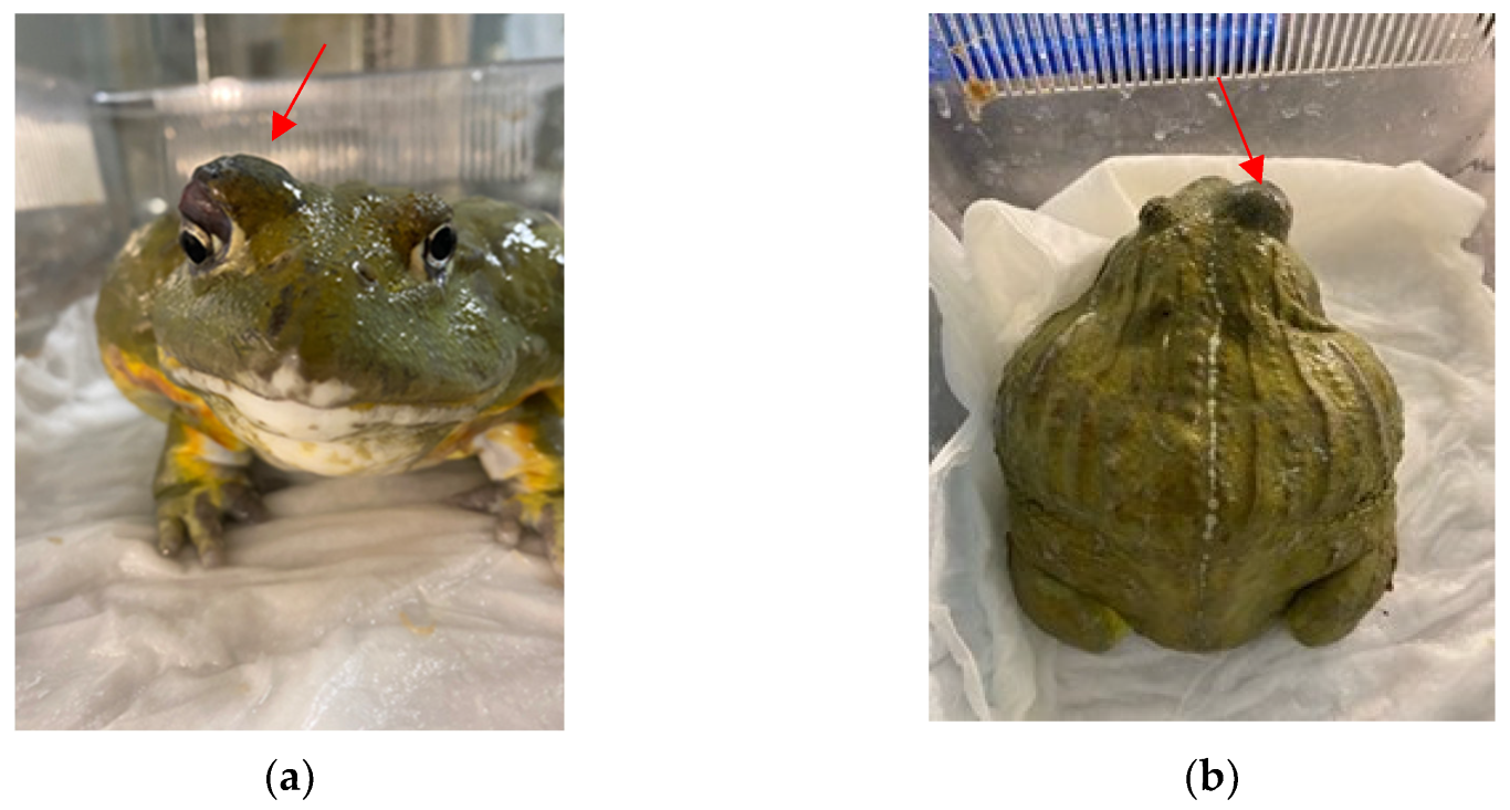
Abstract
1. Introduction
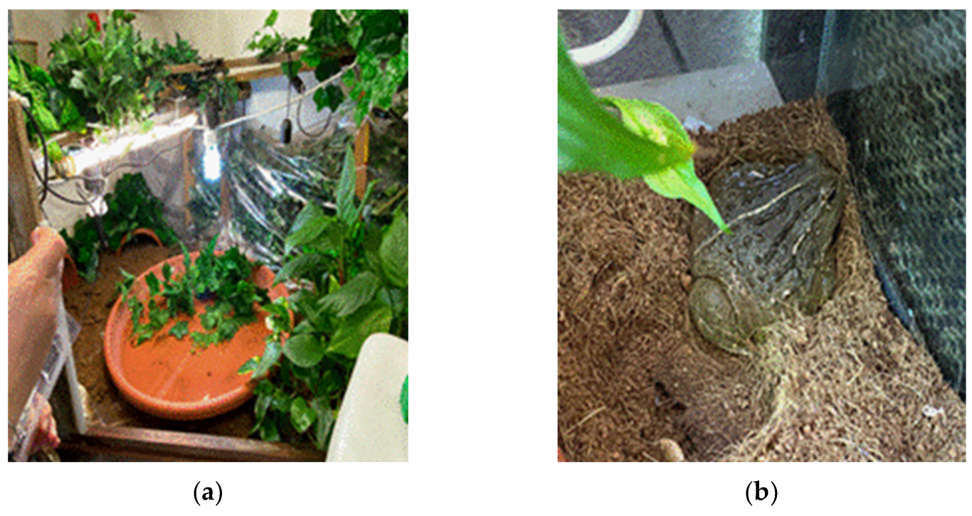
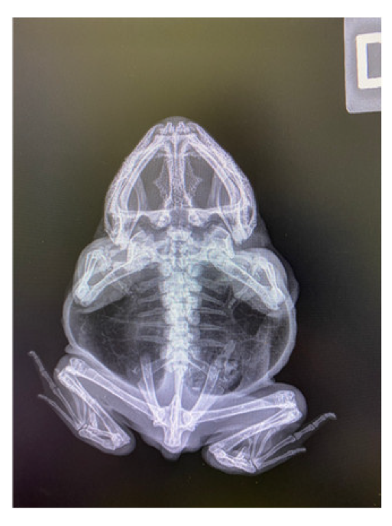
2. Case Report
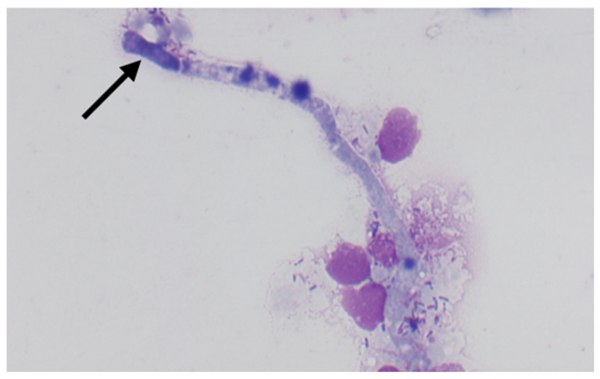
3. Materials and Methods
4. Results
5. Discussion
6. Conclusions
Author Contributions
Funding
Informed Consent Statement
Data Availability Statement
Conflicts of Interest
References
- Bensch, K.; Braun, U.; Groenewald, J.Z. The Genus Cladosporium. Stud. Mycol. 2012, 72, 1–401. [Google Scholar] [CrossRef]
- Bensch, K.; Groenewald, J.Z.; Meijer, M.; Dijksterhuis, J.; Jurjević, Ž.; Andersen, B.; Houbraken, J.; Crous, P.W.; Samson, R.A. Cladosporium species in indoor environments. Stud. Mycol. 2018, 89, 177–301. [Google Scholar] [CrossRef] [PubMed]
- Samson, R.A.; Houbraken, J.; Thrane, U.; Frisvad, J.C.; Andersen, B. Food and Indoor Fungi, 1st ed.; CBS Laboratory Manual Series 2; Westerddijk Fungal Biodiversity Institute: Utrecht, The Netherlands, 2010. [Google Scholar]
- Kulik, T.; Treder, K.; Zaluski, D. Quantification of Alternaria, Cladosporium, Fusarium and Penicillium verrucosum in conventional and organic grains by qPCR. J. Phytopathol. 2014, 163, 522–528. [Google Scholar] [CrossRef]
- Frasz, S.L.; Miller, J.D. Fungi in Ontario maple syrup & some factors that determine the presence of mold damage. Int. J. Food Microbiol. 2015, 207, 66–70. [Google Scholar] [PubMed]
- Black, P.N.; Udy, A.A.; Brodie, S.M. Sensitivity to fungal allergens is a risk factor for life-threatening asthma. Eur. J. Allergy Clin. Immunol. 2000, 55, 501–504. [Google Scholar] [CrossRef] [PubMed]
- Sellart-Altisent, M.; Torres-Rodriguez, J.M.; De Ana, S.G.; Alvarado-Ramirez, E. Nasal fungal microbiota in allergic and healthy subjects. Rev. Iberoam. Micol. 2007, 24, 25–130. [Google Scholar] [CrossRef]
- De Hoog, G.S.; Vicente, V.A.; Najafzadeh, M.J.; Harrak, M.J.; Badali, H.; Seyedmousavi, S. Waterborne Exophiala species causing disease in cold-blooded animals. Persoonia 2011, 27, 46–72. [Google Scholar] [CrossRef]
- Sandoval-Denis, M.; Sutton, D.A.; Martin-Vicente, A.; Cano-Lira, J.F.; Wiederhold, N.; Guarro, J.; Gené, J. Cladosporium Species Recovered from Clinical Samples in the United States. J. Clin. Microbiol. 2015, 53, 2990–3000. [Google Scholar] [CrossRef]
- Gugnani, H.C.; Sood, N.; Singh, B.; Makkar, R. Subcutaneous phaeohyphomycosis due to Cladosporium cladosporioides. Mycoses 2000, 43, 85–87. [Google Scholar] [CrossRef]
- Passero, L.F.D.; Cavallone, I.N.; Belda, W., Jr. Reviewing the Etiologic Agents, Microbe-Host Relationship, Immune Response, Diagnosis, and Treatment in Chromoblastomycosis. J. Immunol. Res. 2021, 2021, 9742832. [Google Scholar] [CrossRef]
- Queiroz-Telles, F.; De Hoog, G.S.; Santos, D.W.; Salgado, C.G.; Vicente, V.A.; Bonifaz, A.; Roilides, E.; Xi, L.; Azevedo, C.M.; Da Silva, M.B.; et al. Chromoblastomycosis. Clin Microbiol. Rev. 2017, 30, 233–276. [Google Scholar] [CrossRef] [PubMed]
- World Health Organization (WHO). Control of Neglected Tropical Diseases, Chromoblastomycosis and Other Deep Mycoses. 2017. Available online: https://www.who.int/teams/control-of-neglected-tropical-diseases/mycetoma-chromoblastomycosis-and-other-deep-mycoses/chromoblastomycosis-and-other-deep-mycoses (accessed on 1 January 2020).
- Schell, W.A. Agents of chromoblastomycosis and sporotrichosis. Topley & Wilson’s Microbiology and Microbial Infections, 9th ed.; Collier, L., Balows, A., Sussman, M., Eds.; Hodder Arnold Publication: London, UK; Oxford University Press: London, UK, 1998; Volume 4, pp. 315–523. [Google Scholar]
- Pindycka-Piaszczyńska, M.; Krzyściak, P.; Piaszczyński, M.; Cieślik, S.; Januszewski, K.; Izdebska-Straszak, G.; Jarząb, J.; De Hoog, S.; Jagielski, T. Chromoblastomycosis as an endemic disease in temperate Europe: First confirmed case and review of the literature. Eur. J. Clin. Microbiol. Infect. Dis. 2014, 33, 391–398. [Google Scholar] [CrossRef] [PubMed]
- Seyedmousavi, S.; Guillot, J.; De Hoog, G.S. Phaeohyphomycoses, emerging opportunistic diseases in animals. Clin. Microbiol. Rev. 2013, 26, 19–35. [Google Scholar] [CrossRef] [PubMed]
- Bube, A.; Burkhardt, E.; Weiß, R. Spontaneous chromomycosis in the marine toad (Bufo marinus). J. Comp. Pathol. 1992, 106, 73–77. [Google Scholar] [CrossRef] [PubMed]
- Hosoya, T.; Hanafusa, Y.; Kudo, T.; Tamukai, K.; Une, Y. First report of Veronaea botryosa as a causal agent of chromomycosis in frogs. Med. Mycol. 2015, 53, 369–377. [Google Scholar] [CrossRef]
- Fukushiro, R. Chromoblastomycosis in Japan. Int. J. Derm. 1983, 22, 221–229. [Google Scholar] [CrossRef]
- Chandramukhi, A.; Ramadevi, M.G.; Shankar, S.K. Cerebral Cladosporiosis—A neuropathological and microbiological study. Clin. Neurol. Neurosurg. 1983, 85, 245–253. [Google Scholar] [CrossRef]
- Heaton, S.M.; Weintrob, A.C.; Downing, K.; Keenan, B.; Aggarwal, D.; Shaikh, F.; Tribble, D.R.; Wells, J.; Infectious Disease Clinical Research Program Trauma Infectious Disease Outcomes Study Group. Histopathological techniques for the diagnosis of combat-related invasive fungal wound infections. BMC Clin. Pathol. 2016, 16, 11. [Google Scholar] [CrossRef]
- De Hoog, G.S.; Guarro, J.; Gené, J.; Figueras, M.J. Atlas of Clinical Fungi, 2nd ed.; Centraalbureau voor Schimmelcultures: Utrecht, The Netherlands, 2000. [Google Scholar]
- Stielow, J.B.; Lévesque, C.A.; Seifert, K.A.; Meyer, W.; Iriny, L.; Smits, D.; Renfurm, R.; Verkley, G.J.M.; Groenewald, M.; Chaduli, D.; et al. One fungus, which genes? Development and assessment of universal primers for potential secondary fungal DNA barcodes. Persoonia 2015, 35, 242–263. [Google Scholar] [CrossRef]
- Basic Local Alignment Search Tool (BLAST) Databases. Available online: https://blast.ncbi.nlm.nih.gov/Blast.cgi (accessed on 1 January 2020).
- GenBank®, National Library of Medicine (NIH) Genetic Sequence Database. Available online: http://www.ncbi.nlm.nih.gov/genbank (accessed on 1 January 2020).
- MycoBank, Fungal Databases, Nomenclature & Species Banks. Available online: http://www.mycobank.org (accessed on 1 January 2020).
- Namratha, N.; Nadgir, S.; Kale, M.; Rathod, R. Chromoblastomycosis due to Cladosporium carrionii. J. Lab. Physicians 2010, 2, 47–48. [Google Scholar] [CrossRef]
- Walsh, T.J.; Hayden, R.T.; Larone, D.H. Front Matter. In Larone’s Medically Important Fungi; Walsh, T.J., Hayden, R., Larone, D., Eds.; Wiley: New York, NY, USA, 2018. [Google Scholar] [CrossRef]
- Dixon, D.M.; Polak-Wyss, A. The medically important dematiaceous fungi and their identification. Mycoses 1991, 34, 1–18. [Google Scholar] [CrossRef] [PubMed]
- Carey, C.; Cohen, N.; Rollins-Smith, L. Amphibian declines: An immunological perspective. Dev. Comp. Immunol. 1999, 23, 459–472. [Google Scholar] [CrossRef] [PubMed]
- Channing, A. Amphibians of Central and Southern Africa; Protea Book House: Pretoria, South Africa, 2001. [Google Scholar]
- Minter, L.R.; Burger, M.; Harrison, J.A.; Braack, H.H.; Bishop, P.J.; Kloepfer, D. Atlas and Red Data Book of the Frogs of South Africa, Lesotho and Swaziland; Smithsonian Institute and Avian Demography Unit: Washington, DC, USA, 2004. [Google Scholar]
- Batra, N.; Kaur, H.; Mohindra, S.; Singh, S.; Shamanth, A.S.; Rudramurthy, S.M. Cladosporium sphaerospermum causing brain abscess, a saprophyte turning pathogen: Case and review of published reports. J. Mycol. Med. 2019, 29, 180–184. [Google Scholar] [CrossRef] [PubMed]
- Indranil, S. Cutaneous, subcutaneous and systemic mycology. In Veterinary Mycology; Springer: New Delhi, India, 2015. [Google Scholar] [CrossRef]
- Queiroz-Telles, F. Chromoblastomycosis: A neglected tropical disease. Rev. Inst. Med. Trop. São Paulo 2015, 57, 46–50. [Google Scholar] [CrossRef]
- Crous, P.W.; Braun, U.; Schubert, K.; Groenewald, J.Z. The genus Cladosporium and similar dematiaceous hyphomycetes. Stud. Mycol. 2007, 58, 253. [Google Scholar]
- Schubert, K.; Groenewald, J.Z.; Braun, U.; Dijksterhuis, J.; Starink, M.; Hill, C.F.; Zalar, P.; De Hoog, G.S.; Crous, P.W. Biodiversity in the Cladosporium herbarum complex (Davidiellaceae, Capnodiales), with standardisation of methods for Cladosporium taxonomy and diagnostics. Stud. Mycol. 2007, 58, 105–156. [Google Scholar] [CrossRef]
- Zalar, P.; De Hoog, G.S.; Schroers, H.-J.; Crous, P.W.; Groenewald, J.Z.; Gunde-Cimerman, N. Phylogeny and ecology of the ubiquitous saprobe Cladosporium sphaerospermum, with descriptions of seven new species from hypersaline environments. Stud. Mycol. 2007, 58, 157–183. [Google Scholar] [CrossRef]
- Bensch, K.; Groenewald, J.Z.; Dijksterhuis, J.; Starink-Willemse, M.; Andersen, B.; Summerell, B.A.; Shin, H.-D.; Dugan, F.M.; Schroers, H.-J.; Braun, U.; et al. Species and ecological diversity within the Cladosporium cladosporioides complex (Davidiellaceae, Capnodiales). Stud. Mycol. 2010, 67, 1–94. [Google Scholar] [CrossRef]
- Kidd, S.; Halliday, C.; Alexiou, H.; Ellis, D. Descriptions of Medical Fungi, 3rd ed.; CABI: Adelaide, Australia, 2016. [Google Scholar]
- Hu, Y.; Qi, X.; Sun, H.; Lu, Y.; Hu, Y.; Chen, X.; Liu, K.; Yang, Y.; Mao, Z.; Wu, Z.; et al. Photodynamic therapy combined with antifungal drugs against chromoblastomycosis and the effect of ALA-PDT on Fonsecaea in vitro. PLoS Negl Trop Dis. 2019, 13, e0007849. [Google Scholar] [CrossRef]


Disclaimer/Publisher’s Note: The statements, opinions and data contained in all publications are solely those of the individual author(s) and contributor(s) and not of MDPI and/or the editor(s). MDPI and/or the editor(s) disclaim responsibility for any injury to people or property resulting from any ideas, methods, instructions or products referred to in the content. |
© 2023 by the authors. Licensee MDPI, Basel, Switzerland. This article is an open access article distributed under the terms and conditions of the Creative Commons Attribution (CC BY) license (https://creativecommons.org/licenses/by/4.0/).
Share and Cite
Grassi, A.; Gambini, M.; Pantoli, M.; Toscano, S.; Albertetti, A.; Del Frassino, D.M.; Ugochukwu, I.C.I.; Romeo, O.; Otranto, D.; Cafarchia, C. A Lethal Case of Disseminated Cladosporium allicinum Infection in a Captive African Bullfrog. J. Fungi 2023, 9, 191. https://doi.org/10.3390/jof9020191
Grassi A, Gambini M, Pantoli M, Toscano S, Albertetti A, Del Frassino DM, Ugochukwu ICI, Romeo O, Otranto D, Cafarchia C. A Lethal Case of Disseminated Cladosporium allicinum Infection in a Captive African Bullfrog. Journal of Fungi. 2023; 9(2):191. https://doi.org/10.3390/jof9020191
Chicago/Turabian StyleGrassi, Andrea, Matteo Gambini, Marianna Pantoli, Simona Toscano, Anna Albertetti, Deborah Maria Del Frassino, Iniobong Chukwuebuka Ikenna Ugochukwu, Orazio Romeo, Domenico Otranto, and Claudia Cafarchia. 2023. "A Lethal Case of Disseminated Cladosporium allicinum Infection in a Captive African Bullfrog" Journal of Fungi 9, no. 2: 191. https://doi.org/10.3390/jof9020191
APA StyleGrassi, A., Gambini, M., Pantoli, M., Toscano, S., Albertetti, A., Del Frassino, D. M., Ugochukwu, I. C. I., Romeo, O., Otranto, D., & Cafarchia, C. (2023). A Lethal Case of Disseminated Cladosporium allicinum Infection in a Captive African Bullfrog. Journal of Fungi, 9(2), 191. https://doi.org/10.3390/jof9020191