Article

Modelling the In Vitro Growth of Phytopathogenic Filamentous Fungi and Oomycetes: The Gompertz Parameters as Robust Indicators of Propolis Antifungal Action

by
Catarina Passão
1,
Cristina Almeida-Aguiar
1,2,* and
Ana Cunha
1,2,*
1
Department of Biology, University of Minho, Campus de Gualtar, 4710-057 Braga, Portugal
2
CBMA—Centre of Molecular and Environmental Biology, University of Minho, Campus of Gualtar, 4710-057 Braga, Portugal
*
Authors to whom correspondence should be addressed.
J. Fungi 2023, 9(12), 1161; https://doi.org/10.3390/jof9121161
Submission received: 31 October 2023 / Revised: 30 November 2023 / Accepted: 30 November 2023 / Published: 3 December 2023

Abstract

:
Propolis is a resinous mixture produced by honeybees, mainly from plant exudates. With a rich chemical composition including many phenolic compounds, mostly responsible for its biological properties, namely antimicrobial ones, propolis may be a promising alternative to synthetic pesticides. The study of propolis from the south of Portugal and of its potential against phytopathogenic agents are still very recent and different methodological approaches hinder a comparison of efficacies. In this context, we aimed to test the value of a mathematical model for the multiparametric characterization of propolis’ antifungal action on solid medium assays. An ethanol extract (EE) of a propolis sample harvested in 2016 from Alves (A16) was characterized in terms of phenolic composition and antimicrobial potential against five phytopathogenic species. A16.EE (500–2000 µg/mL) inhibited the mycelial growth of all the species, with Phytophthora cinnamomi and Biscogniauxia mediterranea being the most susceptible and Colletotrichum acutatum being the least affected. The Gompertz mathematical model proved to be a suitable tool for quantitatively describing the growth profiles of fungi and oomycetes, and its parameters exhibit a high level of discrimination. Our results reveal that propolis extracts may have potential applications beyond traditional uses, particularly within the agri-food sector, allowing beekeepers to make their businesses more profitable and diversified.

Graphical Abstract

1. Introduction

It is estimated that global food production would need to increase by 60% to feed the 9.8 billion people projected for 2050 [1,2]. Considering the limited availability of arable land, farmers worldwide will need to enhance production per hectare on existing agricultural lands [3]. On the other hand, an estimated 20–40% of crop yield is lost because of pests and diseases worldwide, with a huge impact on the sector and the global economy [4,5]. About 70–80% of crop losses derived from microbial diseases are caused by fungi [6,7,8,9], with more than 10,000 species of phytopathogenic fungi being known [10]. Worldwide, 4.6 million tons of synthetic pesticides are applied each year [11], and about 1000 active substances are used in its composition, 30% of which are highly poisonous to the environment [12,13]. Moreover, it has been estimated that less than 0.1% of the pesticides reach their target organisms, while the remainder contaminate the surrounding environment [14,15]. These harmful effects together with the growing interest in environmentally friendly approaches has increased the demand for biologically based viable alternatives [16]. Furthermore, in the context of the European Green Deal, the European Commission proposed binding targets to reduce the use of chemical pesticides and of the more hazardous pesticides by 50% by 2030 [17].
Propolis is a natural resinous product made by honeybees (particularly Apis mellifera L.) from plant exudates such as lipophilic materials on leaves and leaf buds, resins, mucilages and lattices [18,19], which are mixed with salivary enzymes (e.g., β-glucosidase) and bee wax [20]. More than 850 composts have already been identified in propolis from different regions, belonging to such diverse chemical classes as flavonoids, phenylpropanoids, terpenes, stilbenes, lignans and coumarins [21,22]. Their abundance depends upon the flora at the collection site and on the collecting season [18,19,20]. Propolis research over the last decades has proved that it has a broad range of biological properties such as antibacterial, antifungal, antiviral, antioxidant, (anti)genotoxic and antitumoral activities, closely related to its richness in phenolic compounds [20,23,24,25,26,27]. Therefore, propolis is a natural product with biopesticide potential [28,29]. Portuguese propolis is generally included in the poplar European temperate type [26], produced from poplar exudates, mainly Populus nigra L., rich in flavonoid aglycones (flavones and flavanones), phenolic acids and their esters [20,25,30,31,32,33,34]. However, Portugal exhibits a great botanical diversity and two different types of propolis were identified: a common temperate propolis type, observed in the north, central coast and Azores, with a high phenolic content, and a less common propolis, with lower phenolic content and an unusual composition in quercetin and kaempferol glycosides, characteristic of the central interior and south, with Cistus ladanifer L. being the main botanical source content [35,36,37]. Despite the growing demand for propolis-based products in several industries (like pharmaceuticals, cosmetics and food), the Portuguese supply is limited and unstandardized, and still not recognized as profitable or even useful by the majority of the beekeepers [38,39].
Anthracnose caused by Colletotrichum spp. is one of the most limiting diseases to the olive sector. Colletotrichum acutatum J.H. Simmonds is one of the main detrimental factors because it affects fruits at maturity, leading to significant poor fruit and oil quality and consequently significant yield losses [40]. Colletotrichum fioriniae (Marcelino & Gouli) R.G. Shivas and Y.P. Tan is also pathogenic, but less virulent [41,42]. The charcoal and bot canker diseases, caused by Biscogniauxia mediterranea (De Notaris) O. Kuntze and Diplodia corticola A.J.L. Phillips, A. Alves and J. Luque, respectively, cause high economic losses due to the development of symptoms in cork oak trunks that affect cork production and quality [43,44]. The oomycete Phytophthora cinnamomi Rands, responsible for ink disease, is a major soilborne pathogen of the European chestnut [45]. Considering the antifungal potential of propolis and the high incidence of the diseases described above in economically important crops of the Mediterranean basin, but of worldwide relevance, this study aims to evaluate the potential of a propolis sample from the south of Portugal against those five phytopathogenic species, envisaging an alternative to chemical synthetic fungicides.
Mathematical models used to predict microbial growth have been developed based on liquid cultures data, while models describing growth on solid surfaces are scarce [46]. Also, the quantitative characterization of mycelial growth is providential to better study the effect of antifungal agents in toxicological assays. We propose the use of a re-parameterisation of the Gompertz model [47] to describe the growth of filamentous fungi and oomycetes colonies on solid media (PDA), also evaluating the power of its parameters as indicators of toxicity, in this case assisting in the characterization of the antifungal and anti-oomycetal activity of a propolis extract against different phytopathogenic species. The Gompertz growth function describes a sigmoidal growth curve and is one of the most widely used nonlinear models in many aspects of biology, having been fitted to the growth of animals and plants as well as to tumour, bacterial and fungal growth, among others [47,48,49,50]. We aim to contribute with knowledge on the use of propolis in agronomic applications, an approach which has been rarely explored so far, while validating the use of the Gompertz model as an important tool in antifungal and anti-oomycetal assays, and ultimately for the valorisation of Portuguese propolis.

2. Material and Methods

2.1. Chemicals

All the chemicals were of analytical grade purity. Sodium carbonate (Na2CO3), aluminium chloride (AlCl3) and quercetin were obtained from ACROS Organics (Thermo Fisher Scientific, Bridgewater, NJ, USA). The Folin–Ciocalteu reagent, gallic acid and sodium molybdate (Na2MoO4) were purchased from Sigma-Aldrich (Merck KGaA, Darmstadt, Germany). The PDA medium (potato extract 4 g/L, dextrose 20 g/L and agar 15 g/L) was supplied from Thermo Scientific (Thermo Fisher Scientific Inc., Waltham, MA, USA). Ethanol was supplied by Carlo Erba Reagents (DASIT Group, Milan, Italy). Deionized water was treated in a Milli-Q water purification system (Millipore, Merck KGaA, Darmstadt, Germany).

2.2. Propolis Samples Origin and Extraction Procedure

The propolis sample was collected in June 2016 from a Portuguese apiary located in the Baixo Alentejo region, Alves, Beja district (N 37°35′31.2288″ N; O 7°35′54.1248″). It was very elastic and gluey, presenting a dark green colour. The sample designated by A16 was extracted with absolute ethanol (gummy, as previously reported [33]). Briefly, 15 g of raw propolis was added to 80 mL of ethanol (EtOH) and maintained under orbital agitation (110 rpm) for 24 h at room temperature in the dark. The resulting solution was filtered and the solid fraction was subjected to another cycle of extraction and filtration. The filtrates were then mixed and dried in a rotavapor (45 rpm, 40 °C), generating the ethanol extract A16.EE, which was stored at 4 °C until further use. The weight of the raw propolis sample (WS) and of the dried ethanol extract (WEE) were used to determine the yield of extraction (%) with Equation (1):
yield   % =   W E E / W S   ×   100

2.3. Chemical Characterization of A16.EE

2.3.1. Total Phenolic Compounds Content

The total phenolic compounds content (TPC) was determined using the Folin–Ciocalteu colorimetric method [51], with some modifications. For that, 10 µL of propolis solution (50 to 300 µg/mL) was added to 40 µL of 7.5% (w/v) Na2CO3 and 50 µL of 10% (w/v) Folin–Ciocalteu reagent, all in absolute ethanol. The mixtures were incubated for 1 h in the dark at room temperature and the absorbance read at 760 nm against a blank (EtOH). Gallic acid standard solutions (1–50 µg/mL) were used to obtain the calibration curve, and the TPC of the A16.EE was expressed as mg of gallic acid equivalents per gram of dry extract (mg GAE/g).

2.3.2. Total Flavonoids Content

Total flavonoids content (TFC) in A16.EE was estimated using an adaptation of the method of Woisky and Salatino [52], adding 50 µL of 2% (w/v) AlCl3 in EtOH to 50 µL extract solutions (400–1800 µg/mL). After 1 h of incubation, protected from light at room temperature, absorbance was measured at 420 nm. The TFC of A16.EE was calculated by comparison with the standard quercetin (5–200 µg/mL) and expressed as milligrams of quercetin equivalents per gram of dry extract (mg QE/g).

2.3.3. Total Ortho-Diphenols Content

The total ortho-diphenols content (TOC) was evaluated through an adaptation of the method described by Durán et al. [53]. Briefly, 160 µL of A16.EE (150–300 µg/mL) was mixed with 40 µL of 5% (w/v) Na2MoO4 prepared with EtOH 50% (v/v). Mixtures were incubated for 15 min at room temperature in the dark and absorbance measured at 370 nm. As for TPC, gallic acid was used as the standard and TOC of A16.EE was expressed as mg GAE/g.

2.4. In vitro Activity of A16.EE against Phytopathogenic Fungi and Oomycetes

2.4.1. Fungi and Oomycete Strains, Media and Growth Conditions

Strains of filamentous fungi and the oomycete used to assess propolis antifungal activity (Table 1) were grown in PDA medium (Thermo Fisher Scientific Inc., Waltham, MA, USA), at 24 °C (±1 °C), in the dark. Ground olive leaves were added to PDA (4 g/L) for Colletotrichum fioriniae growth.

2.4.2. Evaluation of A16.EE Antifungal and Anti-Oomycetal Activities

In vitro antifungal and anti-oomycetal activities of the propolis were assessed according to an adaptation of the agar dilution method as described by Gallez et al. [54]. Prior to solidification, 50 µL of A16.EE solutions were mixed with 20 mL of PDA medium to final concentrations of 500, 1000 and 2000 µg/mL and immediately poured into Petri dishes. PDA medium alone and PDA with 50 µL of absolute ethanol were used as controls (C-H2O and C-EtOH, respectively). After solidification, a 6 mm diameter mycelial disc from the actively growing margin of a 5- to 10-day-old pure culture of filamentous fungus or oomycete was placed at the centre of the PDA plates. The cultures were incubated at 24 °C (±1 °C), in the dark, until the mycelia reached the edge of Petri dish or stopped growing. Two perpendicular lines intersecting the centre of the inoculum were drawn at the bottom of the plates and the 4 radii were measured once or twice a day, depending on mycelial growth speed (with measurements at least 9 h apart), to calculate the average mycelial diameter. The macroscopic characteristics of the mycelia were examined and registered daily.

2.4.3. Gompertz Model Applied to Filamentous Growth

The Gompertz nonlinear regression model was used to fit and describe mycelial radial growth along time using Equation (2), adapted by Tjørve and Tjørve [47]:
Y t = Y max   ×   exp exp   k G t t i
where Y(t) is the mycelium diameter (mm) as a function of time, t is time (days), Ymax (mm) represents the upper asymptote (maximum diameter reached by the mycelium), kG is a growth-rate coefficient ((Y/Ymax)/t, days−1), which affects the slope of the tangent line at the inflection point, and ti is the time at inflection (days), corresponding to the time that the mycelium reaches its maximum growth rate. The specific growth rate (μ) (mm/day) was calculated with Equation (3):
µ = Y max   ×   k G / e
where e is the Euler’s number. For each culture and condition, the mycelial diameter as a function of time was fitted to the Gompertz function, the respective growth curves were plotted and the growth parameters (Ymax, ti and μ) were calculated. The inhibitory effect of each treatment was also calculated as the percent mycelial growth inhibition compared to the control at the day that the latter colonized the entire plate area, following Equation (4):
Inhibition   of   mycelial   growth   %   =   ( D C     D T ) / D C   ×   100
where DC is the average diameter of the control and DT is the diameter of each replicate colony under treatment at the same time point.

2.5. Statistical Analysis

The results are presented as the mean ± standard deviation (SD). For antifungal and anti-oomycetal assays, three independent experiments were carried out and, in the case of the chemical characterization of A16.EE, there were three independent experiments each with three replicates. Data were analysed using one-way analysis of variance (ANOVA) followed by a post hoc Tukey test for multiple comparisons. The statistical significance of differences between mean values is represented by the lettering notation; means followed by at least one same letter were not significantly different (p > 0.05). All statistical analyses and graphs were made with GraphPad Prism version 7.0.0 for Windows (GraphPad Software, San Diego, CA, USA), www.graphpad.com (accessed on 28 November 2023).

3. Results

3.1. Extraction Yield and Chemical Characterization of A16.EE

The A16 extraction yield and the mean content in total phenolics (TPC), total flavonoids (TFC) and total ortho-diphenols of the extract obtained—A16.EE—are depicted in Table 2.

3.2. Quantitative Characterization of the Antimicrobial Activity of Propolis Extract against Phytopathogenic Filamentous Species

In the first analysis, two main results stood out: the fit of the experimental data to the Gompertz equation was very good for all growth curves (r2 > 0.98), and the three concentrations of A16.EE tested were able to inhibit the mycelial growth of all the fungi and oomycete studied significantly in vitro (Figure 1; Table 3). Although A16.EE exerted an inhibitory effect on the colony growth, the response to increasing concentrations of the propolis extract was different (Figure 1). Just by observation, it is possible to distinguish two types of response regarding the concentration effect, dependent and independent, and two types regarding achieving (or not) the maximum potential growth, similar to the control or a premature growth stop (Figure 1). Quantitatively (Table 3), the estimated parameters translate these two dichotomies very well and further discriminate responses between species and the stringency of the concentration dependency. From Table 3, it is possible to verify that for B. mediterranea, C. acutatum and D. corticola, no significant differences were observed in the specific growth rates (μ) of the cultures when comparing the three A16.EE concentrations; instead, C. fioriniae and P. cinnamomi showed a consistent decrease in μ, with emphasis for the oomycete.
Unlike μ, the inflection time (ti) varied (increased) in a concentration-dependent manner for all the species, reflecting the delay in colony growth when in the presence of A16.EE (Table 3). For the highest concentration tested, the ti value for B. mediterranea and P. cinnamomi cultures was, respectively, 2.7× and 8.8× higher than in the absence of the extract (C-EtOH). D. corticola, C. acutatum and C. fioriniae had a less pronounced increase, with ti values of 2.1×, 1.7× and 1.4× higher, respectively. The profile of the growth curves and the reported ti increase seemed to demonstrate that A16.EE exerts its inhibitory effect from an early stage of mycelial growth. These results suggest that this parameter is more sensitive, having higher discriminant power than μ.
Regarding the maximum colony growth (Ymax) reached in the presence of propolis extract, the two behaviours referred to above were also clearly distinguished by this parameter (Table 3). Thus, while B. mediterranea and C. acutatum ended up filling the entire area of the plate for the three concentrations tested (Figure 1), C. fioriniae, D. corticola and P. cinnamomi revealed a significant decrease in Ymax for all concentrations tested, with the exception of P. cinnamomi at 500 µg/mL (Table 3). P. cinnamomi stood out by taking the longest time to reach maximum growth, and was the smallest one (smaller colonies), and was also the most responsive species regarding the range of concentrations tested. This disparity in relation to the fungi species’ responses was captured by the model being translated into the highest relative variations in all the growth parameters analysed (Table 3).
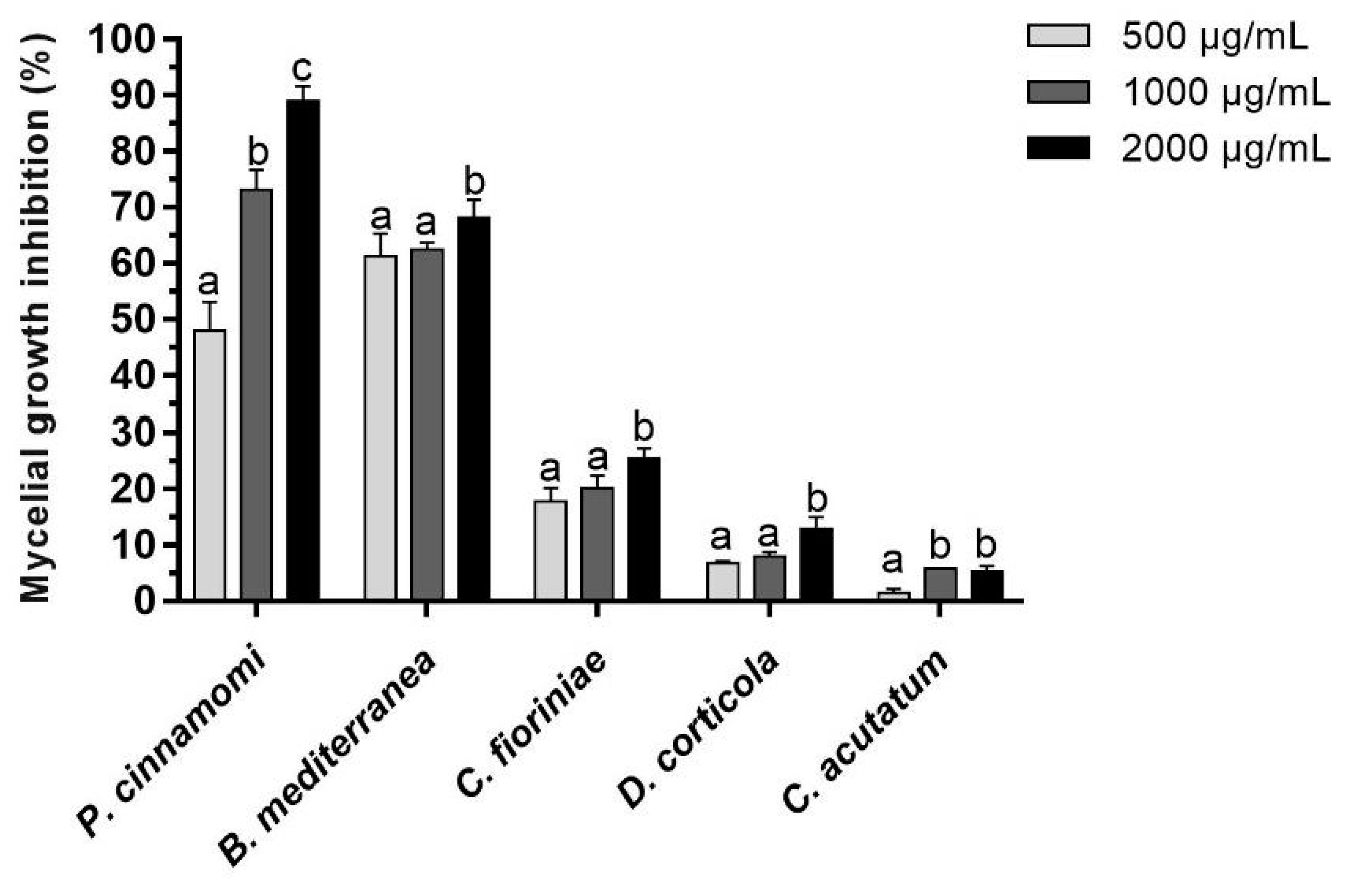
The percentage of mycelial growth inhibition was determined for all the species and concentrations tested, at the day mycelia covered the entire area of the control plate (Figure 2).
B. mediterranea and P. cinnamomi were clearly the most susceptible strains to A16.EE, and C. acutatum the least susceptible. At 500 μg/mL, B. mediterranea showed the highest growth inhibition (61.37%), followed by P. cinnamomi (48.04%). At 1000 and 2000 μg/mL, P. cinnamomi growth was inhibited by 73.14 and 89.02%, respectively, while B. mediterranea showed inhibitions similar to those observed with 500 μg/mL (62.55 and 68.24%, respectively), emphasizing the different behaviours of these two species regarding the dependence towards A16.EE concentration. In turn, C. acutatum showed inhibitions of mycelial growth below 6% for the three concentrations tested, while D. corticola and C. fioriniae registered inhibitions between 6.82 and 12.98% and between 17.84 and 25.49%, respectively. For P. cinnamomi, very significant differences were observed when comparing the three concentrations of A16.EE, in line with those already described for this species in the growth parameter analyses (Table 3).
Regarding the qualitative macroscopic analysis, no noticeable changes were detected in the characteristics of B. mediterranea, D. corticola and P. cinnamomi cultures grown in the presence of propolis (Figure 3). However, the central part of C. acutatum and C. fioriniae colonies showed a more intense brown colour in the presence of A16.EE (at all concentrations) than in its absence.

4. Discussion

4.1. A16 Shows Chemical Characteristics from Propolis Typical from the South and North of Portugal, Suggesting a Gradient Is More Likely than a Dichotomy

The ethanolic extraction yield of sample A16 fits within the range of yields obtained by our research group for Portuguese propolis samples being closer to the upper limit (61.6% to 96.4%) (from a collection of unpublished studies of our investigation group). Lower yield values for propolis samples from the north of Portugal—61.6% [34] and 62% [55]—were found. Similarly, the yield reported in this study is higher in comparison to the values of ethanol extracts from propolis samples of other countries, such as from China (51.03%) [56] and from Taiwanese green propolis (66.75%) [57]. The results of TPC and TFC characterization are in line with the values reported by Silva et al. [58] for an EE from Beja (south of Portugal), but are also comparable to those of other European countries such as Croatia [59]. A16.EE showed a lower TPC in comparison to propolis samples from the north and centre of Portugal (characterized by a dark orange colour and being hard and brittle), which ranged between 202.3 and 415.5 mg GAE/g extract [25,36,58,60], and from other European regions such as the Basque Country (200–340 mg GAE/g extract) [61,62]. Regarding TFC, A16.EE presents similar content to that of an EE from the interior centre of Portugal (30.21 mg QE/g of extract), although showing a lower TPC than this extract (160.4 mg GAE/g extract) [25]. Also, the presence of Cistus ladanifer L. around the apiary may suggest A16 fitting in the less common Portuguese propolis type—typical of the central interior and south of the country, elastic and gummy, with dark green colour such as A16 and less rich in phenolics—for which a TPC range between 54.8 and 189.1 mg GAE/g extract was reported [36,63]. However, TPC and TFC values of A16.EE far exceeded the minimum contents proposed by Falcão et al. [36] as quality standards of the less common Portuguese propolis (6 and 2%, respectively). Together, these results corroborate the idea that intermediate profiles can be found, especially in areas where some overlap/gradient in botanical species may exist. Indeed, a previous study with an extract from Caramulo (interior centre of Portugal) also observed a similar result, noting that a “diversity and some deviation from the reference values can occur even in the same region/country, probably due to the availability of botanical species for propolis production in a given local and time” [64]. TPC and TFC in A16.EE are lower than those reported for Caramulo extract (166.3 mg GAE/g extract and 69.3 mg QE/g extract, respectively) [64]. Unlike for a wide variety of plant extracts [65,66], just one study was found determining TOC in propolis [34]. However, we consider this analysis essential since the ortho-diphenols is the class of phenolic compounds with the highest antioxidant power [67,68,69], proposing the inclusion of this assay in the common set of tests performed with propolis samples. Freitas et al. [34] reported a lower value for a propolis extract from the propolis from the north of Portugal (263.05 ± 15.19 mg GAE/g extract) than the value found in our study.

4.2. In Vitro Activity of Propolis against Phytopathogenic Filamentous Species

Several authors have corroborated the promising potential of propolis extracts against phytopathogenic filamentous fungi/oomycetes [70,71,72,73,74]. However, due to lack of uniform methodologies for the evaluation and characterization of antimicrobial effects, a fair comparison of efficiencies is generally impeded. Loebler et al. [73] reported that a propolis hydroalcoholic (ethanol 70%) extract from the centre of Portugal, at 100 μg/mL, inhibited 30% of the Colletotrichum gloeosporioides mycelial growth after ten days of inoculation, while 500 and 1000 μg/mL induced inhibitions of about 70 and 80%, values much higher than those described here for species of the Colletotrichum genus. Moreover, 100 to 1000 μg/mL of this extract inhibited between 50 and 80% of Botrytis cinerea growth, respectively [73]. Pereira et al. [64] obtained inhibitions of Penicillium expansum, after four days of inoculation, of 36, 42 and 48% for 500, 1000 and 2000 μg/mL for a propolis hydroalcoholic extract (ethanol 70%) from the centre of Portugal, respectively. Yusuf et al. [70] reported that 3 to 10 μg/mL of a methanol extract of Turkish propolis, with an incubation period of four days, completely inhibited the growth of Phytophthora infestans, Phytophthora capsici and Phytophthora parasitica. The latter result, in line with ours, gives support to the hypothesis of oomycetes, at least of the Phytophthora genus, having a much higher susceptibility towards propolis than true filamentous fungi. García et al. [75] observed an approximately 35% mycelial growth inhibition of C. acutatum for a Colombian propolis ethanol extract at 500 and 1000 μg/mL after four days of inoculation. To compare our results obtained for C. acutatum (inhibitions of less than 6%) with those of García et al. [75], the percent inhibition of C. acutatum by A16.EE was calculated for the fourth day of growth, and similar results were obtained with 500 and 1000 μg/mL A16.EE—25.96 (±8.81) and 28.60 (±3.95) %, respectively.
The mechanisms of action of propolis against filamentous fungi have not yet been fully elucidated. Some of the bioactive compounds from propolis extracts seem to induce a reduction in the ergosterol biosynthesis, leading to a loss of cell membrane integrity [74,76]. It is also known that they may inhibit the enzyme 1,3-β-glucan synthase, with consequent interference in the synthesis of 1,3-β-glucan, and they demonstrate an ability to bind to chitin, affecting fungal cell wall integrity [74,76]. Yang et al. [77] reported a high inhibitory activity of an EE of Chinese propolis against Penicillium italicum, having identified pinocembrin, pinobanksin, chrysin and galangin as the compounds probably responsible for such activity. Indeed, pinocembrin isolated from a Chinese propolis sample exhibited strong activity against P. italicum, with Peng et al. [78] concluding that this compound inhibits cellular respiration, causing an imbalance in mitochondrial homeostasis with a consequent disruption in cell membranes and metabolic disorders in the pathogen. Also, Chudapongse et al. [79], in a study with the human pathogenic yeast Candida albicans, pointed out that galangin inhibits NADH oxidation and CCCP (carbonyl cyanide m-chlorophenyl hydrazone)-stimulated respiration in isolated mitochondria, thus suggesting a galangin interference with the electron transport chain of this fungi. A study with propolis from Brazil observed that the antifungal activity against C. albicans was due to induced apoptosis and the disruption of the expression of genes involved in crucial processes such as cell adhesion, biofilm formation and filamentous growth [80]. Prenyl caffeate isolated from French propolis extracts showed significant antifungal activity against Candida glabrata and C. albicans (MIC80 of 16 and 62 µg/mL, respectively); pinocembrin had moderate activity (MIC80 62–125 µg/mL), and pinobanksin-3-acetate, chrysin, and galangin exhibited weak activity (MIC80 ≥ 250 µg/mL) [81]. Agüero et al. [82] isolated and characterised bioactive compounds from methanolic extracts of propolis from Argentina, with 2’,4’-dihydroxy-30’-methoxychalcone and 2’,4’-dihydroxychalcone being the main antifungal compounds.
The colour alteration in C. acutatum and C. fioriniae colonies in the presence of the extract possibly reveals the occurrence of metabolic alterations [83]. Not excluding other causes, since information regarding this aspect is scarce, the brown colour of these colonies could be due to a class of dark/brown pigments called melanins, which are secondary metabolites produced by fungi via the oxidative polymerisation of phenolic compounds. It was reported that these pigments contribute to survival under environmental stress conditions [84,85]. Indeed, Colletotrichum species are known to produce DHN melanins [84,86,87,88].
In the case of sterol-auxotroph oomycete Phytophthora cinnamomi, compounds from propolis extracts may possess sterol-binding activity, possibly by acting as a competitor of sterol sensors or by sequestration sterols from the membrane, inhibiting the growth of this oomycete [89,90]. They may also attack 1,3-β-glucan, which are a major component of oomycete cell walls [91]. Miljanović et al. [92] observed a strong activity of an EE of propolis from Croatia against the pathogenic oomycetes Aphanomyces astaci and Saprolegnia parasitica, having predicted apigenin, chrysin and pinocembrin as the compounds responsible for this activity. With oomycetes being amongst the most important plant pathogens causing plant diseases worldwide [93], this particular susceptibility of oomycete species to propolis extracts and isolated compounds also constitutes a unique experimental tool, with potential high impact in plant disease control. In fact, P. cinnamomi susceptibility to A16.EE stood out, but as the only oomycete of the microbial panel, to know if this particular behaviour/susceptibility to propolis extract is characteristic of this class of filamentous microorganisms, further investigation will be needed. This is especially relevant since several plant extracts with antifungal activity exhibit less efficiency against Phytophtora cinnamoni than true filamentous fungi [94].

4.3. Gompertz-Derived Parameters Are Robust Descriptors and Exhibit High Discriminating Power

Permana et al. [48] and Camenzind et al. [49] applied a different parameterisation of the Gompertz model for modelling filamentous fungal growth. As far as we know, the parameterisation of the model proposed herein to describe the growth profiles of filamentous fungi/oomycetes cultures on solid media is a pioneering approach. Results clearly showed that it is a useful analysis tool, particularly in the context of toxicity assays, since it allowed a quantitative characterisation of mycelial radial growth and of the antifungal/anti-oomycetal effect of antimicrobial agents as the propolis extract. The three growth parameters analysed were concentration- and species-sensitive, revealing a high discriminating power, very useful both in screening and secondary assays.

5. Conclusions

We have demonstrated that the Gompertz equation accurately described the profile of mycelial growth of filamentous fungi/oomycetes colonies in solid media, and its estimated and derived parameters allow for a high discriminating power in antifungal/anti-oomycetal assays. Under the conditions of this study, the whole range of concentrations tested was able to reduce the in vitro growth of four economically important phytopathogenic fungi and an oomycete, with B. mediterranea and P. cinnamomi being the most susceptible to A16.EE. These findings support the potential role of A16.EE as a biopesticide in disease management programs, and thus its potential to reduce the use of synthetic fungicides, although future work would be required to validate the antifungal action of propolis in the field under natural environmental conditions. It is crucial to test the effective A16.EE concentrations in bioassays with the adequate explants (depending on the fungal strain and mode of infection) from susceptible plants species in order to identify the phytotoxicity threshold of A16.EE. After ex situ experiments under controlled conditions were carried out, field experiments could be more rationally designed to validate the effects in relevant crop species. Also, if particular extracts exhibited high phytotoxicity, good perspectives are still open for A16.EE as a source of bioherbicides. These studies are vital to promote propolis as a source of bioproducts, due to the introduction of numerous new legislatives provisions and guides to register newly formulated biopesticides.
The use of propolis In agronomic applications is still poorly explored. To our knowledge, this study is the first evidence for the activity of propolis against B. mediterranea, C. fioriniae, D. corticola and P. cinnamomi, contributing to its acknowledgement as a high-value natural product and to the socio-economic dynamisation of the Portuguese beekeeping sector.

Author Contributions

C.P.: Investigation; Methodology; Software and modelling analysis; Visualization; Formal analysis; Writing—original draft. C.A.-A.: Conceptualization; Data curation; Formal analysis; Funding acquisition; Project administration; Resources; Supervision; Validation; Writing—review and editing. A.C.: Conceptualization; Software and modelling analysis; Data curation; Formal analysis; Funding acquisition; Project administration; Resources; Supervision; Validation; Writing—review and editing. All authors have read and agreed to the published version of the manuscript.

Funding

This work was supported by national funds through FCT—Portuguese Foundation for Science and Technology—via CBMA research units under the project UIDB/04050/2020.

Institutional Review Board Statement

Not applicable.

Informed Consent Statement

Not applicable.

Data Availability Statement

Data are contained within the article.

Acknowledgments

The authors thank Paulo Silva for kindly providing the propolis samples used in this work.

Conflicts of Interest

The authors declare no conflict of interest.

References

  1. Food and Agriculture Organization of the United Nations. Increasing the Resilience of Agricultural Livelihoods. 2016. Available online: http://www.fao.org/3/a-i5615e.pdf (accessed on 6 April 2022).
  2. United Nations. World Population Prospects—Key Findings and Advance Tables. The 2017 Revision. Department of Economic and Social Affairs. 2017. Available online: https://reliefweb.int/sites/reliefweb.int/files/resources/WPP2017_KeyFindings.pdf (accessed on 20 January 2022).
  3. Santos, J.L. Intensificação sustentável: Um novo modelo tecnológico na agricultura. In Cultivar N.º 3—Cadernos de Análise e Prospetiva. Gabinete de Planeamento, Políticas e Administração Geral; Dimas, B., Diniz, E., Grancinho, M., Morais, A.F., Moura, A.R., Miguel, A.C., Lopes, C., Lobo, H., Gaspar, M., Loureiro, M., et al., Eds.; Gabinete de Planeamento, Políticas e Administração Geral: Lisbon, Portugal, 2016; pp. 13–21. [Google Scholar]
  4. Organisation for Economic Co-operation and Development, Food and Agriculture Organization of the United Nations. OECD-FAO Agricultural Outlook 2012–2021. 2012. Available online: https://www.oecd-ilibrary.org/agriculture-and-food/oecd-fao-agricultural-outlook-2012_agr_outlook-2012-en (accessed on 20 November 2022).
  5. Fang, Y.; Ramasamy, R.P. Current and Prospective Methods for Plant Disease Detection. Biosensors 2015, 5, 537–561. [Google Scholar] [CrossRef] [PubMed]
  6. Moore, D.; Robson, G.D.; Trinci, A.P.J. 21st Century Guidebook to Fungi, 2nd ed.; Cambridge University Press: Cambridge, UK, 2011. [Google Scholar]
  7. Ma, L.J.; Geiser, D.M.; Proctor, R.H.; Rooney, A.P.; O’Donnell, K.; Trail, F.; Gardiner, D.M.; Manners, J.M.; Kazan, K. Fusarium Pathogenomics. Annu. Rev. Microbiol. 2013, 67, 399–416. [Google Scholar] [CrossRef] [PubMed]
  8. Doehlemann, G.; Ökmen, B.; Zhu, W.; Sharon, A. Plant Pathogenic Fungi. Microbiol. Spectr. 2017, 5, FUNK-0023-2016. [Google Scholar] [CrossRef] [PubMed]
  9. Kumar, M.; Brar, A.; Yadav, M.; Chawade, A.; Vivekanand, V.; Pareek, N. Chitinases–Potential Candidates for Enhanced Plant Resistance towards Fungal Pathogens. Agriculture 2018, 8, 88. [Google Scholar] [CrossRef]
  10. Khonglah, D.; Kayang, H. Antagonism of indigenous fungal isolates against Botrytis cineria the causal of gray mold disease of tomato (Solanum lycopersicum L.). Int. J. Curr. Res. Life Sci. 2018, 7, 806–812. [Google Scholar]
  11. Zadeh, S.M. Pesticides. In More People, More Food, Worse Water? A Global Review of Water Pollution from Agriculture; Mateo-Sagasta, J., Zadeh, S.M., Turral, H., Eds.; Food and Agriculture Organization of the United Nations: Rome, Italy; International Water Management Institute on behalf of the Water Land and Ecosystems research program of the CGIAR: Colombo, Sri Lanka, 2018; pp. 77–91. [Google Scholar]
  12. Plessis, A.D. Freshwater Challenges of South Africa and its Upper Vaal River: Current State and Outlook, 1st ed.; Springer International Publishing: New York, NY, USA, 2017. [Google Scholar]
  13. Gaberell, L.; Hoinkes, C. Highly Hazardous Profits: How Syngenta Makes Billions by Selling Toxic Pesticides; Public Eye Lausanne: Lausanne, Switzerland, 2019. [Google Scholar]
  14. Darçin, E.S.; Darçin, M. Health effects of agricultural pesticides. Biomed. Res. 2017, S13–S17. [Google Scholar]
  15. Al-Ahmadi, M.S. Pesticides, Anthropogenic Activities, and the Health of Our Environment Safety. In Pesticides, Anthropogenic Activities and the Health of Our Environment; Larramendy, M.L., Ed.; IntechOpen: London, UK, 2019; pp. 73–88. [Google Scholar] [CrossRef]
  16. Czaja, K.; Góralczyk, K.; Struciński, P.; Hernik, A.; Korcz, W.; Minorczyk, M.; Łyczewska, M.; Ludwicki, J.K. Biopesticides—Towards increased consumer safety in the European Union. Pest Manag. Sci. 2015, 71, 3–6. [Google Scholar] [CrossRef]
  17. European Commission. Green Deal: Pioneering Proposals to Restore Europe’s Nature by 2050 and Halve Pesticide Use by 2030. 2022. Available online: https://ec.europa.eu/commission/presscorner/detail/en/ip_22_3746 (accessed on 29 June 2022).
  18. Bankova, V. Chemical diversity of propolis and the problem of standardization. J. Ethnopharmacol. 2005, 100, 114–117. [Google Scholar] [CrossRef]
  19. Bankova, V.; Popova, M.; Trusheva, B. New emerging fields of application of propolis. Maced. J. Chem. Chem. Eng. 2016, 35, 1–11. [Google Scholar] [CrossRef]
  20. Silva-Carvalho, R.; Baltazar, F.; Almeida-Aguiar, C. Propolis: A Complex Natural Product with a Plethora of Biological Activities That Can Be Explored for Drug Development. Evid. Based Complementary Altern. Med. 2015, 2015, 206439. [Google Scholar] [CrossRef]
  21. Huang, S.; Zhang, C.P.; Wang, K.; Li, G.Q.; Hu, F.L. Recent Advances in the Chemical Composition of Propolis. Molecules 2014, 19, 19610–19632. [Google Scholar] [CrossRef]
  22. Šturm, L.; Ulrih, N.P. Advances in the Propolis Chemical Composition between 2013 and 2018: A Review. eFood 2019, 1, 24–37. [Google Scholar] [CrossRef]
  23. Marcucci, M.C. Propolis: Chemical composition, biological properties and therapeutic activity. Apidologie 1995, 26, 83–99. [Google Scholar] [CrossRef]
  24. Burdock, G.A. Review of the biological properties and toxicity of bee propolis (propolis). Food Chem. Toxicol. 1998, 36, 347–363. [Google Scholar] [CrossRef] [PubMed]
  25. Cruz, M.; Antunes, P.; Paulo, L.; Ferreira, A.M.; Cunha, A.; Almeida-Aguiar, C.; Oliveira, R. Antioxidant and dual dose-dependent antigenotoxic and genotoxic properties of an ethanol extract of propolis. RSC Adv. 2016, 6, 49806–49816. [Google Scholar] [CrossRef]
  26. Graikou, K.; Popova, M.; Gortzi, O.; Bankova, V.; Chinou, I. Characterization and biological evaluation of selected Mediterranean propolis samples. Is it a new type? LWT—Food Sci. Technol. 2016, 65, 261–267. [Google Scholar] [CrossRef]
  27. Anjum, S.I.; Ullah, A.; Khan, K.A.; Attaullah, M.; Khan, H.; Ali, H.; Bashir, M.A.; Tahir, M.; Ansari, M.J.; Ghramh, H.A.; et al. Composition and functional properties of propolis (bee glue): A review. Saudi J. Biol. Sci. 2018, 26, 1695–1703. [Google Scholar] [CrossRef]
  28. Curifuta, M.; Vidal, J.; Sánchez-Venegas, J.; Contreras, A.; Salazar, L.A.; Alvear, M. The in vitro antifungal evaluation of a commercial extract of Chilean propolis against six fungi of agricultural importance. Cien. Investig. Agrar. 2012, 39, 347–359. [Google Scholar] [CrossRef]
  29. Cibanal, I.; Fernández, L.; Krepper, G.; Pellegrini, C.; Gallez, L. Avances en el desarrollo de un biofungicida: Caracterización físico-química y actividad antifúngica de propóleos. Agrocienc. Urug. 2019, 23, 99–108. [Google Scholar] [CrossRef]
  30. Bankova, V.S.; Castro, S.L.; Marcucci, M.C. Propolis: Recent advances in chemistry and plant origin. Apidologie 2000, 31, 3–15. [Google Scholar] [CrossRef]
  31. Fokt, H.; Pereira, A.; Ferreira, A.M.; Cunha, A.; Aguiar, C. How do bees prevent hive infections? The antimicrobial properties of propolis. Curr. Res. Technol. Educ. Top. Appl. Microbiol. Microb. Biotechnol. 2010, 1, 481–493. [Google Scholar]
  32. Silva-Carvalho, R.; Miranda-Gonçalves, V.; Ferreira, A.M.; Cardoso, S.M.; Sobral, A.J.F.N.; Almeida-Aguiar, C.; Baltazar, F. Antitumoural and antiangiogenic activity of Portuguese propolis in in vitro and in vivo models. J. Funct. Foods 2014, 11, 160–171. [Google Scholar] [CrossRef]
  33. Freitas, A.S.; Cunha, A.; Cardoso, S.M.; Oliveira, R.; Almeida-Aguiar, C. Constancy of the bioactivities of propolis samples collected on the same apiary over four years. Food Res. Int. 2019, 119, 622–633. [Google Scholar] [CrossRef] [PubMed]
  34. Freitas, A.S.; Cunha, A.; Parpot, P.; Cardoso, S.M.; Oliveira, R.; Almeida-Aguiar, C. Propolis efficacy: The Quest for Eco-Friendly Solvents. Molecules 2022, 27, 7531. [Google Scholar] [CrossRef] [PubMed]
  35. Falcão, S.I.; Vale, N.; Gomes, P.; Domingues, M.R.M.; Freire, C.; Cardoso, S.M.; Vilas-Boas, M. Phenolic Profiling of Portuguese Propolis by LC–MS Spectrometry: Uncommon Propolis Rich in Flavonoid Glycosides. Phytochem. Anal. 2012, 24, 309–318. [Google Scholar] [CrossRef]
  36. Falcão, S.I.; Freire, C.; Vilas-Boas, M. A Proposal for Physicochemical Standards and Antioxidant Activity of Portuguese Propolis. J. Am. Oil Chem. Soc. 2013, 90, 1729–1741. [Google Scholar] [CrossRef]
  37. Falcão, S.I.; Tomás, A.; Vale, N.; Gomes, P.; Freire, C.; Vilas-Boas, M. Phenolic quantification and botanical origin of Portuguese propolis. Ind. Crops Prod. 2013, 49, 805–812. [Google Scholar] [CrossRef]
  38. Casaca, J.D. Manual de Produção de Pólen e Própolis em Portugal, 1st ed.; Federação Nacional dos Apicultores de Portugal: Lisbon, Portugal, 2010. [Google Scholar]
  39. Vilas-Boas, M. Própolis: Um Negócio Por Explorar. Available online: http://esa.ipb.pt/blogs/noticiasesa/2015/propolis-um-negocio-por-explorar/ (accessed on 5 January 2022).
  40. Gouvinhas, I.; Martins-Lopes, P.; Carvalho, T.; Barros, A.; Gomes, S. Impact of Colletotrichum acutatum Pathogen on Olive Phenylpropanoid Metabolism. Agriculture 2019, 9, 173. [Google Scholar] [CrossRef]
  41. Loureiro, A.; Talhinhas, P.; Oliveira, H. Olive anthracnose is caused by different species of fungi, with distinct geographic distribution, virulence and preference for the cultivar [Special issue]. Rev. Ciências Agrárias 2018, 41, 102–109. [Google Scholar]
  42. Talhinhas, P.; Loureiro, A.; Oliveira, H. Olive anthracnose: A yield- and oil quality-degrading disease caused by several species of Colletotrichum that differ in virulence, host preference and geographical distribution. Mol. Plant Pathol. 2018, 19, 1797–1807. [Google Scholar] [CrossRef]
  43. Henriques, J.; Nóbrega, F.; Sousa, E.; Lima, A. Analysis of the genetic diversity and phylogenetic relationships of Biscogniauxia mediterranea isolates associated with cork oak. Phytoparasitica 2016, 44, 19–34. [Google Scholar] [CrossRef]
  44. Moricca, S.; Linaldeddu, B.T.; Ginetti, B.; Scanu, B.; Franceschini, A.; Ragazzi, A. Endemic and Emerging Pathogens Threatening Cork Oak Trees: Management Options for Conserving a Unique Forest Ecosystem. Plant Dis. 2016, 100, 2184–2193. [Google Scholar] [CrossRef] [PubMed]
  45. Santos, C.; Machado, H.; Correia, I.; Gomes, F.; Gomes-Laranjo, J.; Costa, R. Phenotyping Castanea hybrids for Phytophthora cinnamomi resistance. Plant Pathol. 2015, 64, 901–910. [Google Scholar] [CrossRef]
  46. Rivas, E.M.; Gil de Prado, E.; Wrent, P.; Silóniz, M.I.; Barreiro, P.; Correa, E.C.; Conejero, F.; Murciano, A.; Peinado, J.M. A simple mathematical model that describes the growth of the area and the number of total and viable cells in yeast colonies. Lett. Appl. Microbiol. 2014, 59, 594–603. [Google Scholar] [CrossRef] [PubMed]
  47. Tjørve, K.M.C.; Tjørve, E. The use of Gompertz models in growth analyses, and new Gompertz-model approach: An addition to the Unified-Richards family. PLoS ONE 2017, 12, e0178691. [Google Scholar] [CrossRef] [PubMed]
  48. Permana, A.W.; Sampers, I.; Meeren, P.V. Influence of virgin coconut oil on the inhibitory effect of emulsion-based edible coatings containing cinnamaldehyde against the growth of Colletotrichum gloeosporioides (Glomerella cingulata). Food Control 2021, 121, 107622. [Google Scholar] [CrossRef]
  49. Camenzind, T.; Weimershaus, P.; Lehmann, A.; Aguilar-Trigueros, C.; Rillig, M.C. Soil fungi invest into asexual sporulation under resource scarcity, but trait spaces of individual isolates are unique. Environ. Microbiol. 2022, 24, 2962–2978. [Google Scholar] [CrossRef]
  50. Savary, O.; Coton, M.; Jany, J.L.; Coroller, L.; Coton, E. Effect of abiotic factors and culture media on the growth of cheese-associated Nectriaceae species. Int. J. Food Microbiol. 2022, 364, 109509. [Google Scholar] [CrossRef]
  51. Singleton, V.L.; Orthofer, R.; Lamuela-Raventós, R.M. Analysis of Total Phenols and Other Oxidation Substrates and Antioxidants by Means of Folin-Ciocalteu Reagent. Meth. Enzymol. 1999, 299, 152–178. [Google Scholar]
  52. Woisky, R.G.; Salatino, A. Analysis of propolis: Some parameters and procedures for chemical quality control. J. Apic. Res. 1998, 37, 99–105. [Google Scholar] [CrossRef]
  53. Durán, R.M.; Padilla, R.M.; Martín, A.M.; Ursinos, F.R.; Mendoza, A. Biodegradación de los compuestos fenólicos presentes en el alpechín. Grasas Aceites 1991, 42, 71–276. [Google Scholar]
  54. Gallez, L.; Kiehr, M.; Fernández, L.; Delhey, R.; Stikar, D. Antifungal activity in vitro of propolis solutions from Argentina against two plant pathogenic fungi: Didymella bryoniae and Rhizotocnia solani. J. Apic. Res. 2014, 53, 438–440. [Google Scholar] [CrossRef]
  55. Caetano, A.R.; Oliveira, R.D.; Celeiro, S.P.; Freitas, A.S.; Cardoso, S.M.; Gonçalves, M.S.T.; Baltazar, F.; Almeida-Aguiar, C. Phenolic Compounds Contribution to Portuguese Propolis Anti-Melanoma Activity. Molecules 2023, 28, 3107. [Google Scholar] [CrossRef] [PubMed]
  56. Sun, C.; Wu, Z.; Wang, Z.; Zhang, H. Effect of Ethanol/Water Solvents on Phenolic Profiles and Antioxidant Properties of Beijing Propolis Extracts. Evid. Based Complement. Alternat. Med. 2015, 2015, 595393. [Google Scholar] [CrossRef] [PubMed]
  57. Chen, Y.W.; Ye, S.R.; Ting, C.; Yu, Y.H. Antibacterial activity of propolins from Taiwanese green propolis. J. Food Drug Anal. 2018, 26, 761–768. [Google Scholar] [CrossRef] [PubMed]
  58. Silva, J.C.; Rodrigues, S.; Feás, X.; Estevinho, L.M. Antimicrobial activity, phenolic profile and role in the inflammation of propolis. Food Chem. Toxicol. 2012, 50, 1790–1795. [Google Scholar] [CrossRef] [PubMed]
  59. Gajger, I.T.; Pavlović, I.; Bojić, M.; Kosalec, I.; Srečec, S.; Vlainić, T.; Vlainić, J. The Components Responsible for the Antimicrobial Activity of Propolis from Continental and Mediterranean Regions in Croatia. Czech J. Food Sci. 2017, 35, 376–385. [Google Scholar] [CrossRef]
  60. Moreira, L.; Dias, L.G.; Pereira, J.A.; Estevinho, L. Antioxidant properties, total phenols and pollen analysis of propolis samples from Portugal. Food Chem. Toxicol. 2008, 46, 3482–3485. [Google Scholar] [CrossRef]
  61. Kumazawa, S.; Hamasaka, T.; Nakayama, T. Antioxidant activity of propolis of various geographic origins. Food Chem. 2004, 84, 329–339. [Google Scholar] [CrossRef]
  62. Bonvehí, J.S.; Gutiérrez, A.L. Antioxidant Activity and Total Phenolics of Propolis from the Basque Country (Northeastern Spain). J. Am. Oil Chem. Soc. 2011, 88, 1387–1395. [Google Scholar] [CrossRef]
  63. Maia, S.I.C. Avaliação da Atividade Antioxidante de Extratos de Própolis e Determinação de Componentes com Relevância Farmacológica. Master’s Thesis, Master in Laboratory Medicine, Lisbon, Portugal, 2014. [Google Scholar]
  64. Pereira, L.; Cunha, A.; Almeida-Aguiar, C. Portuguese propolis from Caramulo as a biocontrol agent of the apple blue mold. Food Control 2022, 139, 109071. [Google Scholar] [CrossRef]
  65. Gonçalves, A.; Silva, E.; Brito, C.; Martins, S.; Pinto, L.; Dinis, L.T.; Luzio, A.; Martins-Gomes, C.; Fernandes-Silva, A.; Ribeiro, C.; et al. Olive tree physiology and chemical composition of fruits are modulated by different deficit irrigation strategies. J. Sci. Food Agric. 2019, 100, 682–694. [Google Scholar] [CrossRef] [PubMed]
  66. Carbas, B.; Machado, N.; Oppolzer, D.; Ferreira, L.; Queiroz, M.; Brites, C.; Rosa, E.A.S.; Barros, A.I. Nutrients, Antinutrients, Phenolic Composition, and Antioxidant Activity of Common Bean Cultivars and their Potential for Food Applications. Antioxidants 2020, 9, 186. [Google Scholar] [CrossRef] [PubMed]
  67. Cai, Y.Z.; Sun, M.; Xing, J.; Luo, Q.; Corke, H. Structure–radical scavenging activity relationships of phenolic compounds from traditional Chinese medicinal plants. Life Sci. 2006, 78, 2872–2888. [Google Scholar] [CrossRef] [PubMed]
  68. Soufi, O.; Romero, C.; Louaileche, H. Ortho-diphenol profile and antioxidant activity of Algerian black olive cultivars: Effect of dry salting process. Food Chem. 2014, 157, 504–510. [Google Scholar] [CrossRef]
  69. Araújo, S.; Matos, C.; Correia, E.; Antunes, M.C. Evaluation of phytochemicals content, antioxidant activity and mineral composition of selected edible flowers. Qual. Assur. Saf. Crops Foods 2019, 11, 471–478. [Google Scholar] [CrossRef]
  70. Yusuf, Y.; Durdane, Y.; Servet, A. Antifungal Activity of Turkish Propolis Against Phytophthora Species. Plant Pathol. J. 2005, 4, 58–60. [Google Scholar]
  71. Yang, S.; Peng, L.; Cheng, Y.; Chen, F.; Pan, S. Control of Citrus Green and Blue Molds by Chinese Propolis. Food Sci. Biotechnol. 2010, 19, 1303–1308. [Google Scholar] [CrossRef]
  72. Sánchez, C.; Duarte, P.; Vasilenko, P.; Santos, M.; Loebler, M.; Cruz, A.S.; Gonçalves, M. Potential application of Portuguese propolis to control blue mould disease in ‘Rocha’ pear. Acta Hortic. 2016, 1144, 359–364. [Google Scholar] [CrossRef]
  73. Loebler, M.; Sánchez, C.; Santos, M.; Vasilenko, P.; Duarte, P.; Cruz, A.; Gonçalves, M. Aplicação de extratos de própolis para conservação pós-colheita de morangos. Vida Rural 2018, 1837, 38–40. [Google Scholar]
  74. Loebler, M.; Sánchez, C.; Maurício, E.M.; Diogo, E.; Santos, M.; Vasilenko, P.; Cruz, A.S.; Mendes, B.; Gonçalves, M.; Duarte, M.P. Potential Application of Propolis Extracts to Control the Growth of Stemphylium vesicarium in “Rocha” Pear. Appl. Sci. 2020, 10, 1990. [Google Scholar] [CrossRef]
  75. García, L.R.P.; Galán, J.P.M.; Pajón, C.M.G.; González, J.H.G.; Restrepo, D.L.D. Caracterización Fisicoquímica y Actividad Antimicrobiana del Propóleos en el Municipio de La Unión (Antioquia, Colombia). Rev. Fac. Nac. Agron. Medellín 2010, 63, 5373–5383. [Google Scholar]
  76. Teodoro, G.R.; Ellepola, K.; Seneviratne, C.J.; Koga-Ito, C.Y. Potential Use of Phenolic Acids as Anti-Candida Agents: A Review. Front. Microbiol. 2015, 6, 1420. [Google Scholar] [CrossRef] [PubMed]
  77. Yang, S.Z.; Peng, L.T.; Su, X.J.; Chen, F.; Cheng, Y.J.; Fan, G.; Pan, S.Y. Bioassay-guided isolation and identification of antifungal components from propolis against Penicillium italicum. Food Chem. 2011, 127, 210–215. [Google Scholar] [CrossRef]
  78. Peng, L.; Yang, S.; Cheng, Y.J.; Chen, F.; Pan, S.; Fan, G. Antifungal Activity and Action Mode of Pinocembrin from Propolis against Penicillium italicum. Food Sci. Biotechnol. 2012, 21, 1533–1539. [Google Scholar] [CrossRef]
  79. Chudapongse, N.; Klahan, K.; Kamkhunthod, M.; Ratchawong, C.; Nantapong, N. Antifungal activity against Candida albicans and effect on mitochondrial NADH oxidation of galangin. Planta Med. 2010, 76, P415. [Google Scholar] [CrossRef]
  80. De Castro, P.A.; Bom, V.L.P.; Brown, N.A.; De Almeida, R.S.C.; Ramalho, L.N.Z.; Savoldi, M.; Goldman, M.H.S.; Berretta, A.A.; Goldman, G.H. Identification of the cell targets important for propolis-induced cell death in Candida albicans. Fungal Genet. Biol. 2013, 60, 74–86. [Google Scholar] [CrossRef]
  81. Boisard, S.; Le Ray, A.M.; Landreau, A.; Kempf, M.; Cassisa, V.; Flurin, C.; Richomme, P. Antifungal and antibacterial me-tabolites from a French poplar type propolis. Evid. Based Complementary Altern. Med. 2015, 2015, 319240. [Google Scholar] [CrossRef]
  82. Agüero, M.B.; Gonzalez, M.; Lima, B.; Svetaz, L.; Sánchez, M.; Zacchino, S.; Feresin, G.E.; Shmeda-Hirschmann, G.; Palermo, J.; Wunderlin, D.; et al. Argentinean propolis from Zuccagnia punctata Cav.(Caesalpinieae) exudates: Phytochemical characterization and antifungal activity. J. Agric. Food Chem. 2010, 58, 194–201. [Google Scholar] [CrossRef]
  83. López, J.M.; Jensen, H.J. Generic model of morphological changes in growing colonies of fungi. Phys. Rev. E 2002, 65, 021903. [Google Scholar] [CrossRef]
  84. Holmström-Ruddick, B.; Mortensen, K. In vitro formation and survival of appressoria of a mycoherbicide agent, Colletotrichum gloeosporioides f. sp. malvae and a benomyl-resistant strain, HP4.5RR. Mycol. Res. 1995, 99, 1103–1107. [Google Scholar]
  85. Suthar, M.; Dufossé, L.; Singh, S.K. The Enigmatic World of Fungal Melanin: A Comprehensive Review. J. Fungi 2023, 9, 891. [Google Scholar] [CrossRef]
  86. Yu, S.M.; Ramkumar, G.; Lee, Y.H. Light quality influences the virulence and physiological responses of Colletotrichum acutatum causing anthracnose in pepper plants. J. Appl. Microbiol. 2013, 115, 509–516. [Google Scholar] [CrossRef] [PubMed]
  87. Wang, X.; Lu, D.; Tian, C. Analysis of melanin biosynthesis in the plant pathogenic fungus Colletotrichum gloeosporioides. Fungal Biol. 2021, 125, 679–692. [Google Scholar] [CrossRef] [PubMed]
  88. Li, R.; Xu, W.; Zong, Y.; Wang, X.; Li, Y.; Bi, Y.; Prusky, D.B. Melanin synthesis gene Aapks contributes to appressorium formation, stress response, cell well integrity and virulence in Alternaria alternata. Postharvest Biol. Technol. 2023, 198, 112247. [Google Scholar] [CrossRef]
  89. Gamir, J.; Darwiche, R.; Van’t Hof, P.; Choudhary, V.; Stumpe, M.; Schneiter, R.; Mauch, F. The sterol-binding activity of PATHOGENESIS-RELATED PROTEIN 1 reveals the mode of action of an antimicrobial protein. Plant J. 2017, 89, 502–509. [Google Scholar] [CrossRef]
  90. Wang, W.; Liu, X.; Govers, F. The mysterious route of sterols in oomycetes. PLOS Pathog. 2021, 17, e1009591. [Google Scholar] [CrossRef]
  91. Wang, Y.; Tyler, B.M.; Wang, Y. Defense and Counterdefense During Plant-Pathogenic Oomycete Infection. Annu. Rev. Microbiol. 2019, 73, 667–696. [Google Scholar] [CrossRef]
  92. Miljanović, A.; Bhat, R.A.H.; Tandel, R.S.; Pavić, D.; Grbin, D.; Dent, M.; Marijanović, Z.; Jerković, I.; Pedisic, S.; Bielen, A. Bioactive compounds in fluid propolis preparations inhibit different life stages of pathogenic oomycetes Aphanomyces astaci and Saprolegnia parasitica. Aquaculture 2022, 552, 737982. [Google Scholar] [CrossRef]
  93. Robinson, S.M.; Bostock, R.M. β-glucans and eicosapolyenoic acids as MAMPs in plant–oomycete interactions: Past and present. Front. Plant Sci. 2015, 5, 797. [Google Scholar] [CrossRef]
  94. Castillo-Reyes, F.; Hernández-Castillo, F.D.; Clemente-Constantino, J.A.; Gallegos-Morales, G.; Rodríguez-Herrera, R.; Aguilar, C.N. In vitro antifungal activity of polyphenols-rich plant extracts against Phytophthora cinnamomi Rands. Afr. J. Agric. Res. 2015, 10, 4554–4560. [Google Scholar]
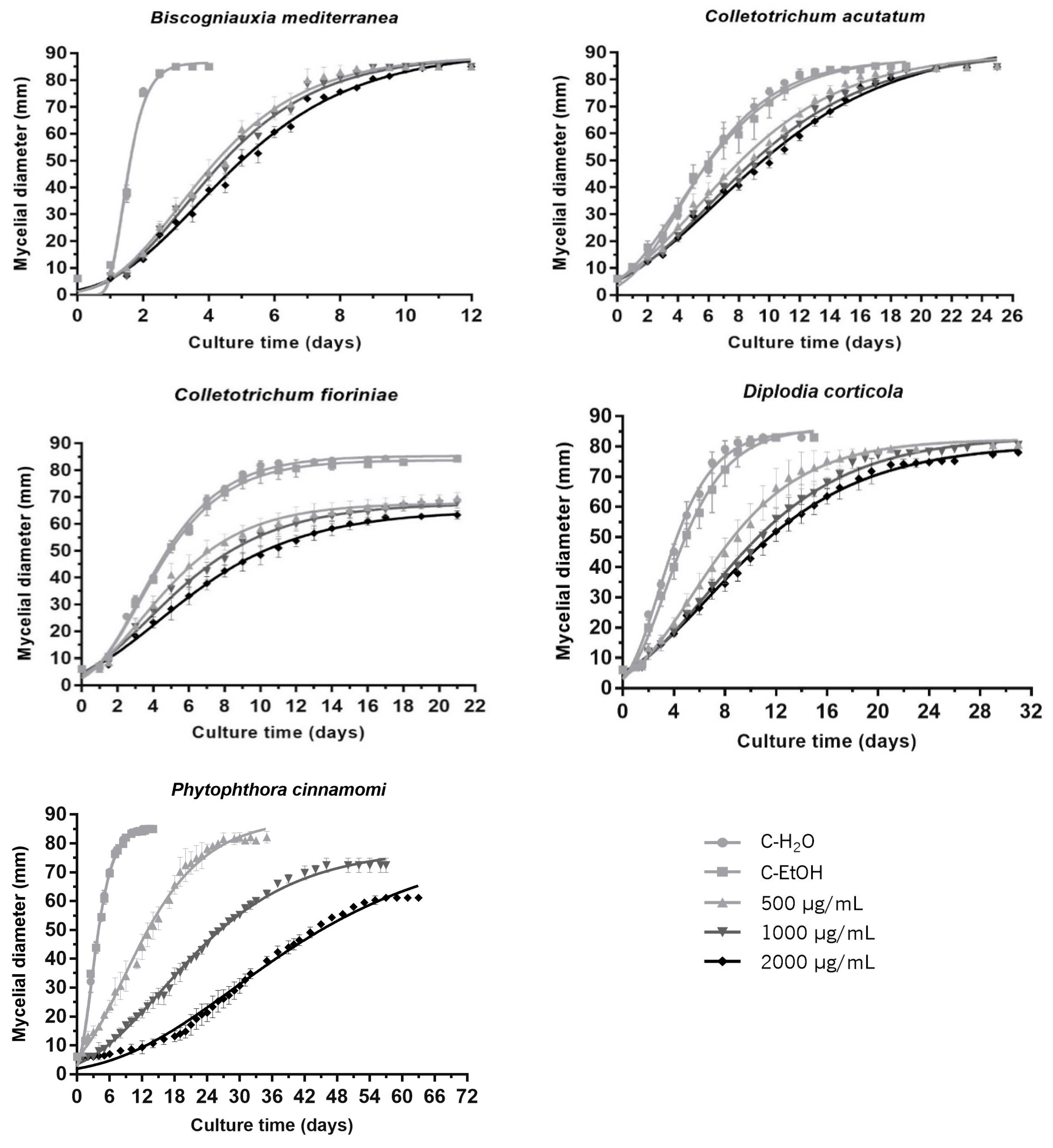
Figure 1. Growth curves estimated using the Gompertz mathematical model for Biscogniauxia mediterranea, Colletotrichum acutatum, Colletotrichum fioriniae, Diplodia corticola and Phytophthora cinnamomi cultures in PDA medium () and PDA supplemented with A16.EE reconstituted in absolute ethanol for final concentrations of 500 (), 1000 (▼) and 2000 μg/mL (◆). The control () was prepared with 50 µL of absolute ethanol (C-EtOH). Each curve represents the mean of three independent experiments, and error bars the respective SD.
Jof 09 01161 g001
Figure 2. Mycelial growth inhibition of Biscogniauxia mediterranea, Colletotrichum acutatum, Colletotrichum fioriniae, Diplodia corticola and Phytophthora cinnamomi in the presence of 500 (), 1000 () and 2000 (■) μg/mL of A16.EE, calculated in relation to growth in C-EtOH at the day that mycelia covered the plate’s entire area. Each bar represents the mean of three independent experiments, and error bars the respective SD. Statistical notation: for each species, mean values with the same letter were not significantly different (p > 0.05).
Jof 09 01161 g002
Figure 3. Macroscopic aspects of Biscogniauxia mediterranea, Colletotrichum acutatum, Colletotrichum fioriniae, Diplodia corticola and Phytophthora cinnamomi cultures at PDA medium supplemented with 500, 1000 and 2000 μg/mL A16.EE. Control (C-EtOH) was prepared with ethanol.
Jof 09 01161 g003
Table 1. Strains of filamentous fungi and the oomycete used to test antifungal/anti-oomycetal activities of the propolis extract. Strains were cultivated in PDA medium at 24 °C, in the dark, and periodically re-cultivated to new PDA medium-containing plates.
Filamentous Fungi/OomyceteReferenceEnvironmental IsolatesOrigin
Biscogniauxia mediterraneaBr41Quercus suber L.Teresa Lino Neto
(University of Minho)
Colletotrichum acutatumPT227Olea europaea LRui Oliveira
(University of Minho)
Colletotrichum fioriniaeunknownOlea europaea L.Paula Baptista
(Polytechnic Institute of Bragança)
Diplodia corticolaCAA008Quercus suber L.Rui Oliveira
(University of Minho)
Phytophthora cinnamomiPH107Castanea sativa Mill.Rui Oliveira
(University of Minho)
Table 2. Ethanolic extraction yield obtained from A16. Total phenolics content (TPC), total flavonoids content (TFC) and total ortho-diphenols content (TOC) of the A16.EE.
ExtractYield (%)TPC
(mg GAE/g Extract)
TFC
(mg QE/g Extract)
TOC
(mg GAE/g Extract)
A16.EE83.489.58 ± 3.4930.47 ± 1.01352.25 ± 3.43
Table 3. Growth parameters estimated by the Gompertz mathematical model (ti and Ymax) and calculated from Equation (3) (μ) after fitting growth data of Biscogniauxia mediterranea, Colletotrichum acutatum, Colletotrichum fioriniae, Diplodia corticola and Phytophthora cinnamomi cultures in PDA medium supplemented with 500, 1000 and 2000 μg/mL of A16.EE. Values represent the mean ± S.D. of three independent experiments. Statistical notation: for each species and parameter, mean values with common letters are not significantly different (p > 0.05).
B. mediterraneaµ (mm/day)ti (days)Ymax (mm)
C-H2O80.74 (±1.53) a1.37 (±0.03) a86.47 (±0.15) a
C-EtOH79.90 (±1.67) a1.36 (±0.03) a86.51 (±0.08) a
500 µg/mL15.01 (±0.92) b3.19 (±0.16) b85.42 (±1.91) b
1000 µg/mL14.51 (±0.35) b3.31 (±0.03) bc85.34 (±1.96) b
2000 µg/mL12.90 (±0.46) b3.61 (±0.20) c85.21 (±0.94) b
C. acutatumµ (mm/day)ti (days)Ymax (mm)
C-H2O9.84 (±0.19) a4.07 (±0.07) a86.10 (±0.17) a
C-EtOH9.05 (±1.45) a3.86 (±0.45) a86.48 (±1.23) a
500 µg/mL6.38 (±0.10) b5.35 (±0.55) b85.70 (±1.04) b
1000 µg/mL6.01 (±0.13) b6.02 (±0.28) bc85.51 (±1.05) b
2000 µg/mL5.50 (±0.24) b6.44 (±0.32) c85.33 (±0.66) b
C. fioriniaeµ (mm/day)ti (days)Ymax (mm)
C-H2O11.78 (±0.40) a3.11 (±0.09) a85.13 (±0.10) a
C-EtOH11.22 (±1.10) a3.14 (±0.09) a84.10 (±0.66) a
500 µg/mL7.54 (±0.89) b3.43 (±0.24) a68.77 (±2.28) b
1000 µg/mL6.30 (±0.65) bc3.75 (±0.33) ab68.14 (±1.93) b
2000 µg/mL5.24 (±0.61) c4.35 (±0.45) b65.09 (±0.71) c
D. corticolaµ (mm/day)ti (days)Ymax (mm)
C-H2O13.69 (±1.23) a2.78 (±0.12) a85.33 (±0.17) a
C-EtOH11.77 (±1.46) a3.23 (±0.30) a86.32 (±0.30) a
500 µg/mL6.45 (±0.94) b5.53 (±0.67) b81.10 (±1.67) b
1000 µg/mL5.06 (±0.51) b6.66 (±0.38) bc80.94 (±1.26) b
2000 µg/mL4.54 (±0.42) b6.83 (±0.57) c78.81 (±0.83) c
P. cinnamomiµ (mm/day)ti (days)Ymax (mm)
C-H2O14.47 (±0.32) a2.62 (±0.12) a86.68 (±0.31) a
C-EtOH14.15 (±0.64) a2.58 (±0.06) a86.03 (±0.57) a
500 µg/mL3.90 (±0.24) b8.54 (±1.14) ab90.23 (±1.41) a
1000 µg/mL2.22 (±0.13) c14.86 (±1.11) b78.91 (±2.72) b
2000 µg/mL1.55 (±0.12) c22.67 (±5.12) c68.68 (±2.34) c
Disclaimer/Publisher’s Note: The statements, opinions and data contained in all publications are solely those of the individual author(s) and contributor(s) and not of MDPI and/or the editor(s). MDPI and/or the editor(s) disclaim responsibility for any injury to people or property resulting from any ideas, methods, instructions or products referred to in the content.