Article

Sporothrix brasiliensis Infection Modulates Antimicrobial Peptides and Stress Management Gene Expression in the Invertebrate Biomodel Galleria mellonella

by
Nathália Faria Reis
1,
Myrela Conceição Santos de Jesus
1,
Lais Cavalcanti dos Santos Velasco de Souza
1,
Lucas Martins Alcântara
1,
Julia Andrade de Castro Rodrigues
1,
Simone Cristina Pereira Brito
1,
Patrícia de Azambuja Penna
2,
Cecília Stahl Vieira
2,
José Rodrigo Santos Silva
3,
Bruno de Araújo Penna
4,
Ricardo Luiz Dantas Machado
1,
Hector M. Mora-Montes
5 and
Andréa Regina de Souza Baptista
1,6,*
1
Center for Microorganisms’ Investigation, Department of Microbiology and Parasitology, Biomedical Institute, Federal Fluminense University, Niterói 24020-141, RJ, Brazil
2
Laboratory of Insect Biology, Federal Fluminense University, Niterói 24220-900, RJ, Brazil
3
Department of Statistics and Actuarial Sciences, Federal University of Sergipe, São Cristóvão 49107-230, SE, Brazil
4
Laboratory of Gram-Positive Cocci, Biomedical Institute, Fluminense Federal University, Niterói 24020-141, RJ, Brazil
5
Department of Biology, Division of Natural and Exact Sciences, Campus Guanajuato, University of Guanajuato, Guanajuato 36050, Mexico
6
Rede Micologia RJ—Fundação de Amparo à Pesquisa do Estado do Rio de Janeiro (FAPERJ), Rio de Janeiro 21941-630, RJ, Brazil
*
Author to whom correspondence should be addressed.
J. Fungi 2023, 9(11), 1053; https://doi.org/10.3390/jof9111053
Submission received: 30 August 2023 / Revised: 25 September 2023 / Accepted: 24 October 2023 / Published: 27 October 2023
(This article belongs to the Section Fungal Pathogenesis and Disease Control)

Abstract

:
Sporothrix brasiliensis is the most pathogenic species, responsible for the Brazilian cat-transmitted sporotrichosis hyperendemic. In this scenario, an investigation of the pathogen–host interaction can provide relevant information for future treatment strategies. To this end, the invertebrate Galleria mellonella has proven to be a suitable alternative for evaluating the virulence of pathogenic fungi, since the insect immune system is similar to the mammalian innate immune response. The aim of this work was to investigate phenotypic and molecular aspects of the immune response of G. mellonella throughout the S. brasiliensis infection. Hemocyte density and the evolution of the fungal load were evaluated. In parallel, RT-qPCR expression analysis of genes encoding antimicrobial peptides (Gallerimycin and Galiomycin) and stress management genes (C7 Contig 15362 and C8 Contig 19101) was conducted. The fungal load and hemocyte densities increased simultaneously and proportionally to the deleterious morphological events and larvae mortality. Gallerimycin, C7 Contig 15362 and C8 Contig 19101 genes were positively regulated (p < 0.05) at distinct moments of S. brasiliensis infection, characterizing a time-dependent and alternately modulated profile. Galiomycin gene expression remained unchanged. Our results contribute to the future proposal of potential alternative pathways for treating and consequently controlling S. brasiliensis zoonosis, a major public health issue in Latin America.

1. Introduction

Sporotrichosis is a disease of great importance to public health, since human infection can occur through direct contact with contaminated soil organic matter (sapronosis) or, more frequently, by the zoonotic route through the domestic feline [1,2]. In the latter scenario, the incriminated fungus is Sporothrix brasiliensis, the most virulent and pathogenic species of the Sporothrix genus, with strong evidence of drug resistance [3,4]. The S. brasiliensis-zoonotic-transmitted sporotrichosis began as an outbreak in the late 1990s in Rio de Janeiro, Brazil, and it is currently considered to be hyperendemic nationwide [5,6], with reports of recent geographical expansion to other Latin American countries [7,8]. Recently, the first three cases of domestic-feline-transmitted S. brasiliensis outside South America were described in the United Kingdom, Europe [9].
Itraconazole is the drug of choice for human and feline sporotrichosis [10,11]. However, cat treatment remains a challenge for the veterinarian, given the limited number of antifungal agents, their high cost and adverse effects, and their commercial presentations. Moreover, little is known about the immune response triggered by the infected hosts, especially the domestic feline [1,11].
In order to address this issue, the expansion of studies aiming to expand information related to the pathogen–host interaction is necessary. To this end, Galleria mellonella stands out as an invertebrate model for the investigation of the immune response triggered by distinct fungal pathogens, such as yeast of the Candida genus [12,13] and Cryptococcus neoformans [14]. Among filamentous fungi, Aspergillus niger, Rhizopus spp. and Rhizomucor spp. have also been challenged against the invertebrate biomodel’s defense [15,16,17], as have also the dimorphic Paracoccidioides lutzii and Histoplasma capsulatum [18,19]. However, few studies have been dedicated to the interaction between G. mellonella and fungi of the Sporothrix genus, all focusing on phenotypic aspects investigating the host–pathogen interaction [20,21,22] or in vivo drug response [23].
The G. mellonella immune system has close structural and functional similarities to the mammalian innate immune system [24,25], and it is divided into two strongly interconnected pathways: the cell-mediated pathway and the humoral pathway [26]. The cellular response of insects is mediated by hemocytes—the phagocytic cells in the hemolymph. These have the function of phagocytosing foreign bodies, capturing and encapsulating them in multicellular structures called nodules or capsules [26,27,28], playing roles analogous to those of human macrophages and neutrophils [24,25]. In parallel, the humoral response of insects includes antimicrobial peptides (AMPs), which act directly against invaders. Among the G. mellonella AMPs, gallerimycin and galiomycin are the most well-described in the literature [29], with evidence of positive regulation on the encoding genes’ expression in fungal infections with this insect, such as Candida spp. and Aspergillus niger [17,30,31].
The transcriptome analysis developed for G. mellonella provided important data related to immunity, notably expanding the knowledge about immune and stress-inducible genes in the invertebrate model. These results provided access to the genetic architecture of the insect’s immunity and elucidated the molecular mechanisms inherent in the immune response against potential pathogens. Hence, it became clear that both AMPs and some stress management genes encode key proteins involved in the insect’s response to pathogens [30,32]. Among the best characterized ones are those responsible for the regulation of cytokines (C7 Contig 15362), those linked to the phagocytosis and related to protein binding (C8 Contig 19101) and those involved in inflammatory responses (C3 Contig 15265, C4 Contig 290595, C5 Contig 21310, C6 Contig 1327) [30,32,33].
Data regarding the cellular and humoral insect defenses during the infectious process can provide new information on the host–pathogen interaction. Furthermore, elucidating the mechanisms of S. brasiliensis infection in the invertebrate model may contribute to the future detection of new therapeutic targets, as well as to the description of promising molecules for controlling this mycosis. Thus, the objective of the present study was to monitor G. mellonella’s phenotypic and genotypic responses against S. brasiliensis. To the best of our knowledge, the present study is the first to evaluate the defense-related gene expression of G. mellonella in response to infection with a dimorphic fungus.

2. Materials and Methods

2.1. Strains and Culture Conditions

The reference strain of Sporothrix brasiliensis used in this work was the American Type Culture Collection isolate, ATCC MYA 4823. The yeast cells were maintained under cryopreservation at −80 °C in the mycotheque of the Center for Microorganisms’ Investigation (CIM-UFF) until their reactivation for the experiments. For this purpose, the cryotubes were defrosted with subsequent replicas in a Yeast Extract Peptone Dextrose (YPD) medium, with incubation under shaking at 37 °C for 5 days. The microbial growth in the broth was then centrifuged at 2000× g for 5 min, washed with sterile Phosphate Buffered Saline (PBS; Fujifilm Irvine Scientific, Santa Ana, CA, USA) and suspended in the same buffer. After these processes, standardized suspensions of 1 × 10⁷ yeast/larva were prepared using a hemocytometer [20,21].

2.2. Galleria mellonella Survival Assays

The invertebrate biomodel investigated was kindly provided from a well-established colony by the Biofilms and Microbial Diversity Laboratory of the Federal University of Rio Grande do Sul. The survival curves were performed with larvae in the final larval stage of development, weighing between 0.2 g and 0.3 g, with uniform coloration, without spots or signs of melanization [34]. The inoculation of the fungus was subcutaneous, with 10 μL of the inoculum applied in PBS with a Hamilton syringe (701N, Caliber 26, Hamilton Company, Reno, NV, USA) in the last left proleg, as recommended [21,34]. The infected larvae were grouped by experimental condition, kept at 37 °C in 14 cm Petri dishes and monitored daily for survival. Lack of movement and extensive melanization of the body were both taken as indicators of animal death. As a control, two groups of animals were selected—one group with only the physical injury of the needle (naive) and the other injected with PBS—in order to assess mortality from animal handling and mechanical injury. Each group, including the control, consisted of 30 larvae. These experiments were conducted in triplicate.

2.3. Determination of Fungal Load during the Survival Curve

To monitor the fungal load during the different phases of infection, an experiment with ten (10) inoculated larvae was conducted with the same inoculum (1 × 10⁷ yeast/larva) and under the same temperature conditions, in triplicate, as detailed earlier in the survival curve. One larva was randomly selected from the pool daily, previously cleaned with ethanol 70%, and then, the hemolymph was collected. For this purpose, the larvae were sectioned with a sterile scalpel on the lower part, and 30 μL of hemolymph was collected from each individual. Then, the hemolymph was immediately placed in an Eppendorf® tube containing physiological saline buffer for insects in a 1:10 proportion (IPS: 150 mM sodium chloride, 5 mM potassium chloride, 10 mM Tris-HCl, pH 6.9, 10 mM EDTA and 30 mM sodium citrate) [35]. Soon after, serial dilutions were performed on Sabouraud agar plates plus Chloramphenicol (Sigma-Aldrich, St. Louis, MO, USA) incubated for seven days at 25 °C. After this period, one of the colonies was chosen for observation of confirmatory micromorphology of the genus Sporothrix using methyl blue staining. Once the genus was detected, all colony-forming units (CFUs) were counted as described by Clavijo-Giraldo and colleagues [21].

2.4. Determination of Hemocyte Density in Hemolymph

Similar to the methodology used for hemolymph collection described in the previous paragraph, hemocyte density was estimated from 30 μL of hemolymph diluted in IPS in a 1:10 proportion. The number of hemocytes was counted using a hemocytometer [36]. The experiment was performed in triplicate, and the results were expressed in hemocytes/mL. In parallel to the quantitative analysis, qualitative notes of phenomena such as nodule formation, as well as yeast adhered to the hemocytes, were made.

2.5. Analysis of Gene Expression

2.5.1. RNA Extraction and cDNA Synthesis

For the RNA extraction procedure, 20 control larvae with only the physical injury and 20 larvae inoculated with 1 × 10⁷ S. brasiliensis/larva were selected. The assays investigating the dynamics of gene expression were performed at three time points of the survival curve: on days one, five and eight (D1, D5 and D8) after infection with S. brasiliensis. For each incubation time (D), six (06) larvae from each group were randomly selected, cooled on dry ice and conditioned in microtubes for total RNA extraction from the tissue. For this purpose, the larvae were macerated using a sterile pestle, and the extraction was performed with SV Total RNA Isolation System kit (Promega Corporation, Madison, WI, USA), following the manufacturer’s recommendations. The total RNA was transcribed into complementary DNA (cDNA) using GoScript Reverse Transcription System Kit (Promega Corporation, Madison, WI, USA), according to the manufacturer’s instructions. In the cDNA synthesis, in addition to the reverse transcriptase, the DNase enzyme was also used to eliminate possible contaminating DNA in the sample. The cDNA was then quantified in the Quantus Fluorometer (Promega Corporation, Madison, WI, USA) using the QuantiFluor One dsDNA System kit (Promega Corporation, Madison, WI, USA). Finally, samples were diluted to the final use concentration of 2.5 ng/μL. All these experiments were performed in triplicate.

2.5.2. Reverse Transcription–Quantitative Polymerase Chain Reaction (RT-qPCR)

The quantification of gene expression was performed by RT-qPCR using the Power SYBR™ Green PCR Master Mix (Thermo Fisher Scientific, Carlsbad, CA, USA). The gene expression levels related to the production of Gallerimycin and Galiomycin AMPs, in addition to C7 Contig 15362 and C8 Contig 19101 genes, were evaluated at the three time points of infection. Gene β-actin was used as a normalizing reference control to monitor the amount of housekeeping gene not involved in the immune response, using the same cDNA preparations. The primer sequences used are specified in Table 1. The group of non-inoculated larvae was used as a control, which was compared with the group inoculated with the fungal pathogen. The samples and controls were tested in triplicate, and all reactions were performed in 96-well plates with a final volume of 15 μL, consisting of 7.5 μL of Power SYBR™ Green PCR Master Mix, 300 nM of each primer, 7.5 ng of cDNA and DEPEC water (InvitrogenTM, Carlsbad, CA, USA) of the total volume.
The reaction was performed in the 7500 Fast Real-Time PCR System (Applied Biosystems, Framingham, MA, USA) with the following conditions: initial heating at 50 °C for 2 min, denaturation at 95 °C for 2 min, followed by 40 cycles of denaturation at 95 °C for 15 s, annealing and extension at temperatures ranging from 55 to 60 °C for 1 min, depending on each primer (Table 1). In the end, a melting curve was performed to verify the specificity of the reaction: 95 °C for 15 s, 60° for 1 min, followed by 30 s at 95 °C. The means of the threshold cycle (cT) values, measured in triplicate, were used to calculate the expression of the target genes. The results were obtained as relative gene expression values (based on the formula: 2−ΔΔCT) compared to the reference gene expression—β-actin—resulting in a value equal to 1.

2.6. Statistical Analyses

The survival analyses of G. mellonella infected with fungal pathogens were estimated using the Kaplan–Meier survival curve, in which the log-rank test was used to compare the groups in terms of survival. The evolution of fungal load over days and the relationship between the hemocyte count and CFU were analyzed using simple linear regression models. Adherence to the normal distribution of the variables was established using the Shapiro–Wilk test. The Kruskal–Wallis test and Dunn’s post hoc test were used to check the dynamics of gene expression as a function of day for each experimental group. The Mann–Whitney test was used to compare the relative quantification of genes on days D1, D5 and D8 as a function of the non-inoculated and inoculated groups. All experiments (survival curves, including larvae—individual and groups—hemocyte counts, fungal loads) were performed in triplicate. The significance level adopted was 5%, and the software used was R, version 4.1.2.

3. Results

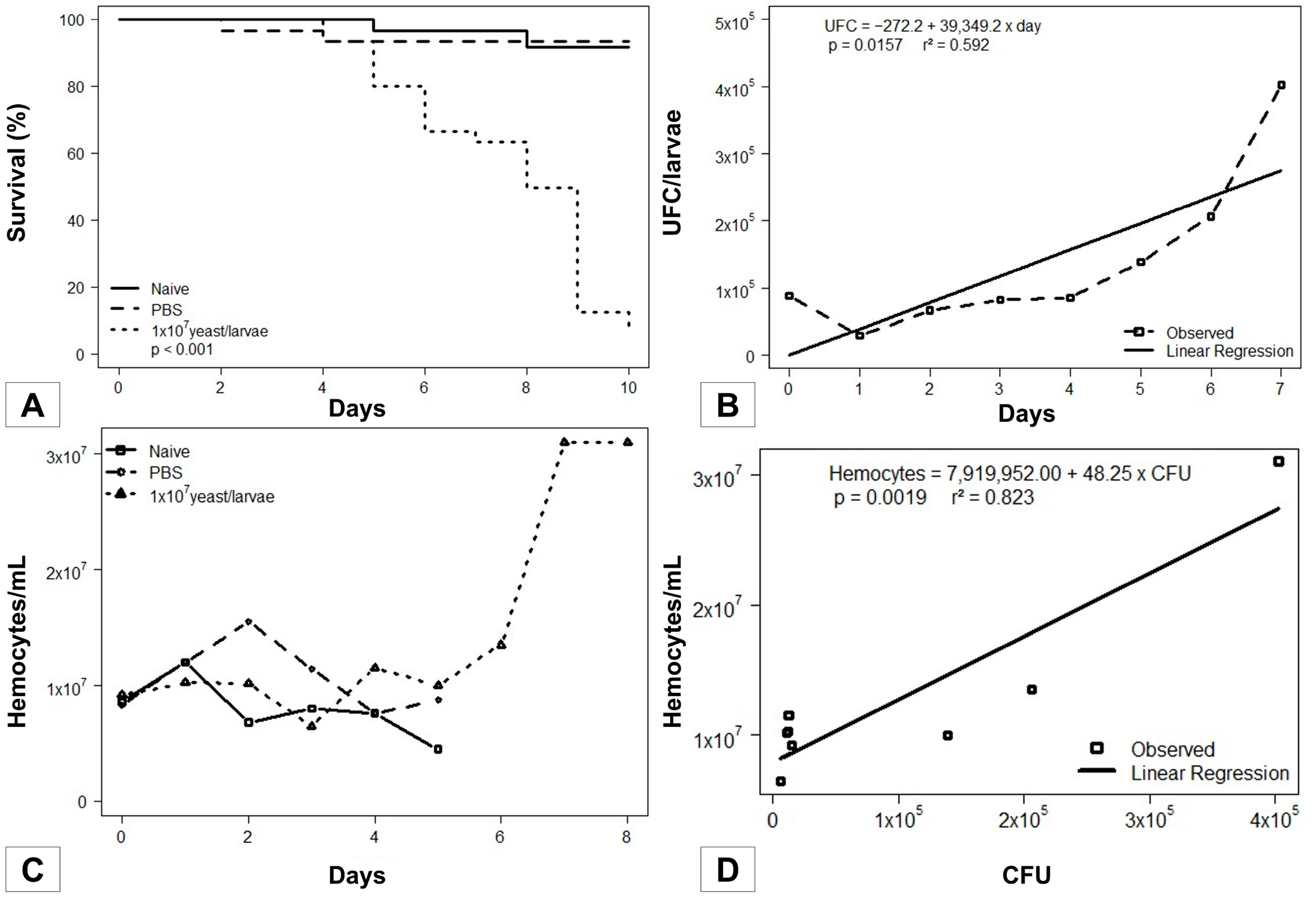
3.1. Galleria mellonella Survival Assays

The survival curve showed that larvae inoculated with 1 × 10⁷ yeast of S. brasiliensis started dying on day 4, while the majority of all naive larvae and those inoculated with PBS only remained unaltered (p < 0.001; Kaplan–Meier), reaching the pupal stage in the last days of the curve. The results are presented in Figure 1A.

3.2. Fungal Load during the Infection

Parallel to the survival curve, daily collection followed by hemolymph serial dilutions and subsequent plating were performed to obtain the CFUs. Colonies whose macromorphology was compatible with the genus Sporothrix were further evaluated using micromorphological analyses, evidencing thin, hyaline, septate, branched hyphae and conidia arranged in flower-like structures. Figure 1B shows the evolution of the fungal load along the survival curve, with increasing values from 6 × 10³ up to 4 × 10⁵ CFU/larva ( x ¯ = 1.01 × 10⁵ s.d. ± 1.4 × 10⁵). A direct correlation of the increase in S. brasiliensis yeasts over the days was observed (p = 0.0157).

3.3. Sporothrix brasiliensis–Galleria mellonella Interaction

Daily hemocyte counting allowed the detection of increasing insect cellular defense generated by the fungus, verified by values from 6.4 × 10⁶ up to 3.0 × 10⁷ hemocytes/mL along the curve ( x ¯ = 1.28 × 10⁷; s.d. ± 7.6 × 10⁶). The hemocyte count of the larvae from control groups (naive and PBS) was performed until D5, since afterward, both began to develop into pupae. In contrast, larvae infected with S. brasiliensis showed slower development in the applied experimental conditions (Figure 1C).
The hemocyte count associated with the determination of the fungal load along the survival curve allowed the evaluation of the Sporothrix brasiliensis–Galleria mellonella interaction dynamics. The progression of infection through the fungal pathogen triggered significant, proportional growth in insect hemocyte density (p = 0.0019; r2 = 0.823; Figure 1D).

3.4. Gene Expression

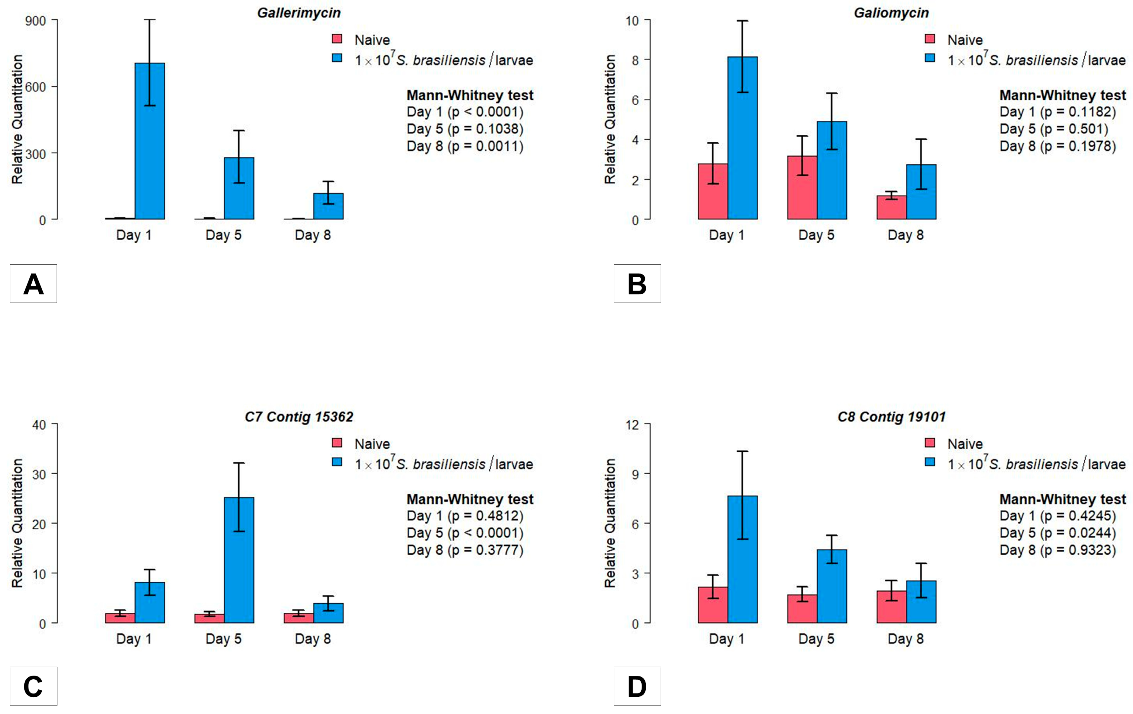
3.4.1. Antimicrobial Peptides

The gene encoding the AMP Gallerimycin showed higher early expression from the first day after infection (D1; Figure 2A) compared to naive larvae (p < 0.0001; Mann–Whitney test). The higher expression in S. brasiliensis infected larvae remained during the subsequent days D5 (p > 0.05) and D8 (p = 0.0011; Mann–Whitney test). In parallel, comparing the intragroup Gallerimycin gene expression, a significant reduction was observed in the infected larvae (p = 0.0055; Kruskal–Wallis test), contrary to the expression observed in the control group (p = 0.9968; Figure S1A; Supplementary Materials).
The expression of the gene encoding Galiomycin showed a trend similar to Gallerimycin, but the difference compared to the controls did not rise to a level of statistical significance among the two experimental groups, as well as following the intragroup analysis (p > 0.05; Mann–Whitney test; Figure 2B and Figure S1B, Supplementary Materials).

3.4.2. Stress Managing Genes

The C7 Contig 15362 mRNA expression analysis differs among the investigated groups at D5 post-inoculation (p < 0.0001; Mann–Whitney test). After this period, the expression decreases and exhibits its lowest values at D8 (Figure 2C). Analysis of the intragroup gene expression showed a significant difference among the three evaluated time periods (p = 0.0024; Kruskal–Wallis test; Figure S1C), with higher levels of mRNA production at D5. This difference was not observed in non-infected larvae among the evaluated days (p > 0.05; Supplementary Materials).
The expression of C8 Contig 19101 showed an initial, non-significant increase in mRNA expression from the first day following S. brasiliensis inoculation (D1; p > 0.05). However, at D5, an important increase in C8 Contig 19101 gene expression was detected compared to naive larvae (Figure 2D). In the intragroup comparison, C8 Contig 19101 mRNA expression did not vary significantly among D1 × D5 × D8 (p > 0.05; Kruskal–Wallis test; Figure S1D).

4. Discussion

Although sporotrichosis is a disease of great importance to public health, numerous aspects of the pathogen–host relationship are yet to be elucidated, mainly for the main host of this zoonosis: the domestic feline [1,39]. As a matter of fact, such investigation requires an appropriate experimental model. For decades, the murine model has been used as the in vivo gold standard model for pathogenicity mechanisms of distinct microorganisms [40], including Sporothrix spp. [41]. However, in recent times, the scientific community has been expressing ethical and social concerns towards the rationalization of animal model use [29,42,43].
Therefore, aiming at obtaining an alternative approach to mammalian models, several invertebrate models of infection have been studied, with emphasis on Galleria mellonella [44,45]. In spite of the previous use of this insect for the investigation of different fungal and bacterial infections [19,46], studies concerning the Sporothrix genus pathogenicity are still limited. Actually, only four previous studies investigated the S. brasiliensis–G. mellonella binomial, based on the description of phenotypic aspects [20,21,22,23]. Therefore, this is the first study to draw a parallel between G. mellonella innate-immunity-related gene expression and deleterious events during Sporothrix infection.
The results obtained in the survival curve of S. brasiliensis infection in G. mellonella standardization showed that the ideal yeast inoculum for observing mortality and infection dynamics was 1 × 10⁷ yeast/larva, since lower fungal loads (1 × 10⁵ and 1 × 10⁶ yeast/larva) did not allow such observations. Furthermore, the best temperature condition was 37 °C. These results were partially similar to those described by Clavijo-Giraldo and colleagues [21]. For more efficient killing with the fungal pathogen, these authors concluded that temperatures closer to the natural mammalian host are required to maintain all virulence attributes expressed by the yeast morphology. However, a less concentrated inoculum of 1 × 10⁵ yeast/larva was sufficient for these authors to obtain an appropriate survival curve. It is believed that this difference may be related mainly to the lineage of the larva used or even uncontrolled environmental conditions. On the other hand, Freitas and co-authors [20] used the same fungal load of 1 × 10⁷ yeast/larva, since when testing lower concentrations (1 × 10⁴ yeast/larva and 1 × 10⁶ yeast/larva), they did not observe mortality in the survival curve. In addition, the temperature of 37 °C was also recommended for the experiment, corroborating the results obtained here in both aspects.
Daily monitoring of S. brasiliensis load during the infection curve provided progressively increasing values, ranging from 1.2 × 10⁴ to 4 × 10⁵ CFU per animal. An average fungal load ranging from 1.8 × 10⁵ to 2.4 × 10⁵ CFU per animal was previously described [22] after 24 h of infection. Gandra and colleagues [31] observed that by inoculating 1 × 10⁷ Candida albicans/larva, the CFUs ranged from 1.0 × 10⁸ to 1.0 × 10¹² at 6, 24 and 48 h. These data reinforce the suitability of G. mellonella as a model of yeast infection, since it differentially mirrors the Candida and Sporothrix fungal load increases, such as those previously described for mammalian hosts [47,48].
It is noteworthy to mention the proportional increase in hemocytes in response to S. brasiliensis’s higher fungal loads, beginning on the fifth day of the curve. As a matter of fact, on the fifth day, the major phenotypic event registered was the beginning of larva death. Thus, it is possible to hypothesize that larva morbidity is a result of higher fungal loads as the immune system—although proportionally stimulated, reaching greater hemocyte recruitment—fails [26,49].
Lozoya-Pérez and collaborators [22] described a similar S. brasiliensisG. mellonella approach at a single specific point of the survival curve. The CFU and hemocyte counts from the insect’s hemolymph were obtained 24 h after yeast inoculation, previously grown in distinct culture media. CFU values of between 1.8 × 10⁵ and 2.4 × 10⁵ and hemocyte values of between 3.9 and 9.2 × 10⁶ hemocytes/mL were found, depending on the culture medium used. Such values were lower than those found in the present study (6.4 × 10⁶ to 3.0 × 10⁷ hemocytes/mL). This difference may be related to the 100x higher inoculum of S. brasiliensis yeast used in the present study protocol.
In spite of the increasing amount of literature involving the use of G. mellonella as an invertebrate biomodel, little is known about its defense strategies against S. brasiliensis. Even though the present study was able to establish a comparison with the previous work concerning the phenotypic events of the SporothrixG. mellonella interaction [21,22,50], we are limited to few previous publications describing the molecular aspects of the S. schenckii infection [50]. In fact, the gene expression data for such experimental scenario are limited to the genus Candida [31,44].
To the best of our knowledge, this is the first study investigating G. mellonella phenotypic and molecular events triggered through infection with the most virulent Sporothrix species, S. brasiliensis. In contrast, a considerable number of published works are available on the G. mellonella humoral response to the Candida species. These works quantify the expression of AMPs promoter genes with distinct purposes—among them, the description of the larval immune response profiles under different infection protocols and the evaluation of potential antimicrobial drugs. By respecting the differences inherent to each fungal species, these findings were used as parameters in order to discuss the findings of the present study.
Given the previous works highlighting the importance of the G. mellonella AMPs encoding genes Gallerimycin and Galiomycin, during both filamentous and yeast fungal infections [29,31], this study sought to quantify these gene expressions after infection with the dimorphic fungus Sporothrix brasiliensis. While Gallerimycin presented an expressive increase in gene expression along the survival curve, Galiomycin remained unaltered. These results suggest that the AMP Gallerimycin plays a relevant role in the insect’s response to S. brasiliensis infection, as previously described during Candida spp. infection [31,51]. Contrarily, Dekkerová-Chupáčová and co-authors [12] observed that the inoculation of 2 × 10⁵ yeast/larva of C. albicans and C. dubliniensis in G. mellonella triggered an expressive increase in AMP coding genes, especially Galiomycin.
The differential relevance of the G. mellonella Galiomycin gene expression during Candida albicans and dubliniensis versus S. brasiliensis infections may be explained by the fact that the first is a well-known microbiota yeast member, interacting with vertebrates over millions of years of evolution [52], while the Sporothrix species evolved from saprophytic mycelial fungi recently adapted to parasitism via thermodimorphism [53]. Likewise, differences in virulence and cell wall composition among these fungi could also be involved. From this perspective, Sporothrix species may be in the process of adapting gene expression in response to animal organic matter [2,53].
In addition to positive regulation, time-dependent regulation was observed, and Gallerimycin evidenced a significant increase on days D1 and D8. Such result partially corroborates that obtained by Dekkerová-Chupáčová and co-authors [12]. They noted that the maximum upregulation of both genes was shown at 24 h post-infection, but as early as 1 h into the infection, a positive regulation of expression was already observed. More specifically, the gene encoding Gallerimycin was up to 1.3-fold higher (1 h post-infection) and 3.2-fold higher (24 h post-infection) in larvae infected with C. albicans compared to C. dubliniensis. Meanwhile, for Galiomycin, we observed up to 3.7-fold (1 h post-infection) and 7.1-fold (24 h post-infection) relative upregulation in C. albicans-infected larvae. The upregulation of both genes decreased at 48 h post-infection with these two species.
In this regard, the difference in gene expression analysis intervals was based on the metabolic characteristics of the fungal species. Candida spp. demonstrated considerably faster growth both In Vitro and in vivo, in the invertebrate model itself, compared to Sporothrix spp. [13]. Moreover, given the scarcity of information regarding the gene expression of G. mellonella against Sporothrix spp., the choice of day (D) for gene expression analysis was defined according to the defense phenomena of the larvae observed in response to the fungus, such as melanization, mobility changes and the onset of mortality.
Concerning the results observed in the expression of stress management genes, C7 Contig 15362 and C8 Contig 19101 showed a significant difference in gene expression among the experimental groups on the fifth day (D5) of infection. Drawing a parallel with the study conducted by Melo and collaborators [30] evaluating G. mellonella gene expression after C. albicans infection versus different antifungal therapies at 24 and 48 h, it was also possible to observe a positive regulation of both genes in the infected, untreated larvae. A relevant point is that in this case, the C8 Contig 19101 gene was about four times overexpressed compared to the C7 Contig 15362 gene.
In the present study, it was possible to observe that the expression of the genes studied is not only time dependent but also alternately modulated, since, when comparing the genes responsible for encoding AMPs with those of stress management, this significant increase D-day varied. C7 Contig 15362 and C8 Contig 19101 are considered stress management genes and showed a significant increase in expression on day 5 post-infection. Interestingly, this moment coincided with the onset of mortality and deleterious phenotypic changes observed during the survival curve, as well as with the beginning of the expressive increase in defense cells and fungal load. In contrast, genes encoding AMPs, especially Gallerimycin, showed an immediate response verified by the intense mRNA encoding as early as the first post-infection day. This finding is compatible with the participation of the AMPs, previously characterized as an early element of humoral immunity against the infectious process [54,55]. For the same reason, different authors determine the evaluation protocols in hours, both in the investigation of the infectious process with Candida spp. [12,31,51] and with Aspergillus niger [17].
This work is the first to evaluate the gene expression related to the defense of the insect G. mellonella in response to infection with a dimorphic fungus. Most authors have dedicated themselves to the investigation of yeasts and, among these, of the genus Candida [12,30,31]. Among the filamentous fungi, previous investigations considered Aspergillus niger [17] and Fusarium oxysporum [56].
The possibility that filamentation occurred during the infectious process in G. mellonella cannot be excluded. Thus, the comparative discussion of some of the parameters evaluated here is limited. It is important to add the fact that the presence of hyphae of S. brasiliensis has been reported In Vitro after feline phagocyte exposure [57] and either during human or animal parasitism (manuscript in preparation). It is known that the fungal cell wall undergoes profound transformation, altering—during the dimorphic transition—β-1-3-glucans to α-glucans, posing an immediate challenge to the host immune response (PAMPs) [17,58]. However, only studies considering the distinct periods within the survival curve and also the evaluation of potential changes in fungal cell morphology will be able to answer whether the verified gene expression was exclusively yeast-triggered and/or yeast-targeted.
Another acknowledged limitation is the absence of data regarding gene expression and protein translation or its antimicrobial activity. However, because this study represents a pioneering investigation in the area, new perspectives and methodologies can be generated and improved in order to overcome the scarcity of data in the literature and elucidate the aspects of this relevant pathogen–host interaction. The present work opens new doors of investigation on the pathogenesis with Sporothrix spp., as well as offering a better characterized model for the research of potential antifungal drugs effective for the control of this important zoonosis.

Supplementary Materials

The following supporting information can be downloaded at: https://www.mdpi.com/article/10.3390/jof9111053/s1. Figure S1: Investigation of the gene expression profile of the invertebrate host Galleria mellonella in infection by the pathogenic fungus Sporothix brasiliensis. Dynamics of gene expression observed according to day, for each experimental group. (A) Gallerimycin, (B) Galliomycin and the stress manager genes (C) C7 Contig 15362 and (D) C8 Contig 19101. The units on the Y axis were calculated based on the 2−ΔΔCT method, and are expressed as mean. Each gene was normalized and compared to the expression of control (naive) insects using the β-actin reference gene. The Mann-Whitney test was used to compare the relative quantification of genes and a p ≤0.05 value was considered significant.

Author Contributions

Conceptualization, N.F.R., R.L.D.M., H.M.M.-M. and A.R.d.S.B.; Methodology, N.F.R., M.C.S.d.J., J.A.d.C.R. and L.C.d.S.V.d.S.; Software, J.R.S.S. and M.C.S.d.J.; Validation, N.F.R., L.M.A. and H.M.M.-M.; Formal Analysis, P.d.A.P., C.S.V., H.M.M.-M. and R.L.D.M.; Investigation, N.F.R., M.C.S.d.J., J.A.d.C.R. and S.C.P.B.; Resources, A.R.d.S.B., P.d.A.P. and R.L.D.M.; Data Curation, N.F.R., M.C.S.d.J. and A.R.d.S.B.; Writing—Original Draft Preparation, N.F.R., M.C.S.d.J. and A.R.d.S.B.; Writing—Review and Editing, A.R.d.S.B., B.d.A.P., P.d.A.P. and H.M.M.-M.; Visualization, H.M.M.-M.; Supervision, A.R.d.S.B., B.d.A.P., P.d.A.P. and C.S.V.; Project Administration, A.R.d.S.B. and R.L.D.M.; Funding Acquisition, A.R.d.S.B., P.d.A.P. and R.L.D.M. All authors have read and agreed to the published version of the manuscript.

Funding

This study was supported by grants from the Brazilian Agencies: Conselho Nacional de Desenvolvimento Científico e Tecnológico (CNPq/PECI: 200536/2022-2 and PIBIC-CNPq-UFF, Brazil); Fundação de Amparo à Pesquisa no Estado do Rio de Janeiro, Brazil (FAPERJ E-26/211.502/2021), Coordenação de Aperfeiçoamento de Pessoal de Nível Superior, Brazil (CAPES)—Financial Code 001, Fopesq/Proppi-UFF and Rede Micologia RJ. ARSB (308986/2022-9) and RLDM (302722/2022-0) are research fellows from the National Council for Scientific and Technological Development.

Institutional Review Board Statement

Not applicable.

Informed Consent Statement

Not applicable.

Data Availability Statement

The data presented in this study are available within the article.

Acknowledgments

The authors wish to express their gratitude to Alexandre Macedo of the Federal University of Rio Grande do Sul for donating Galleria mellonella eggs and to Laura Nunes for technical advice. The authors would also like to thank Centro Integrado de Tradução e Escrita (CITE/UFF) for assistance with English language translation and developmental editing.

Conflicts of Interest

The authors declare no conflict of interest.

References

  1. Gremião, I.D.F.; Miranda, L.H.M.; Reis, E.G.; Rodrigues, A.M.; Pereira, S.A. Zoonotic Epidemic of Sporotrichosis: Cat to Human Transmission. PLoS Pathog. 2017, 13, e1006077. [Google Scholar] [CrossRef] [PubMed]
  2. Rodrigues, A.M.; Della Terra, P.P.; Gremião, I.D.; Pereira, S.A.; Orofino-Costa, R.; de Camargo, Z.P. The Threat of Emerging and Re-Emerging Pathogenic Sporothrix Species. Mycopathologia 2020, 185, 813–842. [Google Scholar] [CrossRef] [PubMed]
  3. Della Terra, P.P.; Rodrigues, A.M.; Fernandes, G.F.; Nishikaku, A.S.; Burger, E.; de Camargo, Z.P. Exploring Virulence and Immunogenicity in the Emerging Pathogen Sporothrix brasiliensis. PLoS Negl. Trop. Dis. 2017, 11, e0005903. [Google Scholar] [CrossRef]
  4. Nakasu, C.C.T.; Waller, S.B.; Ripoll, M.K.; Ferreira, M.R.A.; Conceição, F.R.; Dos Reis Gomes, A.; da Gama Osório, L.; de Faria, R.O.; Cleff, M.B. Feline Sporotrichosis: A Case Series of Itraconazole-Resistant Sporothrix brasiliensis Infection. Braz. J. Microbiol. 2021, 52, 163–171. [Google Scholar] [CrossRef]
  5. Gremião, I.D.F.; Oliveira, M.M.E.; Monteiro de Miranda, L.H.; Saraiva Freitas, D.F.; Pereira, S.A. Geographic Expansion of Sporotrichosis, Brazil. Emerg. Infect. Dis. 2020, 26, 621–624. [Google Scholar] [CrossRef]
  6. Schechtman, R.C.; Falcão, E.M.M.; Carard, M.; García, M.S.C.; Mercado, D.S.; Hay, R.J. Sporotrichosis: Hyperendemic by Zoonotic Transmission, with Atypical Presentations, Hypersensitivity Reactions and Greater Severity. An. Bras. De Dermatol. 2022, 97, 1–13. [Google Scholar] [CrossRef] [PubMed]
  7. Etchecopaz, A.; Toscanini, M.A.; Gisbert, A.; Mas, J.; Scarpa, M.; Iovannitti, C.A.; Bendezú, K.; Nusblat, A.D.; Iachini, R.; Cuestas, M.L. Sporothrix brasiliensis: A Review of an Emerging South American Fungal Pathogen, Its Related Disease, Presentation and Spread in Argentina. J. Fungi 2021, 7, 170. [Google Scholar] [CrossRef] [PubMed]
  8. Thomson, P.; González, C.; Blank, O.; Ramírez, V.; del Río, C.; Santibáñez, S.; Pena, P. Sporotrichosis Outbreak Due to Sporothrix brasiliensis in Domestic Cats in Magallanes, Chile: A One-Health-Approach Study. J. Fungi 2023, 9, 226. [Google Scholar] [CrossRef]
  9. Barnacle, J.R.; Chow, Y.J.; Borman, A.M.; Wyllie, S.; Dominguez, V.; Russell, K.; Roberts, H.; Armstrong-James, D.; Whittington, A.M. The First Three Reported Cases of Sporothrix brasiliensis Cat-Transmitted Sporotrichosis Outside South America. Med. Mycol. Case Rep. 2023, 39, 14–17. [Google Scholar] [CrossRef]
  10. Orofino-Costa, R.; de Macedo, P.M.; Rodrigues, A.M.; Bernardes-Engemann, A.R. Sporotrichosis: An Update on Epidemiology, Etiopathogenesis, Laboratory and Clinical Therapeutics. An. Bras. Dermatol. 2017, 92, 606–620. [Google Scholar] [CrossRef]
  11. Gremião, I.D.F.; Martins da Silva da Rocha, E.; Montenegro, H.; Carneiro, A.J.B.; Xavier, M.O.; de Farias, M.R.; Monti, F.; Mansho, W.; de Macedo Assunção Pereira, R.H.; Pereira, S.A.; et al. Guideline for the Management of Feline Sporotrichosis Caused by Sporothrix brasiliensis and Literature Revision. Braz. J. Microbiol. 2021, 52, 107–124. [Google Scholar] [CrossRef] [PubMed]
  12. Dekkerová-Chupáčová, J.; Borghi, E.; Morace, G.; Bujdáková, H. Up-Regulation of Antimicrobial Peptides Gallerimycin and Galiomicin in Galleria mellonella Infected with Candida Yeasts Displaying Different Virulence Traits. Mycopathologia 2018, 183, 935–940. [Google Scholar] [CrossRef] [PubMed]
  13. García-Carnero, L.C.; Clavijo-Giraldo, D.M.; Gómez-Gaviria, M.; Lozoya-Pérez, N.E.; Tamez-Castrellón, A.K.; López-Ramírez, L.A.; Mora-Montes, H.M. Early Virulence Predictors during the Candida Species–Galleria mellonella Interaction. J. Fungi 2020, 6, 152. [Google Scholar] [CrossRef] [PubMed]
  14. Trevijano-Contador, N.; Herrero-Fernández, I.; García-Barbazán, I.; Scorzoni, L.; Rueda, C.; Rossi, S.A.; García-Rodas, R.; Zaragoza, O. Cryptococcus neoformans Induces Antimicrobial Responses and Behaves as a Facultative Intracellular Pathogen in the Non Mammalian Model Galleria mellonella. Virulence 2015, 6, 66–74. [Google Scholar] [CrossRef] [PubMed]
  15. Jacobsen, I. Animal Models to Study Mucormycosis. J. Fungi 2019, 5, 27. [Google Scholar] [CrossRef] [PubMed]
  16. Maurer, E.; Hörtnagl, C.; Lackner, M.; Grässle, D.; Naschberger, V.; Moser, P.; Segal, E.; Semis, M.; Lass-Flörl, C.; Binder, U. Galleria mellonella as a Model System to Study Virulence Potential of Mucormycetes and Evaluation of Antifungal Treatment. Med. Mycol. 2019, 57, 351–362. [Google Scholar] [CrossRef] [PubMed]
  17. Stączek, S.; Zdybicka-Barabas, A.; Wojda, I.; Wiater, A.; Mak, P.; Suder, P.; Skrzypiec, K.; Cytryńska, M. Fungal α-1,3-Glucan as a New Pathogen-Associated Molecular Pattern in the Insect Model Host Galleria mellonella. Molecules 2021, 26, 5097. [Google Scholar] [CrossRef]
  18. Thomaz, L.; García-Rodas, R.; Guimarães, A.J.; Taborda, C.P.; Zaragoza, O.; Nosanchuk, J.D. Galleria mellonella as a Model Host to Study Paracoccidioides lutzii and Histoplasma capsulatum. Virulence 2013, 4, 139–146. [Google Scholar] [CrossRef]
  19. Singulani, J.L.; Scorzoni, L.; de Oliveira, H.C.; Marcos, C.M.; Assato, P.A.; Fusco-Almeida, A.; Mendes-Giannini, M. Applications of Invertebrate Animal Models to Dimorphic Fungal Infections. J. Fungi 2018, 4, 118. [Google Scholar] [CrossRef]
  20. Freitas, D.F.; Santos, S.S.; Almeida-Paes, R.; de Oliveira, M.M.; do Valle, A.C.; Gutierrez-Galhardo, M.C.; Zancopé-Oliveira, R.M.; Nosanchuk, J.D. Increase in Virulence of Sporothrix brasiliensis over Five Years in a Patient with Chronic Disseminated Sporotrichosis. Virulence 2015, 6, 112–120. [Google Scholar] [CrossRef]
  21. Clavijo-Giraldo, D.M.; Matínez-Alvarez, J.A.; Lopes-Bezerra, L.M.; Ponce-Noyola, P.; Franco, B.; Almeida, R.S.; Mora-Montes, H.M. Analysis of Sporothrix schenckii sensu stricto and Sporothrix brasiliensis Virulence in Galleria mellonella. J. Microbiol. Methods 2016, 122, 73–77. [Google Scholar] [CrossRef] [PubMed]
  22. Lozoya-Pérez, N.E.; Casas-Flores, S.; Martínez-Álvarez, J.A.; López-Ramírez, L.A.; Lopes-Bezerra, L.M.; Franco, B.; Mora-Montes, H.M. Generation of Sporothrix schenckii Mutants Expressing the Green Fluorescent Protein Suitable for the Study of Host-Fungus Interactions. Fungal Biol. 2018, 122, 1023–1030. [Google Scholar] [CrossRef] [PubMed]
  23. Borba-Santos, L.P.; Barreto, T.L.; Vila, T.; Chi, K.D.; dos Santos Monti, F.; de Farias, M.R.; Alviano, D.S.; Alviano, C.S.; Futuro, D.O.; Ferreira, V.; et al. In Vitro and In Vivo Antifungal Activity of Buparvaquone against Sporothrix brasiliensis. Antimicrob. Agents Chemother. 2021, 65, e00699-21. [Google Scholar] [CrossRef]
  24. Browne, N.; Heelan, M.; Kavanagh, K. An Analysis of the Structural and Functional Similarities of Insect Hemocytes and Mammalian Phagocytes. Virulence 2013, 4, 597–603. [Google Scholar] [CrossRef] [PubMed]
  25. Sheehan, G.; Garvey, A.; Croke, M.; Kavanagh, K. Innate Humoral Immune Defences in Mammals and Insects: The Same, with Differences? Virulence 2018, 9, 1625–1639. [Google Scholar] [CrossRef] [PubMed]
  26. Wojda, I. Immunity of the Greater Wax Moth Galleria mellonella: Galleria mellonella Immunity. Insect Sci. 2017, 24, 342–357. [Google Scholar] [CrossRef] [PubMed]
  27. Ratcliffe, N.A.; Gagen, S.J. Studies on the in vivo Cellular Reactions of Insects: An Ultrastructural Analysis of Nodule Formation in Galleria mellonella. Tissue Cell 1977, 9, 73–85. [Google Scholar] [CrossRef] [PubMed]
  28. Lavine, M.D.; Strand, M.R. Insect Hemocytes and Their Role in Immunity. Insect Biochem. Mol. Biol. 2002, 32, 1295–1309. [Google Scholar] [CrossRef]
  29. Pereira, T.; de Barros, P.; Fugisaki, L.; Rossoni, R.; Ribeiro, F.; de Menezes, R.; Junqueira, J.; Scorzoni, L. Recent Advances in the Use of Galleria mellonella Model to Study Immune Responses against Human Pathogens. J. Fungi 2018, 4, 128. [Google Scholar] [CrossRef]
  30. de Melo, N.R.; Abdrahman, A.; Greig, C.; Mukherjee, K.; Thornton, C.; Ratcliffe, N.A.; Vilcinskas, A.; Butt, T.M. Myriocin Significantly Increases the Mortality of a Non-Mammalian Model Host during Candida Pathogenesis. PLoS ONE 2013, 8, e78905. [Google Scholar] [CrossRef]
  31. Gandra, R.M.; McCarron, P.; Viganor, L.; Fernandes, M.F.; Kavanagh, K.; McCann, M.; Branquinha, M.H.; Santos, A.L.S.; Howe, O.; Devereux, M. in vivo Activity of Copper(II), Manganese(II), and Silver(I) 1,10-Phenanthroline Chelates Against Candida Haemulonii Using the Galleria mellonella Model. Front. Microbiol. 2020, 11, 470. [Google Scholar] [CrossRef] [PubMed]
  32. Vogel, H.; Altincicek, B.; Glöckner, G.; Vilcinskas, A. A Comprehensive Transcriptome and Immune-Gene Repertoire of the Lepidopteran Model Host Galleria mellonella. BMC Genom. 2011, 12, 308. [Google Scholar] [CrossRef] [PubMed]
  33. Nale, J.Y.; Chutia, M.; Cheng, J.K.J.; Clokie, M.R.J. Refining the Galleria mellonella Model by Using Stress Marker Genes to Assess Clostridioides difficile Infection and Recuperation during Phage Therapy. Microorganisms 2020, 8, 1306. [Google Scholar] [CrossRef]
  34. Champion, O.; Titball, R.; Bates, S. Standardization of G. mellonella Larvae to Provide Reliable and Reproducible Results in the Study of Fungal Pathogens. J. Fungi 2018, 4, 108. [Google Scholar] [CrossRef]
  35. Senior, N.J.; Titball, R.W. Isolation and Primary Culture of Galleria mellonella Hemocytes for Infection Studies. F1000Research 2021, 9, 1392. [Google Scholar] [CrossRef]
  36. Scorzoni, L.; de Lucas, M.P.; Mesa-Arango, A.C.; Fusco-Almeida, A.M.; Lozano, E.; Cuenca-Estrella, M.; Mendes-Giannini, M.J.; Zaragoza, O. Antifungal Efficacy during Candida krusei Infection in Non-Conventional Models Correlates with the Yeast In Vitro Susceptibility Profile. PLoS ONE 2013, 8, e60047. [Google Scholar] [CrossRef]
  37. Brown, S.E.; Howard, A.; Kasprzak, A.B.; Gordon, K.H.; East, P.D. A Peptidomics Study Reveals the Impressive Antimicrobial Peptide Arsenal of the Wax Moth Galleria mellonella. Insect Biochem. Mol. Biol. 2009, 39, 792–800. [Google Scholar] [CrossRef]
  38. Zhao, H.-X.; Xiao, W.-Y.; Ji, C.-H.; Ren, Q.; Xia, X.-S.; Zhang, X.-F.; Huang, W.-Z. Candidate Chemosensory Genes Identified from the Greater Wax Moth, Galleria mellonella, through a Transcriptomic Analysis. Sci. Rep. 2019, 9, 10032. [Google Scholar] [CrossRef]
  39. Miranda, L.H.M.; Santiago, M.d.A.; Schubach, T.M.P.; Morgado, F.N.; Pereira, S.A.; Oliveira, R.d.V.C.d.; Conceição-Silva, F. Severe Feline Sporotrichosis Associated with an Increased Population of CD8 low Cells and a Decrease in CD4 + Cells. Med. Mycol. 2015, 54, 29–39. [Google Scholar] [CrossRef]
  40. Legrand, N.; Weijer, K.; Spits, H. Experimental Models to Study Development and Function of the Human Immune System In Vivo. J. Immunol. 2006, 176, 2053–2058. [Google Scholar] [CrossRef]
  41. Arrillaga-Moncrieff, I.; Capilla, J.; Mayayo, E.; Marimon, R.; Marine, M.; Genis, J.; Cano, J.; Guarro, J. Different Virulence Levels of the Species of Sporothrix in a Murine Model. Clin. Microbiol. Infect. 2009, 15, 651–655. [Google Scholar] [CrossRef] [PubMed]
  42. Borman, A.M. Of Mice and Men and Larvae: Galleria mellonella to Model the Early Host-Pathogen Interactions after Fungal Infection. Virulence 2018, 9, 9–12. [Google Scholar] [CrossRef] [PubMed]
  43. Gómez-Gaviria, M.; Martínez-Álvarez, J.A.; Mora-Montes, H.M. Current Progress in Sporothrix brasiliensis Basic Aspects. J. Fungi 2023, 9, 533. [Google Scholar] [CrossRef] [PubMed]
  44. Piatek, M.; Sheehan, G.; Kavanagh, K. Utilising Galleria mellonella Larvae for Studying in vivo Activity of Conventional and Novel Antimicrobial Agents. Pathog. Dis. 2020, 78, ftaa059. [Google Scholar] [CrossRef]
  45. Dinh, H.; Semenec, L.; Kumar, S.S.; Short, F.L.; Cain, A.K. Microbiology’s next Top Model: Galleria in the Molecular Age. Pathog. Dis. 2021, ftab006. [Google Scholar] [CrossRef]
  46. Binder, U.; Maurer, E.; Lass-Flörl, C. Galleria mellonella: An Invertebrate Model to Study Pathogenicity in Correctly Defined Fungal Species. Fungal Biol. 2016, 120, 288–295. [Google Scholar] [CrossRef]
  47. Fakhim, H.; Vaezi, A.; Dannaoui, E.; Chowdhary, A.; Nasiry, D.; Faeli, L.; Meis, J.F.; Badali, H. Comparative Virulence of Candida Auris with Candida haemulonii, Candida glabrata and Candida albicans in a Murine Model. Mycoses 2018, 61, 377–382. [Google Scholar] [CrossRef]
  48. Corrêa-Moreira, D.; Menezes, R.C.; Romeo, O.; Borba, C.M.; Oliveira, M.M.E. Clinical and Anatomopathological Evaluation of BALB/c Murine Models Infected with Isolates of Seven Pathogenic Sporothrix Species. Pathogens 2021, 10, 1647. [Google Scholar] [CrossRef]
  49. Trevijano-Contador, N.; Zaragoza, O. Immune Response of Galleria mellonella against Human Fungal Pathogens. J. Fungi 2018, 5, 3. [Google Scholar] [CrossRef]
  50. Tamez-Castrellón, A.K.; van der Beek, S.L.; López-Ramírez, L.A.; Martínez-Duncker, I.; Lozoya-Pérez, N.E.; van Sorge, N.M.; Mora-Montes, H.M. Disruption of Protein Rhamnosylation Affects the Sporothrix schenckii-Host Interaction. Cell Surf. 2021, 7, 100058. [Google Scholar] [CrossRef]
  51. Rossoni, R.D.; Fuchs, B.B.; de Barros, P.P.; Velloso, M.d.S.; Jorge, A.O.C.; Junqueira, J.C.; Mylonakis, E. Lactobacillus Paracasei Modulates the Immune System of Galleria mellonella and Protects against Candida albicans Infection. PLoS ONE 2017, 12, e0173332. [Google Scholar] [CrossRef] [PubMed]
  52. Mancera, E.; Nocedal, I.; Hammel, S.; Gulati, M.; Mitchell, K.F.; Andes, D.R.; Nobile, C.J.; Butler, G.; Johnson, A.D. Evolution of the complex transcription network controlling biofilm formation in Candida species. eLife 2021, 10, e64682. [Google Scholar] [CrossRef] [PubMed]
  53. de Beer, Z.W.; Duong, T.A.; Wingfield, M.J. The Divorce of Sporothrix and Ophiostoma: Solution to a Problematic Relationship. Stud. Mycol. 2016, 83, 165–191. [Google Scholar] [CrossRef]
  54. Meister, M.; Lemaitre, B.; Hoffmann, J.A. Antimicrobial Peptide Defense in Drosophila. Bioessays 1997, 19, 1019–1026. [Google Scholar] [CrossRef]
  55. Bulet, P.; Hetru, C.; Dimarcq, J.-L.; Hoffmann, D. Antimicrobial Peptides in Insects; Structure and Function. Dev. Comp. Immunol. 1999, 23, 329–344. [Google Scholar] [CrossRef]
  56. Lee, Y.S.; Yun, E.K.; Jang, W.S.; Kim, I.; Lee, J.H.; Park, S.Y.; Ryu, K.S.; Seo, S.J.; Kim, C.H.; Lee, I.H. Purification, CDNA Cloning and Expression of an Insect Defensin from the Great Wax Moth, Galleria mellonella. Insect Mol. Biol. 2004, 13, 65–72. [Google Scholar] [CrossRef]
  57. Mothé, G.B. Estudo Da Interação Entre Fagócitos Do Felis Catus (LINNAEUS,1758) e os Principais Agentes Etiológicos Da Esporotricose. Ph.D. thesis, Universidade Federal Fluminense, Rio de Janeiro, Brasil, 2021. Available online: https://app.uff.br/riuff/handle/1/24482 (accessed on 29 August 2023).
  58. Złotko, K.; Wiater, A.; Waśko, A.; Pleszczyńska, M.; Paduch, R.; Jaroszuk-Ściseł, J.; Bieganowski, A. A Report on Fungal (1→3)-α-d-Glucans: Properties, Functions and Application. Molecules 2019, 24, 3972. [Google Scholar] [CrossRef]
Figure 1. Phenotypic investigation of the infection dynamics in the invertebrate host Galleria mellonella against the fungal pathogen Sporothix brasiliensis. (A) Survival curve performed up to the tenth day of infection. (B) Monitoring of the fungal load from the insect’s hemolymph during the infection process. (C) Hemocyte count performed among insects in the control groups and the group infected with S. brasiliensis. (D) Parallel drawn between insect cellular immune response and fungal load along the survival curve.
Jof 09 01053 g001
Figure 2. Investigation of the gene expression profile of the invertebrate host Galleria mellonella following infection with the pathogenic fungus Sporothix brasiliensis. Relative quantification of genes encoding antimicrobial peptides (A) Gallerimycin, (B) Galliomycin. Additionally, the stress manager genes (C) C7 Contig 15362 and (D) C8 Contig 19101. The units on the Y-axis were calculated based on the 2−ΔΔCT method and are expressed as mean and standard error of the mean (s.e.m.). Each gene was normalized and compared to the expression of control (naive) insects using the β-actin reference gene. The Mann–Whitney test was used to compare the relative quantification of genes, and a p ≤ 0.05 value was considered significant.
Jof 09 01053 g002
Table 1. Genes quantified during the fungal infection process of Galleria mellonella with Sporothrix brasiliensis. The references used, as well as the primer sequences.
GeneNCBI Genbank
References
Sequence
(5′–3′)
Annealing Temperature
GaliomycinAY528421.1F-TCCAGTCCGTTTTGTTGTTG60 °C
[37]R-CAGAGGTGTAATTCGTCGCA
GallerimycinAF453824.1F-GAAGATCGCTTTCATAGTCGC60 °C
[37]R-TACTCCTGCAGTTAGCAATGC
C7 Contig 15362Contig 15362F-CGAGCTAAAGACAGGCGATT58 °C
[30]R-TCACCTGCGGTTGAATCATA
C8 Contig 19101Contig 19101F-ATTGCTAGCCAGGTTCAGGA60 °C
[30]R-AGCTATTTGGCGGAAACTCA
β-actin[38]F-GGACTTGTACGCCAACACAG
R-CCACATCTGCTGGAATGTCG
55 °C
Disclaimer/Publisher’s Note: The statements, opinions and data contained in all publications are solely those of the individual author(s) and contributor(s) and not of MDPI and/or the editor(s). MDPI and/or the editor(s) disclaim responsibility for any injury to people or property resulting from any ideas, methods, instructions or products referred to in the content.

