Article

MoMaf1 Mediates Vegetative Growth, Conidiogenesis, and Pathogenicity in the Rice Blast Fungus Magnaporthe oryzae

College of Biology and Food Engineering, Changshu Institute of Technology, Changshu 215500, China
*
Author to whom correspondence should be addressed.
J. Fungi 2023, 9(1), 106; https://doi.org/10.3390/jof9010106
Submission received: 7 December 2022 / Revised: 3 January 2023 / Accepted: 10 January 2023 / Published: 12 January 2023

Abstract

:
In eukaryotes, Maf1 is an essential and specific negative regulator of RNA polymerase (Pol) III. Pol III, which synthesizes 5S RNA and transfer RNAs (tRNAs), is suppressed by Maf1 under the conditions of nutrient starvation or environmental stress. Here, we identified M. oryzae MoMaf1, a homolog of ScMaf1 in budding yeast. A heterogeneous complementation assay revealed that MoMaf1 restored growth defects in the ΔScmaf1 mutant under SDS stress. Destruction of MoMAF1 elevated 5S rRNA content and increased sensitivity to cell wall agents. Moreover, the ΔMomaf1 mutant exhibited reduced vegetative growth, conidiogenesis, and pathogenicity. Interestingly, we found that MoMaf1 underwent nuclear-cytoplasmic shuffling, through which MoMaf1 accumulated in nuclei under nutrient deficiency or upon the interaction of M. oryzae with rice. Therefore, this study can help to elucidate the pathogenic molecular mechanism of M. oryzae.

1. Introduction

Protein synthesis requires coding and noncoding RNAs, and the synthesis process is regulated by multiple growth conditions. The transcription of protein synthesis involves regulation of all three RNA polymerases: ribosomal RNAs are mostly transcribed by polymerase I (Pol I), ribosomal protein-coding mRNAs are transcribed by Pol II, and 5S rRNA and tRNAs are transcribed by Pol III [1,2].
In eukaryotes, dividing cells always contain more tRNAs and 5S rRNAs than resting cells, but the presence of too many tRNAs often has adverse effects in cells [3]. For example, excess tRNAs in mammalian cells can increase the risk of carcinogenesis [3,4]. Pol III repressor, Maf1, is required for the attenuation of the Pol III system in unfavorable growth conditions: nutrient starvation, oxidative stress, and cell wall stress [5].
In yeast, Maf1 is a phosphoprotein, and under favorable conditions, Maf1 is phosphorylated and mostly cytoplasmic, allowing Pol III transcription [6]. Phosphorylation by TOR-regulated kinase Sch9 and protein kinase A (PKA), and nuclear export by Msn5 [7,8,9], are important for Maf1 maintaining its cytoplasmic localization. Upon nutrient deprivation, Maf1 is dephosphorylated and moves into the nucleus, where it binds Pol III and represses tRNA transcription [7]. To activate tRNA transcription again, casein kinase II phosphorylates Maf1 to promote the dissociation of Maf1 from Pol III [10].
The rice blast fungus Magnaporthe oryzae causes rice blast, a devastating global disease, and is also a widely adopted model organism for studying plant–pathogen interactions [11]. M. oryzae is a hemi-biotrophic fungus undergoing an initial biotrophic stage prior to switching to a necrotrophic stage that promotes plant cell death [12]. During the infection process, how the fungus adapts to the nutrient starvation stress in rice cells is always a key question. Studies have revealed that the modulation of TOR signaling pathway was important for M. oryzae to adapt to nutrient starvation [13]. Under nutrient limitations, TOR signaling is inhibited and leads to the separation of Tap42 and MoPpe1 [13,14]. The released MoPpe1 promotes the utilization of non-preferred nutrients (such as nitrate) and activates the CWI pathway to regulate the virulence of M. oryzae [13,14]. In this study, we established a link between infection and protein synthesis by monitoring the localization of MoMaf1, which is essential for the pathogenicity of M. oryzae.

2. Materials and Methods

2.1. Fungal Strains and Culture Conditions

All tested strains were cultured on CM at 28 °C, and the wild-type Guy11 strain was used for transformation in this study. Vegetative growth of ΔMomaf1 mutant, complemented strain, and Guy11 was measured on complete medium (CM) (50 mL 20× nitrate salts, 1 mL trace elements, 10 g glucose, 2 g peptone, 1 g yeast extract, 1 g casamino acids, 1 mL vitamin solution, 15 g agar in 1 L distilled water), minimal medium (MM) (6 g NaNO3, 0.52 g KCl, 0.152 g MgSO4·7 H2O, 1.52 g KH2PO4, 0.01 g VB1, 1 mL trace elements, 10 g glucose in 1 L distilled water), oatmeal agar medium (OM) (30 g oatmeal and 10 g agar in 1 L distilled water), and straw decoction and corn agar medium (SDC) (100 g straw, 40 g corn powder, 15 g agar in 1 L distilled water) for 7 days as described previously [15].
For conidiation, mycelial blocks were inoculated on (SDC) at 28 °C for 7 days in the dark, followed by 3 days of illumination under fluorescent light [16]. Mycelia were harvested from liquid CM and then used for DNA and protein extraction.

2.2. Quantification of Gene Expression during Different Phases

Total RNA samples were extracted from mycelia, conidia, and infected rice leaves, respectively. Reverse transcriptase HiScript III RT SuperMix for qPCR (Vazyme Biotech Co., Ltd., Nanjing, China) was used to prepare cDNA. qRT-PCR was run on an Applied Biosystems (Foster City, CA, USA) 7500 Real-time PCR System with SYBR Premix Ex Taq (Vazyme Biotech Co., Ltd., Nanjing, China). The 2−ΔΔCT method [17,18] was used to calculate the relative quantification of each transcript with the M. oryzae actin gene as the internal control. The experiment was repeated three times with three biological replicates.

2.3. Nucleic Acid Manipulation and qRT-PCR

Total RNA was extracted from each sample using the RNeasy mini kit from Qiagen (Shanghai, China). RNA were manipulated using standard procedures.
For qRT-PCR, 5 μg of total RNA was reverse transcribed into cDNA. qRT-PCR was performed following previously established procedures [19] and the relative abundance of target transcripts was normalized to that of the actin gene (MGG_03982). Three independent replicates were performed for each experiment; the qRT-PCR primers used were listed in Table S1. Data analysis was performed using the delta delta-CT (2−ΔΔCT) method [20,21].

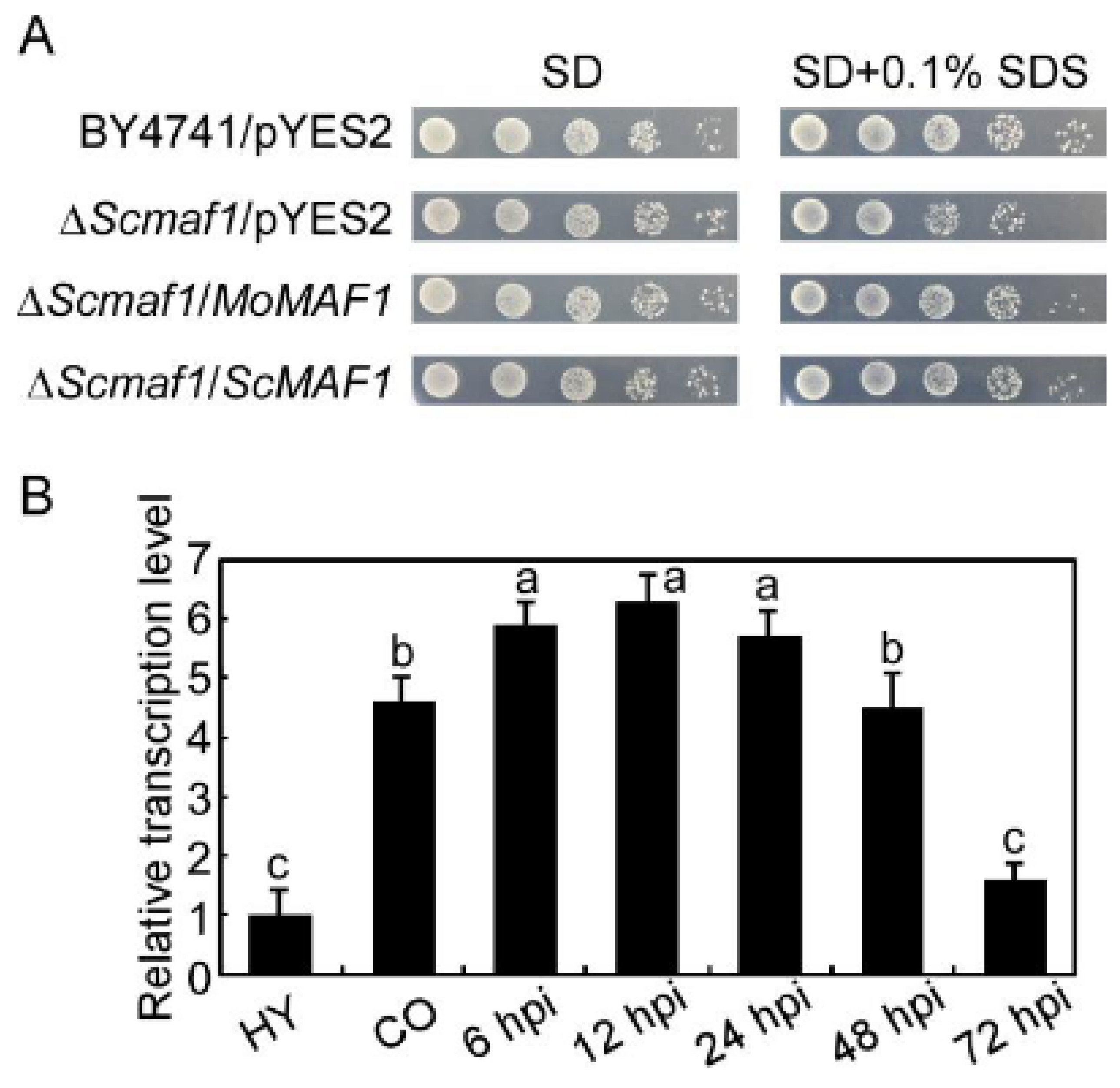
2.4. The Complementation of S. cerevisiae Δmaf1 Mutant

The full-length cDNA of MoMAF1 was amplified with primers (Supporting Information Table S1), digested with XbaI and SacI, and then cloned into the yeast expression vector pYES2 (Invitrogen). After verification by sequencing and selection on SD medium lacking uracil, the MoMAF1-pYES2 vector was transformed into the yeast ΔMomaf1 mutant (BY4741, DYRE122C). Yeast strains were cultured on YPD medium and diluted to an OD600 of 0.1, after which 5 µL of 10-fold serial dilutions grew on SD-Met-Leu-His-Ura (galactose) and SD-Met-Leu-His-Ura supplemented with 0.1% SDS plates at 30 °C for 4 days before being photographed.

2.5. MoMAF1 Gene Deletion and Complementation

The ΔMomaf1 mutants were generated by the one-step gene replacement strategy. Two 1.0 kb fragments flanking the targeted gene were PCR amplified with primer pairs (Supplementary Table S1). The two 1.0 kb fragments were ligated to the two ends of the hygromycin resistance cassette (HPH 1.4 kb) by overlap PCR to form a 3.4 kb fragment. Then, the 3.4 kb fragment was transformed into Guy11 protoplasts by transformation [22]. The putative mutants were screened by PCR after 7–10 days of incubation at 28 °C and further confirmed by Southern blot analysis.
To generate a complementary pYF11-MoMAF1-GFP fusion construct, the gene sequence containing the MoMAF1 gene and 1.5 kb native promoter was amplified by PCR. Yeast strain XK125 was cotransformed with this sequence and the XhoI-digested pYF11 plasmid (containing a bleomycin resistance gene and GFP sequence) by the yeast gap repair approach. Then, obtained yeast plasmid was expressed in E. coli. To generate the complementary strain, the pYF11-MoMAF1 construct containing the bleomycin resistance gene for the M. oryzae transformants screen was introduced into the ΔMomaf1 mutant.

2.6. Conidial Germination and Appressorium Formation

Conidial germination and appressorium formation were measured on a hydrophobic surface. Conidial suspensions of 25 μL (5 × 10⁴ spores/mL) were dropped onto a hydrophobic surface and placed in a humidified box at 28 °C. The appressorium formation rate was counted at 24 h postinoculation (hpi) under a microscope, and more than 200 appressoria were counted for each strain.

2.7. Host Penetration and Pathogenicity Assay

For the spraying assay, two-week-old rice seedlings (O. sativa cv. CO39) were sprayed with 5 mL of the conidial suspension and kept in a growth chamber at 28 °C with high humidity (>80%) in the dark for the first 24 h, followed by incubation under a 12 h light:12 h dark cycle for 7 days. For the barley infection assay, 7-day-old barley leaves were inoculated with three droplets (25 μL) of the conidial suspension, and photographs were taken on day 5 after infection. Each experiment was repeated at least three times. To assess rice sheath penetration and invasive hyphal expansion, the conidial suspension (1 × 10⁵ spores/mL) was inoculated into the sheaths. After incubation at 28 °C for 30 h, the sheath cuticle cells were observed under a Zeiss Axio Observer A1 inverted microscope.

2.8. Western Blot Analysis of Protein Phosphorylation

The ΔMomaf1 mutant and wild-type strains were cultured in liquid CM for 2 days and then harvested, and 1 mL of protein lysis buffer and 10 µL of protease inhibitor cocktail (Sangon, Shanghai, China) were added. After vortexing and homogenization, the lysate was centrifuged at 12,000 rpm for 10 min at 4 °C. Then, 200 μL of the supernatant was mixed with 50 μL loading buffer and boiled for 5 min. Obtained proteins were separated on SDS–PAGE gels and transferred onto a polyvinylidene fluoride membrane using a Bio-Rad blotting apparatus. The intensity of the phosphorylated Mps1 signal was detected by the addition of an anti-phospho-p44/42 MAP kinase antibody (Cell Signaling Technology, Boston, MA, USA), with an anti-MAPK1 antibody (N-terminal anti-Mpk1) used as a control.

2.9. Chitin (N-acetylglucosamine, GlcNAc) Content Assay

The chitin (N-acetylglucosamine, GlcNAc) content was analyzed as follows. First, mycelial samples were freeze-dried, and then 5 mg of the dried mycelia was resuspended in 1 mL of 6% KOH and heated at 80 °C for 90 min. The samples were centrifuged (16,000× g, 10 min), and the pellets were washed with PBS over three cycles of centrifugation and resuspension (16,000× g, 10 min) before the final suspension in 0.5 mL of McIlvaine’s buffer (pH 6). An aliquot of 100 mL (13 units) of Streptomyces plicatus chitinase (Sigma, St. Louis, MO, USA) was added, and the mixture was incubated for 16 h at 37 °C with gentle mixing; 100 mL samples were then combined with 100 mL of 0.27 M sodium borate (pH 9) and heated for 10 min at 100 °C with the final addition of 1 mL of freshly diluted (1:10) Ehrlich’s reagent (10 g of p-dimethylaminobenzaldehyde in 1.25 mL of HCl and 8.75 mL of glacial acetic acid). After incubation at 37 °C for 20 min, 1 mL of the sample was transferred to a 2.5 mL plastic cuvette (Greiner, Frickenhausen, Germany), and the absorbance at 585 nm was recorded. Standard curves were prepared with GlcNAc (Sigma). The experiment was repeated three times.

2.10. The Observation of Subcellular Localization

To observe the subcellular localization of MoMaf1, we fused MoMaf1 with a GFP tag and a nuclear marker with an RFP tag. The green and red fluorescence signals in vegetative hyphae and infectious hyphae were observed by dual fluorescence (Zeiss LSM710, 63× oil).

2.11. Statistical Analysis

Each experiment was performed with three replicates and obtained data were represented as mean ± standard deviation (SD). The significant differences between treatments were statistically determined by one-way analysis of variance (ANOVA) comparison and followed by Duncan’s new multiple-range tests.

3. Results

3.1. Identification and Expression of MoMAF1

Examination of the M. oryzae genome database revealed that MGG_15675 and S. cerevisiae ScMaf1 exhibited high amino acid sequence homology, and we named the MGG_15675 sequence MoMaf1. We first expressed MoMAF1 in the ΔScmaf1 mutant using the yeast expression vector pYES2 and found that ΔScmaf1/MoMAF1 suppressed the growth defect in sensitivity to SDS in the ΔScmaf1 mutant, indicating that MoMaf1 is a functional paralog of ScMaf1 (Figure 1A). In addition, transcription profile analysis of MoMAF1 at different developmental stages in M. oryzae showed that MoMAF1 was more highly expressed during the infection phase than during the mycelia stage, suggesting that MoMAF1 participated in the M. oryzae–rice interaction (Figure 1B).

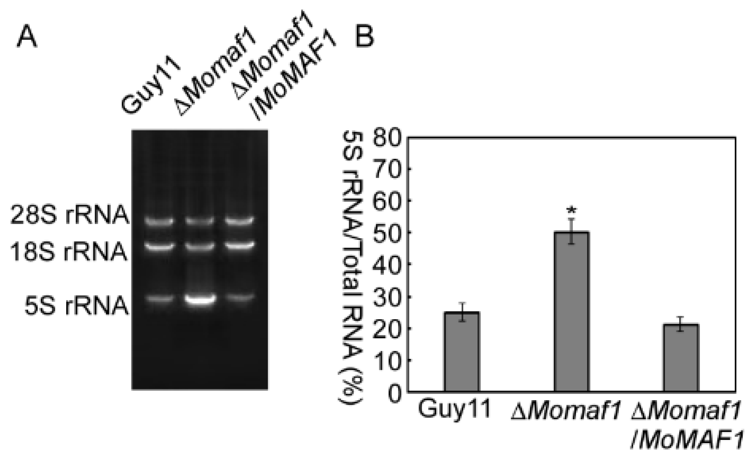
3.2. MoMAF1-Regulated RNA Synthesis

In Saccharomyces cerevisiae, Maf1 is a negative regulator of Pol III that represses the synthesis of 5S rRNA and tRNA. Deletion of MAF1 causes a substantial increase in 5S rRNA and tRNA [23]. To investigate the roles of MoMaf1 in M. oryzae, we generated ∆Momaf1 mutants and verified them by PCR amplification and Southern blot hybridization (Supplementary Figure S1). We then further examined the function of MoMAF1 in RNA synthesis. RNA extraction successfully yielded one small 5S rRNA species and two large rRNA species (18S and 28S). The ∆Momaf1 mutant showed dramatically elevated 5S rRNA level compared with that of the wild-type and complemented strain (Figure 2A,B). These results suggested that MoMAF1 regulated RNA synthesis.

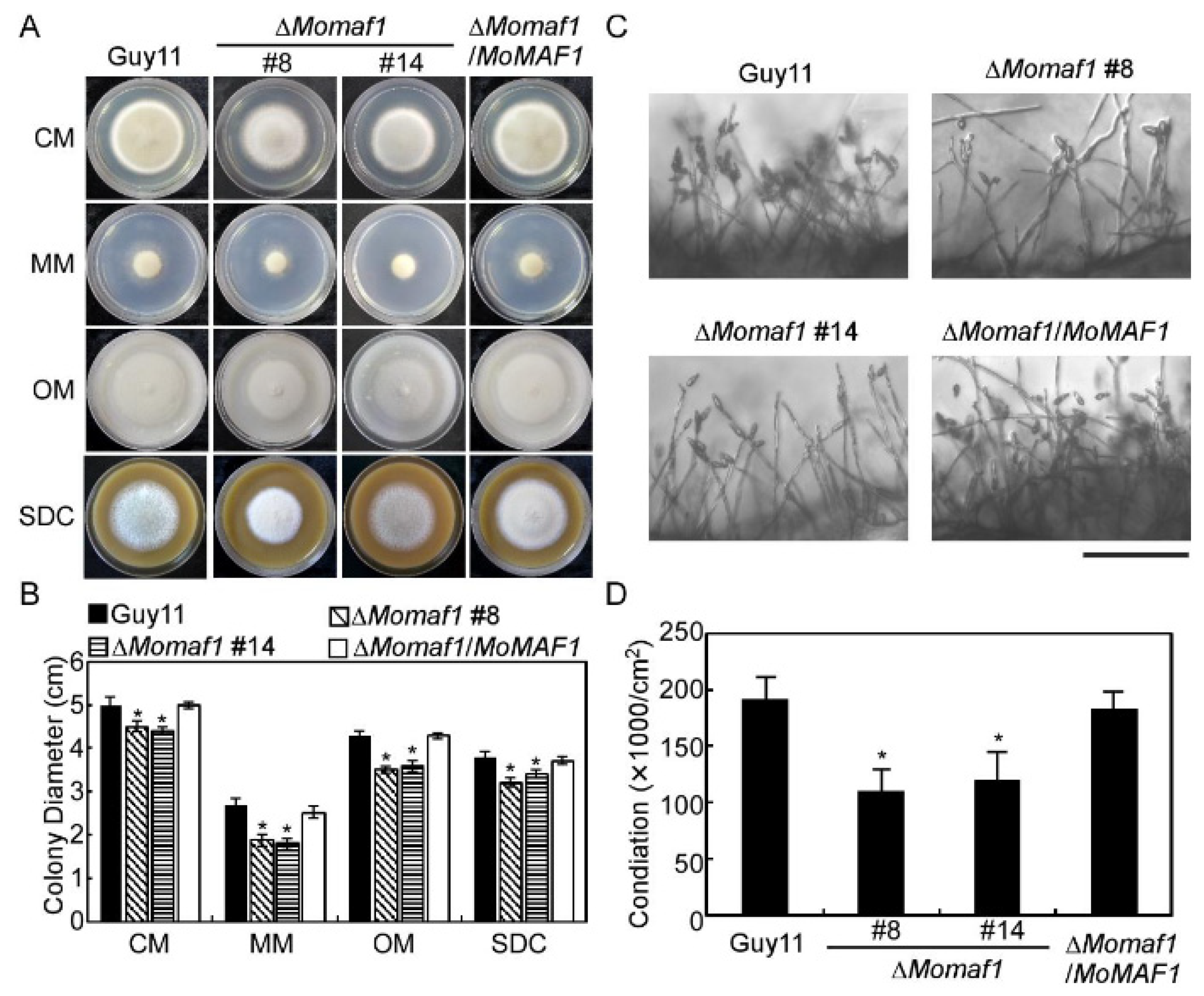
3.3. MoMaf1 Was Involved in Vegetative Growth and Conidiation

Since we determined that MoMaf1 is a homolog of ScMaf1, we then observed whether the loss of MoMAF1 in M. oryzae led to a considerable defect in vegetable growth. As shown in (Figure 3A,B), destruction of MoMAF1 leads to defective vegetative growth. Additionally, the ΔMomaf1 mutant produced fewer conidia than the wild-type strain (Guy11) and the complemented strain (ΔMomaf1/MoMAF1) (Figure 3C,D). Due to the function of Maf1 in inhibiting transcription, we examined the expression of six conidiation-related genes in the ΔMomaf1 mutant and Guy11 strain and found that the expression levels of MoCOM1, MoCON2, MoHOX2, and MoSTUA were significantly lower in the ΔMomaf1 mutant, while MoCOS1 and MoCON7 levels were not significantly different from those in the Guy11 strain (Supplementary Figure S2), indicating that MoMaf1 was involved in regulating conidiation-related genes.

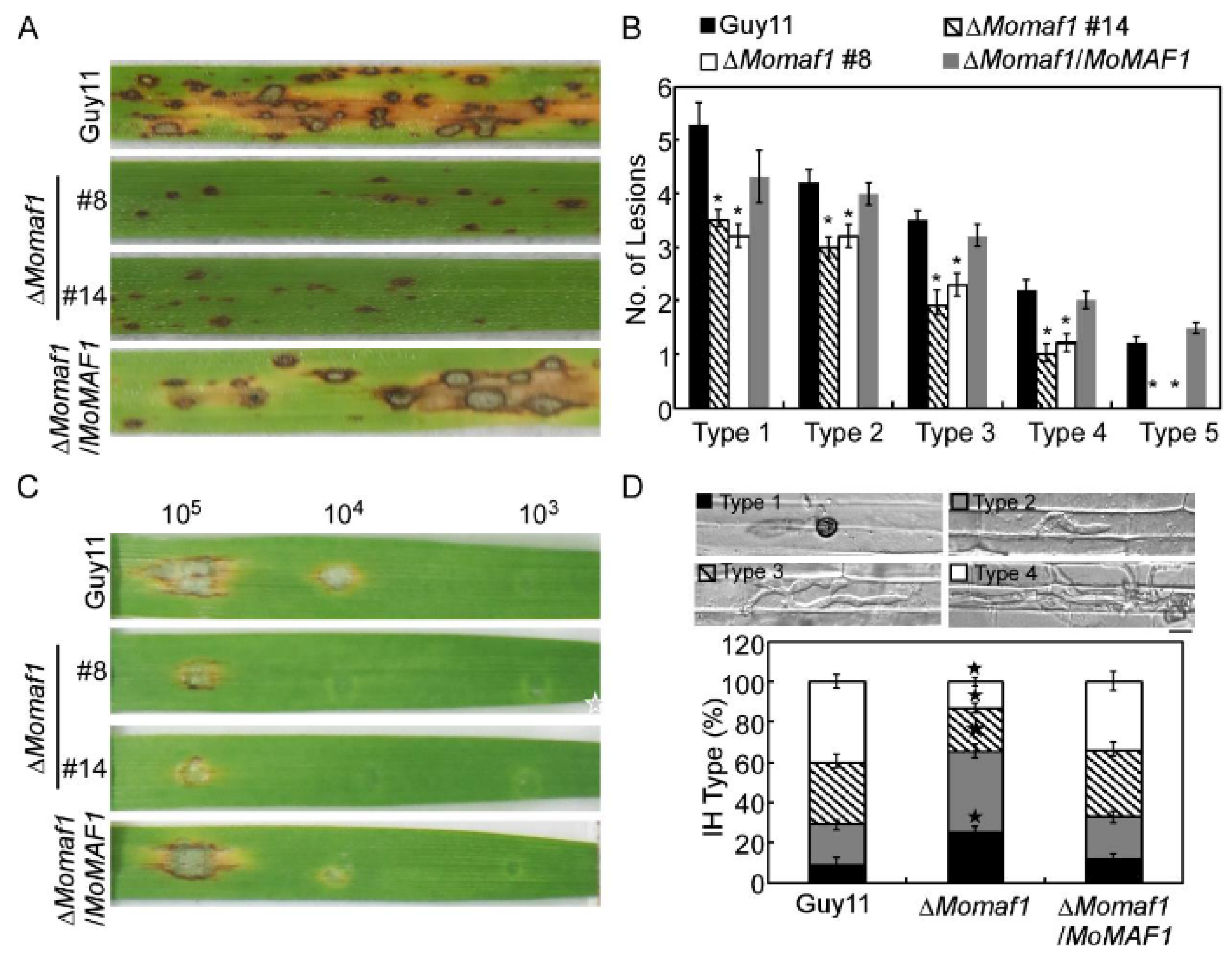
3.4. MoMaf1 Was Required for Penetration and Infectious Growth

To further examine the role of MoMaf1 in virulence, conidial suspensions of the Guy11 strain, the ΔMomaf1 mutant, and the complemented strain were sprayed onto two-week-old rice seedlings (Oryza sativa cv. CO-39). After 7 days of inoculation, the mutant showed reduced virulence, with fewer and smaller lesions on the rice leaves in comparison to the numerous typical lesions caused by the wild-type strain (Guy11) and the complemented strain. A “lesion-type” scoring assay [24] revealed that the numbers of all five types of lesions caused by the ΔMomaf1 mutant were significantly decreased (Figure 4A,B). Similar results were obtained after the inoculation of conidial suspensions dropped on detached barley leaves, in which the ΔMomaf1 mutant caused more restricted lesions to form (Figure 4C).
As the ΔMomaf1 mutant caused fewer and smaller lesions, we further investigated the role of MoMaf1 in penetration and infectious hyphal growth. Statistical analysis of the results showed that approximately 25% of the appressoria formed by the ΔMomaf1 mutant were unable to penetrate the rice cuticle (type 1), 40% of the penetration sites formed infectious hyphae (IH), but these IH were restricted to one cell with no branches or 1–2 branches (type 2 and type 3, respectively), and less than 15% of the IH extended to the neighboring cells (type 4). In contrast, there were only approximately 10% type 1 and over 70% type 3 and type 4 IH in the Guy11 strain and the complemented strain (Figure 4D). These results indicated that MoMaf1 played a critical role in penetration and infectious growth in rice blast fungus.

3.5. MoMaf1 Regulated the Generation of Appressorium Turgor Pressure

Appressoria are critical structures for M. oryzae infection. The entire spore can be trafficked into the appressorium, where it undergoes maturation [25]. These coupled processes generate enormous hydrostatic turgor pressure in the appressorium, which has been measured at up to 8.0 MPa, to breach the rice leaf cuticle [26,27]. As appressorium formation in the ΔMomaf1 mutant was no different from that in the wild-type strain (Supplementary Figure S3), we then examined whether the defect in turgor pressure generation resulted in a reduction in pathogenicity. An appressorium collapse assay was performed to test the appressorial turgor pressure using 1–4 M glycerol solutions [28]. The appressoria of the ΔMomaf1 mutant exhibited higher collapse ratios than those of the wild-type and complemented strain (Supplementary Figure S4), suggesting that the reduced pathogenicity of the ΔMomaf1 mutants may be related to the aberrant development of functional appressoria.

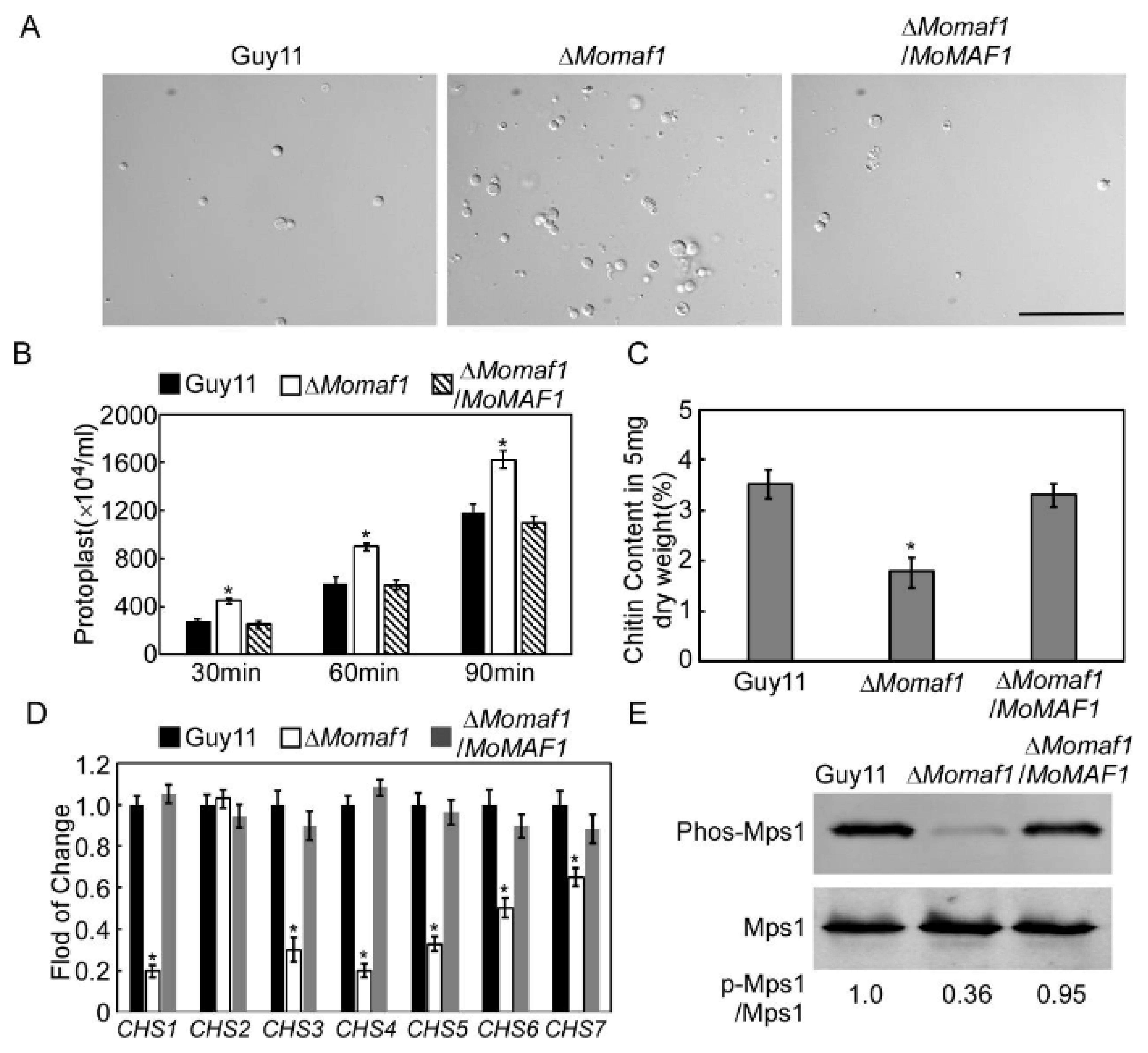
3.6. MoMaf1 Was Involved in Cell Wall Integrity (CWI)

We further investigated whether MoMaf1 was involved in modulating CWI. First, we assessed the effect of cell wall-degrading enzymes on mycelia in all tested strains [29]. Under the same conditions, the hyphae of the ΔMomaf1 mutant released more protoplasts than those of the wild-type strain or the complemented strain (Figure 5A,B). Then, we further quantified the chitin that accumulated in the cell wall and found that the ΔMomaf1 mutant had a higher chitin content than the wild-type strain (Guy11) (Figure 5C). We also examined the expression levels of chitin synthase (CHS) genes and found that the expression of six CHS genes was significantly reduced in the ΔMomaf1 mutant, but this was not the case for CHS2 (Figure 5D). Additionally, the phosphorylation of MoMps1 was clearly decreased when compared with that of the wild-type Guy11 strain (Figure 5E). In addition, the ΔMomaf1 mutants were more sensitive to the cell wall-perturbing agents CFW and Congo red (CR) (Supplementary Figure S5). Taken together, these results indicated that MoMaf1 was involved in maintaining CWI.

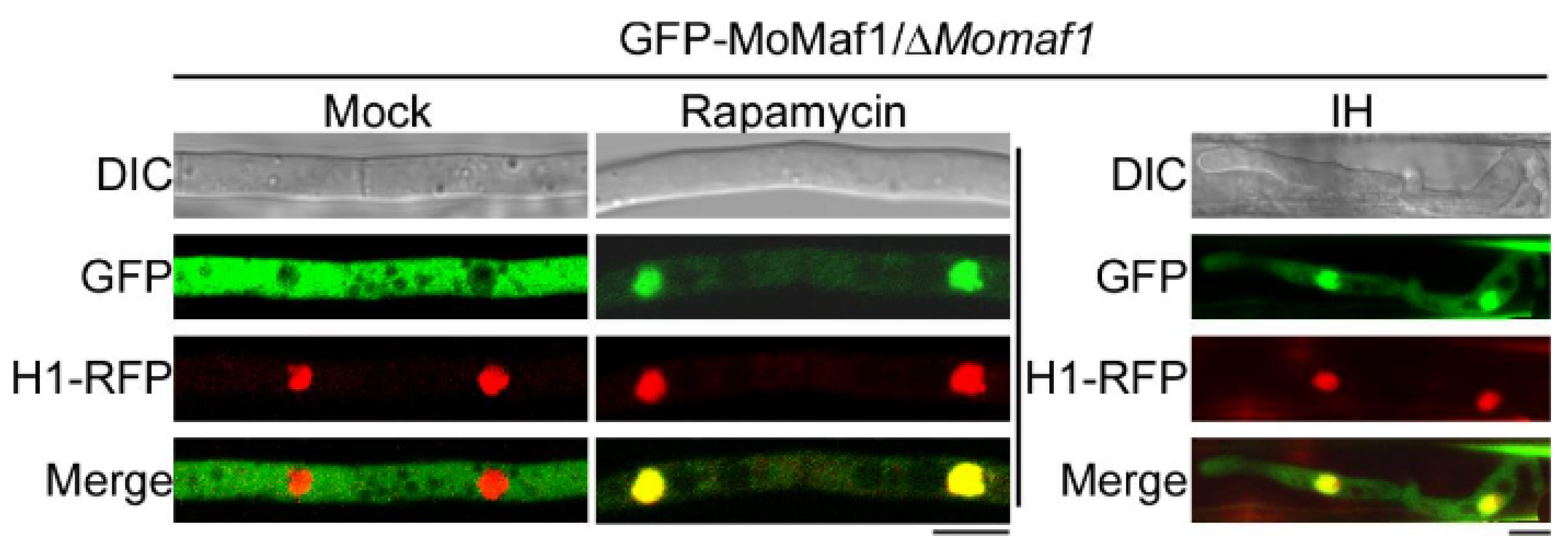
3.7. The Subcellular Localization of MoMaf1

To examine the subcellular localization of MoMaf1, we monitored the GFP-MoMaf1 fusion protein in the wild-type strain under different nutrient conditions. The GFP-MoMaf1 fluorescence signal was localized to the cytoplasm in a nutrient-rich complete medium (CM), and GFP-MoMaf1 was then translocated to the nucleus upon treatment with rapamycin to simulate nitrogen stress (Figure 6). During infection, the GFP fluorescence signal in the cytosol was weaker than that in the nucleus, suggesting that MoMaf1 was transferred from the cytoplasm to the nucleus (Figure 6). These results illustrated that the translocation of MoMaf1 from the cytosol to the nucleus was nutrient-dependent and that tRNA transcription needed to be properly balanced to infect the host.

4. Discussion

In eukaryotes, there are three RNA polymerases, designated Pol I, II, and III. Pol II is responsible for the transcription of all protein-coding mRNAs and many non-protein-coding RNAs, while Pol I and Pol III are specialized in the high-level synthesis of non-coding RNA species, rRNA and tRNA, respectively, which are fundamental components of the translation machinery [30]. As the global negative effector of Pol III, Maf1 was originally identified in S. cerevisiae, and its orthologues were subsequently characterized in other species [31]. Like previous observations in many species [23,32,33], we have demonstrated in this study that MoMaf1 is also a repressor of Pol III in M. oryzae, adding a new member to the Maf1 family.
In this study, we found MoMaf1 was essential for the growth, conidiation, and pathogenicity of M. oryzae. As observed in Candida albicans and Fusarium graminearum [34,35], deletion of MAF1 both led to a significantly reduced growth rate, indicating that the functions of the Maf1 were conserved. We also found that the ΔMomaf1 mutant exhibited high sensitivity to cell wall stressors (CFW and CR) and impaired cell wall integrity. In M. oryzae, the CWI MAP kinase pathway, consisting of MoMck1, MoMkk1, and MoMps1, was important for appressorium function and virulence [36]. These results indicated that the impaired CWI in ΔMomaf1 mutant may result in attenuated virulence.
Similar to that of other pathogenic fungi, the infection cycle of M. oryzae starts with conidia [37]. The ΔMomaf1 mutant produced fewer conidia than the wild-type strain, and we also found that the expression of four conidiation-related genes, MoCOM1, MoCON2, MoHOX2, and MoSTUA, were significantly reduced in the ΔMomaf1 mutant, which was consistent with the MoMaf1 protein expression data and indicated that MoMaf1 was involved in sporulation and conidial morphology by regulating the expression of these genes.
In S. cerevisiae, several signaling pathways, including those mediated by TOR, modulate the phosphorylation status of Maf1 and mediate various stress signals to Pol III [38]. However, in contrast to the well-studied phosphorylation process of Maf1, little is known about the dephosphorylation of Maf1. By far, studies only revealed that the PP4 complex, with Pph3 as the catalytic subunit, is the major and direct phosphatase of Maf1 [7]. Other PP2A phosphatase complexes and a set of alternative PP2A catalytic subunits that were involved in Maf1 dephosphorylation need further study.
Importantly, we found that MoMaf1 undergoes cytoplasmic-to-nuclear translocation during infection or in response to nitrogen stress. Similar interaction-dependent translocation has been reported before. Liu and colleagues revealed that MoYvh1 was translocated into the nucleus following oxidative stress to control the maturation of ribosomes, which promoted extracellular protein synthesis and secretion to scavenge rice reactive oxygen species (ROS) [39]. Mature ribosomes carry out extracellular protein synthesis and secretion to scavenge ROS and modulate the rice defense response. Moreover, many transcription factors such as MoMsn2 and MoHac1 both undergo cytoplasmic-to-nuclear translocation when faced with stress or during infection [28,40,41]. So, these studies suggested that a switch of gene expression and protein synthesis was needed for successful infection. In summary, in this study, we identified a pathogenic factor, MoMaf1, which played an important role in growth, conidiation, and pathogenicity in M. oryzae, and revealed a novel link between tRNA synthesis and fungal virulence that was mediated by MoMaf1. We concluded that during early infection, M. oryzae may need tRNA synthesis to be properly balanced for its pathogenicity.

Supplementary Materials

The following supporting information can be downloaded at: https://www.mdpi.com/article/10.3390/jof9010106/s1, Figure S1. Gene knockout strategy and southern blot analyses in M. oryzae. Figure S2. Transcriptional analysis of six conidiation related genes. Figure S3. Statistical analysis of the appressorium formation rate. Figure S4. MoMaf1 is involved in appressorium turgor generation. Figure S5. MoMaf1 is involved in cell wall stress response. Table S1. Primers used in this study.

Author Contributions

Conceptualization, L.G.; methodology, B.Q. and C.S.; software, B.Q.; validation, L.G.; formal analysis, H.J.; investigation, L.G.; resources, B.Q.; data curation, L.G.; writing—original draft preparation, B.Q.; writing—review and editing, C.S.; visualization, L.G. and C.S.; supervision, C.S.; project administration, B.Q. and H.J.; funding acquisition, B.Q. All authors have read and agreed to the published version of the manuscript.

Funding

This research was supported by the program of the Natural Science Foundation of China Youth Program for NSFC (32202240), Youth Program of the Natural Science Foundation of Jiangsu Province (BK20200543). We thank Prof. Zhengguang Zhang of Nanjing Agricultural University for providing help with this study.

Data Availability Statement

Not applicable.

Conflicts of Interest

The authors declare no conflict of interest.

References

  1. Dieci, G.; Fiorino, G.; Castelnuovo, M.; Teichmann, M.; Pagano, A. The expanding RNA polymerase III transcriptome. Trends Genet. 2007, 23, 614–622. [Google Scholar] [CrossRef] [PubMed]
  2. Vannini, A.; Ringel, R.; Kusser, A.G.; Berninghausen, O.; Kassavetis, G.A.; Cramer, P. Molecular Basis of RNA Polymerase III Transcription Repression by Maf1. Cell 2010, 143, 59–70. [Google Scholar] [CrossRef] [Green Version]
  3. Berns, A. A tRNA with oncogenic capacity. Cell 2008, 133, 29–30. [Google Scholar] [CrossRef] [PubMed] [Green Version]
  4. Seton-Rogers, S. tRNA supply and demand. Nat. Rev. Cancer 2022, 22, 129. [Google Scholar] [CrossRef]
  5. Boguta, M.; Graczyk, D. RNA polymerase III under control: Repression and de-repression. Trends Biochem. Sci. 2011, 36, 451–456. [Google Scholar] [CrossRef]
  6. Karkusiewicz, I.; Turowski, T.W.; Graczyk, D.; Towpik, J.; Dhungel, N.; Hopper, A.K.; Boguta, M. Maf1 Protein, Repressor of RNA Polymerase III, Indirectly Affects tRNA Processing. J. Biol. Chem. 2011, 286, 39478–39488. [Google Scholar] [CrossRef] [PubMed] [Green Version]
  7. Oler, A.J.; Cairns, B.R. PP4 dephosphorylates Maf1 to couple multiple stress conditions to RNA polymerase III repression. EMBO J. 2012, 31, 1440–1452. [Google Scholar] [CrossRef]
  8. Moir, R.D.; Lee, J.; Haeusler, R.A.; Desai, N.; Engelke, D.R.; Willis, I.M. Protein kinase A regulates RNA polymerase III transcription through the nuclear localization of Maf1. Proc. Natl. Acad. Sci. USA 2006, 103, 15044–15049. [Google Scholar] [CrossRef] [PubMed] [Green Version]
  9. Lee, J.; Moir, R.D.; Willis, I.M. Regulation of RNA polymerase III transcription involves SCH9-dependent and SCH9-independent branches of the target of rapamycin (TOR) pathway. J. Biol. Chem. 2009, 284, 12604–12608. [Google Scholar] [CrossRef] [Green Version]
  10. Graczyk, D.; Dębski, J.; Muszyńska, G.; Bretner, M.; Lefebvre, O.; Boguta, M. Casein kinase II-mediated phosphorylation of general repressor Maf1 triggers RNA polymerase III activation. Proc. Natl. Acad. Sci. USA 2011, 108, 4926–4931. [Google Scholar] [CrossRef]
  11. Zhang, H.; Zheng, X.; Zhang, Z. The Magnaporthe grisea species complex and plant pathogenesis. Mol. Plant Pathol. 2016, 17, 796–804. [Google Scholar] [CrossRef] [PubMed] [Green Version]
  12. Yin, Z.; Feng, W.; Chen, C.; Xu, J.; Li, Y.; Yang, L.; Wang, J.; Liu, X.; Wang, W.; Gao, C.; et al. Shedding light on autophagy coordinating with cell wall integrity signaling to govern pathogenicity of Magnaporthe oryzae. Autophagy 2020, 16, 900–916. [Google Scholar] [CrossRef] [PubMed]
  13. Qian, B.; Liu, X.; Jia, J.; Cai, Y.; Chen, C.; Zhang, H.; Zheng, X.; Wang, P.; Zhang, Z. MoPpe1 partners with MoSap1 to mediate TOR and cell wall integrity signalling in growth and pathogenicity of the rice blast fungus Magnaporthe oryzae. Environ. Microbiol. 2018, 20, 3964–3979. [Google Scholar] [CrossRef]
  14. Qian, B.; Liu, X.; Ye, Z.; Zhou, Q.; Liu, P.; Yin, Z.; Wang, W.; Zheng, X.; Zhang, H.; Zhang, Z. Phosphatase-associated protein MoTip41 interacts with the phosphatase MoPpe1 to mediate crosstalk between TOR and cell wall integrity signalling during infection by the rice blast fungus Magnaporthe oryzae. Environ. Microbiol. 2021, 23, 791–809. [Google Scholar] [CrossRef] [PubMed]
  15. Qian, B.; Su, X.; Ye, Z.; Liu, X.; Liu, M.; Shen, D.; Chen, H.; Zhang, H.; Wang, P.; Zhang, Z. MoErv29 promotes apoplastic effector secretion contributing to virulence of the rice blast fungus Magnaporthe oryzae. New Phytol. 2021, 233, 1289–1302. [Google Scholar] [CrossRef]
  16. Feng, W.; Yin, Z.; Wu, H.; Liu, P.; Liu, X.; Liu, M.; Yu, R.; Gao, C.; Zhang, H.; Zheng, X.; et al. Balancing of the mitotic exit network and cell wall integrity signaling governs the development and pathogenicity in Magnaporthe oryzae. PLoS Pathog. 2021, 17, e1009080. [Google Scholar] [CrossRef]
  17. Chen, D.; Liang, J.; Jiang, C.; Wu, D.; Huang, B.; Teng, X.; Tang, Y. Mitochondrion Participated in Effect Mechanism of Manganese Poisoning on Heat Shock Protein and Ultrastructure of Testes in Chickens. Biol. Trace Elem. Res. 2022, 1–10. [Google Scholar] [CrossRef]
  18. Sun, Q.; Liu, Y.; Teng, X.; Luan, P.; Yin, X. Immunosuppression participated in complement activation-mediated inflammatory injury caused by 4-octylphenol via TLR7/IkappaBalpha/NF-kappaB pathway in common carp (Cyprinus carpio) gills. Aquat. Toxicol. 2022, 249, 106211. [Google Scholar] [CrossRef]
  19. Guo, M.; Chen, Y.; Du, Y.; Dong, Y.; Guo, W.; Zhai, S.; Zhang, H.; Dong, S.; Zhang, Z.; Wang, Y.; et al. The bZIP Transcription Factor MoAP1 Mediates the Oxidative Stress Response and Is Critical for Pathogenicity of the Rice Blast Fungus Magnaporthe oryzae. PLoS Pathog. 2011, 7, e1001302. [Google Scholar] [CrossRef] [Green Version]
  20. Cui, J.; Zhou, Q.; Yu, M.; Liu, Y.; Teng, X.; Gu, X. 4-tert-butylphenol triggers common carp hepatocytes ferroptosis via oxidative stress, iron overload, SLC7A11/GSH/GPX4 axis, and ATF4/HSPA5/GPX4 axis. Ecotoxicol. Environ. Saf. 2022, 242, 113944. [Google Scholar] [CrossRef]
  21. Miao, Z.; Teng, X.; Xu, S. Melatonin alleviates lead-induced intestinal epithelial cell pyroptosis in the common carps (Cyprinus carpio) via miR-17-5p/TXNIP axis. Fish Shellfish Immunol. 2022, 131, 127–136. [Google Scholar] [CrossRef] [PubMed]
  22. Yin, Z.; Chen, C.; Yang, J.; Feng, W.; Liu, X.; Zuo, R.; Zhang, Z. Histone acetyltransferase MoHat1 acetylates autophagy-related proteins MoAtg3 and MoAtg9 to orchestrate functional appressorium formation and pathogenicity in Magnaporthe oryzae. Autophagy 2019, 15, 1234–1257. [Google Scholar] [CrossRef]
  23. Pluta, K.; Lefebvre, O.; Martin, N.C.; Smagowicz, W.J.; Stanford, D.R.; Ellis, S.R.; Hopper, A.K.; Sentenac, A.; Boguta, M. Maf1p, a Negative Effector of RNA Polymerase III in Saccharomyces cerevisiae. Mol. Cell. Biol. 2001, 21, 5031–5040. [Google Scholar] [CrossRef] [PubMed] [Green Version]
  24. Zhong, K.; Li, X.; Le, X.; Kong, X.; Zhang, H.; Zheng, X.; Wang, P.; Zhang, Z. MoDnm1 Dynamin Mediating Peroxisomal and Mitochondrial Fission in Complex with MoFis1 and MoMdv1 Is Important for Development of Functional Appressorium in Magnaporthe oryzae. PLoS Pathog. 2016, 12, e1005823. [Google Scholar] [CrossRef] [PubMed] [Green Version]
  25. Veneault-Fourrey, C.; Barooah, M.; Egan, M.; Wakley, G.; Talbot, N.J. Autophagic fungal cell death is necessary for infection by the rice blast fungus. Science 2006, 312, 580–583. [Google Scholar] [CrossRef] [Green Version]
  26. deJong, J.C.; MCCormack, B.J.; Smirnoff, N.; Talbot, N.J. Glycerol generates turgor in rice blast. Nature 1997, 389, 244–245. [Google Scholar] [CrossRef]
  27. Howard, R.J.; Valent, B. Breaking and entering: Host penetration by the fungal rice blast pathogen Magnaporthe grisea. Annu. Rev. Microbiol. 1996, 50, 491–512. [Google Scholar] [CrossRef]
  28. Tang, W.; Ru, Y.; Hong, L.; Zhu, Q.; Zuo, R.; Guo, X.; Wang, J.; Zhang, H.; Zheng, X.; Wang, P.; et al. System-wide characterization of bZIP transcription factor proteins involved in infection-related morphogenesis of Magnaporthe oryzae. Environ. Microbiol. 2014, 17, 1377–1396. [Google Scholar] [CrossRef] [Green Version]
  29. Yin, Z.; Tang, W.; Wang, J.; Liu, X.; Yang, L.; Gao, C.; Zhang, J.; Zhang, H.; Zheng, X.; Wang, P.; et al. Phosphodiesterase MoPdeH targets MoMck1 of the conserved mitogen-activated protein (MAP) kinase signalling pathway to regulate cell wall integrity in rice blast fungus Magnaporthe oryzae. Mol. Plant Pathol. 2016, 17, 654–668. [Google Scholar] [CrossRef]
  30. Lesniewska, E.; Boguta, M. Novel layers of RNA polymerase III control affecting tRNA gene transcription in eukaryotes. Open Biol. 2017, 7, 170001. [Google Scholar] [CrossRef]
  31. Oficjalska-Pham, D.; Harismendy, O.; Smagowicz, W.J.; de Peredo, A.G.; Boguta, M.; Sentenac, A.; Lefebvre, O. General Repression of RNA Polymerase III Transcription Is Triggered by Protein Phosphatase Type 2A-Mediated Dephosphorylation of Maf1. Mol. Cell 2006, 22, 623–632. [Google Scholar] [CrossRef]
  32. Johnson, S.S.; Zhang, C.; Fromm, J.; Willis, I.M.; Johnson, D.L. Mammalian Maf1 is a negative regulator of transcription by all three nuclear RNA polymerases. Mol. Cell 2007, 26, 367–379. [Google Scholar] [CrossRef] [PubMed]
  33. Reina, J.H.; Azzouz, T.N.; Hernandez, N. Maf1, a New Player in the Regulation of Human RNA Polymerase III Transcription. PLoS ONE 2006, 1, e134. [Google Scholar] [CrossRef] [PubMed] [Green Version]
  34. Asghar, F.; Yan, H.; Jiang, L. The putative transcription factor CaMaf1 controls the sensitivity to lithium and rapamycin and represses RNA polymerase III transcription in Candida albicans. FEMS Yeast Res. 2018, 18, foy068. [Google Scholar] [CrossRef] [PubMed]
  35. Gu, Q.; Zhang, C.; Yu, F.; Yin, Y.; Shim, W.B.; Ma, Z. Protein kinase FgSch9 serves as a mediator of the target of rapamycin and high osmolarity glycerol pathways and regulates multiple stress responses and secondary metabolism in Fusarium graminearum. Environ. Microbiol. 2015, 17, 2661–2676. [Google Scholar] [CrossRef]
  36. Yang, J.; Liu, M.; Liu, X.; Yin, Z.; Sun, Y.; Zhang, H.; Zhang, Z. Heat-Shock Proteins MoSsb1, MoSsz1, and MoZuo1 Attenuate MoMkk1-Mediated Cell-Wall Integrity Signaling and Are Important for Growth and Pathogenicity of Magnaporthe oryzae. Mol. Plant-Microbe Interact. 2018, 31, 1211–1221. [Google Scholar] [CrossRef] [PubMed] [Green Version]
  37. Li, C.; Yang, J.; Zhou, W.; Chen, X.L.; Huang, J.G.; Cheng, Z.H.; Peng, Y.L. A spindle pole antigen gene MoSPA2 is important for polar cell growth of vegetative hyphae and conidia, but is dispensable for pathogenicity in Magnaporthe oryzae. Curr. Genet. 2014, 60, 255–263. [Google Scholar] [CrossRef]
  38. Michels, A.A. MAF1: A new target of mTORC1. Biochem. Soc. Trans. 2011, 39, 487–491. [Google Scholar] [CrossRef]
  39. Liu, X.; Yang, J.; Qian, B.; Cai, Y.; Zou, X.; Zhang, H.; Zheng, X.; Wang, P.; Zhang, Z. MoYvh1 subverts rice defense through functions of ribosomal protein MoMrt4 in Magnaporthe oryzae. PLoS Pathog. 2018, 14, e1007016. [Google Scholar] [CrossRef] [Green Version]
  40. Xiao, Y.; Liu, L.; Zhang, T.; Zhou, R.; Ren, Y.; Li, X.; Zhang, H. Transcription factor MoMsn2 targets the putative 3-methylglutaconyl-CoA hydratase-encoding gene MoAUH1 to govern infectious growth via mitochondrial fusion/fission balance in Magnaporthe oryzae. Environ. Microbiol. 2021, 23, 774–790. [Google Scholar] [CrossRef]
  41. Zhang, H.; Zhao, Q.; Guo, X.; Guo, M.; Qi, Z.; Tang, W.; Dong, Y.; Ye, W.; Zheng, X.; Wang, P.; et al. Pleiotropic Function of the Putative Zinc-Finger Protein MoMsn2 in Magnaporthe oryzae. Mol. Plant-Microbe Interact. 2014, 27, 446–460. [Google Scholar] [CrossRef] [PubMed]
Figure 1. Expression profiles of MoMAF1 at different developmental stages in M. oryzae determined by yeast complementation assays: (A) MoMAF1 partially suppressed the growth defect of ΔScmaf1 mutant yeast under 0.1% SDS stress. Photographs were taken at 4 days. (B) Expression levels of MoMAF1 at different stages in M. oryzae. Expression in the hyphal stage was used as an internal reference, and error bars represent the standard deviation (SD). Values on the bars followed by the same letter are not significantly different at (p < 0.05).
Jof 09 00106 g001
Figure 2. Total-RNA analysis of the test strains: (A) The Magnaporthe oryzae wild-type strain Guy11, ΔMomaf1 mutant strain, and ΔMomaf1/MoMAF1 strain were grown in liquid CM at 28 °C for 36 h and then collected for total-RNA extraction. Total-RNA samples of 5 μg were loaded onto a 1% agarose gel and stained with ethidium bromide before visualization. (B) Statistical analysis of the 5S rRNA/total RNA ratio in the different strains. Error bars represent ±SD, and asterisks indicate significant differences (p < 0.01). Relative band intensity was quantified by IMAGEJ software. The experiment was repeated three times.
Jof 09 00106 g002
Figure 3. MoMaf1 is involved in vegetative growth and conidiation: (A) The Guy11, ΔMomaf1 mutant, and ΔMomaf1/MoMAF1 strains were inoculated on CM, MM, OM, and SDC medium; incubated at 28 °C for 7 days; and photographed. (B) Statistical analysis of the colony diameter for the strains. Error bars represent ±SD (standard deviation), and asterisks denote statistical significance (p < 0.01). (C) Conidia were observed and photographed under a light microscope after illumination for 24 h. Scale bar, 50 μm. (D) Statistical analysis of the conidial number of the indicated strains. Error bars represent ±SD, and asterisks denote significant differences (p < 0.01).
Jof 09 00106 g003
Figure 4. MoMaf1 is required for the virulence of M. oryzae: (A) Two-week-old rice seedlings (Oryza sativa cv. CO-39) were sprayed with 4 mL of a conidial suspension (5 × 10⁴ conidia/mL). The diseased leaves were harvested and photographed at 7 days post-inoculation (dpi). Three independent experiments were performed. (B) Quantification of lesions of different types (per 1.5 cm2) on diseased rice leaves. Type 1, dark brown pinpoint lesions; type 2, 1.5 mm brown spots; type 3, 2–3 mm lesions with brown margins; type 4, eyespot lesions longer than 3 mm; type 5, coalesced lesions that infected 50% or more of the maximum leaf size (Qian et al., 2021). Error bars represent ±SD, and asterisks denote significant differences (p < 0.01). (C) Detached barley leaves were drop-inoculated with serial dilutions (1 × 10⁵, 1 × 10⁴, 1 × 10³ spores/mL) of conidial suspensions, and the diseased leaves were photographed at 5 dpi. (D) Conidial suspensions (1 × 10⁵ spores/mL) were injected into rice sheaths, and infection severity was observed at 36 hpi. The percentages of different types of infectious hyphae (IH) in the rice cells were quantified at 30 hpi. Error bars represent the SD, and asterisks denote the significant differences of each type (p < 0.01). Type 1, no penetration; type 2, only a single invasive hypha (IH) without branches; type 3, 1–3 branches but restricted to one cell; type 4, more than three branches and extended to the neighboring cell. Fifty infected cells were observed for each strain and the experiment was repeated 3 times.
Jof 09 00106 g004
Figure 5. MoMaf1 is involved in cell wall integrity: (A) Light microscopic examination of protoplast release after treatment with cell wall-degrading enzymes for 90 min at 30 °C. Scale bar, 50 μm. (B) Statistical analysis of the protoplast number. The protoplast number was calculated at 30, 60, and 90 min. Error bars represent ±SD, and asterisks denote significant differences (p < 0.01). (C) The chitin content was measured in the wild-type Guy11 and ΔMomaf1 mutant hyphae. The experiment was repeated at least three times with three replicates each time. (D) The expression profiles of seven CHS genes in the Guy11, ΔMomaf1 mutant, and complemented strain. (E) Total proteins of the Guy11 and ΔMomaf1 mutant strains were isolated from mycelia to detect the MoMps1 phosphorylation level using the anti-phospho-p44/42 MAP kinase antibody, and the anti-p44/42 MAP kinase antibody was used as a control. The numerical values indicate the ratio of phosphorylated MAPK/endogenous MAPK, and the ratio of phosphorylated MAPK/endogenous MAPK in the Guy11 strain was defined as 1. Relative band intensity was quantified by IMAGEJ software. Three independent experiments that showed similar results were carried out. Asterisks represent signifigant differences (p < 0.01).
Jof 09 00106 g005
Figure 6. Subcellular localization of GFP-MoMaf1 at different phases. The subcellular localization of GFP-MoMaf1 was observed using the complemented strain with or without rapamycin (30 ng/mL) treatment for 40 min, and nuclei were stained with DAPI (left). Scale bar, 10 μm. Subcellular localization of GFP-MoMaf1 during the early infection phase (right). Scale bar, 5 μm.
Jof 09 00106 g006
Disclaimer/Publisher’s Note: The statements, opinions and data contained in all publications are solely those of the individual author(s) and contributor(s) and not of MDPI and/or the editor(s). MDPI and/or the editor(s) disclaim responsibility for any injury to people or property resulting from any ideas, methods, instructions or products referred to in the content.

