Article

Systematic Identification of Essential Genes Required for Yeast Cell Wall Integrity: Involvement of the RSC Remodelling Complex

Departamento de Microbiología y Parasitología, Facultad de Farmacia, Universidad Complutense de Madrid, Instituto Ramón y Cajal de Investigaciones Sanitarias (IRYCIS), 28040 Madrid, Spain
*
Author to whom correspondence should be addressed.
Current address: Centro Nacional de Biotecnología, CSIC, 28049 Madrid, Spain.
J. Fungi 2022, 8(7), 718; https://doi.org/10.3390/jof8070718
Submission received: 29 April 2022 / Revised: 5 July 2022 / Accepted: 6 July 2022 / Published: 8 July 2022
(This article belongs to the Special Issue The Fungal Cell Wall Integrity Pathway)

Abstract

:
Conditions altering the yeast cell wall lead to the activation of an adaptive transcriptional response mainly governed by the cell wall integrity (CWI) mitogen-activated protein kinase (MAPK) pathway. Two high-throughput screenings were developed using the yTHC collection of yeast conditional mutant strains to systematically identify essential genes related to cell wall integrity, and those required for the transcriptional program elicited by cell wall stress. Depleted expression of 52 essential genes resulted in hypersensitivity to the dye Calcofluor white, with chromatin organization, Golgi vesicle transport, rRNA processing, and protein glycosylation processes, as the most highly representative functional groups. Via a flow cytometry-based quantitative assay using a CWI reporter plasmid, 97 strains exhibiting reduced gene-reporter expression levels upon stress were uncovered, highlighting genes associated with RNA metabolism, transcription/translation, protein degradation, and chromatin organization. This screening also led to the discovery of 41 strains displaying a basal increase in CWI-associated gene expression, including mainly putative cell wall-related genes. Interestingly, several members of the RSC chromatin remodelling complex were uncovered in both screenings. Notably, Rsc9 was necessary to regulate the gene expression of CWI-related genes both under stress and non-stress conditions, suggesting distinct requirements of the RSC complex for remodelling particular genes.

1. Introduction

The fungal cell wall is an essential structure responsible for cell morphology; it contributes to preserving osmostability, protects cells against environmental stresses, and delineates cellular immunogenicity. In the eukaryotic model Saccharomyces cerevisiae, the cell wall consists of three major components: an inner layer of glucans (β-1,3 and β-1,6-glucan); a small amount of chitin (polymer of N-acetylglucosamine) and an outer layer of mannoproteins (such as agglutinins and flocculins involved in cell adhesion; and others related to enzymatic or structural functions). All these components must be correctly assembled to build a functional structure (for review, see [1,2]). Despite its apparent rigidity, the cell wall is very dynamic, so its composition and organization vary during growth and development. Additionally, when yeast cells face conditions where the cell wall integrity is compromised an adaptive response is triggered to maintain cell viability. This response includes changes in the synthesis and crosslinking of different polymers, and increased expression levels of genes functionally related to cell wall maintenance [3,4]. Cell wall stress adaptive responses in yeast are mainly regulated by the cell wall integrity (CWI) MAPK pathway, which includes a conserved MAPK module [5,6]. This module is activated through a cascade of phosphorylation events, which ultimately lead to the phosphorylation of the MAPK of the pathway, Slt2/Mpk1. In turn, phosphorylated Slt2 activates the transcription factor Rlm1, the main factor responsible for the transcriptional reprogramming elicited under these conditions [7,8,9]. CWI signalling is triggered by a variety of cell wall stressors (such as cell wall-interfering compounds) [4] or mutations in genes affecting proteins involved in cell wall homeostasis. Yeast mutants deleted for functionally relevant cell wall-related genes display constitutive Slt2 hyperactivation [10,11]. It was recently claimed that nucleosome remodelling at the promoters of Rlm1-dependent genes through cooperation between the ATP-dependent chromatin-remodelling SWI/SNF and acetyltransferase SAGA complexes is required for the binding of phosphorylated Rlm1, to target promoters and assembly of the transcription initiation machinery under cell wall stress [9,12]. In fact, mutants in different SWI and SAGA subunits display cell wall-related phenotypes. Additionally, various works also reported the association of cell wall integrity with mutants in some subunits of the RSC (Remodel the Structure of Chromatin) complex [13,14,15,16], which belongs to the SWI/SNF family of ATP-dependent chromatin remodellers [17]. However, information is lacking regarding their possible connection with CWI-dependent genes.
Largely due to its potential as a selective target for antifungal drugs, in addition to other biotechnological purposes, a great effort has been made to characterize genes involved in biogenesis, regulation, and maintenance of the yeast cell wall. This objective was fuelled by the availability of the complete collection of haploid yeast mutant strains individually deleted in each of the approximately 4800 non-essential genes identified in this organism [18]. To date, several genome-wide phenotypic or chemical-genomic profiling screens have been conducted using this yeast knock-out collection to identify cell wall-related mutants. Hypersensitivity to K1 and K2 killer toxins that form lethal pores in the plasma membrane [19,20], Congo red (chitin-binding dye), caspofungin (β-1,3-glucan synthase inhibitor), zymolyase (enzymatic cocktail including β-1,3-glucanase and chitinase activities) [21,22], freeze-thaw or heat stress [23,24] are some examples of the screenings developed to date. These experiments have permitted the identification of a broad functional catalogue of genes potentially related to maintaining cellular integrity under cell wall stress conditions. Moreover, from their comparative analysis, it has been possible to infer damage-specific signalling mechanisms and uncover genes required to withstand both specific and general cell wall injuries.
Following a complementary approach, we performed large-scale screenings using the whole collection of haploid deletion strains in all non-essential genes of S. cerevisiae transformed with a reporter system based on transcriptional fusion of the MLP1/KDX1 (YKL161C) promoter to the coding sequence of the nat1 gene encoding nourseothricin N-acetyltransferase from Streptomyces noursei [25]. Firstly, we used this collection of mutant strains to identify genes whose absence produces constitutive activation of the CWI pathway under non-stress growth conditions [11]. Globally, this strategy uncovered mutants related to signalling and potential effectors required to maintain cell viability upon cell wall stress. Additionally, we used the same collection in a second screening to identify elements involved in the adaptive transcriptional response elicited upon cell wall stress and required for proper gene expression under stress conditions affecting cellular integrity [9]. This approach allowed the identification of several protein complexes related to the regulation of gene expression upon zymolyase-mediated cell wall damage conditions, including the SWI/SNF ATP-dependent chromatin-remodelling complex.
All these functional studies did not include the approximately one thousand yeast genes essential for viability under standard laboratory growth conditions, which account for nearly 19% of the genes identified in the S. cerevisiae genome [18,26]. Considering that around 20% of yeast genes may show a direct or indirect functional relationship with cell wall homeostasis [27], and the fact that this structure is essential for cell survival, this functional group would be expected to be significantly represented within the group of essential genes.
In practice, functional genomics applied to essential genes is scarce due to the difficulty of handling these yeast strains. In this context, several strategies have been developed to facilitate these studies, particularly temperature-sensitive mutations or conditionally degradable fusion proteins containing a degron sequence [28]. These approaches show, as the main drawbacks, that repression conditions can affect cell physiology (heat shock) and sequence alteration of the ORFs under study. As an alternative, the yeast Tet-promoters Hughes collection (yTHC) [29] is available. This collection, which has been successfully used in several large-scale screens [28] including those related to MAPK signalling [30], encompasses approximately 75% of essential yeast genes. It is based on replacing the native promoter region of essential genes with the tetracycline (tet)-regulatable promoter to create mutant conditional alleles. In this way, repression of expression is controlled by adding doxycycline to the growth medium, without affecting yeast physiology.
Using the yTHC collection, we developed two different types of screenings to identify cell wall-related genes. One aimed to identify essential genes required to cope with cell wall damage conditions imposed by treatment with the chitin-binding dye Calcofluor white (CW). The second screening was designed to identify elements required for the adaptive transcriptional response induced by cell wall stress. Using both strategies, we uncovered yeast genes related to cell wall maintenance, including those associated with the biogenesis of this structure and the regulatory elements required for this process. We have delved deeper into the study of the participation of the RSC chromatin remodelling complex in regulating the expression of CWI pathway-dependent genes. All this information increases our basic knowledge of the gene catalogue required to withstand cell wall damage. In addition, it will facilitate the establishment of the mode of action of antifungal drugs that target products encoded by essential genes.

2. Materials and Methods

2.1. Yeast Strains and Media

All the experiments in this work were performed using the Saccharomyces cerevisiae Tet-promoters collection (yTHC) from the Hughes Laboratory, University of Toronto (Canada), provided by Open Biosystems (Dharmacon Inc., GE Healthcare, UK). It includes 821 essential yeast genes whose expression is regulated by doxycycline, since in each strain the endogenous promoter has been replaced with the TetO7 promoter. This collection was created using the strain R1158 (BY4741 background) URA3::CMV-tTA MATa his3-1 leu2-0 met15-0.
Yeast strains were grown on YPD medium (2% glucose, 2% peptone, 1% yeast extract). For selection of yeast transformants bearing the pBS1 plasmid, SD-His medium was used (0.17% yeast nitrogen base, 0.5% ammonium sulphate, 2% glucose, supplemented with the required amino acids). When required, doxycycline (Calbiochem, Darmstadt, Germany) was added to the culture medium at a final concentration of 10 μg/mL.

2.2. Calcofluor White Sensitivity Screening

The complete yTHC collection, including the wild-type strain, was grown in 96-well microtiter plates in 200 μL of YDP medium for 24–48 h at 30 °C. Afterwards, 5 μL of these cultures (optical density ~0.15 at 595 nm) were inoculated in a new set of four 96-well microtiter plates containing 145 μL of YPD, YPD plus 10 μg/mL of doxycycline (DOX), YPD plus 40 μg/mL of Calcofluor white (CW, Fluorescent brightener 28, Sigma-Aldrich, Merck, Darmstadt, Germany) and YPD+DOX+CW. After 48 h of static incubation at 30 °C, the level of growth was determined by measuring the absorbance at 595 nm in each well using a microplate reader (Model 680, Bio-Rad, Hercules, CA, USA). The level of sensitivity of each mutant was calculated as follows. First, we obtained the absorbance ratios after growing in YPD+DOX+CW vs. YPD+CW and YPD+DOX vs. YPD (as an indicator of non-CW mediated effects). Finally, both ratios were divided (presence vs. absence of CW) to obtain a numerical value corresponding to the level of sensitivity of each mutant allele to CW when gene expression is blocked in the presence of doxycycline. After an initial global screening, a mutant strain was considered hypersensitive to the drug when this final ratio was ≤0.2. To focus on mutants hypersensitive to CW when gene expression is turned off by doxycycline, those mutants largely affected in growth in the presence of the drug in the absence of doxycycline were not further considered. Finally, those mutants selected from the first screening (CW hypersensitive) were retested in two additional independent sensitivity assays as described above. Eventually, those mutants showing mean ratios ≤ 0.2 were selected as positive hits of the screening.

2.3. Screening for Altered MLP1-GFP Expression Using Flow Cytometry

Initially, the full collection of Tet-promoter mutant strains was individually transformed following the lithium acetate protocol with the pBS1 plasmid using SC-His plates as selection medium. This plasmid is a variant of the MLP1pro-MLP1-GFP plasmid [25] in which the SmaI-digested HIS3 ORF (1.7 Kb) from plasmid p34H-HIS3 [31] was introduced into the StuI site within the URA3 gene, thus changing the selection marker. To identify mutants showing alterations in MLP1 expression upon cell wall stress, one transformant from each mutant strain was grown in YPD medium supplemented with 10 μg/mL of doxycycline at 24 °C overnight and then divided into two parts. One part continued growing under the same conditions (non-treated culture), whereas the other was supplemented with CW (10 μg/mL). Cells were harvested after 3 h of incubation, washed twice with PBS, and finally resuspended in PBS supplemented with 0.4 μg/mL propidium iodide (PI) (Sigma Aldrich, St. Louis, MO, USA) and analysed using a FACSCan flow cytometer (Becton Dickinson, Bergen County, NJ, USA) equipped with an argon-ion laser emitting at 488 nm by acquiring green fluorescence emission through a 530/30 bandpass (BP) filter. For each sample, the mean fluorescence intensities (MFIs) and the percentage of GFP-positive cells within the PI-negative population (dead cells are stained with PI) were obtained using the BD CellQuest 3.3 software (Becton Dickinson) on Mac® OS 9.2.2. Following this, the MFI ± CW ratio was calculated for each mutant strain (R1) and the wild-type strain (R2). Finally, those mutants displaying a quotient R1/R2 ≤ 0.25 and/or a percentage of GFP-positive cells in the presence of CW lower than 15% (mean value for the wild-type strain is around 75%) were initially selected as positive hits from the screening. To confirm these data, these mutants were retested, as described above, using two additional independent transformants. Eventually, those mutants showing a mean MFI R1/R2 from the three replicates ≤0.25 and/or lower than 15% of GFP-positive cells (as mean value) were considered positive hits.

2.4. Western Blotting Assays

The procedures used for immunoblotting analyses, including cell collection and lysis, collection of proteins, fractionation by SDS–PAGE, and transfer to nitrocellulose membranes were performed as previously described [32] using the Odyssey Infrared Imaging System (LI-COR). The detection of phosphorylated Slt2 was accomplished using the anti-phospho-p44/p42 MAPK monoclonal antibody (thr202/tyr204; Cat. No. 4370; Cell Signaling Technology, Beverly, MA, USA). To monitor protein loading, Glucose-6-phosphate dehydrogenase levels were determined using an anti-G6PDH polyclonal antibody (Cat. No. A9521; Sigma-Aldrich Corp, St. Louis, MO, USA). The secondary antibodies used were IRDye 800CW goat anti-rabbit (Cat. No. 926-32211) and IRDye 680LT goat anti-mouse (Cat. No. 926-68020), both from LI-COR Biosciences (Lincoln, NE, USA). All antibodies were used at the dilutions recommended by the manufacturers. Protein band quantification was carried out by densitometric analysis using the software Image Studio Lite (LI-COR Biosciences). Phospho-Slt2 protein levels were normalized against the loading control.

2.5. Quantitative RT-PCR Assays

RNA isolation and RT-qPCR assays were performed as previously described [8]. For quantification, the abundance of each transcript was determined using the amount of the standard transcript ACT1 for input cDNA normalization, and final data on relative gene expression between the conditions tested were calculated following the 2−ΔΔCt method [33]. Oligonucleotide sequences are available upon request.

3. Results and Discussion

3.1. High-Throughput Screening for Essential Genes Required to Cope with Cell Wall Damage

We designed a screening using the yTHC collection of yeast conditional mutant strains containing 821 of the 1105 total reported essential genes to obtain the complete repertoire of essential genes related to cell wall maintenance. The rationale of this screening was that those strains with a defective cell wall would display a phenotype of hypersensitivity to agents that disturb this essential fungal structure [27]. To identify strains hypersensitive to the cell wall-interfering compound CW, under restrictive conditions (repression of gene expression due to the presence of doxycycline in the culture medium), we first set up the optimal growth conditions for the yeast collection in a 96 multi-well plate format using sublethal concentrations of CW. Eventually, the assay was performed at static incubation at 30 °C, in the presence or not of 10 μg/mL of doxycycline and 40 μg/mL CW. The effect on cell growth was quantified by measuring the optical density at 595 nm after 48 h (see Figure 1). As explained in detail in Materials and Methods, the mutant strains were considered hypersensitive to CW when their growth was reduced by this agent in the presence of doxycycline in the culture medium in comparison with the same conditions but in the absence of CW. Following this approach, for each conditional allele we obtained a growth ratio (equivalent to sensitivity ratio) from 0 (maximum sensitivity due to down-regulation of gene expression) to ~1 (absence of effect on growth, as observed for the wild-type strain), indicative of the relative hypersensitivity level to CW (Figure 1 and Table S1). The mutant strains that exhibited higher sensitivity levels (arbitrary cut-off growth ratio ≤ 0.2) were selected as preliminary positive hits from this first round of screening. Next, this group of mutants underwent two additional screening rounds under the same experimental conditions to finally select those with a reproducible CW hypersensitivity phenotype (see Material and Methods for details). Finally, 52 mutant strains were considered positive hits of the screening (Table 1 and Table S2). The CW hypersensitivity of some selected mutants was validated by serial dilution assays in the presence or absence of doxycycline (Figure S1).
To determine the functional categories of the genes selected in the screening, we manually grouped the gene set using the functional annotation deposited in the Saccharomyces Genome Database (SGD) tool “GO Slim Mapper: process”. For the 52 CW hypersensitive mutants, the more represented groups corresponded to chromatin organization (8), Golgi vesicle transport (8), rRNA processing (8), and protein glycosylation/glycosylphosphatidylinositol (GPI) anchor biosynthesis (7) (Table 1). In other cell wall-related screenings carried out on the complete collection of viable haploid yeast deletion mutants, similar functional groups were uncovered [22,34]. Moreover, these screenings including treatments with agents interfering with cell-wall construction via different mechanisms of action, such as Congo red, zymolyase, or caspofungin, uncovered a high percentage of specific mutants for each drug [22]. We can speculate that the situation may be similar in the case of mutants in essential genes. In any case, additional work will be necessary, using alternative cell-wall interfering compounds to confirm this hypothesis.
It is reasonable to speculate that some of the mutants identified in our screening could be ascribed to the so-called “Type I” of essential genes, which are those postulated to execute “core” functions indispensable to the organism [35], like those included in the group of rRNA processing. The hypersensitivity to cell wall interfering agents, in this case, could be somehow associated with pleiotropic effects on cell wall architecture. However, other genes seem more directly related to cell wall biogenesis or maintenance, such as those involved in protein glycosylation or GPI biosynthesis, which are required for the correct function of cell wall proteins or those related to vesicle transport required for the homeostasis of this structure (see next sections). In addition, recent studies have shown that the MAPK CWI signalling pathway regulates the expression of genes necessary for survival in the presence of agents that damage the cell wall in collaboration with the chromatin remodelling complexes SWI/SNF [9] and SAGA [12]. In fact, we found components of SWI/SNF (ARP7) and SAGA (TAF5 and TAF10), and, remarkably, we also identified many members of the RSC chromatin remodelling complex (ARP7, RSC6, RSC8, RSC58, and STH1). This result implies that the RSC complex plays an important role under cell wall stress conditions. In this respect, previous studies have reported that some rsc mutants show cell wall-related phenotypes [13,14,15,16].

3.2. Identification of Essential Genes Required for the Transcriptional Adaptive Cell Wall Stress Response

In yeast, cell wall damage triggers a compensatory mechanism principally based on the transcriptional induction of a group of effector genes, finely regulated via the CWI pathway governed by the MAPK Slt2 [4]. Therefore, identifying essential genes involved in this process is of great interest to complete the catalogue of genes associated with this adaptive cellular response. To this end, complementary to the phenotypic study, we designed and developed a flow cytometry screening to discover yeast mutants for essential genes affected in the transcriptional induction caused by treatment with CW (Figure 1). The yTHC collection was individually transformed with a plasmid incorporating the fusion of the MLP1/KDX1 gene coding sequence (encoding a pseudokinase paralog of Slt2) to that of the GFP protein, expressed under the control of the native MLP1 promoter [25]. MLP1 shows low basal gene expression levels but is highly expressed under cell wall stress, making this gene of particular usefulness for transcriptional studies. Moreover, it is controlled via the CWI pathway, and therefore its up-regulation is largely dependent on Slt2 and the transcription factor Rlm1 [8,25,36,37]. The wild-type strain of this collection (R1158 background) transformed with the plasmid containing the MLP1-GFP fusion presented, under non-stress conditions, 3.09 ± 1.92% of positive cells for GFP fluorescence with a mean fluorescence intensity (MFI) of 22.68 ± 3.5. Upon induction conditions (3 h exposure to CW), these values rose sharply to 73.69 ± 7.0% of GFP positive cells and MFI values around 154.50 ± 39.20. These data indicate that the MLP1-GFP construction offers optimum fluorescence levels to identify mutant strains affected in the expression of this CWI-reporter under basal and stress conditions. As an example, GFP fluorescence images of a wild-type and a mutant strain in which MLP1 induction is blocked are shown (Figure S2).
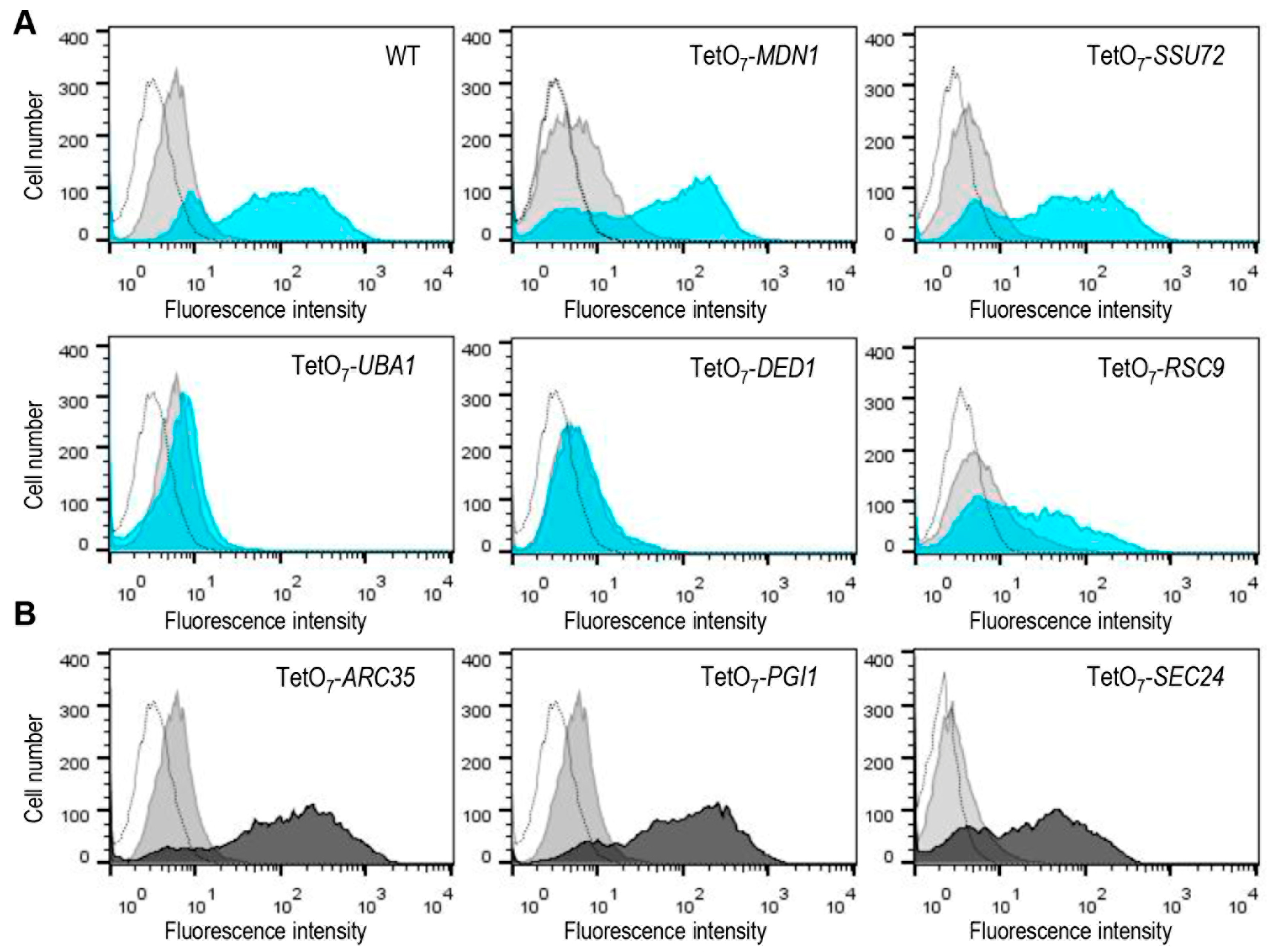
As described in Materials and Methods and illustrated in Figure 1, the complete collection of yeast mutants expressing Mlp1-GFP was grown in a rich medium including 10 µg/mL of doxycycline and then exposed, or not, to CW for 3 h. Next, GFP fluorescence levels, as the readout of Mlp1-GFP amounts for each mutant tested, were quantified by flow cytometry. The wild-type strain was always included as a control of MLP1 induction in each experiment. Thus, the effect of the down-regulation of the expression of each conditional allele on Mlp1-GFP levels after CW treatment was determined, calculating first the ratio of MFIs between treatment and control conditions (absence of CW) for the wild-type and the mutant strains. Finally, the ratio obtained for each mutant strain was divided by that calculated for the wild-type strain. Flow cytometry data analyses uncovered mutants with changes in global MFIs and mutants in which the number of cells expressing detectable fluorescence signals was affected. Representative examples of the different flow cytometry patterns obtained are shown in Figure 2A. From these observations, in a first round of screening, we preliminarily selected those mutants showing at least a 75% decrease in Mlp1-GFP levels with respect to those of the wild-type strain (fluorescence ratio mutant/fluorescence ratio WT ≤ 0.25) and/or showing a percentage of GFP positive cells lower than 15% (Table S3). Next, two additional experiments were conducted using different yeast transformants of this group of mutants to finally define the positive hits of the screening (see Material and Methods for details and Table S4).
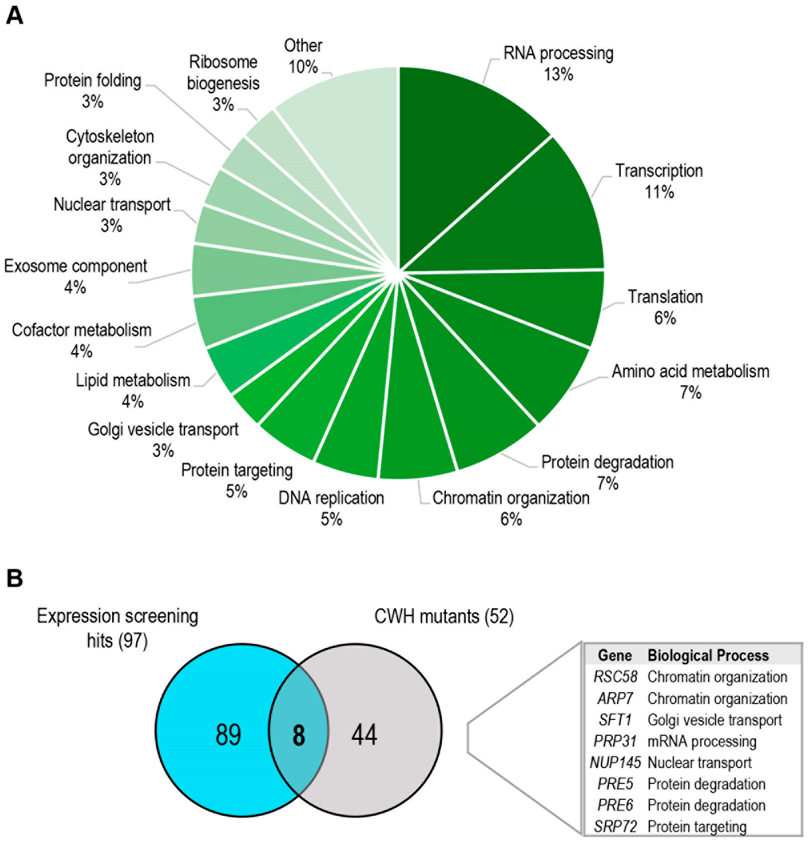
We sought functional categories within the group of 97 mutants selected (Figure 3A and Table S4). The most represented group (17%) corresponded to that associated with RNA metabolism, including RNA processing and degradation (exosome component). The identification of this group of genes, in conjunction with those associated with transcription, ribosome biogenesis, translation, and protein folding, could be explained as a consequence of alterations in the cellular machinery required for the correct expression and translation of the gene reporter Mlp1 under cell wall stress. However, it cannot be ruled out that CWI pathway signalling or regulatory defects could also be present in some mutants. Other groups of interest corresponded to those related to lipid metabolism, protein degradation, nucleocytoplasmic RNA transport, or chromatin remodelling. Within the first group, we found genes involved in ergosterol biosynthesis (ERG1 and ERG26) or sphingolipid metabolism (LCB2 and TSC13). Their identification is in accordance with the fact that sphingolipids are a structurally diverse class of lipids implicated in many cell signalling functions and interconnected with the ergosterol pathway [38]. The protein degradation group includes several subunits of the 26S proteasome (PRE5, PRE6, RPN11, and RPT2) and some ubiquitin-related enzymes (UBA1, CDC34, and HRT1). Interestingly, proteasome up-regulation has been proposed under different stress conditions, such as heat shock or oxidative stress [39,40]. Moreover, a control of proteasome abundance mediated by the MAPK Slt2 has also been reported [41]. Since proteasome-mediated degradation regulates the turnover of numerous cellular proteins involved in the majority of cellular processes [42], it is difficult, in these mutants, to discern between the existence of direct effects on CWI pathway regulation, mainly due to ubiquitination, or pleiotropic effects that indirectly alter the adaptive transcriptional response. Interestingly, proteasome inhibition leads to a slight increase in CW sensitivity (our unpublished results). Additional work will be required to clarify the molecular mechanisms involved in the CWI pathway-proteosome interaction. Still, the identification of only certain elements of this large protease complex allows us to hypothesize that they are particularly important under cell wall stress conditions.
Other mutants of interest are those related to nucleocytoplasmic RNA transport (NUP145 or MEX67). Considering that, under heat shock stress, a selective nuclear export of heat-shock mRNAs and retention of regular transcripts mediated by Mex67 have recently been reported [43], similar or alternative mechanisms may take place under other stress conditions. Finally, it is important to highlight the presence of genes functionally related to chromatin remodelling, including essential subunits of the SWI/SNF (ARP7) and SAGA (TAF9) chromatin-modifying complexes, which were previously associated with the regulation of the expression of CWI-dependent genes upon cell wall stress [9,12]. Interestingly, within this group of chromatin-associated mutants, we also found essential elements of the RSC remodelling complex (ARP7, RSC58, and RSC9). In agreement, different ATP-dependent chromatin remodelling complexes work together to regulate the expression of stress-responsive genes [44,45].
Comparative analyses of the genes identified in both the phenotypic and transcriptional screenings under the imposed cut-off thresholds revealed a reduced number of mutants, including two subunits of the RSC complex and two proteasome members (Figure 3B). This low overlap agrees with previous studies with the complete collection of viable deletion mutants after zymolyase treatment. In these assays, 154 mutants exhibited hypersensitivity to zymolyase [22], and 159 mutants showed a reduction in the MLP1-reporter expression under zymolyase treatment [9], but only 35 mutants were found common in both studies. In agreement, most of the genes included in the cell wall damage compensatory mechanism work cooperatively to support cell wall integrity, but they are not essential individually to maintain cell viability under stress situations, except for those involved in signalling or transcriptional regulation, where the overall response is impaired. The elements identified of the RSC complex would belong to this category.

3.3. Impact of the RSC Complex on the Expression of CWI Pathway-Dependent Genes

We uncovered several elements of the RSC chromatin remodelling complexes showing CW hypersensitivity and/or alteration of Mlp1-GFP levels under cell wall stress conditions (Table S2 and Table S4). The RSC complex consists of 17 subunits (both essential and dispensable for cell viability) that can work in smaller subcomplexes, hindering its functional characterization [46]. This complex is mainly involved in transcription regulation, remodelling nucleosomes in promoter and transcribed coding sequences [47,48]. It also plays a role in chromosomal transactions such as DNA replication and repair and chromosome segregation [47,49]. In agreement with the functional complexity of RSC, previous large-scale transcriptional studies carried out under basal growth conditions of several yeast RSC subunit mutants (rsc3, rsc30, rsc4 or rsc14) have demonstrated a small overlap in the genes regulated by the different subunits [13,50,51]. Additionally, in these studies, some RSC subunits modulated the expression of a reduced group of cell wall-related genes.
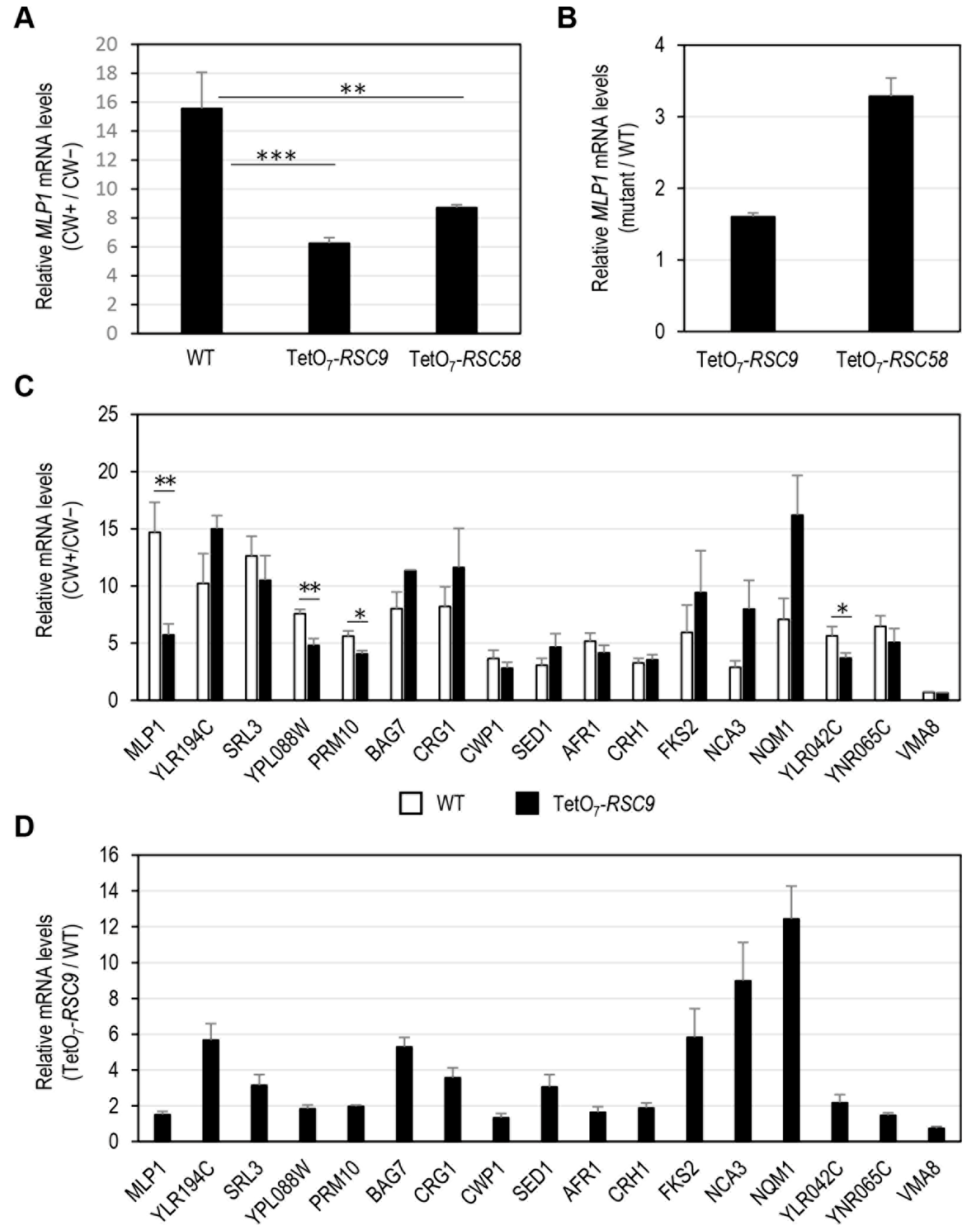
We aimed to further investigate the relationship between possible alterations in the cell wall of some essential rsc mutants identified in our screening and modulation of the adaptive transcriptional response mediated by the CWI pathway when cell wall integrity is compromised. Firstly, mRNA levels of the CWI reporter gene MLP1 were quantified via RT-qPCR in rsc9 and rsc58 mutants. These subunits belong to the substrate recruitment module (SRM) of the RSC complex [46]. In the case of Rsc9, this element has been involved both in the repression and activation of specific stress-regulated genes (i.e., hydrogen peroxide and rapamycin [52] or osmostress [53]). The transcriptional induction of MLP1 in the presence of CW was significantly affected in these mutants under shut-off gene conditions (Figure 4A), validating the experimental approach developed in our screening and suggesting that the RSC complex regulates transcription upon cell wall stress. In the absence of stress, both subunits negatively regulate MLP1 transcription, as inferred from the increase in the amount of MLP1 transcripts in both mutants, particularly in the rsc58 strain (Figure 4B). Next, we investigated the impact of rsc mutations on the expression of additional CWI-dependent genes. To this end, the mRNA levels of 15 other genes induced by cell wall stress [8,36,37,54,55] were analysed under the conditions indicated above, focusing on the rsc9 mutant (Figure 4C). Interestingly, we observed two types of genes regarding the effect caused by the absence of Rsc9. One group, including YPL088W, PRM10, and YLR042C, showed a behaviour similar to MLP1 in terms of the impact on their expression levels after treatment with CW compared with the up-regulation observed in the wild-type strain; and a second, more extensive group, in which no significant alteration in transcript levels was observed. Quantification of the transcript levels of these genes under non-stress conditions in the rsc9 mutant relative to those of the wild-type strain showed a derepression of genes YLR194C, SRL3, BAG7, CRG1, SED1, FKS2, NCA3 and NQM1 in the absence of RSC9 (Figure 4D). This basal regulation was not manifested in the case of the VMA8 gene, which encodes a subunit of vacuolar ATPase non-transcriptionally regulated by cell wall stress. Therefore, our results indicate that the RSC complex regulates the gene expression of certain CWI-related genes both under stress and non-stress conditions. As described above, positive or negative transcriptional modulation of specific genes under different stress conditions, probably a consequence of their interaction with other regulators (repressors or activators), has been previously described for Rsc9 [52] and other RSC subunits [13,51]. Under basal (non-stress) conditions, RSC could also be involved in stabilizing the nucleosomes present at some CWI-dependent genes. Similarly, some UPR genes show high basal level expression when RSC is disrupted, indicating the presence of inherently altered chromatin in their absence [45]. Upon cell wall stress conditions, Slt2 phosphorylates and activates the transcription factor Rlm1 to recruit both elements to the promoters of CWI-responsive genes in complex with SWI/SNF [9]. SWI/SNF activity is necessary to evict nucleosomes positioned in this region and permit pre-initiation complex (PIC) assembly in cooperation with the SAGA complex, which mediates histone acetylation for nucleosome reorganization [6,12]. The results described here suggest that the RSC complex could cooperate with SWI/SNF and SAGA complexes for the chromatin remodelling necessary for the transcriptional activation of CWI-dependent genes upon stress. There seems to be distinct requirements of the RSC complex for remodelling particular genes in a stress-dependent manner. Thus, the influence of the RSC complex via its subunit Rsc9 on the expression levels of genes regulated by the CWI pathway shows a highly gene-specific regulatory pattern, probably a consequence of each particular promoter architecture, chromatin status, and interaction with the transcriptional machinery.

3.4. Identification of Essential Genes Associated with the Constitutive Activation of the Cell Wall Integrity Pathway

One of the objectives of this work was to uncover essential genes that could be directly or indirectly related to the presence of alterations in the fungal cell wall. On many occasions, they were revealed by the constitutive activation of the CWI route without the need to apply any type of external stimulus. Presumably, this type of mutant should be included among those that exhibited an increment in Mlp1-GFP levels in the absence of CW when the essential gene expression is blocked (presence of doxycycline). We arbitrarily ascribed to this group those mutants exhibiting a relevant level of MFI (higher than three-fold) with respect to the value obtained for the wild-type strain (Table 2 and Table S5). Representative flow cytometry profiles of these mutants are shown in Figure 2B. Eventually, 41 yeast strains met these criteria. After functional analyses, as described above, we found that approximately 68% belonged to four functional groups closely related to cell wall homeostasis: Protein glycosylation, Golgi vesicle transport, Glycosylphosphatidylinositol (GPI)-anchor assembly, and Cytoskeleton organization. GPI-anchored proteins (GPI-APs) are luminal secretory cargos that are attached by a post-translational glycolipid modification, the GPI anchor, to the plasma membrane and/or cell wall in yeast, consisting of a process conserved among eukaryotes [56]. A number of the GPI proteins in yeast serve enzymatic functions required for the biosynthesis and continuous shape adaptations of the cell wall, some seem to be structural elements of the cell wall, and others mediate cell adhesion [57]. The secretory pathway plays a key role in cell wall construction and remodelling, which explains the identification of this type of mutant in our screening. In fact, some of them, Sec24 and Sar1 belonging to the coat protein complex II (COPII), are involved in the specialized transport of GPI-APs to the ER [56]. The relationship between protein glycosylation and cell wall integrity is well established since most cell wall proteins are glycoproteins that have passed through the secretory pathway in transit to the cell wall. Thus, the isolation of this type of mutant is not unexpected given the importance of glycosylation for the proper conformation, localization, and function of cell wall proteins [58]. Regarding actin cytoskeleton-related genes, potential cell wall alterations could be a consequence of the requirement of this structure for correct spatiotemporal vesicular trafficking and cell wall assembly [59]. In accordance with the putative existence of alterations in the cell wall of mutants displaying incremented basal levels of Mlp1-GFP, about 17% were also selected in the screening for CW hypersensitivity (Table 2). In agreement with this idea, this value was significantly higher (50%) when a less strict cut-off point (0.4) was considered for CW hypersensitivity (Table 2).
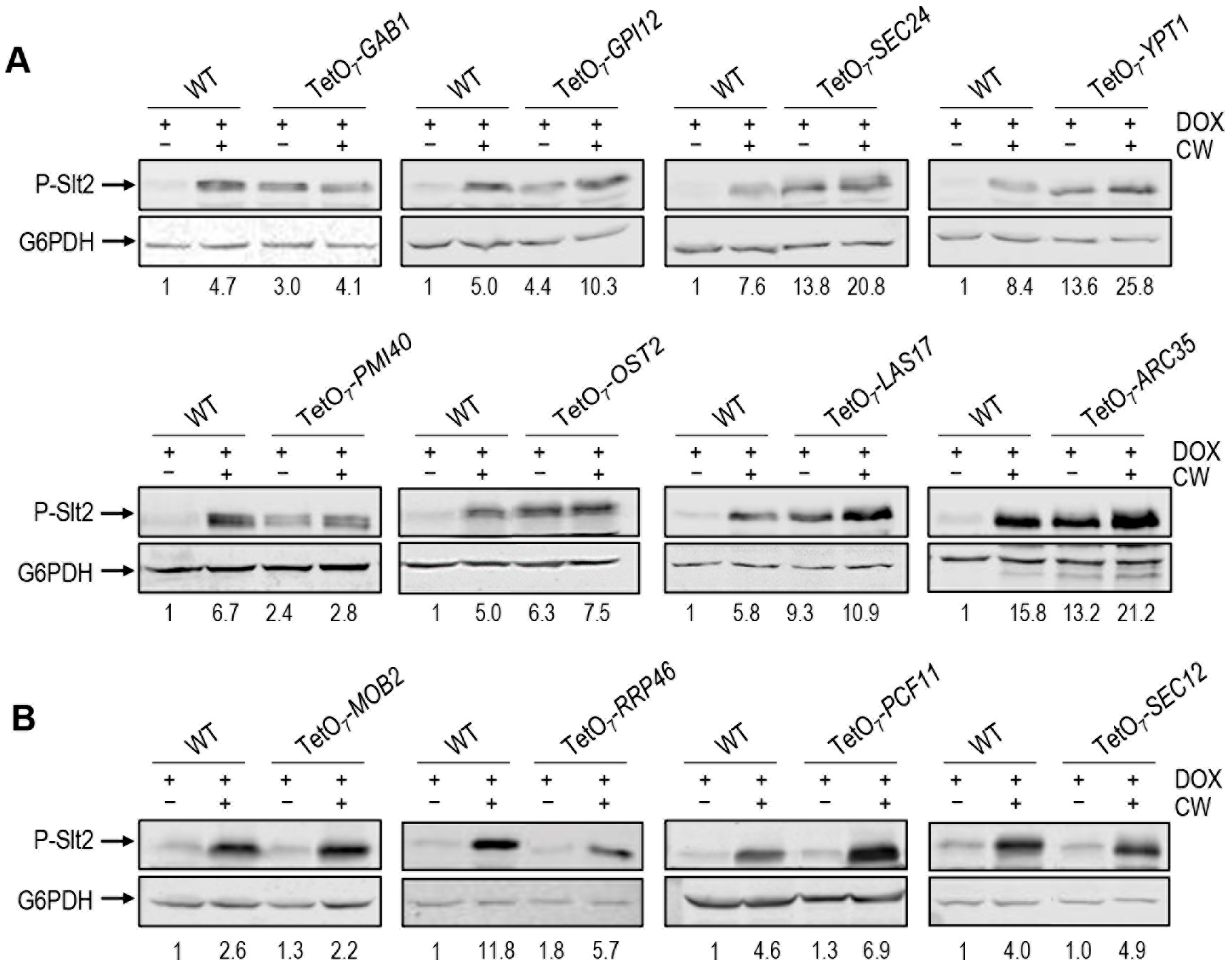
To further investigate whether the increase in Mlp1 expression in these mutants could be associated with the activation of the CWI pathway, we analysed the levels of phospho-Slt2 (typical readout of the level of activation of this pathway) in two mutants from each of the four functional groups described above. To this end, we obtained total protein extracts from these strains grown in the presence of doxycycline and treated or not with CW. Next, these extracts were analysed by Western blotting using an antibody that recognizes the dually phosphorylated (active) form of Slt2. As shown in Figure 5A, in the mutant strains tested, the levels of phospho-Slt2 under basal growth conditions were clearly higher than those observed in the wild-type strain. These results support the notion that the increase in Mlp1 levels detected in these mutants, when the conditional allele is off, is a consequence of the activation of the CWI pathway. Interestingly, as shown in Figure 5A, in the presence of CW, in some mutants the activation level (phospho-Slt2) does not increase beyond basal levels, while in others the ability to over-activate the pathway seems to be maintained. As expected, this effect was not observed in other mutants not affecting Mlp1-GFP levels under non-stress conditions (Figure 5B). Future gene-focused studies will be required to decipher whether the constitutive activation of the CWI pathway is due to signalling via the cell surface CWI pathway associated sensors or is mediated by downstream elements of the pathway, which eventually induces Slt2 phosphorylation.
Globally, it is noteworthy that the percentage of essential genes directly or indirectly related to cell wall integrity revealed in this work is similar to that predicted from pioneering cell wall phenotypic studies using viable knock-out mutant strains [27]; this supports the importance of maintaining the integrity of this fungal structure. The results described herein improve our knowledge regarding the putative connection between essential genes and cell wall integrity. This will greatly facilitate further in-depth studies directed towards investigating at which cellular level (signalling, transcriptional or post-transcriptional) a specific essential gene impacts on the adaptive response to withstand cell wall injuries. Moreover, the results of this work will be useful for the development of targeted studies on specific genes, particularly in the case of the search and design of antifungal agents directed against the cell wall.

Supplementary Materials

The following supporting information can be downloaded at: https://www.mdpi.com/article/10.3390/jof8070718/s1, Table S1: Calcofluor white hypersensitivity screening dataset; Table S2: Positive hits from the Calcofluor white hypersensitivity screening; Table S3: Mlp1-GFP expression screening dataset; Table S4: Positive hits from the Mlp1-GFP expression screening; Table S5: Mutant strains showing increased basal levels of Mlp1-GFP; Figure S1: CW susceptibility assays of some selected hits from the CW sensitivity screening. Figure S2: Fluorescence microscopy images of strains expressing Mlp1-GFP.

Author Contributions

Conceptualization, J.M.R.-P., A.B.S. and J.A.; methodology, S.D.-M., A.B.S. and J.M.R.-P.; formal analysis, A.B.S. and J.M.R.-P., investigation, S.D.-M., J.M., Y.P. and A.B.S.; writing–original draft preparation, J.M.R.-P. and A.B.S.; Writing-Review & Editing, J.A. and C.N.; visualization, A.B.S. Supervision, J.M.R.-P., C.N. and J.A.; project administration, J.A. Funding Acquisition, J.A. and C.N. All authors have read and agreed to the published version of the manuscript.

Funding

This work was supported by the grants BIO2016-79289-P and PID2019-105223GB-I00 (Ministerio de Ciencia e Innovación, MICINN, Spain) and S2017/BMD3691-InGEMICS (Comunidad de Madrid and European Structural and Investment Funds) to J.A and the Special Chair on Fermented Beverages and Health to C.N., J.M. and Y.P. were the recipient of the research contracts (Laboratory technician) PEJ15/BIO/TL-0538 and PEJ-2018-TL/BMD-11487 from Consejería de Educación e Investigación de la Comunidad de Madrid y del Fondo Social Europeo, respectively.

Institutional Review Board Statement

Not applicable.

Informed Consent Statement

Not applicable.

Data Availability Statement

The data presented in this study are available on request from the corresponding author.

Acknowledgments

All members of our research group (UCM-920640: Yeast Functional Genomics) at the Department of Microbiology and Parasitology (UCM, Madrid, Spain) are acknowledged for their support. Thanks are due to all members of the Genomics Unit (UCM, Spain) for their help with RT-qPCR and Carlos Pérez Martín and Amalia Vázquez Gutierrez of the Flow Cytometry and Fluorescence Microscopy Unit (UCM, Spain) for expert help in flow cytometry.

Conflicts of Interest

The authors declare no conflict of interest.

References

  1. Orlean, P. Architecture and biosynthesis of the Saccharomyces cerevisiae cell wall. Genetics 2012, 192, 775–818. [Google Scholar] [CrossRef] [PubMed] [Green Version]
  2. Cabib, E.; Arroyo, J. How carbohydrates sculpt cells: Chemical control of morphogenesis in the yeast cell wall. Nat. Rev. Microbiol. 2013, 11, 648–655. [Google Scholar] [CrossRef] [PubMed]
  3. Popolo, L.; Gualtieri, T.; Ragni, E. The yeast cell-wall salvage pathway. Med. Mycol. 2001, 39 (Suppl. 1), 111–121. [Google Scholar] [CrossRef]
  4. Sanz, A.B.; García, R.; Rodríguez-Peña, J.M.; Arroyo, J. The CWI Pathway: Regulation of the Transcriptional Adaptive Response to Cell Wall Stress in Yeast. J. Fungi 2017, 4, 1. [Google Scholar] [CrossRef] [PubMed] [Green Version]
  5. Levin, D.E. Regulation of cell wall biogenesis in Saccharomyces cerevisiae: The cell wall integrity signaling pathway. Genetics 2011, 189, 1145–1175. [Google Scholar] [CrossRef] [PubMed] [Green Version]
  6. Sanz, A.B.; García, R.; Pavon-Verges, M.; Rodriguez-Pena, J.M.; Arroyo, J. Control of Gene Expression via the Yeast CWI Pathway. Int. J. Mol. Sci. 2022, 23, 1791. [Google Scholar] [CrossRef] [PubMed]
  7. Jung, U.S.; Levin, D.E. Genome-wide analysis of gene expression regulated by the yeast cell wall integrity signalling pathway. Mol. Microbiol. 1999, 34, 1049–1057. [Google Scholar] [CrossRef]
  8. García, R.; Bermejo, C.; Grau, C.; Pérez, R.; Rodríguez-Peña, J.M.; Francois, J.; Nombela, C.; Arroyo, J. The global transcriptional response to transient cell wall damage in Saccharomyces cerevisiae and its regulation by the cell integrity signaling pathway. J. Biol. Chem. 2004, 279, 15183–15195. [Google Scholar] [CrossRef] [Green Version]
  9. Sanz, A.B.; García, R.; Rodríguez-Peña, J.M.; Díez-Muñiz, S.; Nombela, C.; Peterson, C.L.; Arroyo, J. Chromatin remodeling by the SWI/SNF complex is essential for transcription mediated by the yeast cell wall integrity MAPK pathway. Mol. Biol. Cell 2012, 23, 2805–2817. [Google Scholar] [CrossRef] [Green Version]
  10. Lagorce, A.; Hauser, N.C.; Labourdette, D.; Rodríguez, C.; Martin-Yken, H.; Arroyo, J.; Hoheisel, J.D.; Francois, J. Genome-wide analysis of the response to cell wall mutations in the yeast Saccharomyces cerevisiae. J. Biol. Chem. 2003, 278, 20345–20357. [Google Scholar] [CrossRef] [Green Version]
  11. Arias, P.; Díez-Muñiz, S.; García, R.; Nombela, C.; Rodríguez-Peña, J.M.; Arroyo, J. Genome-wide survey of yeast mutations leading to activation of the yeast cell integrity MAPK pathway: Novel insights into diverse MAPK outcomes. BMC Genom. 2011, 12, 390. [Google Scholar] [CrossRef] [PubMed] [Green Version]
  12. Sanz, A.B.; García, R.; Rodríguez-Peña, J.M.; Nombela, C.; Arroyo, J. Cooperation between SAGA and SWI/SNF complexes is required for efficient transcriptional responses regulated by the yeast MAPK Slt2. Nucleic Acids Res. 2016, 44, 7159–7172. [Google Scholar] [CrossRef] [PubMed] [Green Version]
  13. Angus-Hill, M.L.; Schlichter, A.; Roberts, D.; Erdjument-Bromage, H.; Tempst, P.; Cairns, B.R. A Rsc3/Rsc30 zinc cluster dimer reveals novel roles for the chromatin remodeler RSC in gene expression and cell cycle control. Mol. Cell 2001, 7, 741–751. [Google Scholar] [CrossRef]
  14. Chai, B.; Hsu, J.M.; Du, J.; Laurent, B.C. Yeast RSC function is required for organization of the cellular cytoskeleton via an alternative PKC1 pathway. Genetics 2002, 161, 575–584. [Google Scholar] [CrossRef]
  15. Wilson, B.; Erdjument-Bromage, H.; Tempst, P.; Cairns, B.R. The RSC chromatin remodeling complex bears an essential fungal-specific protein module with broad functional roles. Genetics 2006, 172, 795–809. [Google Scholar] [CrossRef] [Green Version]
  16. Wang, S.L.; Cheng, M.Y. The defects in cell wall integrity and G2-M transition of the htl1 mutant are interconnected. Yeast 2012, 29, 45–57. [Google Scholar] [CrossRef]
  17. Rando, O.J.; Winston, F. Chromatin and transcription in yeast. Genetics 2012, 190, 351–387. [Google Scholar] [CrossRef] [Green Version]
  18. Giaever, G.; Chu, A.M.; Ni, L.; Connelly, C.; Riles, L.; Veronneau, S.; Dow, S.; Lucau-Danila, A.; Anderson, K.; Andre, B.; et al. Functional profiling of the Saccharomyces cerevisiae genome. Nature 2002, 418, 387–391. [Google Scholar] [CrossRef]
  19. Page, N.; Gerard-Vincent, M.; Menard, P.; Beaulieu, M.; Azuma, M.; Dijkgraaf, G.J.; Li, H.; Marcoux, J.; Nguyen, T.; Dowse, T.; et al. A Saccharomyces cerevisiae genome-wide mutant screen for altered sensitivity to K1 killer toxin. Genetics 2003, 163, 875–894. [Google Scholar] [CrossRef]
  20. Serviene, E.; Luksa, J.; Orentaite, I.; Lafontaine, D.L.; Urbonavicius, J. Screening the budding yeast genome reveals unique factors affecting K2 toxin susceptibility. PLoS ONE 2012, 7, e50779. [Google Scholar] [CrossRef] [Green Version]
  21. Rodríguez-Peña, J.M.; Díez-Muñiz, S.; Bermejo, C.; Nombela, C.; Arroyo, J. Activation of the yeast cell wall integrity MAPK pathway by zymolyase depends on protease and glucanase activities and requires the mucin-like protein Hkr1 but not Msb2. FEBS Lett. 2013, 587, 3675–3680. [Google Scholar] [CrossRef] [PubMed] [Green Version]
  22. García, R.; Botet, J.; Rodríguez-Peña, J.M.; Bermejo, C.; Ribas, J.C.; Revuelta, J.L.; Nombela, C.; Arroyo, J. Genomic profiling of fungal cell wall-interfering compounds: Identification of a common gene signature. BMC Genom. 2015, 16, 683. [Google Scholar] [CrossRef] [PubMed] [Green Version]
  23. Ando, A.; Nakamura, T.; Murata, Y.; Takagi, H.; Shima, J. Identification and classification of genes required for tolerance to freeze-thaw stress revealed by genome-wide screening of Saccharomyces cerevisiae deletion strains. FEMS Yeast Res. 2007, 7, 244–253. [Google Scholar] [CrossRef] [PubMed] [Green Version]
  24. Auesukaree, C.; Damnernsawad, A.; Kruatrachue, M.; Pokethitiyook, P.; Boonchird, C.; Kaneko, Y.; Harashima, S. Genome-wide identification of genes involved in tolerance to various environmental stresses in Saccharomyces cerevisiae. J. Appl. Genet. 2009, 50, 301–310. [Google Scholar] [CrossRef] [PubMed]
  25. Rodríguez-Peña, J.M.; Díez-Muñiz, S.; Nombela, C.; Arroyo, J. A yeast strain biosensor to detect cell wall-perturbing agents. J. Biotechnol. 2008, 133, 311–317. [Google Scholar] [CrossRef]
  26. Giaever, G.; Nislow, C. The yeast deletion collection: A decade of functional genomics. Genetics 2014, 197, 451–465. [Google Scholar] [CrossRef] [Green Version]
  27. De Groot, P.W.; Ruiz, C.; Vázquez de Aldana, C.R.; Duenas, E.; Cid, V.J.; Del Rey, F.; Rodríguez-Peña, J.M.; Pérez, P.; Andel, A.; Caubín, J.; et al. A genomic approach for the identification and clasification of genes involved in cell wall formation and its regulation in Saccharomyces cerevisiae. Comp. Funct. Genom. 2001, 2, 124–142. [Google Scholar] [CrossRef] [Green Version]
  28. Norman, K.L.; Kumar, A. Mutant power: Using mutant allele collections for yeast functional genomics. Brief Funct. Genom. 2016, 15, 75–84. [Google Scholar] [CrossRef] [Green Version]
  29. Mnaimneh, S.; Davierwala, A.P.; Haynes, J.; Moffat, J.; Peng, W.T.; Zhang, W.; Yang, X.; Pootoolal, J.; Chua, G.; Lopez, A.; et al. Exploration of essential gene functions via titratable promoter alleles. Cell 2004, 118, 31–44. [Google Scholar] [CrossRef] [Green Version]
  30. Cappell, S.D.; Baker, R.; Skowyra, D.; Dohlman, H.G. Systematic analysis of essential genes reveals important regulators of G protein signaling. Mol. Cell 2010, 38, 746–757. [Google Scholar] [CrossRef] [Green Version]
  31. Tatjer, L.; Sacristan-Reviriego, A.; Casado, C.; Gonzalez, A.; Rodriguez-Porrata, B.; Palacios, L.; Canadell, D.; Serra-Cardona, A.; Martin, H.; Molina, M.; et al. Wide-Ranging Effects of the Yeast Ptc1 Protein Phosphatase Acting Through the MAPK Kinase Mkk1. Genetics 2016, 202, 141–156. [Google Scholar] [CrossRef] [PubMed]
  32. Bermejo, C.; Rodríguez, E.; García, R.; Rodríguez-Peña, J.M.; Rodríguez de la Concepcion, M.L.; Rivas, C.; Arias, P.; Nombela, C.; Posas, F.; Arroyo, J. The sequential activation of the yeast HOG and SLT2 pathways is required for cell survival to cell wall stress. Mol. Biol. Cell. 2008, 19, 1113–1124. [Google Scholar] [CrossRef] [Green Version]
  33. Livak, K.J.; Schmittgen, T.D. Analysis of relative gene expression data using real-time quantitative PCR and the 2(-Delta Delta C(T)) Method. Methods 2001, 25, 402–408. [Google Scholar] [CrossRef] [PubMed]
  34. Zhao, F.; Li, J.; Lin, K.; Chen, H.; Lin, Y.; Zheng, S.; Liang, S.; Han, S. Genome-wide screening of Saccharomyces cerevisiae deletion mutants reveals cellular processes required for tolerance to the cell wall antagonist calcofluor white. Biochem. Biophys. Res. Commun. 2019, 518, 1–6. [Google Scholar] [CrossRef] [PubMed]
  35. Chen, P.; Wang, D.; Chen, H.; Zhou, Z.; He, X. The nonessentiality of essential genes in yeast provides therapeutic insights into a human disease. Genome Res. 2016, 26, 1355–1362. [Google Scholar] [CrossRef] [PubMed] [Green Version]
  36. García, R.; Rodríguez-Peña, J.M.; Bermejo, C.; Nombela, C.; Arroyo, J. The high osmotic response and cell wall integrity pathways cooperate to regulate transcriptional responses to zymolyase-induced cell wall stress in Saccharomyces cerevisiae. J. Biol. Chem. 2009, 284, 10901–10911. [Google Scholar] [CrossRef] [Green Version]
  37. García, R.; Bravo, E.; Díez-Muñiz, S.; Nombela, C.; Rodríguez-Peña, J.M.; Arroyo, J. A novel connection between the Cell Wall Integrity and the PKA pathways regulates cell wall stress response in yeast. Sci. Rep. 2017, 7, 5703. [Google Scholar] [CrossRef]
  38. Montefusco, D.J.; Matmati, N.; Hannun, Y.A. The yeast sphingolipid signaling landscape. Chem. Phys. Lipids 2014, 177, 26–40. [Google Scholar] [CrossRef] [Green Version]
  39. Hahn, J.S.; Neef, D.W.; Thiele, D.J. A stress regulatory network for co-ordinated activation of proteasome expression mediated by yeast heat shock transcription factor. Mol. Microbiol. 2006, 60, 240–251. [Google Scholar] [CrossRef]
  40. Ma, M.; Liu, Z.L. Comparative transcriptome profiling analyses during the lag phase uncover YAP1, PDR1, PDR3, RPN4, and HSF1 as key regulatory genes in genomic adaptation to the lignocellulose derived inhibitor HMF for Saccharomyces cerevisiae. BMC Genom. 2010, 11, 660. [Google Scholar] [CrossRef] [Green Version]
  41. Rousseau, A.; Bertolotti, A. An evolutionarily conserved pathway controls proteasome homeostasis. Nature 2016, 536, 184–189. [Google Scholar] [CrossRef] [PubMed]
  42. Livneh, I.; Cohen-Kaplan, V.; Cohen-Rosenzweig, C.; Avni, N.; Ciechanover, A. The life cycle of the 26S proteasome: From birth, through regulation and function, and onto its death. Cell Res. 2016, 26, 869–885. [Google Scholar] [CrossRef] [Green Version]
  43. Zander, G.; Hackmann, A.; Bender, L.; Becker, D.; Lingner, T.; Salinas, G.; Krebber, H. mRNA quality control is bypassed for immediate export of stress-responsive transcripts. Nature 2016, 540, 593–596. [Google Scholar] [CrossRef] [PubMed]
  44. Erkina, T.Y.; Zou, Y.; Freeling, S.; Vorobyev, V.I.; Erkine, A.M. Functional interplay between chromatin remodeling complexes RSC, SWI/SNF and ISWI in regulation of yeast heat shock genes. Nucleic Acids Res. 2010, 38, 1441–1449. [Google Scholar] [CrossRef] [Green Version]
  45. Sahu, R.K.; Singh, S.; Tomar, R.S. The ATP-dependent SWI/SNF and RSC chromatin remodelers cooperatively induce unfolded protein response genes during endoplasmic reticulum stress. Biochim. Biophys. Acta Gene Regul. Mech. 2021, 1864, 194748. [Google Scholar] [CrossRef] [PubMed]
  46. Ye, Y.; Wu, H.; Chen, K.; Clapier, C.R.; Verma, N.; Zhang, W.; Deng, H.; Cairns, B.R.; Gao, N.; Chen, Z. Structure of the RSC complex bound to the nucleosome. Science 2019, 366, 838–843. [Google Scholar] [CrossRef]
  47. Lorch, Y.; Kornberg, R.D. Chromatin-remodeling for transcription. Q. Rev. Biophys. 2017, 50, e5. [Google Scholar] [CrossRef]
  48. Biernat, E.; Kinney, J.; Dunlap, K.; Rizza, C.; Govind, C.K. The RSC complex remodels nucleosomes in transcribed coding sequences and promotes transcription in Saccharomyces cerevisiae. Genetics 2021, 217, iyab021. [Google Scholar] [CrossRef]
  49. Sing, T.L.; Hung, M.P.; Ohnuki, S.; Suzuki, G.; San Luis, B.J.; McClain, M.; Unruh, J.R.; Yu, Z.; Ou, J.; Marshall-Sheppard, J.; et al. The budding yeast RSC complex maintains ploidy by promoting spindle pole body insertion. J. Cell Biol. 2018, 217, 2445–2462. [Google Scholar] [CrossRef] [Green Version]
  50. Kasten, M.; Szerlong, H.; Erdjument-Bromage, H.; Tempst, P.; Werner, M.; Cairns, B.R. Tandem bromodomains in the chromatin remodeler RSC recognize acetylated histone H3 Lys14. EMBO J. 2004, 23, 1348–1359. [Google Scholar] [CrossRef] [Green Version]
  51. Conde, R.; Cueva, R.; Larriba, G. Rsc14-controlled expression of MNN6, MNN4 and MNN1 regulates mannosylphosphorylation of Saccharomyces cerevisiae cell wall mannoproteins. FEMS Yeast Res. 2007, 7, 1248–1255. [Google Scholar] [CrossRef] [PubMed] [Green Version]
  52. Damelin, M.; Simon, I.; Moy, T.I.; Wilson, B.; Komili, S.; Tempst, P.; Roth, F.P.; Young, R.A.; Cairns, B.R.; Silver, P.A. The genome-wide localization of Rsc9, a component of the RSC chromatin-remodeling complex, changes in response to stress. Mol. Cell 2002, 9, 563–573. [Google Scholar] [CrossRef] [Green Version]
  53. Mas, G.; de Nadal, E.; Dechant, R.; Rodríguez de la Concepcion, M.L.; Logie, C.; Jimeno-Gonzalez, S.; Chavez, S.; Ammerer, G.; Posas, F. Recruitment of a chromatin remodelling complex by the Hog1 MAP kinase to stress genes. EMBO J. 2009, 28, 326–336. [Google Scholar] [CrossRef] [PubMed]
  54. Garcia, R.; Itto-Nakama, K.; Rodriguez-Pena, J.M.; Chen, X.; Sanz, A.B.; de Lorenzo, A.; Pavon-Verges, M.; Kubo, K.; Ohnuki, S.; Nombela, C.; et al. Poacic acid, a beta-1,3-glucan-binding antifungal agent, inhibits cell-wall remodeling and activates transcriptional responses regulated by the cell-wall integrity and high-osmolarity glycerol pathways in yeast. FASEB J. 2021, 35, e21778. [Google Scholar] [CrossRef] [PubMed]
  55. Rodríguez-Peña, J.M.; Pérez-Diaz, R.M.; Alvarez, S.; Bermejo, C.; García, R.; Santiago, C.; Nombela, C.; Arroyo, J. The ‘yeast cell wall chip’—A tool to analyse the regulation of cell wall biogenesis in Saccharomyces cerevisiae. Microbiology 2005, 151, 2241–2249. [Google Scholar] [CrossRef] [Green Version]
  56. Muñiz, M.; Riezman, H. Trafficking of glycosylphosphatidylinositol anchored proteins from the endoplasmic reticulum to the cell surface. J. Lipid Res. 2016, 57, 352–360. [Google Scholar] [CrossRef] [Green Version]
  57. Pittet, M.; Conzelmann, A. Biosynthesis and function of GPI proteins in the yeast Saccharomyces cerevisiae. Biochim. Biophys. Acta 2007, 1771, 405–420. [Google Scholar] [CrossRef] [Green Version]
  58. Bowman, S.M.; Free, S.J. The structure and synthesis of the fungal cell wall. Bioessays 2006, 28, 799–808. [Google Scholar] [CrossRef]
  59. Mishra, M.; Huang, J.; Balasubramanian, M.K. The yeast actin cytoskeleton. FEMS Microbiol. Rev. 2014, 38, 213–227. [Google Scholar] [CrossRef] [Green Version]
  60. Curwin, A.J.; von Blume, J.; Malhotra, V. Cofilin-mediated sorting and export of specific cargo from the Golgi apparatus in yeast. Mol. Biol. Cell 2012, 23, 2327–2338. [Google Scholar] [CrossRef]
  61. Li, H.; Page, N.; Bussey, H. Actin patch assembly proteins Las17p and Sla1p restrict cell wall growth to daughter cells and interact with cis-Golgi protein Kre6p. Yeast 2002, 19, 1097–1112. [Google Scholar] [CrossRef] [PubMed]
  62. Sukegawa, Y.; Negishi, T.; Kikuchi, Y.; Ishii, K.; Imanari, M.; Ghanegolmohammadi, F.; Nogami, S.; Ohya, Y. Genetic dissection of the signaling pathway required for the cell wall integrity checkpoint. J. Cell Sci. 2018, 131, jcs219063. [Google Scholar] [CrossRef] [PubMed] [Green Version]
  63. Mahfouz, H.; Ragnini-Wilson, A.; Venditti, R.; De Matteis, M.A.; Wilson, C. Mutational analysis of the yeast TRAPP subunit Trs20p identifies roles in endocytic recycling and sporulation. PLoS ONE 2012, 7, e41408. [Google Scholar] [CrossRef] [PubMed] [Green Version]
  64. Guo, Q.; Duan, Y.; Meng, N.; Liu, Y.; Luo, G. The N-terminus of Sec3 is required for cell wall integrity in yeast. Biochimie 2020, 177, 30–39. [Google Scholar] [CrossRef]
  65. Suda, Y.; Tachikawa, H.; Inoue, I.; Kurita, T.; Saito, C.; Kurokawa, K.; Nakano, A.; Irie, K. Activation of Rab GTPase Sec4 by its GEF Sec2 is required for prospore membrane formation during sporulation in yeast Saccharomyces cerevisiae. FEMS Yeast Res. 2018, 18, fox095. [Google Scholar] [CrossRef]
  66. Bialek-Wyrzykowska, U.; Bauer, B.E.; Wagner, W.; Kohlwein, S.D.; Schweyen, R.J.; Ragnini, A. Low levels of Ypt protein prenylation cause vesicle polarization defects and thermosensitive growth that can be suppressed by genes involved in cell wall maintenance. Mol. Microbiol. 2000, 35, 1295–1311. [Google Scholar] [CrossRef] [Green Version]
  67. Orlowski, J.; Machula, K.; Janik, A.; Zdebska, E.; Palamarczyk, G. Dissecting the role of dolichol in cell wall assembly in the yeast mutants impaired in early glycosylation reactions. Yeast 2007, 24, 239–252. [Google Scholar] [CrossRef] [Green Version]
  68. Bickle, M.; Delley, P.A.; Schmidt, A.; Hall, M.N. Cell wall integrity modulates RHO1 activity via the exchange factor ROM2. EMBO J. 1998, 17, 2235–2245. [Google Scholar] [CrossRef] [Green Version]
  69. Machi, K.; Azuma, M.; Igarashi, K.; Matsumoto, T.; Fukuda, H.; Kondo, A.; Ooshima, H. Rot1p of Saccharomyces cerevisiae is a putative membrane protein required for normal levels of the cell wall 1,6-beta-glucan. Microbiology 2004, 150, 3163–3173. [Google Scholar] [CrossRef] [Green Version]
  70. Absmanner, B.; Schmeiser, V.; Kampf, M.; Lehle, L. Biochemical characterization, membrane association and identification of amino acids essential for the function of Alg11 from Saccharomyces cerevisiae, an alpha1,2-mannosyltransferase catalysing two sequential glycosylation steps in the formation of the lipid-linked core oligosaccharide. Biochem. J. 2010, 426, 205–217. [Google Scholar] [CrossRef] [Green Version]
  71. Rodríguez-Peña, J.M.; Rodríguez, C.; Alvarez, A.; Nombela, C.; Arroyo, J. Mechanisms for targeting of the Saccharomyces cerevisiae GPI-anchored cell wall protein Crh2p to polarised growth sites. J. Cell Sci. 2002, 115, 2549–2558. [Google Scholar] [CrossRef] [PubMed]
  72. Rodríguez-Pachon, J.M.; Martin, H.; North, G.; Rotger, R.; Nombela, C.; Molina, M. A novel connection between the yeast Cdc42 GTPase and the Slt2-mediated cell integrity pathway identified through the effect of secreted Salmonella GTPase modulators. J. Biol. Chem. 2002, 277, 27094–27102. [Google Scholar] [CrossRef] [PubMed] [Green Version]
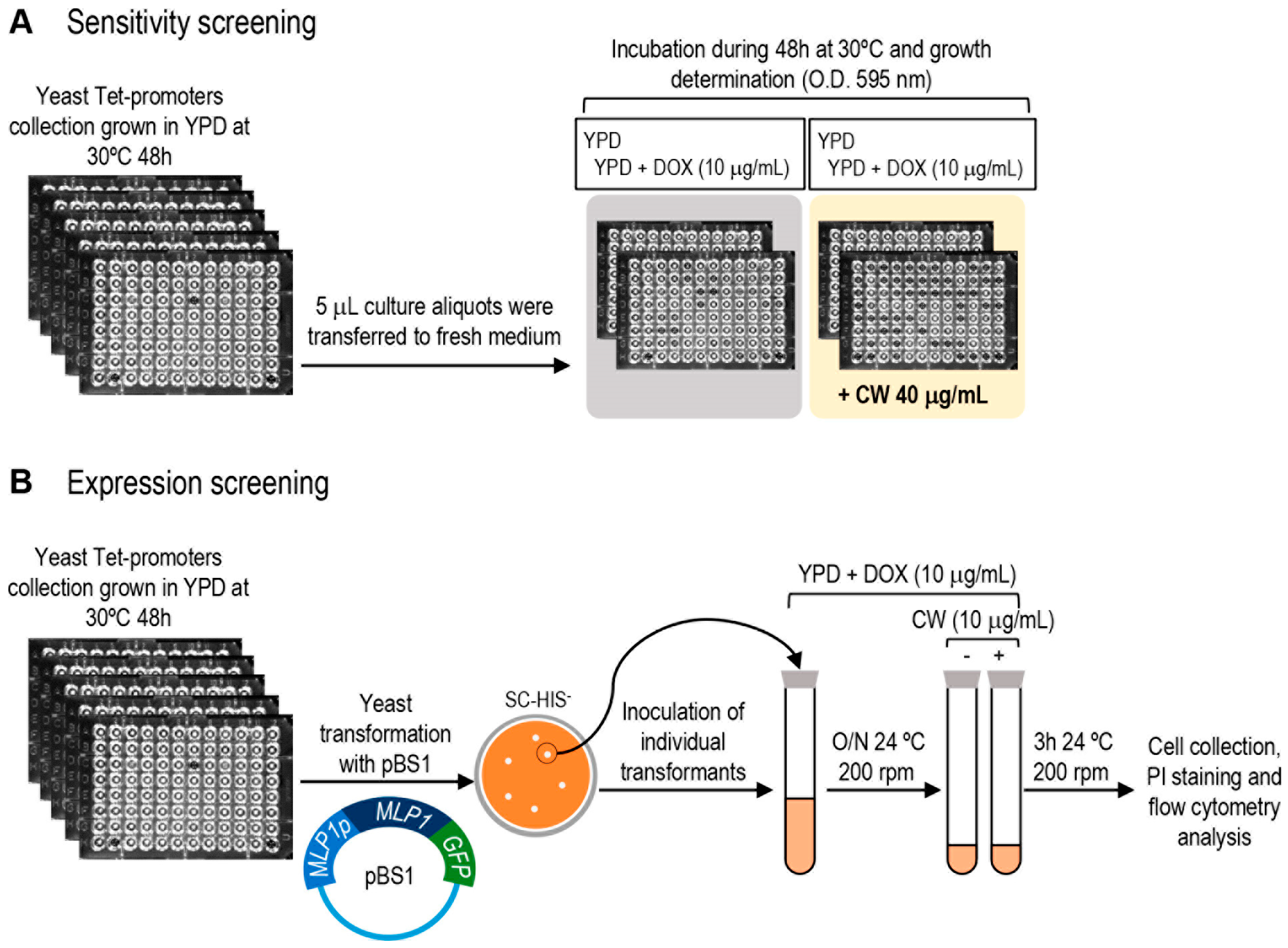
Figure 1. Overview of the screening strategies designed to identify cell wall-related mutants in essential genes. (A) Hypersensitive phenotype to Calcofluor white was studied in a 96-well microtiter plate format. (B) Detection of altered expression levels of the CWI-reporter gene KDX1/MLP1. The complete yeast Tet-promoter collection was transformed with the plasmid pBS1 (MLP1p-MLP1-GFP) and individual transformants were tested for Mlp1-GFP expression in the presence or absence of Calcofluor white (CW) by flow cytometry. DOX: exposure to doxycycline at the indicated concentration. PI: Propidium Iodide.
Jof 08 00718 g001
Figure 2. Identification of mutants from the yTHC collection that exhibit altered Mlp1-GFP levels by flow cytometry analysis. (A) Representative experiments of mutants in which Mlp1-GFP fluorescence levels were found to be affected (lower panels), or not affected (upper panels), compared with those of the wild-type strain. For each strain, histograms show the distribution of GFP fluorescence under non-stress conditions (grey-filled) and after treatment with Calcofluor white (10 μg/mL for 3 h) (blue-filled) in the presence of doxycycline (10 μg/mL). (B) Examples of mutants displaying increased GFP signal under non-stress conditions (absence of CW) in the presence of doxycycline are shown. For each conditional allele, histograms show basal GFP fluorescence corresponding to wild-type (grey-filled) and mutant (black-filled) strains handled in parallel. Control cells that do not express GFP are shown in each histogram overlay (unfilled peaks).
Jof 08 00718 g002
Figure 3. Functional classification of genes identified in the expression screening. (A) Relative distribution of functional categories based on Gene Ontology (biological process) from the 97 positive hits. (B) The common group of essential gene mutants uncovered in both the phenotypic and expression screenings are shown. CWH: Calcofluor white hypersensitive.
Jof 08 00718 g003
Figure 4. Gene expression analysis of CWI-responsive genes in the wild-type strain and mutants in essential subunits of the RSC complex. (A) MLP1 mRNA levels of the wild-type (WT), TetO7-RSC9, and TetO7-RSC58 strains treated with doxycycline (DOX, 10 μg/mL) and exposed or not to CW (10 μg/mL for 3 h) were analysed by RT-qPCR. Values represent the ratio between CW-treated and nontreated cells grown in the presence of doxycycline. (B) Basal mRNA levels of MLP1 in TetO7-RSC9 and TetO7-RSC58 strains in the presence of DOX. Values represent the ratio between mutant and WT cells. (C) Analysis of mRNA levels of several selected CWI-responsive genes in the WT and TetO7-RSC9 strains as indicated in A. A CWI-independent gene, VMA8, was included as a control. (D) Impact of depletion of Rsc9 on the basal expression levels of the indicated CWI-related genes, represented as indicated in panel B. Data correspond to the mean and standard deviation of at least three independent experiments. Statistical significance was determined using a two-tailed, unpaired, Student’s t-test, comparing it with data from the WT strain (* p ≤ 0.05, ** p ≤ 0.01, *** p ≤ 0.001).
Jof 08 00718 g004
Figure 5. Detection of increased levels of Slt2 phosphorylation (P-Slt2) in the absence of cell wall stress. (A) Example of mutants displaying increased P-Slt2 basal levels. (B) Examples of mutants without P-Slt2 alteration. Cells exposed to doxycycline 10 μg/mL (DOX) treated or not with Calcofluor white 10 μg/mL for 3 h (CW) of the indicated yeast strains were taken and processed. Western blots detecting P-Slt2 with G6PDH as loading control are shown. Numbers correspond to the P-Slt2 fold-change obtained from densitometric quantification of the P-Slt2 bands normalized against G6PDH bands, using the values of the wild-type strain (WT) grown in the absence of CW as a reference (fold-change set to 1.0).
Jof 08 00718 g005
Table 1. List of genes selected from the screening for Calcofluor white (CW) hypersensitivity, ordered according to their Gene Ontology annotations. CW sensitivity indicates the level of growth affectation in the presence of CW, from 0.0 (maximum effect) to 1.0 (absence of effect).
GeneCW SensitivityBiological ProcessGeneCW SensitivityBiological Process
CDC200.10Cell cycleCFT10.14mRNA processing
CKS10.09Cell cycleDCP20.14mRNA processing
GUK10.16Cell wallPRP310.16mRNA processing
ARP70.19Chromatin organizationNUP1450.18Nuclear transport
RSC580.20Chromatin organizationPRE50.18Protein degradation
RSC60.08Chromatin organizationPRE60.18Protein degradation
RSC80.20Chromatin organizationRPT60.17Protein degradation
SPT160.20Chromatin organizationOST20.07Protein glycosylation
STH10.12Chromatin organizationRFT10.10Protein glycosylation
TAF100.15Chromatin organizationROT10.11Protein glycosylation
TAF50.20Chromatin organizationSWP10.18Protein glycosylation
ARC400.18Cytoskeleton organizationVRG40.06Protein glycosylation
PFY10.06Cytoskeleton organizationSRP680.19Protein targeting
MCM70.18DNA replicationSRP720.13Protein targeting
SLD50.17DNA replicationYPP10.17Protein targeting
BET30.13Golgi vesicle transportBRX10.15rRNA processing
DOP10.15Golgi vesicle transportDBP90.20rRNA processing
SEC140.14Golgi vesicle transportEBP20.17rRNA processing
SEC180.12Golgi vesicle transportFAP70.04rRNA processing
SEC20.11Golgi vesicle transportFCF10.17rRNA processing
SFT10.09Golgi vesicle transportSPB10.19rRNA processing
TIP200.14Golgi vesicle transportNOP20.15rRNA processing
YIP10.16Golgi vesicle transportNOP150.19rRNA processing
GAB10.12GPI biosynthesisMOT10.18Transcription
MCD40.18GPI biosynthesisRPL18A0.17Translation
PHS10.10Lipid metabolismTIF350.14Translation
Table 2. List of genes corresponding to those mutants showing increased Mlp1-GFP levels under non-stress conditions. Mlp1-GFP ratio refers to the MFI (mean fluorescence intensity) value for each mutant respect to the wild-type. CW sensitivity denotes the sensitivity level to CW obtained from the phenotypic screen previously described. Those mutants with ratio ≤ 0.2 are highlighted in red and those with ratio ≤ 0.4 in orange. N.M.: non-measurable. References including previously described cell wall associations are listed.
GeneMlp1-GFP RatioCW SensitivityBiological ProcessCell Wall Association
PGI15.230.22Carbohydrate metabolism
CDC125.290.30Cell cycle
MET304.840.93Cell cycle
MTW14.65N.M.Chromosome segregation
SPC347.091.41Chromosome segregation
ARP27.631.48Cytoskeleton organization
COF17.170.73Cytoskeleton organization[60]
ARC356.290.37Cytoskeleton organization
LAS173.570.87Cytoskeleton organization[61,62]
TRS203.010.38Golgi vesicle transport[63]
SEC53.950.44Golgi vesicle transport
SEC35.070.24Golgi vesicle transport[64]
SEC44.940.27Golgi vesicle transport[65]
YPT13.780.41Golgi vesicle transport[66]
SEC156.93N.M.Golgi vesicle transport
SEC243.640.32Golgi vesicle transport
SEC109.460.50Golgi vesicle transport
SAR13.27N.M.Golgi vesicle transport
GPI173.440.21GPI biosynthesis
GPI166.270.51GPI biosynthesis
GAB16.470.12GPI biosynthesis
GPI123.030.39GPI biosynthesis
PGA112.99N.M.GPI biosynthesis
RER23.280.27Lipid metabolism[67]
ERO14.62N.M.Protein folding
ROT17.970.11Protein folding[68,69]
RFT14.390.10Protein glycosylation
ALG145.720.23Protein glycosylation
WBP15.950.42Protein glycosylation
PMI403.040.91Protein glycosylation
SEC533.140.44Protein glycosylation
VRG42.960.06Protein glycosylation
SWP13.360.18Protein glycosylation
ALG113.070.21Protein glycosylation[70]
OST25.740.07Protein glycosylation
DPM13.340.46Protein glycosylation[67]
SEC634.220.82Protein targeting
CDC429.022.19Signaling[71,72]
RPA1903.600.33Transcription from RNA pol I
MOT13.820.18Transcription from RNA pol II
YNL171C5.451.23Unknown
Publisher’s Note: MDPI stays neutral with regard to jurisdictional claims in published maps and institutional affiliations.