Article

Neopestalotiopsis siciliana sp. nov. and N. rosae Causing Stem Lesion and Dieback on Avocado Plants in Italy

1
Department of Agriculture, Food and Environment (Di3A), University of Catania, Via S. Sofia 100, 95123 Catania, Italy
2
Department of Botany and Biodiversity Research, University of Vienna, Rennweg 14, 1030 Vienna, Austria
*
Author to whom correspondence should be addressed.
J. Fungi 2022, 8(6), 562; https://doi.org/10.3390/jof8060562
Submission received: 26 April 2022 / Revised: 17 May 2022 / Accepted: 19 May 2022 / Published: 25 May 2022

Abstract

:
Avocado (Persea americana) represents an important emerging tropical crop in Italy, especially in the southern regions. In this study, young plants of avocado showing symptoms of stem and wood lesion, and dieback, were investigated. Isolations from symptomatic tissues consistently yielded colonies of Neopestalotiopsis-like species. The characterization of representative isolates was based on the observation of morphological characters, the effect of temperature on mycelial growth rate, and on the sequencing of three different gene regions, specifically ITS, TEF1, and TUB2. Phylogenetic analyses were conducted based on maximum parsimony and maximum likelihood approaches. The results showed the presence of two species, viz. Neopestalotiopsis rosae and N. siciliana, the latter of which is here described as a new species. Pathogenicity tests were conducted using the mycelial plug technique on young potted avocado trees for both Neopestalotiopsis species. The results showed that both species were pathogenic to avocado. This study represents the first report of these two species affecting avocado and results in the description of a new species within the genus Neopestalotiopsis. Based on phylogeny, Pestalotiopsis coffeae-arabicae is combined in Neopestalotiopsis.

1. Introduction

Avocado (Persea americana Mill.) is a tree native to Central America and is widely cultivated, especially in tropical and subtropical regions. In the recent years, the consumption and new plantings of this tropical fruit are increasing world-wide. Global top producers (1000 metric tons unit, year 2020) include Mexico (2390), Colombia (876), and the Dominican Republic (676) [1]. Of European countries, Spain is the main avocado producer (99), followed by Greece (10) [1]. In southern Italy, specifically in Sicily, avocado represents an emerging crop in terms of economic opportunity for the growers. In recent years, an increasing consumer interest towards tropical fruits has been observed. Within this new trend, avocado fruit presents great potential due to its high nutritional value and peculiar quality characteristics to achieve requirements desired by consumers [2]. In Italy, since this crop is emerging, few studies have been conducted on the phytopathological situation. Regarding the fungal diseases affecting avocado, several fungal taxa have been reported associated with different symptoms [3], and some of those pose a severe threat for its production around the world. Among these, Phytophthora cinnamomi is considered the most important and widely spread pathogen of avocado, causing significant economic losses [4]. Rosellinia necatrix, the causal agent of white root rot, is a serious threat in the Mediterranean area and is considered the main cause of avocado losses in Spain [5], and recently it has also been reported in Italy [6]. In Italy, as well as around the world [7], several species belonging to the Nectriaceae family (i.e., Cylindrocladiella peruviana, Ilyonectria macrodidyma, and Pleiocarpon algeriense) have been studied in the last few years, and have been associated with root and crown rot [8,9]. Several fungal species belonging to Botryosphaeriaceae and Diaporthaceae families are known to be causal agents of branch canker and fruit stem-end rot on avocado [10,11,12,13]. In addition, in 2018, a new species named Neocosmospora perseae was described as causing trunk cankers in Italy and more recently in Greece [14,15]. Colletotrichum spp. are reported as important pre- and post-harvest pathogens [16,17,18,19]. Moreover, pestalotioid fungi, known as major agents of leaf spot diseases, were also reported on avocado [20,21,22]. During December of 2020, surveys conducted in a commercial avocado orchard in Sicily (Italy) revealed the presence of young plants showing external symptoms of dieback and stem lesions on scion at the grafting point or slightly above. Since avocado is considered an emerging crop in Italy, especially in the southern regions, it is crucial to investigate the etiology of the diseases that could represent an important limiting factor. The aim of the present study was to investigate the etiology of the symptoms observed in the field and to identify the causal fungal agents to species by morphology and molecular data.

2. Materials and Methods

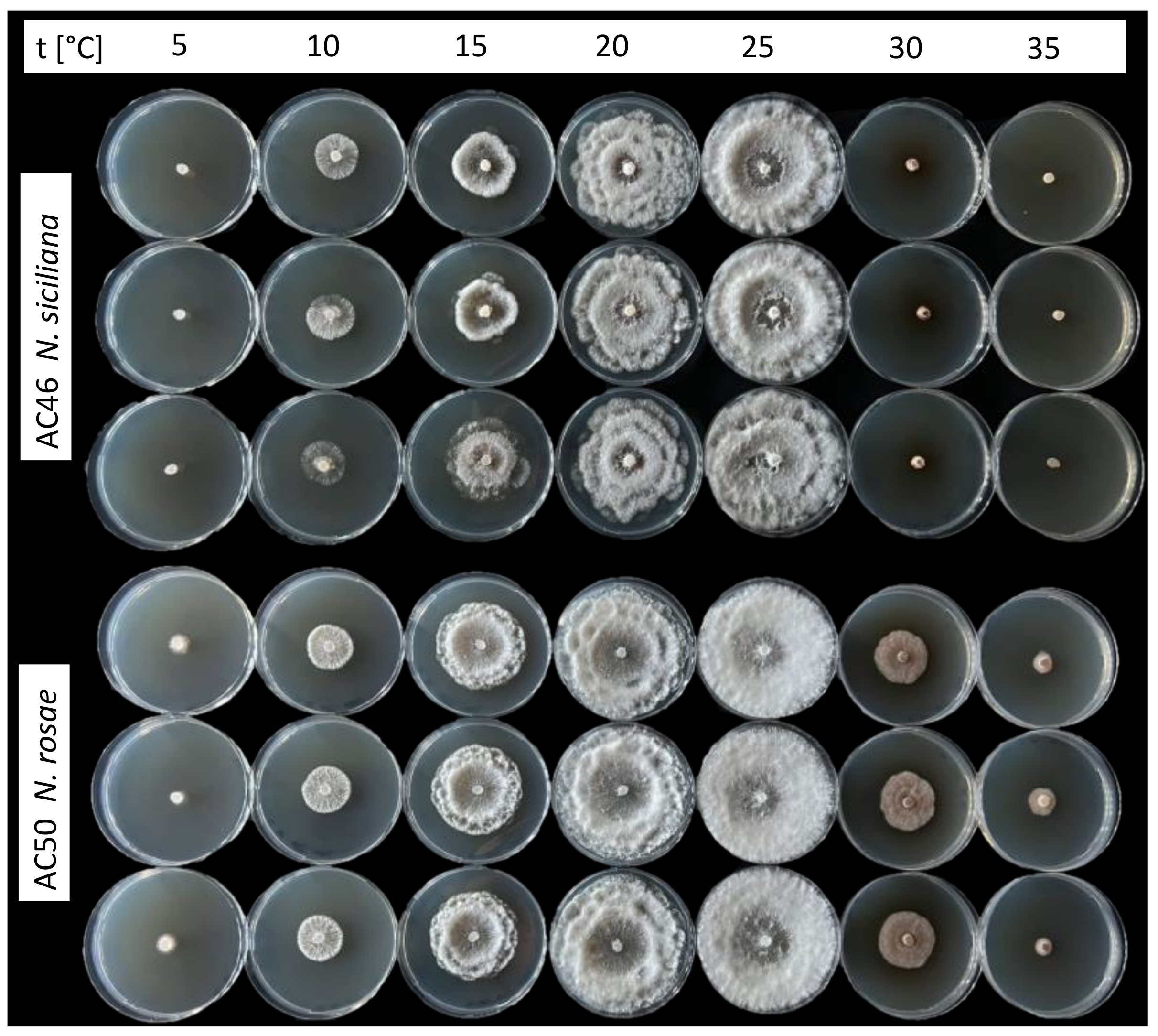
2.1. Isolation and Morphological Characterization

Samples were collected in the field on approximately 20 plants of Persea americana cv. “Hass” grafted on “Zutano”, randomly selected, and were brought to the Plant Pathology laboratory of the Department of Agriculture, Food, and Environment at the University of Catania for further investigations. One hundred small sections (5 mm × 5 mm) of the stems were surface disinfected for 1 min in 1.5% sodium hypochlorite (NaOCl), rinsed in sterile distilled water, dried on sterile absorbent paper, and placed on potato dextrose agar (PDA; Lickson, Vicari, Italy) amended with 100 mg/liter of streptomycin sulphate (Sigma-Aldrich, St. Louis, MO, USA) to prevent bacterial growth, and then incubated at 25 ± 1 °C for five to seven days. Single-spore isolates were obtained from conidia produced in pure cultures grown on PDA. To determine the effect of temperature on mycelial growth and the optimal growth temperature, the representative isolates AC46 and AC50 were cultured on PDA for further assays. After seven days of incubation at 25 °C, 5 mm diam. mycelial plugs were transferred from the edge of the colonies to the center of PDA Petri plates. The plates were incubated at 5–10–15–20–25–30–35 °C in the dark. Three Petri plates were used for each temperature as replicates. The experiment was repeated once. After seven days of incubation, two perpendicular diameters of the colonies were measured with a scale ruler. The isolates used in this study are maintained in the culture collection of the Department of Agriculture, Food, and Environment, University of Catania. Moreover, representative isolates were deposited at the Westerdijk Fungal Biodiversity Institute (CBS), Utrecht, the Netherlands, and dried sporulating cultures were deposited as vouchers in the fungarium of the Department of Botany and Biodiversity Research, University of Vienna (WU-MYC).
Study of macromorphology of conidiomata was performed by using a Nikon SMZ 1500 stereomicroscope equipped with a Nikon DS-U2 digital camera or with a Keyence VHX-6000 digital microscope (Mechelen, Belgium). Microscopic preparations were mounted in water. For light microscopy, a Zeiss Axio Imager.A1 compound microscope (Oberkochen, Germany), equipped with Nomarski differential interference contrast (DIC) optics and a Zeiss Axiocam 506 color digital camera, was used. Photographs and measurements were taken by using the NIS-Elements D v. 3.0 or Zeiss ZEN Blue Edition software. For certain images of conidia, the stacking software Zerene Stacker version 1.04 (Zerene Systems LLC, Richland, WA, USA) was used. Measurements are reported as maxima and minima in parentheses and the mean plus and minus the standard deviation of a number of measurements given in parentheses.

2.2. DNA Extraction, PCR, and Phylogenetic Analysis

The genomic DNA was extracted from surface mycelium scraped off from pure cultures, using the Wizard Genomic DNA Purification Kit (Promega Corporation, Madison, WI, USA). The following three loci were amplified and sequenced: the complete internal transcribed spacer region (ITS1-5.8S-ITS2) with primers ITS5 and ITS4 [23]; a ca. 0.5 kb fragment of the translation elongation factor 1-alpha (TEF1) gene with primers EF1-728F [24] and TEFD_iR [25]; and a ca. 0.95 kb fragment of the beta-tubulin (TUB2) gene with primer pairs T1 [26] and BtHV2r [27]. The PCR products were purified using an enzymatic PCR cleanup [28], as described by Voglmayr and Jaklitsch [29], and sequenced in both directions by Macrogen Inc. (Seoul, South Korea) or at the Department of Botany and Biodiversity Research, University of Vienna, using the ABI PRISM Big Dye Terminator Cycle Sequencing Ready Reaction Kit v. 3.1 (Applied Biosystems, Warrington, UK) and the original PCR primers; sequencing was performed on an automated DNA sequencer (3730xl Genetic Analyser, Applied Biosystems). The DNA sequences generated were assembled with Lasergene SeqMan Pro (DNASTAR, Madison, WI, USA). Sequences generated during the present study were deposited in Genbank (Table 1). The newly generated sequences were aligned to a representative matrix of Neopestalotiopsis, selecting two species of Pestalotiopsis as an outgroup. For Neopestalotiopsis, all 70 accepted species were included in the matrix, preferentially with ex-type sequences. The GenBank accession numbers of sequences used in these analyses are given in Table 1.
Sequence alignments for phylogenetic analyses were produced with the server version of MAFFT (http://mafft.cbrc.jp/alignment/server/, accessed on 22 March 2022), and checked and refined using BioEdit v. 7.2.6 [57]. The ITS rDNA, TEF1, and TUB2 matrices were combined for subsequent phylogenetic analyses, and the combined data matrix contained 2265 characters (545 nucleotides of ITS, 900 nucleotides of TEF1, and 820 nucleotides of TUB2). Maximum likelihood (ML) analyses were performed with RAxML [58], as implemented in raxmlGUI 1.3 [59], using the ML + rapid bootstrap setting and the GTRGAMMA substitution model with 1000 bootstrap replicates. The matrix was partitioned for the different gene regions. For evaluation and discussion of bootstrap support, values below 70% were considered low, between 70 and 90% medium/moderate, and above 90% high and 100% maximum. Maximum parsimony (MP) bootstrap analyses were performed with PAUP v. 4.0a169 [60], with 1000 bootstrap replicates using five rounds of heuristic search replicates with random addition of sequences and subsequent TBR branch swapping (MULTREES option in effect, steepest descent option not in effect, COLLAPSE command set to MINBRLEN, and each replicate limited to 1 million rearrangements) during each bootstrap replicate. All molecular characters were unordered and given equal weight; analyses were performed with gaps treated as missing data; and the COLLAPSE command was set to minbrlen.

2.3. Pathogenicity Test
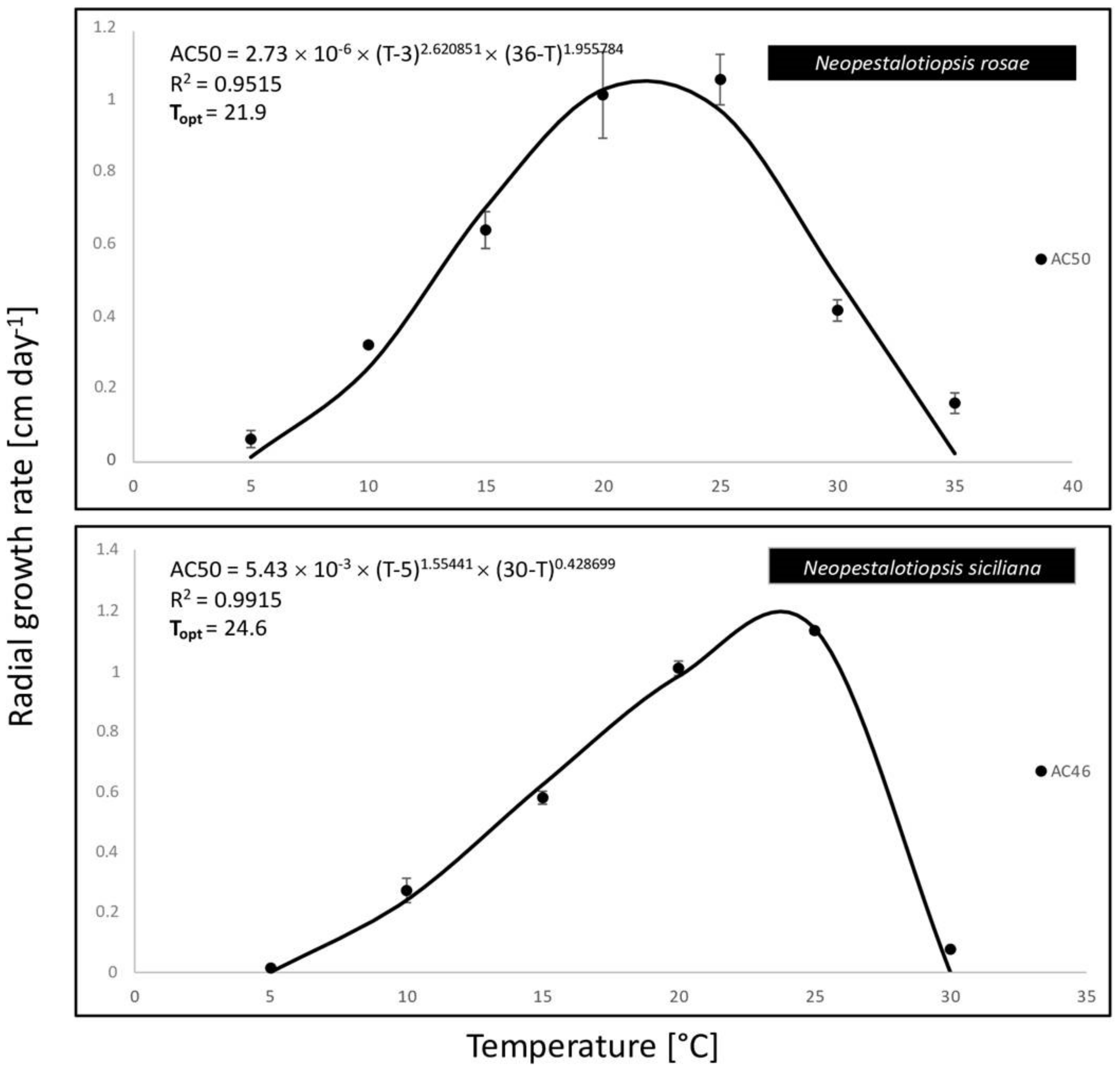
Pathogenicity tests were carried out by artificial inoculations using the isolates AC50 (N. rosae) and AC46 (N. siciliana). Three potted 1-year-old plants of avocado cv. “Hass” grafted on “Zutano” were inoculated with each isolate. Inoculations were made on the stem after removing a piece of bark with a sterile scalpel blade, placing a mycelial plug (0.3 cm2) from a 15-day-old culture of each isolate onto the wound and covering it with Parafilm® (American National Can, Chicago, IL, USA) to prevent desiccation. The same number of plants was inoculated with sterile PDA plugs to serve as control. All the inoculated plants were grown in a growth chamber with a 12 h photoperiod and maintained at 25 ± 1 °C. The inoculated plants were monitored weekly for development of symptoms, and a final assessment was conducted 50 days after the inoculations. Re-isolations were performed as described above to fulfill Koch’s postulates.

2.4. Data Analysis

Data derived from the effect of temperature on mycelial growth rate assay and the lesion length measurements were analyzed in Statistix 10 (Analytical Software 2013) [61]. For analysis of the effect of temperature on mycelial growth, variances of the two assays were tested for the homogeneity using Levene’s test and then combined in one dataset. Data of the mycelial growth were first transformed to radial growth rate (cm day−1) and then a nonlinear regression adjustment of the dataset was applied through the generalized Analytis β model, using the equation described by Moral et al. [62]. Optimum growth temperature was also calculated according to the equation provided by the same authors [62]. For pathogenicity test, analysis of variance (ANOVA) of the lesions length was performed and the mean differences were compared with Fisher’s protected least significance difference (LSD) test at α = 0.05.

3. Results

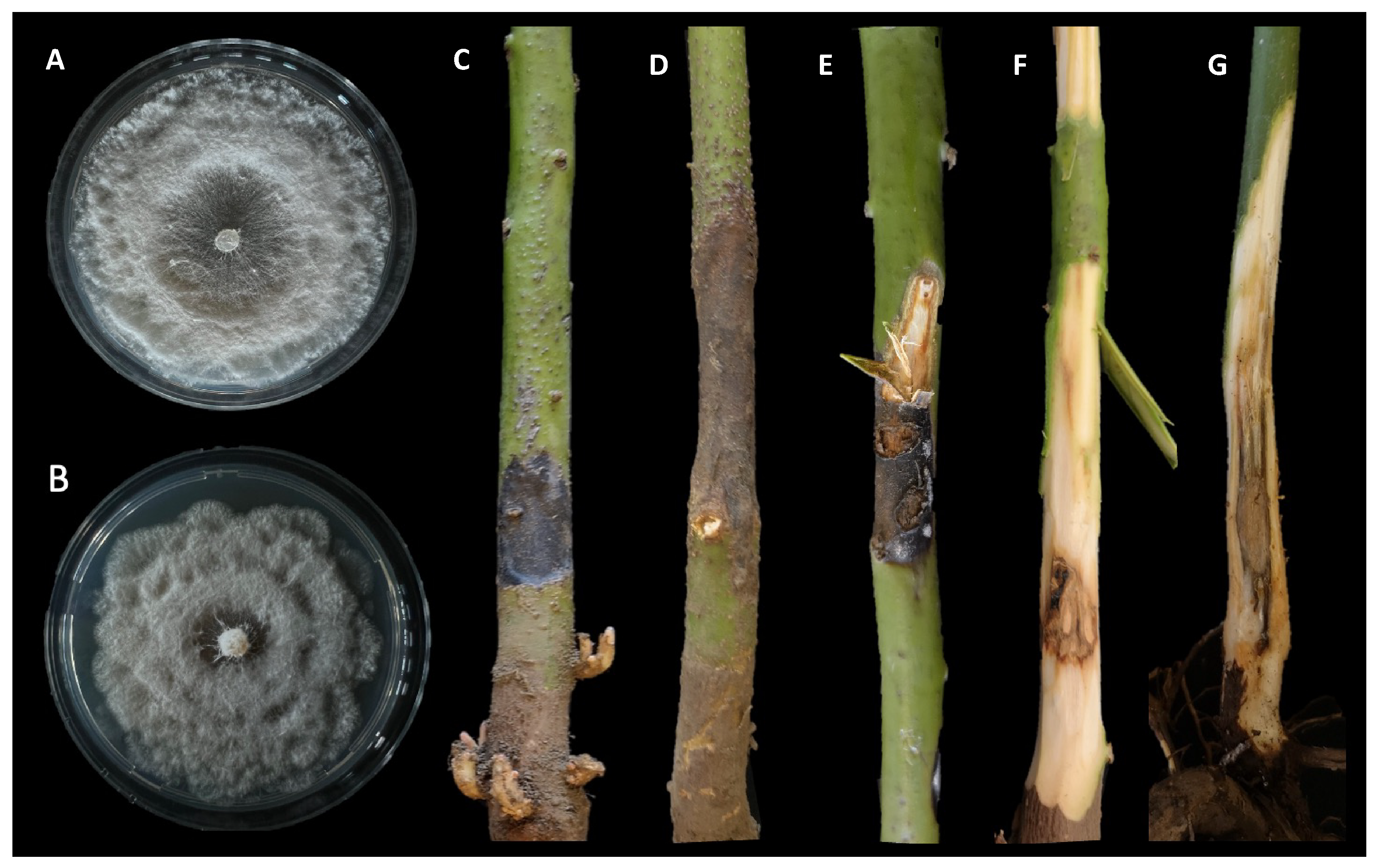
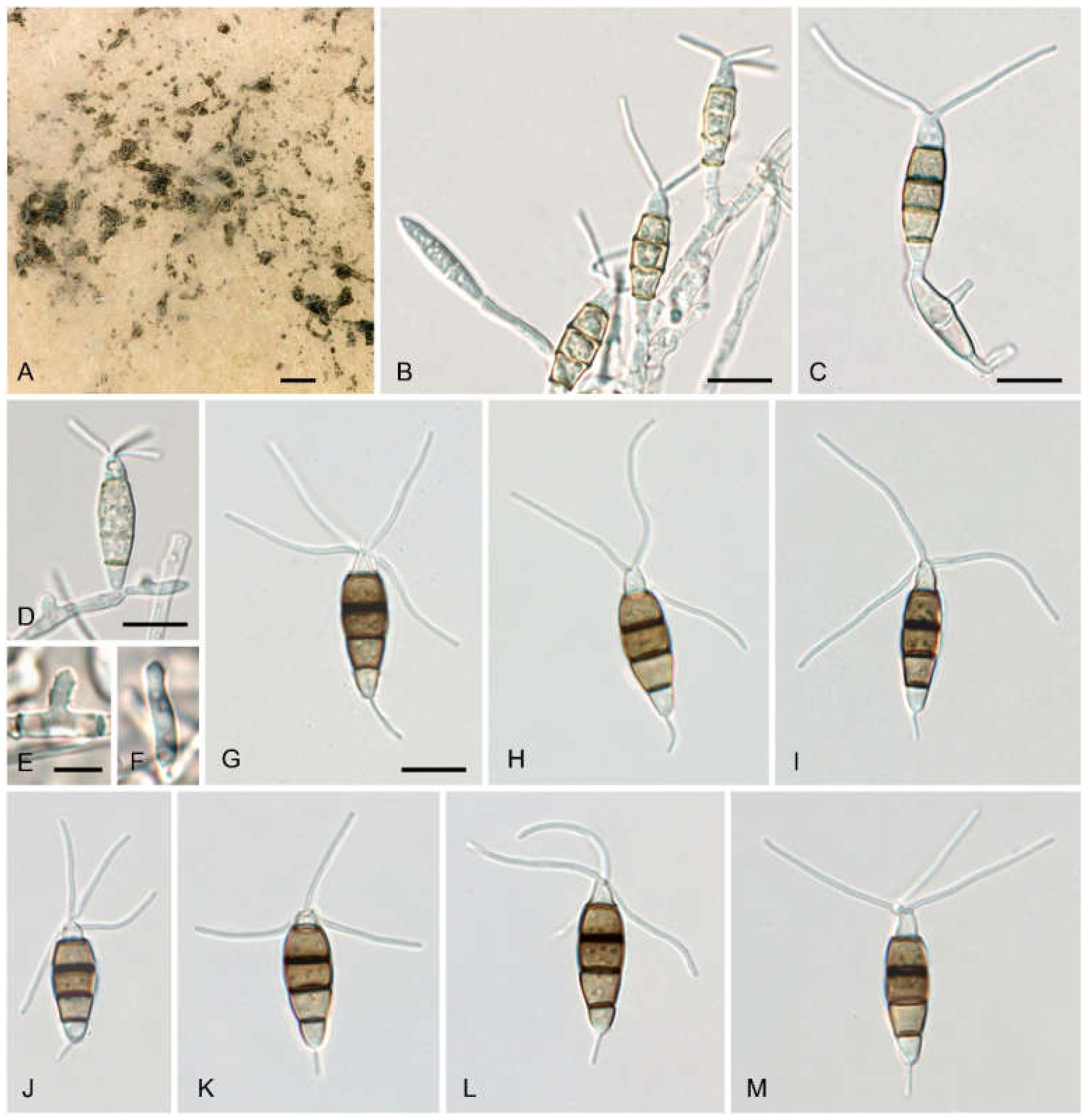
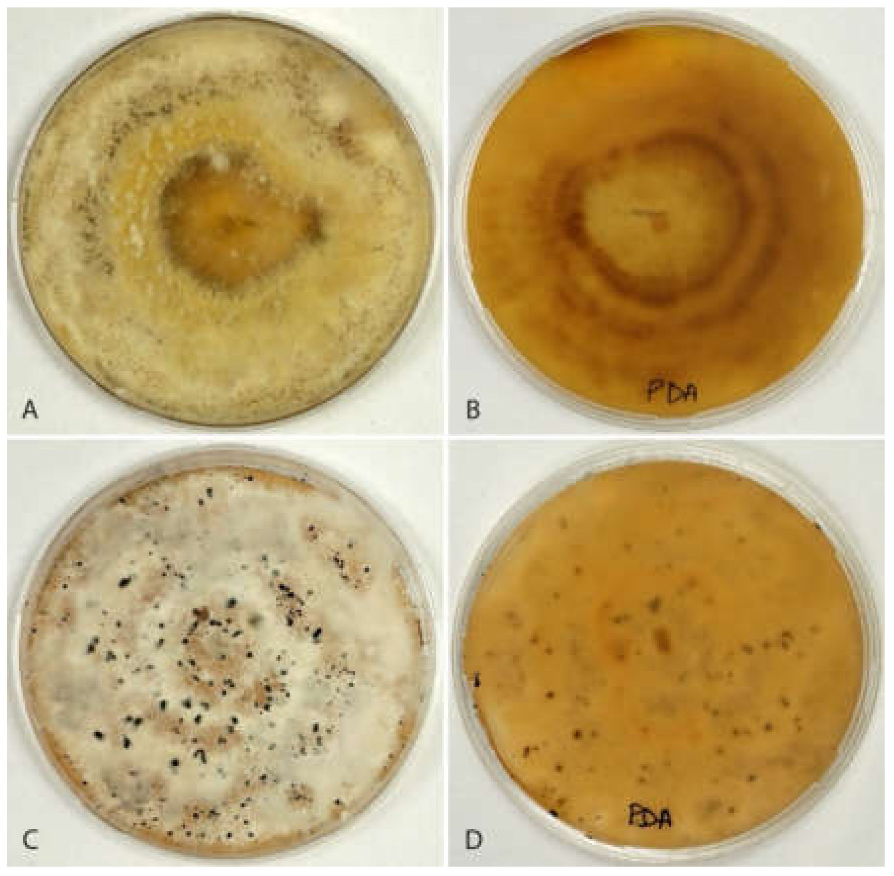
3.1. Isolation and Morphological Characterization

The disease was observed in a commercial avocado orchard located in Giarre (Catania province) on young plants (two years old, 2–3 months after transplanting). Symptoms observed in the field included stem lesions, wood discoloration with brownish streaking, bark cracking, and dieback. Necrotic lesions were characterized by a shrinkage of the affected tissues and internally the wood appeared darkened and dry (Figure 1). Internal lesions started more frequently from the grafting point. The rootstock showed no symptoms. Isolations frequently yielded Neopestalotiopsis-like fungi. A total of eight single-spore isolates were collected and kept in our fungal collection. The highest growth rate for the isolate AC46 (1.13 cm day−1) was observed at 25 °C. According to the Analytis β model, the optimal growth temperature resulted at 24.6 °C. After 7 days of incubation, no mycelial growth was observed at 35 °C. Isolate AC50 showed the highest growth rate (1.06 cm day−1) at 25 °C, and the optimal growth temperature resulted at 21.9 °C. All results of the effects of temperature on mycelial growth rate are shown in Figure 2 and Figure 3.

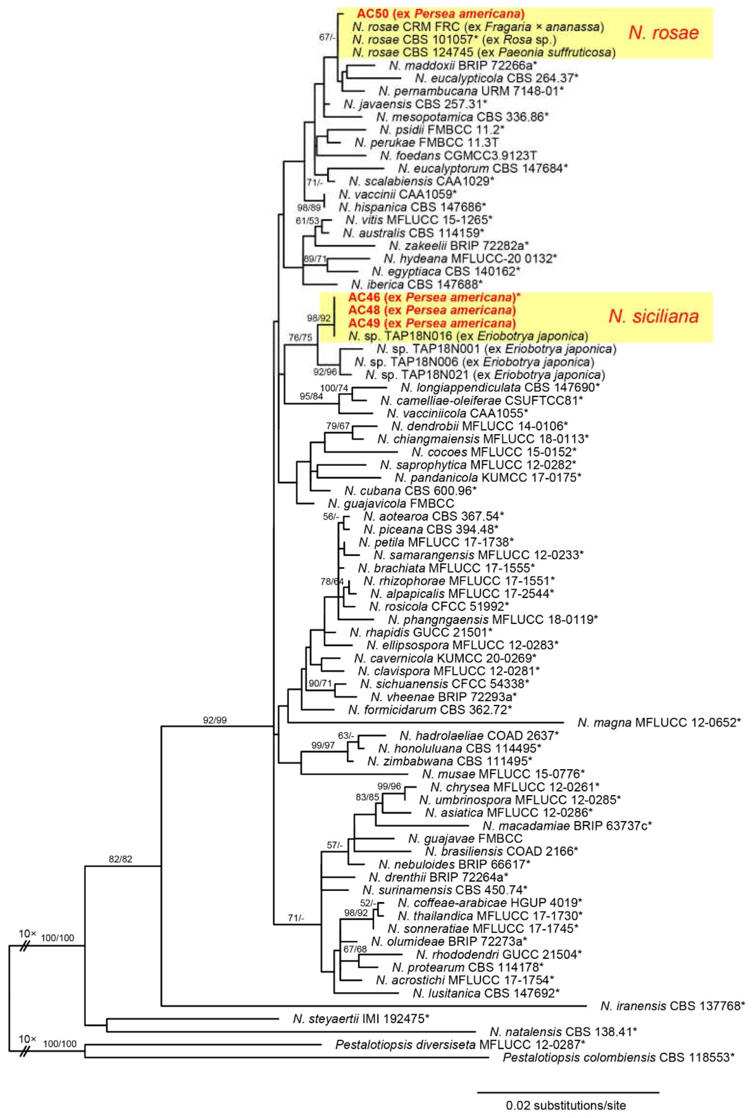
3.2. Phylogenetic Analysis

PCR amplification of the ITS, TEF1, and TUB2 generated 549, 549–550, and 957 bp fragments, respectively. Of the 2261 characters included in the phylogenetic analyses, 334 were parsimony informative (58 from the ITS, 153 from TEF1, and 123 from TUB2). The best ML tree (−lnL = 8699.596) revealed by RAxML is shown as a phylogram in Figure 4. While backbone support of deeper nodes was mostly absent, several terminal nodes received medium to high support. Of the four Neopestalotiopsis isolates of the current study, one was placed within the N. rosae clade, while the other three isolates were contained within a moderately supported clade together with four unnamed Japanese isolates from Eriobotrya japonica. The latter clade was further subdivided into two subclades: a highly supported subclade containing the three isolates of the present study and one isolate from Eriobotrya japonica, and a second, moderately supported subclade containing the residual isolates from Eriobotrya japonica.

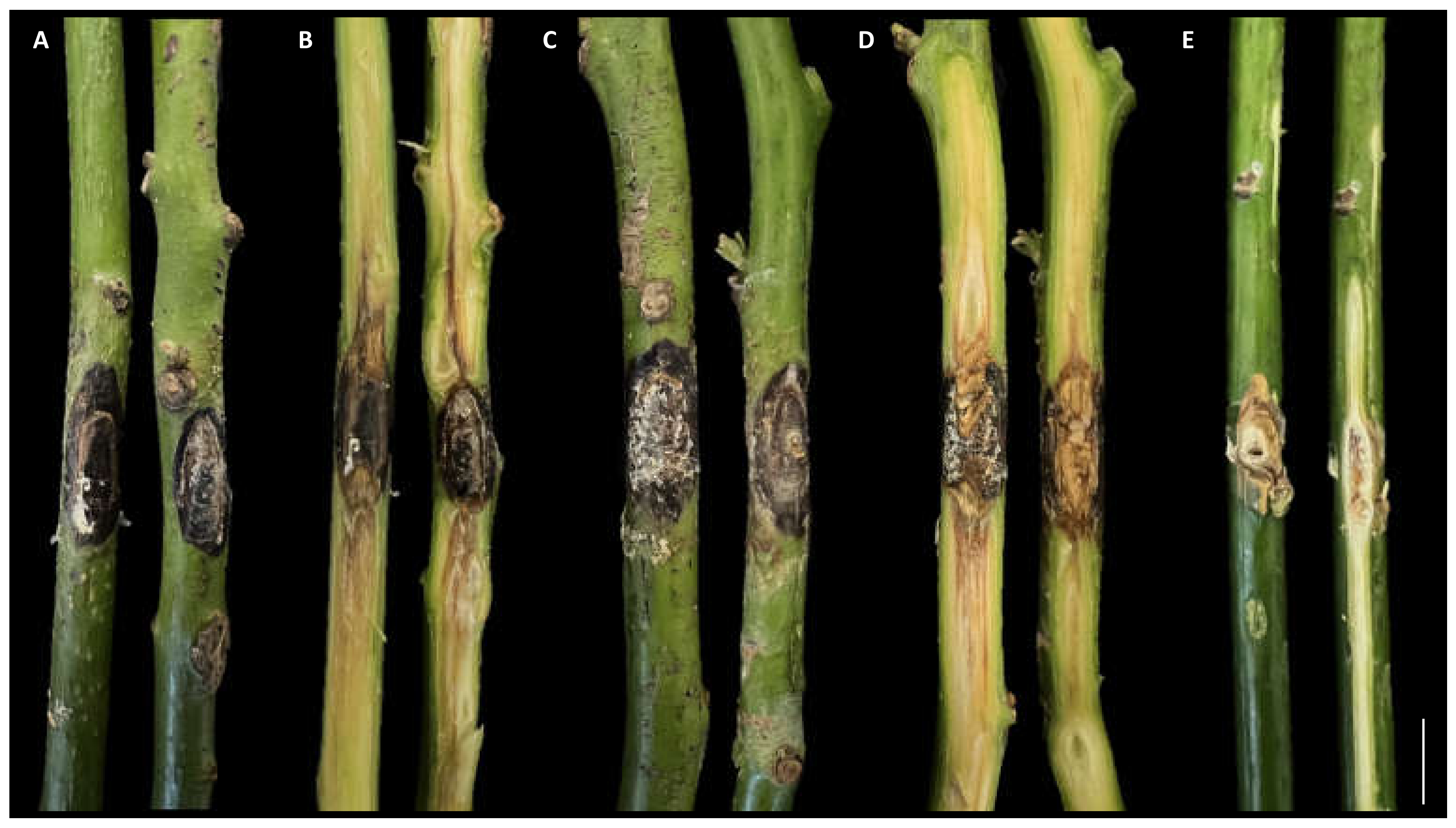
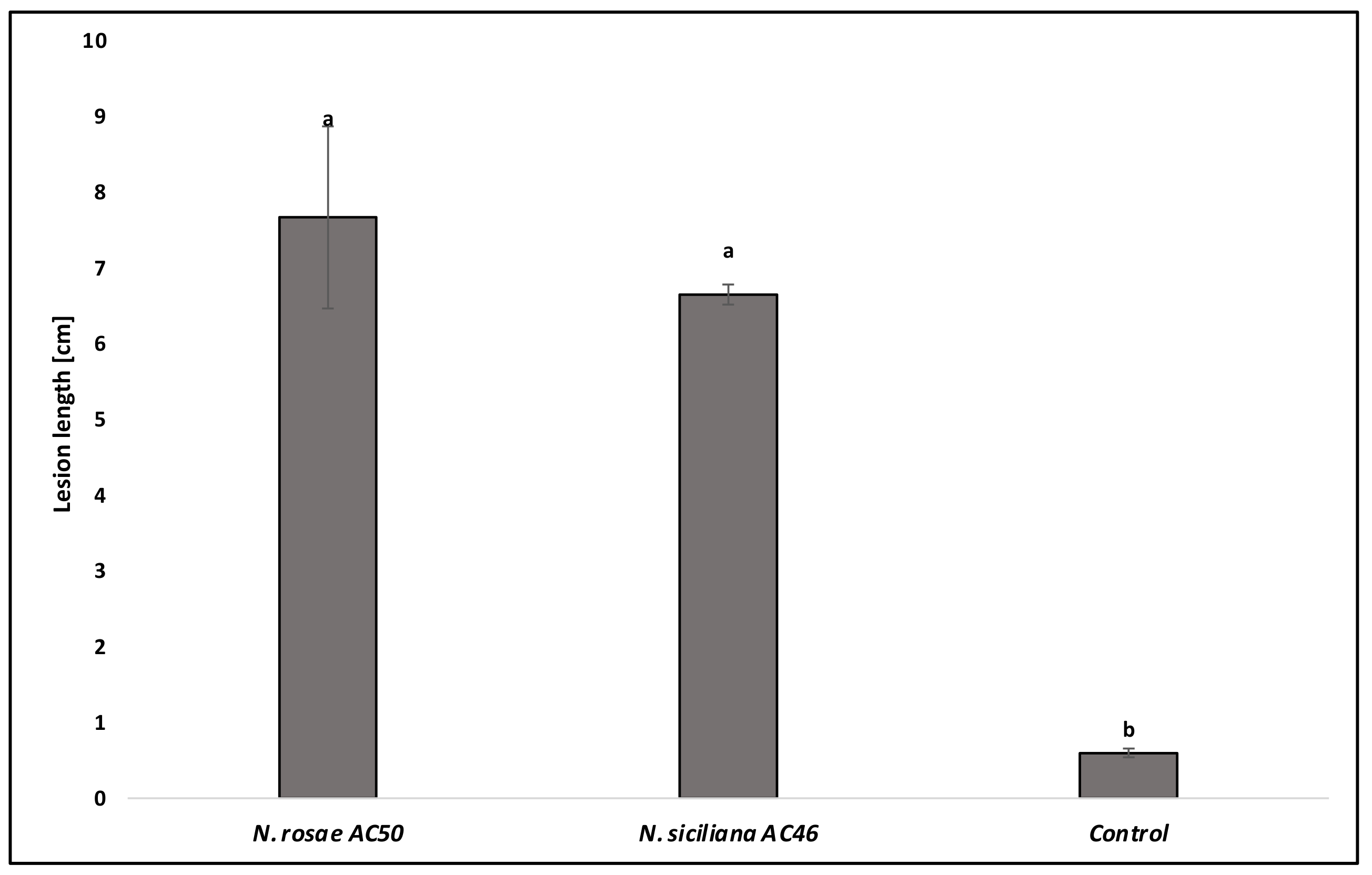
3.3. Pathogenicity Test

The results of the pathogenicity test showed that both species of Neopestalotiopsis identified in this study were pathogenic to avocado and produced the same symptoms similar to those observed in the field. All inoculated trees showed severe external and internal wood discoloration. Controls did not show any symptoms (Figure 5). For both species, the presence of acervuli on the inoculated wounds was observed. The mean lesion lengths of N. rosae (7.76 cm) and N. siciliana (6.65 cm) were significantly different from the control (0.6 cm) (p < 0.05), but not significantly different between them (Figure 6). Re-isolations showed the presence of colonies with the same morphological characteristics as the inoculated species, so Koch’s postulates were fulfilled.

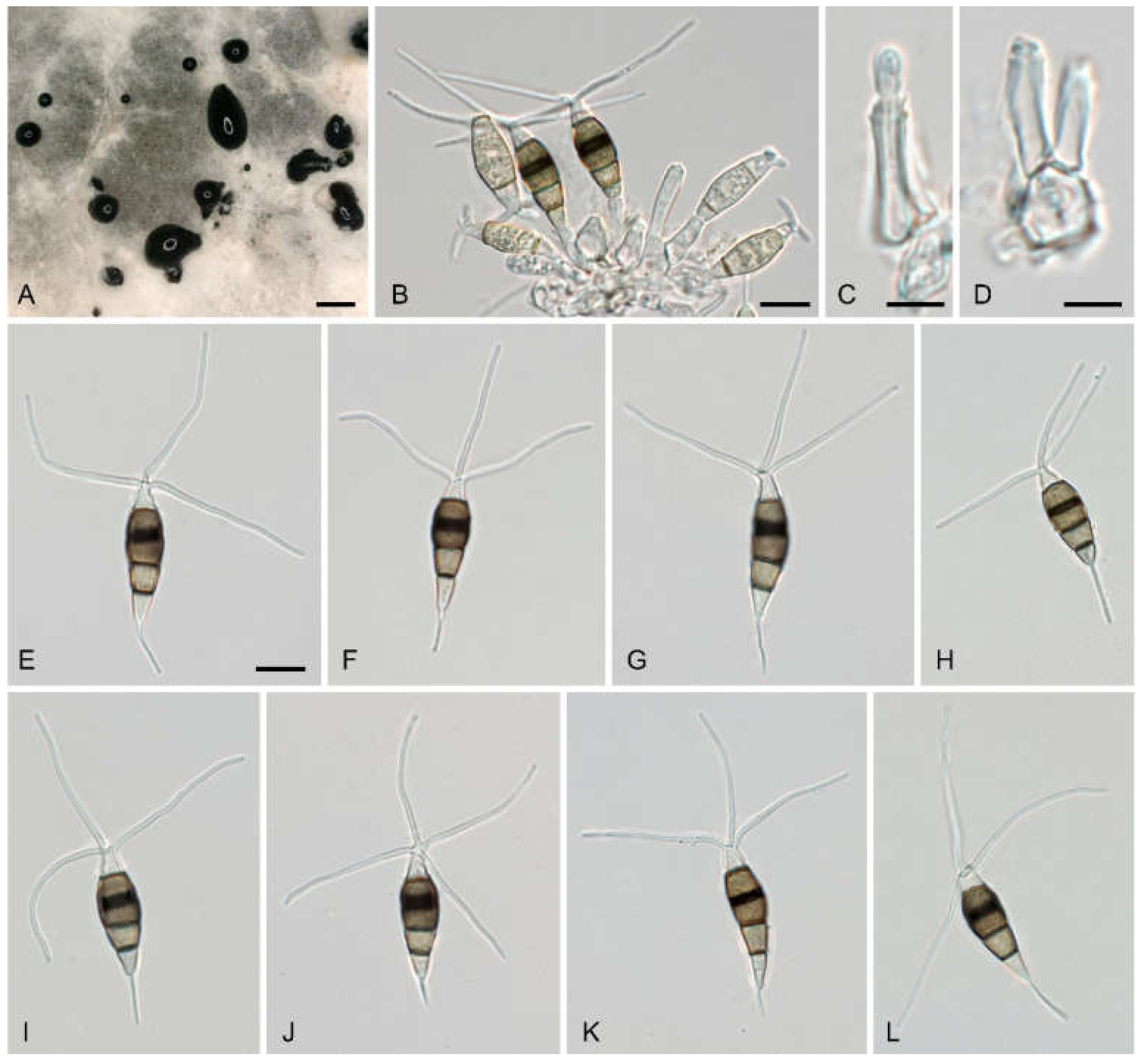
3.4. Morphological Description of Neopestalotiopsis rosae Isolates from Avocado

Neopestalotiopsis rosae Maharachch., K.D. Hyde and Crous, in Maharachchikumbura, Hyde, Groenewald, Xu and Crous, Stud. Mycol. 79: 147 (2014) (Figure 7).
Description—sexual morph unknown. Asexual morph: conidiomata on natural substrate acervular, in culture on PDA sporodochial; solitary, pulvinate, black, 30–100(–150) μm diam., and exuding black conidial masses. Conidiophores indistinct and usually reduced to conidiogenous cells. Conidiogenous cells 1–33 × 1–3.7 μm, discrete, either short-cylindrical, sitting laterally on hyphae, or cylindrical, ampulliform to lageniform, hyaline, smooth, thin-walled, simple, holoblastic-annelidic, proliferating one to two times percurrently, with collarette present and not flared. Conidia (20–)22–24(–25) × (6.2–)6–8.7(–15.2) μm, l/w = (1.6–)2.9–3.6(–3.9) (n = 40), fusoid, straight or slightly curved, four-septate, smooth, slightly constricted at the septa; the basal cell obconic with a truncate base, thin-walled, hyaline or pale brown, and (3.3–)3.8–4.8(–5.4) μm long; three median cells trapezoid or subcylindrical, (8–)14–17(–17) μm long, smooth-walled, versicolored, with septa darker than the rest of the cell; the second cell from the base pale brown and (3.8–)4.6–5.5(–6.1) μm long; the third cell s medium brown and (4–)4.5–5.1(–5.7) μm long; the fourth cell medium brown and (4.4–)5–5.6(–6.1) μm long, septum between the third and fourth cell more distinct, broader, and darker brown than the other septa; the apical cell conic with the subacute apex thin-walled, smooth, hyaline, (2.8–)3.4–4.4(–4.8) μm long, with two to four apical appendages (mostly three) arising from the apical crest; apical appendages unbranched, tubular, centric, and straight or slightly bent, (15–)19–28(–33) μm long, and (0.8–)0.9–1.1(–1.3) μm wide (n = 60); basal appendage single, filiform, unbranched, centric or eccentric, (3–)3.5–5.8(–8.1) μm long and 0.5–0.9 μm wide (n = 85).
Culture characteristics. The colony on PDA attaining 90 mm diameter after 7 days at 21.9 °C, yellowish, with a fluffy whitish aerial mycelium, secreting a yellowish pigment in the culture medium, with isolated conidiomata scattered on the aerial mycelium (Figure 8A). The reverse is pale yellowish brown (Figure 8B).
Habitat. On stems of Persea americana Mill.
Distribution. Sicily, Italy.
Specimens examined. ITALY, Sicily, Catania Province, Giarre, 15 December 2020, Alberto Fiorenza (WU-MYC 0045984; culture AC50 = CBS 149120).
Notes. Our strain AC50 has identical ITS and TEF1 and highly similar TUB2 (99.8%; 1 nt difference) sequences to the type strain of N. rosae (CBS 101057). Neopestalotiopsis rosae has been recorded as a pathogen of various fruit crops, e.g., blueberry (Vaccinium corymbosum; [53,63]), pomegranate (Punica granatum; [64]), and in particular strawberry (Fragaria × ananassa), on which it was reported to cause severe disease outbreaks around the world in recent years (e.g., Australia [65], China [66], Mexico [52], Taiwan [67], and the USA [65]). In the protologue of N. rosae, it was characterized by three to five tubular apical appendages not arising from the apical crest but at different regions in the upper half of the apical cell. This does not agree with our observations, as in our strain, the apical appendages arise from the apical crest. However, the descriptions and illustrations of the other reports of N. rosae cited above agree well with our strain, as do the molecular data.

4. Taxonomy

Neopestalotiopsis coffeae-arabicae (Yu Song, K. Geng, K.D. Hyde and Yong Wang bis) Voglmayr, comb. nov. MycoBank No.: MB 844083.
Basionym: Pestalotiopsis coffeae-arabicae Yu Song, K. Geng, K.D. Hyde and Yong Wang bis, in Song, Geng, Zhang, Hyde, Zhao, Wei, Kang and Wang, Phytotaxa 126(1): 26 (2013)
Notes: This species is clearly a member of Neopestalotiopsis according to the results of phylogenetic analyses (Figure 4). Although it was listed as N. coffeae-arabicae in various phylogenies [30,35,41,53,63,68], this combination is neither present in the Index of Fungi nor Mycobank, and could also not be traced in the literature, indicating that it has not been validly published, which is therefore performed here.
Neopestalotiopsis siciliana Voglmayr, Fiorenza and Aiello, sp. nov.—MycoBank MB 844082; (Figure 9).
Etymology. Named after the region where it was found (Sicily).
Holotype. ITALY, Sicily, Catania Province, Giarre, on stems of Persea americana, 15 December 2020, Alberto Fiorenza (WU-MYC 0045982; culture AC46 = CBS 149117).
Description—Sexual morph unknown. Asexual morph: Conidiomata on natural substrate acervular, in culture on PDA sporodochial, solitary, pulvinate, black, and (100–)300–2000(–2800) μm diam., exuding black, globose, and glistening conidial masses. Conidiophores indistinct, usually reduced to conidiogenous cells. Conidiogenous cells 7.7–15.2 × 2.8–6.7 μm, discrete, cylindrical, ampulliform to lageniform, hyaline, smooth, thin-walled, simple, holoblastic-annelidic, and proliferating one to two times percurrently, with collarette present and not flared. Conidia (20–)23–27(–32) × (6–)6.8–7.9(–8.8) μm, l/w = (2.8–)3.1–3.8(–4.9) (n = 102), fusoid, straight or slightly curved, four-septate, smooth, and slightly constricted at the septa; the basal cell obconic with a truncate base, thin-walled, hyaline or pale brown, (3–)4.3–6(–7.2) μm long; three median cells trapezoid or subcylindrical, (12–)14–17(–23) μm long, smooth-walled, versicolored, with septa darker than the rest of cell; the second cell from the base pale brown and (3.8–)4.5–5.4(–6.4) μm long; the third cell medium brown and (4.1–)4.5–5.5(–7) μm long; the fourth cell medium brown and (3.9–)4.6–5.7(–6.5) μm long; with septum between the third and fourth cell more distinct, broader, and darker brown than the other septa; the apical cell conic with a subacute apex, thin-walled, smooth, hyaline, (3.1–)4.1–5.3(–7) μm long, and with two to four apical appendages (mostly three) arising from the apical crest; apical appendages unbranched, tubular, centric, and straight or slightly bent, (19–)24–34(–38) μm long and (0.9–)1.1–1.5(–1.7) μm wide (n = 105); basal appendage single, filiform, unbranched, centric, (2.8–)4.6–9.3(–15.3) μm long, and (0.5–)0.7–0.9(–1.1) μm wide (n = 85).
Culture characteristics. Colony on PDA attaining 90 mm diameter after 7 days at 24.6 °C, dirty white, with fluffy white aerial mycelium, conidiomata scattered, isolated (Figure 8C). Reverse pale buff (Figure 8D).
Habitat. On stems of Persea americana Mill.
Distribution. Sicily, Italy.
Specimens examined. ITALY, Sicily, Catania Province, Giarre, 15 December 2020, collector Alberto Fiorenza (WU-MYC 0045983; culture AC48 = CBS CBS 149118); Giarre, 15 December 2020, collector Alberto Fiorenza (culture AC49 = CBS 149119).
Notes. The phylogenetic analyses revealed a highly supported, distinct phylogenetic position within Neopestalotiopsis, confirming its status as a new species. Remarkably, it clusters with four unnamed Neopestalotiopsis accessions isolated from the leaves and fruits of Eriobotrya japonica in Japan [55]. While one of these strains has sequences identical to our isolates, with which it clustered within a highly supported subclade, the three other strains from Eriobotrya form a sister clade to the former (Figure 4). As there are some molecular differences between these two subclades, it is yet unclear whether one or two species are involved; considering the uncertainties in species circumscription in the genus and the lack of morphological data, we here maintain these isolates as Neopestalotiopsis sp. As usually observed in Neopestalotiopsis, it is impossible to identify N. siciliana by conidial morphology alone, and sequence data are necessary for reliable species identification.

5. Discussion

The results of this study confirm the presence of Neopestalotiopsis species causing disease on young avocado plants in southern Italy. The fungal species obtained from symptomatic tissues were identified based on the morphological characteristics and molecular phylogenetic analyses of the ITS, TEF1, and TUB2 gene regions. The phylogenetic analyses showed that two species are involved in avocado stem and wood lesions, resulting in the dieback of the plants. Of the four isolates sequenced, one was identified as N. rosae, while another three isolates formed a clade distinct from the other described Neopestalotiopsis species, which is therefore here described as a new species, N. siciliana. Remarkably, these three isolates were phylogenetically close to four unnamed Neopestalotiopsis strains isolated from Eriobotrya japonica in Japan [55]; one even had identical sequences to our isolates. This demonstrates that N. siciliana is widespread and has a wider host range. There are a few reports from avocados attributable to the genus Neopestalotiopsis, for which ITS sequences are available. Valencia et al. [20] recorded N. clavispora (as Pestalotiopsis clavispora; ITS sequence HQ659767) as a causal agent of post-harvest stem-end rot in Chile, while Shetty et al. [69] identified one of their endophytic isolates from organically grown avocado trees in South Florida as Neopestalotiopsis foedans (ITS sequence KU593530). A sequence comparison of the ITS sequences with our matrix showed that the ITS sequence HQ659767 was identical and that KU593530 was almost identical (one gap difference) to our isolate AC50 representing N. rosae. While this could indicate that N. rosae is regularly found on avocado, it needs to be noted that the ITS alone is not suitable for species identification, as several species (e.g., N. hispanica, N. longiappendiculata, N. mesopotamica, N. scalabiensis, and N. vaccinii) have ITS sequences identical to those of N. rosae. Therefore, the species identity of these avocado isolates remains uncertain in the lack of TEF1 and TUB2 sequences.
Pestalotiopsis sensu lato was recently revised by Maharachchikumbura et al. [21] and segregated into three distinct genera, viz. Pestalotiopsis, Pseudopestalotiopsis, and Neopestalotiopsis. During our field surveys, we often encountered young plants of avocado showing stunted growth. Monitoring the plants after the transplant from the nurseries to the open field, it was noticed that some of these were not able to survive. Deeper observations of the internal tissues revealed frequent necrosis and cankers at the grafting point. Likely, propagation processes represent relevant infection courts for pestalotioid fungi. Most of the avocado plants transplanted in Sicily derive from Spanish nurseries where the propagation steps are performed. It is not unusual that symptoms such as stunting, shoot blight, and cankers observed by the growers after the first years from the transplant in the open field are the results of previous infections that had occurred in the nurseries, especially during the grafting. In fact, wounds and injuries are crucial for penetration of the host tissue and subsequent development of the infection, especially for pestalotioid species [70]. Our observations are indeed in accordance with other reports. In China, 30% of symptomatic avocado plants in a nursery plantation showed the presence of Pestalotiopsis longiseta [71] and other authors also reported the presence of Pestalotiopsis spp. at the graft union in different hosts [72,73,74]. Species of these genera are widely distributed in tropical and temperature areas. This group of fungi is commonly found as endophytes and plant pathogens on different hosts, causing stem-end rot, stem and leaf blight, trunk disease, and cankers [21,75]. Previous investigation conducted in Sicily already reported the presence of pestalotioid fungi, including Pestalotiopsis clavispora and P. uvicola, causing diseases on tropical, as well as on ornamental, hosts [22,75]. This study represents a new step forward in the insight of this complex and still understudied group of fungi, especially within the genus of Neopestalotiopsis, providing new information on the ecology of these two species.

6. Conclusions

Two fungal species, Neopestalotiopsis rosae and N. siciliana sp. nov. are described and illustrated. These fungi were isolated from the stem tissues of diseased young avocado plants in Sicily, Italy. Pathogenicity tests were performed, and Koch’s postulates were fulfilled. The result of this study provided new information regarding this still understudied group of phytopathogenic fungi and its wide host range. Neopestalotiopsis rosae and N. siciliana could be a new threat to the Italian avocado industry, especially in the southern regions where avocado represents an emerging crop. The presence of these species in the internal tissues at the graft union corroborates the fact that the propagation processes represent crucial steps to obtain healthy material. Further investigations are needed in order to ascertain the diffusion and epidemiology of these species in the Sicilian avocado orchards, and to evaluate the effective risk for the industry. Therefore, it will be important to carry out additional studies on the pathogenicity and susceptibility of the different cultivars of avocado in the future. To our knowledge, this is the first report of N. rosea and of the fungus here described as N. siciliana affecting avocado.

Author Contributions

Conceptualization, H.V., A.F. and D.A.; methodology, H.V. and A.F.; software, G.G. and H.V.; validation, H.V., G.P. and D.A.; formal analysis, H.V., A.F. and G.G.; investigation, H.V. and A.F.; resources, H.V. and G.P.; data curation, H.V. and A.F.; writing—original draft preparation, H.V. and A.F.; writing—review and editing, H.V., G.P., D.A., A.F. and G.G.; visualization, H.V., D.A. and G.P.; supervision, G.P. and D.A.; project administration, G.P.; funding acquisition, G.P. All authors have read and agreed to the published version of the manuscript.

Funding

This research was funded and supported by following grants: PSR Sicilia 2014–2020 Misura 16—Sottomisura 16.2—Bando 2019, CUP G79J2100558009 Avocado biologico siciliano: superfood per la valorizzazione delle aree ionico-tirreniche (Acronimo SUPERAVOCADO) rappresentato dal Capofila Passanisi Andrea; Programma di ricerca di Ateneo MEDIT-ECO UNICT 2020–2022 Linea 2—University of Catania (Italy).

Institutional Review Board Statement

Not applicable.

Informed Consent Statement

Not applicable.

Data Availability Statement

The data presented in this study are available on request from the corresponding author.

Conflicts of Interest

The authors declare no conflict of interest.

References

  1. Food and Agriculture Organization of the United Nations. FAOSTAT Statistical Database; FAO: Rome, Italy, 2022; Available online: https://www.fao.org/faostat/en/#data/QCL (accessed on 11 April 2022).
  2. Migliore, G.; Farina, V.; Guccione, G.D.; Schifani, G. Quality determinants of avocado fruit consumption in Italy. Implications for small farms. Calitatea 2018, 19, 148–153. [Google Scholar]
  3. Zentmyer, G.A. Avocado. In Compendium of Tropical Fruit Diseases; Ploetz, R.C., Zentmyer, G.A., Nishijima, W.T., Rohrbach, K.G., Ohr, H.D., Eds.; APS Press: St. Paul, MA, USA, 1994; pp. 71–72. [Google Scholar]
  4. Zentmyer, G.A. Phytophthora Cinnamomi and the Disease it Causes; APS Press: St Paul, MA, USA, 1980. [Google Scholar]
  5. López-Herrera, C.J.; Melero-Vara, J.M. Diseases of avocado caused by soil fungi in the southern Mediterranean coast of Spain. In Proceedings of the Second World Avocado Congress California Avocado Society: The Shape of Things to Come, Orange, CA, USA, 21–26 April 1991; Lovatt, C.J., Holthe, P.A., Arpaia, M.L., Eds.; California Avocado Society: Riverside, CA, USA, 1992; Volume 1, pp. 119–121. [Google Scholar]
  6. Fiorenza, A.; Aiello, D.; Leonardi, G.R.; Continella, A.; Polizzi, G. First report of Rosellinia necatrix causing white root rot on avocado in Italy. Plant Dis. 2021, 105, 3294. [Google Scholar] [CrossRef]
  7. Parkinson, L.E.; Shivas, R.G.; Dann, E.K. Pathogenicity of nectriaceous fungi on avocado in Australia. Phytopathology 2017, 107, 1479–1485. [Google Scholar] [CrossRef] [PubMed]
  8. Aiello, D.; Gusella, G.; Vitale, A.; Guarnaccia, V.; Polizzi, G. Cylindrocladiella peruviana and Pleiocarpon algeriense causing stem and crown rot on avocado (Persea americana). Eur. J. Plant Pathol. 2020, 158, 419–430. [Google Scholar] [CrossRef]
  9. Vitale, A.; Aiello, D.; Guarnaccia, V.; Perrone, G.; Stea, G.; Polizzi, G. First report of root rot caused by Ilyonectria (= Neonectria) macrodidyma on avocado (Persea americana) in Italy. J. Phytopathol. 2012, 160, 156–159. [Google Scholar] [CrossRef]
  10. McDonald, V.; Eskalen, A. Botryosphaeriaceae species associated with avocado branch cankers in California. Plant Dis. 2011, 95, 1465–1473. [Google Scholar] [CrossRef] [Green Version]
  11. Menge, J.A.; Ploetz, R.C. Diseases of avocado. In Diseases of Tropical Fruit Crops; Ploetz, R.C., Ed.; CABI Publishing: Wallingford, UK, 2003; pp. 35–71. [Google Scholar]
  12. Hartill, W.F.T.; Everett, K.R. Inoculum sources and infection pathways of pathogens causing stem-end rots of ‘Hass’ avocado (Persea americana). N. Zeal. J. Crop Hortic. Sci. 2002, 30, 249–260. [Google Scholar] [CrossRef]
  13. Guarnaccia, V.; Vitale, A.; Cirvilleri, G.; Aiello, D.; Susca, A.; Epifani, F.; Perrone, G.; Polizzi, G. Characterisation and pathogenicity of fungal species associated with branch cankers and stem-end rot of avocado in Italy. Eur. J. Plant Pathol. 2016, 146, 963–976. [Google Scholar] [CrossRef]
  14. Guarnaccia, V.; Sandoval-Denis, M.; Aiello, D.; Polizzi, G.; Crous, P.W. Neocosmospora perseae sp. nov., causing trunk cankers on avocado in Italy. Fungal Syst. Evol. 2018, 1, 131–140. [Google Scholar] [CrossRef] [Green Version]
  15. Guarnaccia, V.; Aiello, D.; Papadantonakis, N.; Polizzi, G.; Gullino, M.L. First report of branch cankers on avocado (Persea americana) caused by Neocosmospora (syn. Fusarium) perseae in Crete (Greece). J. Plant Pathol. 2021, 104, 419–420. [Google Scholar] [CrossRef]
  16. Freeman, S.; Katan, T.; Shabi, E. Characterization of Colletotrichum species responsible for anthracnose diseases of various fruits. Plant Dis. 1998, 82, 596–605. [Google Scholar] [CrossRef] [Green Version]
  17. Silva-Rojas, H.V.; Ávila-Quezada, G.D. Phylogenetic and morphological identification of Colletotrichum boninense: A novel causal agent of anthracnose in avocado. Plant Pathol. 2011, 60, 899–908. [Google Scholar] [CrossRef]
  18. Sharma, G.; Maymon, M.; Freeman, S. Epidemiology, pathology and identification of Colletotrichum including a novel species associated with avocado (Persea americana) anthracnose in Israel. Sci. Rep. 2017, 7, 15839. [Google Scholar] [CrossRef]
  19. Kimaru, S.K.; Monda, E.; Cheruiyot, R.C.; Mbaka, J.; Alakonya, A. Morphological and molecular identification of the causal agent of anthracnose disease of avocado in Kenya. Int. J. Microbiol. 2018, 2018, 4568520. [Google Scholar] [CrossRef] [Green Version]
  20. Valencia, A.L.; Torres, R.; Latorre, B.A. First report of Pestalotiopsis clavispora and Pestalotiopsis spp. causing postharvest stem end rot of avocado in Chile. Plant Dis. 2011, 95, 492. [Google Scholar] [CrossRef]
  21. Maharachchikumbura, S.S.; Hyde, K.D.; Groenewald, J.Z.; Xu, J.; Crous, P.W. Pestalotiopsis revisited. Stud. Mycol. 2014, 79, 121–186. [Google Scholar] [CrossRef] [Green Version]
  22. Vitale, A.; Polizzi, G. Occurrence of Pestalotiopsis uvicola causing leaf spots and stem blight on bay laurel (Laurus nobilis) in Sicily. Plant Dis. 2005, 89, 1362. [Google Scholar] [CrossRef]
  23. White, T.J.; Bruns, T.; Lee, S.J.W.T.; Taylor, J. Amplification and direct sequencing of fungal ribosomal RNA genes for phylogenetics. In PCR Protocols: A Guide to Methods and Applications; Innes, M.A., Gelfand, D.H., Sninsky, J.J., White, T.J., Eds.; Academic Press: New York, NY, USA, 1990; pp. 315–322. [Google Scholar]
  24. Carbone, I.; Kohn, L.M. A method for designing primer sets for speciation studies in filamentous ascomycetes. Mycologia 1999, 91, 553–555. [Google Scholar] [CrossRef]
  25. Voglmayr, H.; Friebes, G.; Gardiennet, A.; Jaklitsch, W.M. Barrmaelia and Entosordaria in Barrmaeliaceae (fam. nov., Xylariales) and critical notes on Anthostomella-like genera based on multigene phylogenies. Mycol. Prog. 2018, 17, 155–177. [Google Scholar] [CrossRef] [Green Version]
  26. O’Donnell, K.; Cigelnik, E. Two divergent intragenomic rDNA ITS2 types within a monophyletic lineage of the fungus Fusarium are nonorthologous. Mol. Phylogenet. Evol. 1997, 7, 103–116. [Google Scholar] [CrossRef]
  27. Voglmayr, H.; Gardiennet, A.; Jaklitsch, W.M. Asterodiscus and Stigmatodiscus, two new apothecial dothideomycete genera and the new order Stigmatodiscales. Fungal Divers. 2016, 80, 271–284. [Google Scholar] [CrossRef] [Green Version]
  28. Werle, E.; Schneider, C.; Renner, M.; Völker, M.; Fiehn, W. Convenient single-step, one tube purification of PCR products for direct sequencing. Nucleic Acids Res. 1994, 22, 4354. [Google Scholar] [CrossRef]
  29. Voglmayr, H.; Jaklitsch, W.M. Prosthecium species with Stegonsporium anamorphs on Acer. Mycol. Res. 2008, 112, 885–905. [Google Scholar] [CrossRef]
  30. Norphanphoun, C.; Jayawardena, R.S.; Chen, Y.; Wen, T.C.; Meepol, W.; Hyde, K.D. Morphological and phylogenetic characterization of novel pestalotioid species associated with mangroves in Thailand. Mycosphere 2019, 10, 531–578. [Google Scholar] [CrossRef]
  31. Kumar, V.; Cheewangkoon, R.; Gentekaki, E.; Maharachchikumbura, S.S.N.; Brahmange, R.S.; Hyde, K.D. Neopestalotiopsis alpapicalis sp. nov., a new endophyte from tropical mangrove trees in Krabi Province (Thailand). Phytotaxa 2019, 393, 251–262. [Google Scholar] [CrossRef]
  32. Maharachchikumbura, S.S.N.; Guo, L.D.; Cai, L.; Chukeatirote, E.; Wu, W.P.; Sun, X.; Hyde, K.D. A multi-locus backbone tree for Pestalotiopsis, with a polyphasic characterization of 14 new species. Fungal Divers. 2012, 56, 95–129. [Google Scholar] [CrossRef] [Green Version]
  33. Bezerra, J.D.P.; Machado, A.R.; Firmino, A.L.; Rosado, A.W.C.; Souza, C.A.F.; Souza-Motta, C.M.; Freire, K.T.L.S.; Paiva, L.M.; Magalhaes, O.M.C.; Pereira, O.L.; et al. Mycological diversity description I. Acta Bot. Bras. 2018, 32, 656–666. [Google Scholar] [CrossRef] [Green Version]
  34. Li, L.; Yang, Q.; Li, H. Morphology, phylogeny, and pathogenicity of pestalotioid species on Camellia oleifera in China. J. Fungi 2021, 7, 1080. [Google Scholar] [CrossRef] [PubMed]
  35. Liu, X.; Tibpromma, S.; Zhang, F.; Xu, J.; Chethana, K.W.T.; Karunarathna, S.C.; Mortimer, P.E. Neopestalotiopsis cavernicola sp. nov. from Gem Cave in Yunnan Province, China. Phytotaxa 2021, 512, 1–27. [Google Scholar] [CrossRef]
  36. Tibpromma, S.; Hyde, K.D.; McKenzie, E.H.C.; Bhat, D.J.; Phillips, A.J.L.; Wanasinghe, D.N.; Samarakoon, M.C.; Jayawardena, R.S.; Dissanayake, A.J.; Tennakoon, D.S.; et al. Fungal diversity notes 840–928: Micro-fungi associated with Pandanaceae. Fungal Divers. 2018, 93, 1–160. [Google Scholar] [CrossRef]
  37. Song, Y.; Geng, K.; Hyde, K.D.; Zhao, W.; Wei, J.G.; Kang, J.C.; Wang, Y. Two new species of Pestalotiopsis from Southern China. Phytotaxa 2013, 126, 22–30. [Google Scholar] [CrossRef]
  38. Ma, X.Y.; Maharachchikumbura, S.S.; Chen, B.W.; Hyde, K.D.; Mckenzie, E.H.; Chomnunti, P.; Kang, J.C. Endophytic pestalotiod taxa in Dendrobium orchids. Phytotaxa 2019, 419, 268–286. [Google Scholar] [CrossRef]
  39. Prasannath, K.; Shivas, R.G.; Galea, V.J.; Akinsanmi, O.A. Neopestalotiopsis species associated with flower diseases of Macadamia integrifolia in Australia. J. Fungi 2021, 7, 771. [Google Scholar] [CrossRef]
  40. Crous, P.W.; Wingfield, M.J.; Le Roux, J.J.; Richardson, D.M.; Strasberg, D.; Shivas, R.G.; Alvarado, P.; Edwards, J.; Moreno, G.; Sharma, R.; et al. Fungal Planet description sheets: 371–399. Persoonia 2015, 35, 264–327. [Google Scholar] [CrossRef]
  41. Diogo, E.; Gonçalves, C.I.; Silva, A.C.; Valente, C.; Bragança, H.; Phillips, A.J. Five new species of Neopestalotiopsis associated with diseased Eucalyptus spp. in Portugal. Mycol. Prog. 2021, 20, 1441–1456. [Google Scholar] [CrossRef]
  42. Ul Haq, I.; Ijaz, S.; Khan, N.A. Genealogical concordance of phylogenetic species recognition-based delimitation of Neopestalotiopsis species associated with leaf spots and fruit canker disease affected guava plants. Pak. J. Agric. Sci. 2021, 58, 1301–1313. [Google Scholar] [CrossRef]
  43. Freitas, E.F.S.; Da Silva, M.; Barros, M.V.P.; Kasuya, M.C.M. Neopestalotiopsis hadrolaeliae sp. nov., a new endophytic species from the roots of the endangered orchid Hadrolaelia jongheana in Brazil. Phytotaxa 2019, 416, 211–220. [Google Scholar] [CrossRef]
  44. Huanluek, N.; Jjayawardena, R.S.; Maharachchikumbura, S.S.N.; Harishchandra, D.L. Additions to pestalotioid fungi in Thailand: Neopestalotiopsis hydeana sp. nov. and Pestalotiopsis hydei sp. nov. Phytotaxa 2021, 479, 23–43. [Google Scholar] [CrossRef]
  45. Ayoubi, N.; Soleimani, M.J. Strawberry fruit rot caused by Neopestalotiopsis iranensis sp. nov., and N. mesopotamica. Curr. Microbiol. 2016, 72, 329–336. [Google Scholar] [CrossRef]
  46. Akinsanmi, O.A.; Nisa, S.; Jeff-Ego, O.S.; Shivas, R.G.; Drenth, A. Dry flower disease of macadamia in Australia caused by Neopestalotiopsis macadamiae sp. nov. and Pestalotiopsis macadamiae sp. nov. Plant Dis. 2017, 101, 45–53. [Google Scholar] [CrossRef] [Green Version]
  47. Maharachchikumbura, S.S.; Guo, L.D.; Chukeatirote, E.; Hyde, K.D. Improving the backbone tree for the genus Pestalotiopsis; addition of P. steyaertii and P. magna sp. nov. Mycol. Prog. 2014, 13, 617–624. [Google Scholar] [CrossRef]
  48. Crous, P.W.; Wingfield, M.J.; Chooi, Y.H.; Gilchrist, C.L.M.; Lacey, E.; Pitt, J.I.; Roets, F.; Swart, W.J.; Cano-Lira, J.F.; Valenzuela-Lopez, N.; et al. Fungal Planet description sheets: 1042–1111. Persoonia 2020, 44, 301–459. [Google Scholar] [CrossRef] [PubMed]
  49. Silvério, M.L.; Cavalcanti, M.A.Q.; Silva, G.A.; Oliveira, R.J.V.; Bezerra, J.L. A new epifoliar species of Neopestalotiopsis from Brazil. Agrotropica 2016, 28, 151–158. [Google Scholar] [CrossRef]
  50. Crous, P.W.; Summerell, B.A.; Swart, L.; Denman, S.; Taylor, J.E.; Bezuidenhout, C.M.; Palm, M.E.; Marincowitz, S.; Groenewald, J.Z. Fungal pathogens of Proteaceae. Persoonia 2011, 27, 20–45. [Google Scholar] [CrossRef] [Green Version]
  51. Yang, Q.; Zeng, X.Y.; Yuan, J.; Zhang, Q.; He, Y.K.; Wang, Y. Two new species of Neopestalotiopsis from southern China. Biodivers. Data J. 2021, 9, e70446. [Google Scholar] [CrossRef]
  52. Rebollar-Alviter, A.; Silva-Rojas, H.V.; Fuentes-Aragón, D.; Acosta-González, U.; Martínez-Ruiz, M.; Parra-Robles, B.E. An emerging strawberry fungal disease associated with root rot, crown rot and leaf spot caused by Neopestalotiopsis rosae in Mexico. Plant Dis. 2020, 104, 2054–2059. [Google Scholar] [CrossRef]
  53. Santos, J.; Hilário, S.; Pinto, G.; Alves, A. Diversity and pathogenicity of pestalotioid fungi associated with blueberry plants in Portugal, with description of three novel species of Neopestalotiopsis. Eur. J. Plant Pathol. 2022, 162, 539–555. [Google Scholar] [CrossRef]
  54. Jiang, N.; Fan, X.; Tian, C. Identification and characterization of leaf-inhabiting fungi from Castanea plantations in China. J. Fungi 2021, 7, 64. [Google Scholar] [CrossRef]
  55. Nozawa, S.; Uchikawa, K.; Suga, Y.; Watanabe, K. Infection sources of Pestalotiopsis sensu lato related to loquat fruit rot in Nagasaki Prefecture, Japan. J. Gen. Plant. Pathol. 2020, 86, 173–179. [Google Scholar] [CrossRef]
  56. Jayawardena, R.S.; Liu, M.; Maharachchikumbura, S.S.N.; Zang, W.; Xing, Q.K.; Hyde, K.D.; Nilthong, S.; Li, X.; Yan, J. Neopestalotiopsis vitis sp. nov. causing grapevine leaf spot in China. Phytotaxa 2016, 258, 63–74. [Google Scholar] [CrossRef]
  57. Hall, T.A. BioEdit: A user-friendly biological sequence alignment editor and analysis program for Windows 95/98/NT. Nucleic Acids Symp. Ser. 1999, 41, 95–98. [Google Scholar]
  58. Stamatakis, A. RAxML-VI-HPC: Maximum likelihood-based phylogenetic analyses with thousands of taxa and mixed models. Bioinformatics 2006, 22, 2688–2690. [Google Scholar] [CrossRef]
  59. Silvestro, D.; Michalak, I. raxmlGUI: A graphical front-end for RAxML. Org. Divers. Evol. 2012, 12, 335–337. [Google Scholar] [CrossRef]
  60. Swofford, D.L. PAUP* 4.0b10: Phylogenetic Analysis Using Parsimony (*and Other Methods); Sinauer Associates: Sunderland, MA, USA, 2003. [Google Scholar] [CrossRef]
  61. Analytical Software. Statistix 10: User’s Manual; Analytical Software: Tallahassee, FL, USA, 2013. [Google Scholar]
  62. Moral, J.; Jurado-Bello, J.; Sánchez, M.I.; de Oliveira, R.; Trapero, A. Effect of temperature, wetness duration, and planting density on olive anthracnose caused by Colletotrichum spp. Phytopathology 2012, 102, 974–981. [Google Scholar] [CrossRef] [Green Version]
  63. Rodríguez-Gálvez, E.; Hilário, S.; Lopes, A.; Alves, A. Diversity and pathogenicity of Lasiodiplodia and Neopestalotiopsis species associated with stem blight and dieback of blueberry plants in Peru. Eur. J. Plant Pathol. 2020, 157, 89–102. [Google Scholar] [CrossRef]
  64. Xavier, K.V.; Yu, X.; Vallad, G.E. First report of Neopestalotiopsis rosae causing foliar and fruit spots on pomegranate in Florida. Plant Dis. 2020, 105, 504. [Google Scholar] [CrossRef]
  65. Baggio, J.S.; Forcelini, B.B.; Wang, N.Y.; Ruschel, R.G.; Mertely, J.C.; Peres, N.A. Outbreak of leaf spot and fruit rot in Florida strawberry caused by Neopestalotiopsis spp. Plant Dis. 2021, 105, 305–315. [Google Scholar] [CrossRef]
  66. Sun, Q.; Harishchandra, D.; Jia, J.; Zuo, Q.; Zhang, G.; Wang, Q.; Yan, J.; Zhang, W.; Li, X. Role of Neopestalotiopsis rosae in causing root rot of strawberry in Beijing, China. Crop Prot. 2021, 147, 105710. [Google Scholar] [CrossRef]
  67. Wu, H.Y.; Tsai, C.Y.; Wu, Y.M.; Ariyawansa, H.A.; Chung, C.L.; Chung, P.C. First report of Neopestalotiopsis rosae causing leaf blight and crown rot on strawberry in Taiwan. Plant Dis. 2021, 105, 487. [Google Scholar] [CrossRef]
  68. Senanayake, I.C.; Lian, T.T.; Mai, X.M.; Jeewon, R.; Maharachchikumbura, S.S.N.; Hyde, K.D.; Zeng, Y.J.; Tian, S.L.; Xie, N. New geographical records of Neopestalotiopsis and Pestalotiopsis species in Guangdong Province, China. Asian J. Mycol. 2020, 3, 510–530. [Google Scholar] [CrossRef]
  69. Shetty, K.G.; Rivadeneira, D.V.; Jayachandran, K.; Walker, D.M. Isolation and molecular characterization of the fungal endophytic microbiome from conventionally and organically grown avocado trees in South Florida. Mycol. Prog. 2016, 15, 977–986. [Google Scholar] [CrossRef]
  70. Espinoza, J.G.; Briceño, E.X.; Keith, L.M.; Latorre, B.A. Canker and twig dieback of blueberry caused by Pestalotiopsis spp. and a Truncatella sp. in Chile. Plant Dis. 2008, 92, 1407–1414. [Google Scholar] [CrossRef] [PubMed] [Green Version]
  71. Lin, C.H.; Dong, P.P.; Fang, S.Q.; Li, M.F.; Liu, W.B.; Miao, W.G. First report of avocado dieback disease caused by Pestalotiopsis longiseta in China. Plant Dis. 2018, 102, 2660. [Google Scholar] [CrossRef]
  72. Cardoso, J.E.; Maia, C.B.; Pessoa, M.N. Occurrence of Pestalotiopsis psidii and Lasiodiplodia theobromae causing stem rot of guava plants in the State of Ceará, Brazil. Fitopatol. Bras. 2002, 27, 320. [Google Scholar] [CrossRef]
  73. Gibson, I.A.S.; Howland, P. Graft failure in young Cupressus lusitanica. East Afr. Agric. For. J. 1969, 35, 52–54. [Google Scholar] [CrossRef]
  74. Rego, C.; Nascimento, T.; Cabral, A.; Oliveira, H. Fungi associated with young vine decline in Portugal: Results of nine years surveys. IOBC/WPRS Bull. 2006, 29, 123–126. [Google Scholar]
  75. Ismail, A.M.; Cirvilleri, G.; Polizzi, G. Characterisation and pathogenicity of Pestalotiopsis uvicola and Pestalotiopsis clavispora causing grey leaf spot of mango (Mangifera indica L.) in Italy. Eur. J. Plant Pathol. 2013, 135, 619–625. [Google Scholar] [CrossRef]
Figure 1. Symptoms caused by Neopestalotiopsis spp. on avocado. (A) Colony of N. rosae isolate AC50 grown on PDA for 7 days; (B) colony of N. siciliana isolate AC46 grown on PDA for 7 days; (C) external lesion; (D) shrinkage of the necrotic tissue; (E) external lesion with bark cracking; (F,G) wood discoloration and brownish streaking.
Jof 08 00562 g001
Figure 2. Effect of temperature on mycelial growth rate of two Neopestalotiopsis spp. isolated from avocado after 7 days of incubation.
Jof 08 00562 g002
Figure 3. Effect of temperature on mycelial growth rate of two Neopestalotiopsis spp. isolated from avocado. The averages of radial growth rate and temperatures were adjusted to a nonlinear regression curve through the Analytis β model. Data points are the means of two independent experiments of three replicated Petri dishes. Vertical bars are the standard error of the means.
Jof 08 00562 g003
Figure 4. Phylogram of the best ML tree (−lnL = 8699.596) revealed by RAxML from an analysis of the combined ITS-TEF1-TUB2 matrix of Neopestalotiopsis, showing the phylogenetic position of the isolates obtained from diseased avocado stem tissue (bold red). Strains marked by an asterisk (*) represent ex-type strains. ML and MP bootstrap support above 50% are given above or below the branches. The broken branches were scaled to one tenth.
Jof 08 00562 g004
Figure 5. Results of pathogenicity test after 50 days. (A,B) External and internal lesions caused by Neopestalotiopsis rosae; (C,D) external and internal lesions caused by N. siciliana; (E) control. Scale bar = 2 cm.
Jof 08 00562 g005
Figure 6. Comparisons of average lesion length (cm) resulting from pathogenicity tests among Neopestalotiopsis rosae and N. siciliana on potted plants. Columns are the means of 6 inoculation points (2 per plants) for each fungal species. Control consisted of 6 inoculation points. Vertical bars represent the standard error of the means. Bars topped with different letters indicate treatments that were significantly different according to Fisher’s protected LSD test (α = 0.05).
Jof 08 00562 g006
Figure 7. Neopestalotiopsis rosae (strain AC50). (A) PDA culture with sporodochial conidiomata and black conidial masses; (BD) conidiogenous cells giving rise to conidia; (E,F) holoblastic-annelidic conidiogenous cells; (GM) conidia. All in tap water. Scale bars: (A) = 200 μm; (BD,GM) = 10 μm, (E,F) = 5 μm.
Jof 08 00562 g007
Figure 8. Cultures of Neopestalotiopsis spp. from avocado on PDA after 4 weeks. (A,B) N. rosae isolate AC50 from top (A) and reverse (B); (C,D) N. siciliana isolate AC46 from top (C) and reverse (D).
Jof 08 00562 g008
Figure 9. Neopestalotiopsis siciliana (AG,IL) strain AC46, holotype; (H) strain AC48). (A) PDA culture with sporodochial conidiomata and drops of black conidial masses; (B) conidiogenous cells giving rise to conidia; (C,D) holoblastic-annelidic conidiogenous cells; (EL) conidia. All in tap water. Scale bars: (A) = 1 mm; (B,EL) = 10 μm, (C,D) = 5 μm.
Jof 08 00562 g009
Table 1. Information of fungal isolates used in the phylogenetic analysis and their corresponding GenBank accession numbers. Isolates in bold are from this study.
SpeciesStrain 1Host/SubstrateOriginGenBank Accession Numbers 2Reference
ITSTEF1TUB2
Neopestalotiopsis acrostichiMFLUCC 17-1754 TAcrostichum aureumThailandMK764272MK764316MK764338[30]
N. alpapicalisMFLUCC 17-2544 TRhizophora mucronataThailandMK357772MK463547MK463545[31]
N. aotearoaCBS 367.54 TCanvasNew ZealandKM199369KM199526KM199454[32]
N. asiaticaMFLUCC 12-0286 TUnidentified treeChinaJX398983JX399049JX399018[32]
N. australisCBS 114159 TTelopea sp.AustraliaKM199348KM199537KM199348[21]
N. brachiataMFLUCC 17-1555 TRhizophora apiculataThailandMK764274MK764318MK764340[30]
N. brasiliensisCOAD 2166 TPsidium guajavaBrazilMG686469MG692402MG692400[33]
N. camelliae-oleiferaeCSUFTCC81 TCamellia oleiferaChinaOK493585OK507955OK562360[34]
N. cavernicolaKUMCC 20-0269 TCave rock surfaceChinaMW545802MW550735MW557596[35]
N. chiangmaiensisMFLUCC 18-0113 TDead leavesThailandN/AMH388404MH412725[36]
N. chryseaMFLUCC 12-0261 TPandanus sp.ChinaJX398985JX399051JX399020[32]
N. clavisporaMFLUCC 12-0281 TMagnolia sp.ChinaJX398979JX399045JX399014[32]
N. cocoesMFLUCC 15-0152 TCocos nuciferaThailandNR_156312KX789689N/A[30]
N. coffeae-arabicaeHGUP 4019 TCoffea arabicaChinaKF412649KF412646KF412643[37]
N. cubanaCBS 600.96 TLeaf litterCubaKM199347KM199521KM199438[21]
N. dendrobiiMFLUCC 14-0106 TDendrobium cariniferumThailandMK993571MK975829MK975835[38]
N. drenthiiBRIP 72264a TMacadamia integrifoliaAustraliaMZ303787MZ344172MZ312680[39]
N. egyptiacaCBS 140162 TMangifera indicaEgyptKP943747KP943748KP943746[40]
N. ellipsosporaMFLUCC 12-0283 TDead plant materialsChinaJX398980JX399047JX399016[32]
N. eucalypticolaCBS 264.37 TEucalyptus globulusN/AKM199376KM199551KM199431[21]
N. eucalyptorumCBS 147684 TEucalyptus globulusPortugalMW794108MW805397MW802841[41]
N. foedansCGMCC 3.9123 TMangrove plantChinaJX398987JX399053JX399022[32]
N. formicidarumCBS 362.72 TDead Formicidae (ant)GhanaKM199358KM199517KM199455[21]
N. guajavaeFMBCC 11.1 TPsidium guajavaPakistanMF783085MH460868MH460871[42]
N. guajavicolaFMBCC 11.4 TPsidium guajavaPakistanMH209245MH460870MH460873[42]
N. hadrolaeliaeCOAD 2637 THadrolaelia jongheanaBrazilMK454709MK465122MK465120[43]
N. hispanicaCBS 147686 TEucalyptus globulusPortugalMW794107MW805399MW802840[41]
N. honoluluanaCBS 114495 TTelopea sp.USAKM199364KM199548KM199457[21]
N. hydeanaMFLUCC 20-0132 TArtocarpus heterophyllusThailandMW266069MW251129MW251119[44]
N. ibericaCBS 147688 TEucalyptus globulusPortugalMW794111MW805402MW802844[41]
N. iranensisCBS 137768 TFragaria × ananassaIranKM074048KM074051KM074057[45]
N. javaensisCBS 257.31 TCocos nuciferaIndonesiaKM199357KM199548KM199457[21]
N. longiappendiculataCBS 147690 TEucalyptus globulusPortugalMW794112MW805404MW802845[41]
N. lusitanicaCBS 147692 TEucalyptus globulusPortugalMW794110MW805406MW802843[41]
N. macadamiaeBRIP 63737c TMacadamia integrifoliaAustraliaKX186604KX186627KX186654[46]
N. maddoxiiBRIP 72266a TMacadamia integrifoliaAustraliaMZ303782MZ344167MZ312675[39]
N. magnaMFLUCC 12-0652 TPteridium sp.FranceKF582795KF582791KF582793[47]
N. mesopotamicaCBS 336.86 TPinus brutiaTurkeyKM199362KM199555KM199441[21]
N. musaeMFLUCC 15-0776 TMusa sp.ThailandNR_156311KX789685KX789686[30]
N. natalensisCBS 138.41 TAcacia mollissimaSouth AfricaNR_156288KM199552KM199466[47]
N. nebuloidesBRIP 66617 TSporobolus jacquemontiiAustraliaMK966338MK977633MK977632[48]
N. olumideaeBRIP 72273a TMacadamia integrifoliaAustraliaMZ303790MZ344175MZ312683[39]
N. pandanicolaKUMCC 17-0175 TPandanus sp.ChinaN/AMH388389MH412720[36]
N. pernambucanaURM 7148-01 TVismia guianensisBrazilKJ792466KU306739N/A[49]
N. perukaeFMBCC 11.3 TPsidium guajavaPakistanMH209077MH523647MH460876[42]
N. petilaMFLUCC 17-1738 TRhizophora apiculataThailandMK764276MK764320MK764342[30]
N. phangngaensisMFLUCC 18-0119 TPandanus sp.ThailandMH388354MH388390MH412721[36]
N. piceanaCBS 394.48 TPicea sp.UKKM199368KM199527KM199453[21]
N. protearumCBS 114178 TLeucospermum cuneiformeZimbabweJN712498KM199542KM199463[50]
N. psidiiFMBCC 11.2 TPsidium guajavaPakistanMF783082MH460874MH477870[42]
N. rhapidisGUCC 21501 TRhododendron simsiiChinaMW931620MW980442MW980441[51]
N. rhizophoraeMFLUCC 17-1551 TRhizophora mucronataThailandMK764277MK764321MK764343[30]
N. rhododendriGUCC 21504 TRhododendron simsiiChinaMW979577MW980444MW980443[51]
N. rosaeCBS 101057 TRosa sp.New ZealandKM199359KM199523KM199429[21]
N. rosaeCBS 124745Paeonia suffruticosaUSAKM199360KM199524KM199430[21]
N. rosaeCRM-FRCFragaria × ananassaMexicoMN385718MN268532MN268529[52]
N. rosaeAC50Persea americanaItalyON117810ON107276ON209165this study
N. rosicolaCFCC 51992 TRosa chinensisChinaKY885239KY885243KY885245[30]
N. samarangensisMFLUCC 12-0233 TSyzygium samarangenseThailandJQ968609JQ968611JQ968610[30]
N. saprophyticaMFLUCC 12-0282 TMagnolia sp.ChinaJX398982JX399048JX399017[21]
N. scalabiensisCAA1029 TVaccinium corymbosumPortugalMW969748MW959100MW934611[53]
N. sichuanensisCFCC 54338 TCastanea mollissimaChinaMW166231MW199750MW218524[54]
N. sicilianaAC46Persea americanaItalyON117813ON107273ON209162this study
N. sicilianaAC48Persea americanaItalyON117812ON107274ON209163this study
N. sicilianaAC49Persea americanaItalyON117811ON107275ON209164this study
N. sonneratiaeMFLUCC 17-1745 TSonneronata albaThailandMK764280MK764324MK764346[30]
N. sp.TAP18N001Eriobotrya japonicaJapanLC427126LC427128LC427127[55]
N. sp.TAP18N006Eriobotrya japonicaJapanLC427141LC427143LC427142[55]
N. sp.TAP18N016Eriobotrya japonicaJapanLC427168LC427170LC427169[55]
N. sp.TAP18N021Eriobotrya japonicaJapanLC427183LC427185LC427184[55]
N. steyaertiiIMI 192475 TEucalytpus viminalisAustraliaKF582796KF582792KF582794[47]
N. surinamensisCBS 450.74 TSoil under Elaeis guineensisSurinameKM199351KM199518KM199465[21]
N. thailandicaMFLUCC 17-1730 TRhizophora mucronataThailandMK764281MK764325MK764347[30]
N. umbrinosporaMFLUCC 12-0285 Tunidentified plantChinaJX398984JX399050JX399019[32]
N. vacciniiCAA1059 TVaccinium corymbosumPortugalMW969747MW959099MW934610[53]
N. vacciniicolaCAA1055 TVaccinium corymbosumPortugalMW969751MW959103MW934614[53]
N. vheenaeBRIP 72293a TMacadamia integrifoliaAustraliaMZ303792MZ344177MZ312685[39]
N. vitisMFLUCC 15-1265 TVitis viniferaChinaKU140694KU140676KU140685[56]
N. zakeeliiBRIP 72282a TMacadamia integrifoliaAustraliaMZ303789MZ344174MZ312682[39]
N. zimbabwanaCBS 111495 TLeucospermum cuneiformeZimbabweMH554855KM199545KM199456[21]
Pestalotiopsis colombiensisCBS 118553 TEucalyptus grandis × urophyllaColombiaKM199307KM199488KM199421[21]
Pestalotiopsis diversisetaMFLUCC 12-0287 TDead plant materialChinaNR_120187JX399073JX399040[32]
1 BRIP: Queensland Plant Pathology Herbarium, Australia; CAA: Personal culture collection of Artur Alves, Department of Biology, University of Aveiro; CBS: Culture collection of the Westerdijk Fungal Biodiversity Institute, Utrecht, The Netherlands; CFCC: China Forestry Culture Collection Center, Research Institute of Forest Ecology, Environment and Protection, Beijing, China; CGMCC: China General Microbiological Culture Collection Center, Institute of Microbiology, Chinese Academy of Sciences, Beijing, China; COAD: Culture collection of Coleção Octávio Almeida Drummond of the Universidade Federal de Viçosa, Viçosa, Brazil; CRM: Universidad Autónoma Chapingo, Centro Regional Morelia, Morelia, Michoacán, México; CSUFTCC: Central South University of Forestry and Technology culture collection, Hunan, China; FMBCC: Fungal Molecular Biology Laboratory Culture Collection, University of Agriculture Faisalabad, Pakistan; GUCC: Department of Plant Pathology culture collection, Agriculture College, Guizhou University, China; HGUP: Plant Pathology Herbarium of Guizhou University, Guizhou, China; IMI: Culture collection of CABI Europe UK Centre, Egham, UK; KUMCC: Culture collection of Kunming Institute of Botany, Chinese Academy of Sciences, Kunming, China; MFLUCC: Mae Fah Luang University culture collection, Chiang Rai, Thailand; TAP: Culture collection of Tamagawa University, Tokyo, Japan; URM: The Father Camille Torrend Herbarium, Pernambuco, Brazil. Ex-type strains are labeled with T. 2 ITS: internal transcribed spacer; TEF1: translation elongation factor 1-α; TUB2: β-tubulin. N/A: Not available.
Publisher’s Note: MDPI stays neutral with regard to jurisdictional claims in published maps and institutional affiliations.

