2. Materials and Methods
2.1. General
NMR spectral data were obtained with an AVANCE-500 spectrometer (Bruker, Bremen, Germany) (CDCl3, δH 7.26/δC 77.16, and CD3OD, δH 3.30/δC 49.9). High-resolution electrospray ionization mass spectrometry (HRESIMS) data and LC-MS/MS measurements were procured on a Q Exactive Orbitrap mass spectrometer (Thermo Fisher Scientific, Waltham, MA, USA) coupled with a LC-30AD series UPLC (Shimadzu, Kyoto, Japan) equipped with an ACQUITY BEH C18 column (Waters, MA, USA; 2.1 × 100 mm, 1.7 μm). UV data, optical rotation, and IR data, were recorded on Genesys-10S UV-Vis spectrophotometer (Thermo Fisher Scientific, Waltham, MA, USA), MCP 200 Automatic Polarimeter (Anton Paar, Graz, Austria) and IS5 FT-IR spectrophotometer (Thermo Fisher Scientific, Waltham, MA, USA) respectively. The CD spectra were measured by a J-815 spectropolarimeter (JASCO, Tsukuba, Japan). Silica gel (Qingdao Haiyang Chemical Co., Ltd., Qingdao, China, 200–300 mesh), Sephadex LH-20 (GE Healthcare, Uppsala, Sweden), and ODS (50 μm, YMC Co., Ltd., Kyoto, Japan) were used for column chromatography. Semi-preparative HPLC was performed on an Agilent 1200 HPLC system equipped with a DAD UV−vis spectrometric detector (Agilent Technologies Inc., CA, USA) using a reversed-phase Eclipse XDB-C8 column (5 μm, 9.4 × 250 mm, Agilent) with a flow rate of 2.0 mL/min and a CHIRALPAK IC column (5 μm, 4.6 × 250 mm, Daicel, Osaka, Japan) with a flow rate of 0.8 mL/min. For gas chromatography-mass spectrometry (GC-MS) a Shimadzu GCMS-QP2010 Ultra system (Shimadzu, Kyoto, Japan) was used.
2.2. Fungal Material
The strain
Penicillium sp. YM1 used in this work was isolated from mildewed corn, collected in China, in September 2017. The sequences of RPB2 (MT898427), Ben A (MT898428), and CaM (MT898429) of our fungus were deposited in GenBank and employed for phylogenetic analysis. The fungus is similar to
P. cellarum in forming hyaline, roughened stipes with bearing terminal terverticillate penicillii; and producing typically two rami per stipe, which are usually hyaline, roughened, appressed or only narrowly divergent; and having four to five metulae typically per ramus, which are usually hyaline, roughened, appressed or only narrowly divergent as well; and producing typically six to eight per metula phialides, which are usually hyaline, smooth, ampulliform, slender; and with pale green conidia that were typically smooth, globose to sometimes subglobose [
17,
18]. The phylogenetic analyses based on a combined dataset of RPB2, Ben A, and CaM was conducted by using PhyML v.3.0, with 1000 bootstrap replicates presented that our taxon grouped with the other taxa of
P. cellarum with strongly maximum likelihood bootstrap proportions value (
Figure S3). In consideration of the morphological features and phylogeny, this fungus was identified as
P. cellarum YM1.
2.3. Fermentation and Extraction
P. cellarum was cultured on slant of PDA at 28 °C for 10 days. To prepare inoculum, the spores of the strain on the plate were collected with 0.01% sterile solution of Tween 80 (BTL, Warsaw, Poland) and adjusted to 1 × 10⁶ CFU/mL. A large-scale fermentation was done in 40 × 500 mL Fernbach culture flasks containing 80 g of rice in 110 mL of distilled water (each with 0.5 mL of spore suspension) and incubated at 28 °C for 3 weeks. The fermented rice substrates were extracted with EtOAc (3 × 4 L) with the aid of ultrasonication, and the organic solvent was filtered and evaporated to dryness under vacuum to afford the crude extract (33.7 g).
2.4. LC-MS/MS and Molecular Metworking Analysis
LC-MS/MS (MS/MS scan 100−1500 Da) was performed with a Waters ACQUITY BEH C
18 column (2.1 × 100 mm, 1.7 μm particles) eluted by MeCN−H
2O (0.005% TFA) (0.01−8 min 5−80% 8−12 min 80−99%, 12−15 min 99%) in a gradient manner. All the MS/MS data files were converted to “.mzML” format files using MSConver software and uploaded on the GNPS Web platform (
http://gnps.ucsd.edu (accessed on 6 October 2021)) for MN analysis using Classic mode. For the network creation, a parent mass tolerance of 0.02 Da and a fragment ion tolerance of 0.05 Da were applied. The generated molecular network was visualized in Cytoscape 3.8.2 (
www.cytoscape.org (accessed on 6 October 2021)) and guided the isolation of
1–12. The MS/MS molecular network can be browsed and downloaded on the GNPS Web site with the following link:
https://gnps.ucsd.edu/ProteoSAFe/status.jsp?task=8716192add914a1fb3bd8f469f7d2d81 (accessed on 6 October 2021).
2.5. Isolation and Characterization Data
The EtOAc fraction was subjected to a silica gel column chromatography (CC) eluting witH-N-hexane/ether-ethyl acetate (v/v, 100:0, 100:1, 100:2, 100:4, 100:10) and dichloromethane/methanol (v/v, 100:0, 100:1, 100:2, 100:4, 100:8, 100:12, 100:20, 0:100) to give 13 fractions (PC.1–PC.13). Fractions PC.6, PC.8, and PC.12 containing secondary metabolites with similar UV spectra were selected for further purification.
Fraction PC.6 (1.5 g) was further separated on silica gel column by a gradient elution with methanol-dichloromethane to give 25 fractions (PC.6-1–PC.6-25). PC.6–8 (60 mg) was purified finally by RP-HPLC with acetonitrile-water (63:37) to give 1 (13.5 mg, tR 42.3 min) and 2 (6.6 mg, tR 31.5 min). Compound 12 (44.5 mg) was obtained from subfractions PC.6–11 (65 mg) by Sephadex LH-20 chromatography eluting with methanol. Compounds 7 (8.6 mg, tR 31.1 min), 8 (9.5 mg, tR 40.5 min), and 9 (1.6 mg, tR 42.5 min) were obtained from PC.6–20 (75 mg) by RP-HPLC using 86% acetonitrile in acidic water (0.005% TFA).
Fraction PC.8 (3.5 g) was separated on an ODS column using a gradient elution with methanol (35%, 55%, 70%, and 100%) in acidic water (0.005%TFA) to afford 15 subfractions (PC.8-1–PC.8–15). Compounds 5 (14.2 mg) and 6 (12.8 mg) were obtained from subfractions PC.8–9 and PC.8–10 by Sephadex LH-20 chromatography eluting with methanol, respectively.
Fraction PC.12 (4.3 g) eluted with CH2Cl2-acetone (v/v 20:1) was first separated by ODS using a gradient of increasing methanol (35%, 55%, 70%, and 100%) in water to afford 21 subfractions (PC.12-1–PC.12-21). Compounds 3 (2.5 mg, tR 62.5 min) and 4 (3.5 mg, tR 30.5 min) were yielded from PC.12-15 (35 mg) by RP-HPLC using 23% acetonitrile in acidic water (0.005% TFA). Subfractions PC.12-6 (60 mg) was purified by RP-HPLC using 27% acetonitrile in water to afford a mixture of 10 and 11 (35.0 mg, tR 28.3 min). Enantioseparation of the mixture was carried out on CHIRALPAK IC using isopropanol/n-hexane (15:85) as mobile phase to afford 10 (10.0 mg, tR 21.5 min) and 11 (11.5 mg, tR 23.5 min).
Penicicellarusin A (3). light yellow oil, [α
+ 55.6 (c 0.1 MeOH); UV (MeOH)
λmax (log
ε) 240 (3.11), 302 (1.24) nm; IR (neat)
vmax 3429, 2972, 2931, 1703, 1574, 1450, 1378, 1210, 1042, 811 cm
−1; Positive HRESIMS:
m/z 455.2041 [M+Na]
+ (calcd. for C
24H
32O
7Na, 455.2040).
1H-NMR and
13C-NMR, see
Table 1.
Penicicellarusin B (4). light yellow oil, [α
+ 51.0 (c 0.1 MeOH); UV (MeOH)
λmax (log
ε) 241 (3.35), 297 (1.50) nm; IR (neat)
vmax 3420, 2971, 2931, 1700, 1572, 1450, 1377, 1209, 1054, 812 cm
−1; Positive HRESIMS:
m/z 441.1890 [M+Na]
+ (calcd. for C
23H
30O
7Na, 441.1884).
1H-NMR and
13C-NMR, see
Table 1.
Penicicellarusin C (5). light yellow oil, [α
+ 45.0 (c 0.1 MeOH); UV (MeOH)
λmax (log
ε) 236 (3.56), 303 (2.57) nm; IR (neat)
vmax 3410, 2974, 2930, 1685, 1560, 1450, 1378, 1224, 1089, 1043, 812 cm
−1; Positive HRESIMS:
m/z 443.2047 [M+Na]
+ (calcd. for C
23H
32O
7Na, 443.2040).
1H-NMR and
13C-NMR, see
Table 2.
Penicicellarusin D (6). light yellow oil, [α
+ 32.9 (c 0.1 MeOH); UV (MeOH)
λmax (log
ε) 235 (3.56), 299 (2.54) nm; IR (neat)
vmax 3412, 2974, 2932, 1683, 1559, 1450, 1378, 1225, 1089, 1043, 812 cm
−1; Positive HRESIMS:
m/z 457.2190 [M+Na]
+ (calcd. for C
24H
34O
7Na, 457.2197).
1H-NMR and
13C-NMR, see
Table 2.
Penicicellarusin E (7). light yellow oil, [α
+7 8.5 (c 0.1 MeOH); UV (MeOH)
λmax (log
ε) 235 (3.65), 299 (2.58) nm; IR (neat)
vmax 2975, 2933, 1695, 1555, 1450, 1378, 1224, 1086, 1043, 812 cm
−1; Positive HRESIMS:
m/z 695.4490 [M+Na]
+ (calcd. for C
40H
64O
8Na, 695.4493).
1H-NMR and
13C-NMR, see
Table 3.
Penicicellarusin F (8). light yellow oil, [α
+ 81.0 (c 0.1 MeOH); UV (MeOH)
λmax (log
ε) 236 (3.38), 290 (2.54) nm; IR (neat)
vmax 2972, 2932, 1689, 1557, 1450, 1378, 1225, 1088, 1045, 811 cm
−1; Positive HRESIMS:
m/z 721.4658 [M+Na]
+ (calcd. for C
42H
66O
8Na, 721.4650).
1H-NMR and
13C-NMR, see
Table 3.
Penicicellarusin G (9). light yellow oil, [α
+ 76.5 (c 0.1 MeOH); UV (MeOH)
λmax (log
ε) 236 (3.38), 290 (2.54) nm; IR (neat)
vmax 2974, 2932, 1720, 1570, 1455, 1378, 1209, 1054, 812 cm
−1; Positive HRESIMS:
m/z 719.4500 [M+Na]
+ (calcd. for C
42H
64O
8Na, 719.4493).
1H-NMR and
13C-NMR, see
Table 3.
2.6. X-ray Crystallographic Analysis of Compound 1 and 10
2.6.1. Penicicellarusin A (1)
Colorless needles of compound
1 were obtained from ethyl ether. Data collection was performed on a Eos CCD (Bruker, Bremen, Germany) using graphite-monochromated Cu K
α radiation,
λ = 1.54184 Å at 100.00(10) K. Crystal data: C
24H
32O
6,
M = 416.49, space group orthorhombic,
P212121; unit cell dimensions were determined to be a = 5.70420(10) Å, b = 11.7760(2) Å, c = 33.3228(6) Å,
α =
β =
γ = 90.00°,
V = 2238.38(7) Å
3, Z = 4, ρ
calc =1.236 mg/mm
3,
F (000) = 896.0,
μ (Cu K
α) = 0.715 mm
−1. 16083 unique reflections were collected to 2
θmax = 144.15°, in which 4334 reflections were observed [F
2 > 4
σ (F
2)]. The structure refinements were conducted by a previously reported method [
19]. The final refinement gave R
1 = 0.0357,
wR
2 = 0.0852 (
w = 1/
σ|F|
2), and S = 1.047. CCDC 2039557 contains the supplementary crystallographic data for
1. These data can be obtained from The Cambridge Crystallographic Data Centre via
www.ccdc.cam.ac.uk/data_request/cif (accessed on 20 October 2020).
2.6.2. Penicyrone A (10)
Colorless needles of compound
10 from methanol were obtained. Data collection was performed on a Eos CCD using graphite-monochromated Cu K
α radiation,
λ = 1.54184 Å at 100.00(10) K. Crystal data: C
24H
34O
7,
M = 434.519, space group monoclinic,
P21; unit cell dimensions were determined to be a = 5.96230(10) Å, b = 14.1879(3) Å, c = 14.0680(6) Å,
α =
γ = 90.00°,
β = 95.885°
V = 1183.78(4) Å
3, Z = 2, ρ
calc =1.219 mg/mm
3,
F (000) = 468.0,
μ (Cu K
α) = 0.728 mm
−1. 12357 unique reflections were collected to 2
θmax = 140.124°, in which 4418 reflections were observed [F
2 > 4
σ (F
2)]. The structure refinements were conducted by the same method as described for compound
1. The final refinement gave R
1 = 0.0341,
wR
2 = 0.0842 (
w = 1/
σ|F|
2), and S = 1.048. CCDC 2039558 contains the supplementary crystallographic data for
10. These data can be obtained from The Cambridge Crystallographic Data Centre via
www.ccdc.cam.ac.uk/data_request/cif (accessed on 20 October 2020).
2.7. Alkaline Hydrolysis of Compound 8 and 9
Alkaline hydrolysis reaction was carried out following a previously described method [
20]. Each compound (2.0 mg) was dissolved and hydrolyzed with 2 M NaOH/MeOH at 25 °C for 3 h. Then neutralized with 1 N HCl/MeOH and extracted with chloroform for two times (10 mL × 2). Methyl esters of the fatty acids were identified by GC-MS. The GC-MS was operated in EI mode (70 eV) scanning from 40 to 500 amu.
2.8. Bioinformatic Analyses
To identify Biosynthetic Gene Clusters (BGCs) in the genomes of
P. cellarum YM1, antiSMASH 6.2 was used and only clusters containing a putative PKS similar to both VerA and CtvA protein were further considered [
21,
22]. The proteins in these clusters were additionally blasted against
P. polonicum and
Aspergillus terreus var
. aureus to verify their presence. To find functional domains and predict a putative function, we resort to NCBI BLAST using Non-Redundant database and Interproscan.
2.9. Computation Section
Systematic conformational analyses for
5a,
5b,
5c and
5d were performed using the CONFLEX softwre (version 7 Rev. A; CONFLEX Corporation, Tokyo, Japan) via the MMFF94 molecular mechanics force field. Using TDDFT at B3LYP/6-31+G(d,p) basis set level, the MMFF94 conformers were further optimized in methanol with PCM model. The stationary points have been checked as the true minima of the potential energy surface by verifying that they do not exhibit vibrational imaginary frequencies. ECD spectra were calculated by TD-DFT using a Gaussian function at the PBE1PBE/6-311G* level. Using Boltzmann statistics, equilibrium populations of conformers at 298.15 K were calculated from their relative free energies (∆G). According to Boltzmann weighting of main conformers, the overall ECD spectra were then generated [
23].
2.10. Evaluation of Biological Activities
2.10.1. Antimicrobial Bioassay
Assay for antibacterial activities including
Staphylococcus aureus (ATCC 6538 and CGMCC 1.2465), meticillin-resistant
S. aureus (MRSA, clinical isolates, Beijing Chao-yang Hospital, Beijing, China),
Enterococcus faecalis (clinical isolates, Beijing Chao-yang Hospital),
Bacillus subtilis (ATCC 6633), and antifungal activities including
Candida albicans (ATCC 18804), and
Aspergilus fumigatus (CGMCC 3.5835) were carried out as previously described method [
24]. The inhibition rate was calculated and plotted versus test concentrations to afford the MIC. MIC values were defined as the minimum concentration of compounds that inhibited visible microbial growth. All the experiments were performed in triplicate.
2.10.2. Cytotoxicity Assay
Cytotoxicity test against A549, HepG2, and K562 cell lines was carried out as previously described method [
25]. Taxol, 5-flourouracil, and cisplatin were used as the positive controls.
2.10.3. 2-[N-(7-Nitrobenz-2-oxa-1,3-diazol-4-yl)amino]-2-deoxy-d-glucose (2-NBDG) Glucose Uptake Assay
This experiment was consistent with those reported in our previous work [
26]. The HepG2 hepatoma cells were cultured in DMEM supplemented with 10% fetal bovine serum (FBS; Gibco, NY, USA), 100 U/mL penicillin/streptomycin. The cells reaching confluence were treated with 10
−6 M insulin for 24 h to generate insulin resistance. Compounds or positive drug (rosiglitazone) were mixed and incubated for 24 h, with the final concentration of 100, 50, 25, and 12.5 μM; then, 100 nM of insulin was added and incubated for 30 min at 37 °C followed by addition of 50 μM (2-NBDG). After that, cells were washed with ice-cold PBS and 100 μL FBS-free DMEM was added to each well. The level of 2-NBDG uptake was determined on microplate reader (Bio-Tek Instruments, VT, USA) at 485 nm excitation and 528 nm emission. All data were handled with GraphPad Prism 5 and reported as mean ± SD of three independent experiments.
3. Results
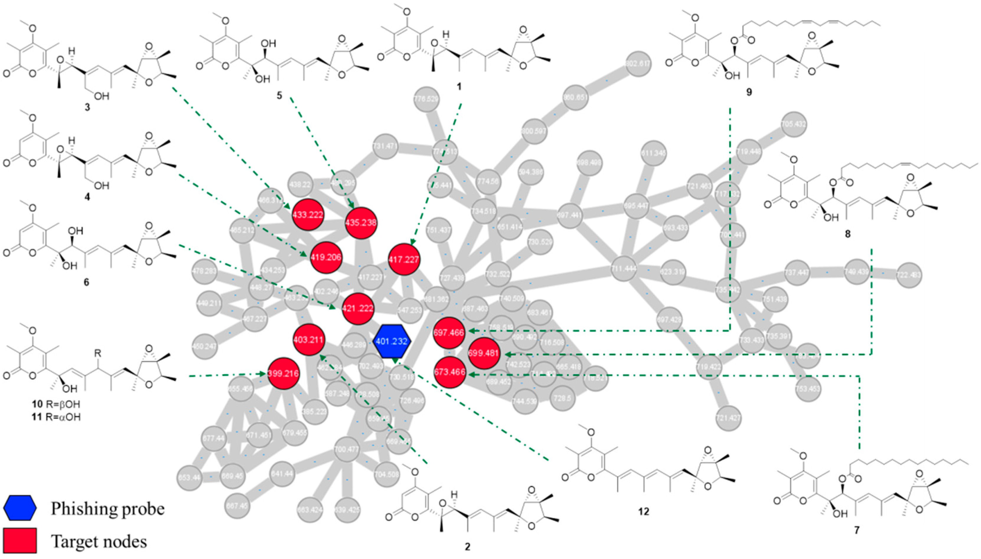
In this study, the MS/MS-based molecular networking strategy was applied for target isolation of new verrucosidins. First,
Penicillium strains were cultured on rice substrates and the resulting EtOAc extracts was screened by HPLC-UV-DAD analysis (
Figure S1). Then, the ethyl acetate extract of
P. cellarum YM1 that produced secondary metabolites with similar retention time and UV characteristics to those of deoxyverrucosidin was further investigated by UPLC-HRMS/MS. The LC-MS/MS data were used to generate a visualized molecular networking that was further annotated by Cytoscape 3.8.2 (
Figure S2).
In details, the HPLC-HRMS/MS analysis in the positive ion mode was conducted on the ethyl acetate extract from
P. cellarum YM1 with deoxyverrucosidin as the phishing probe. The obtained fragmentation data were organized by molecular networking, yielding a metabolite-level view of the data. Individual MS/MS spectrum was organized into 106 clusters consisting of 899 connected nodes (
Figure S2). Using the MS/MS data of deoxyverrucosidin as “seed” spectra, an initial focal point (a blue hexagon with
m/z 401.232) was generated in the global molecular networking. A close examination of the molecular network indicated some nodes connected to deoxyverrucosidin (
Figure 2), which predicted the presence of potential natural analogs. Under the guidance of MS/MS-based molecular networkings, seven new verrucosidins, namely penicicellarusins A-I (
3–9), in addition to five known polyketides verrucosidin (
1) [
4,
27], normethylverrucosidin (
2) [
27], penicyrone A-B (
10–11) [
28], and deoxyverrucosidin (
12) [
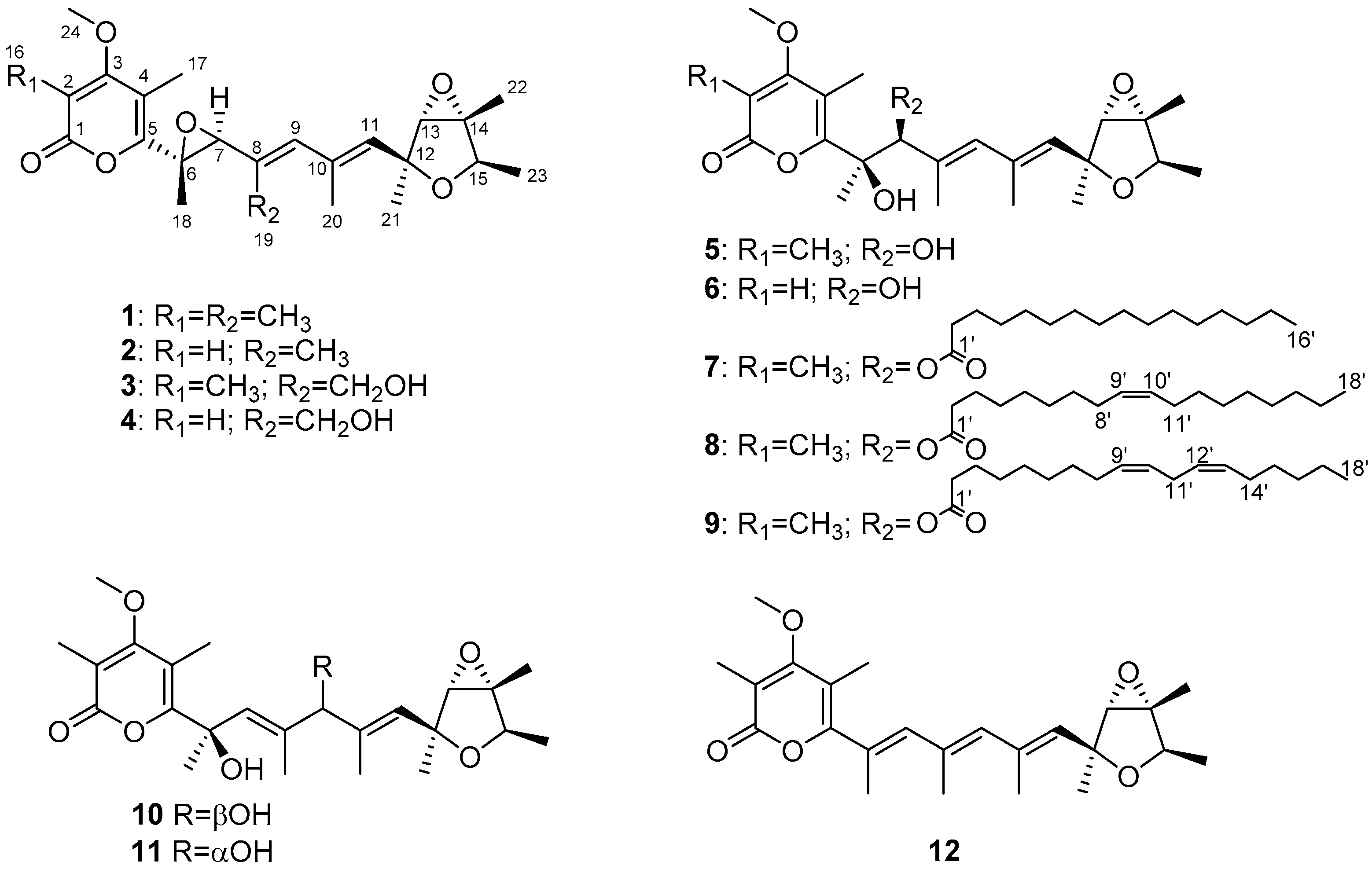
29] were obtained by the isolation workflow. The structures of known compounds were determined by comparing their NMR and MS data with literature data.
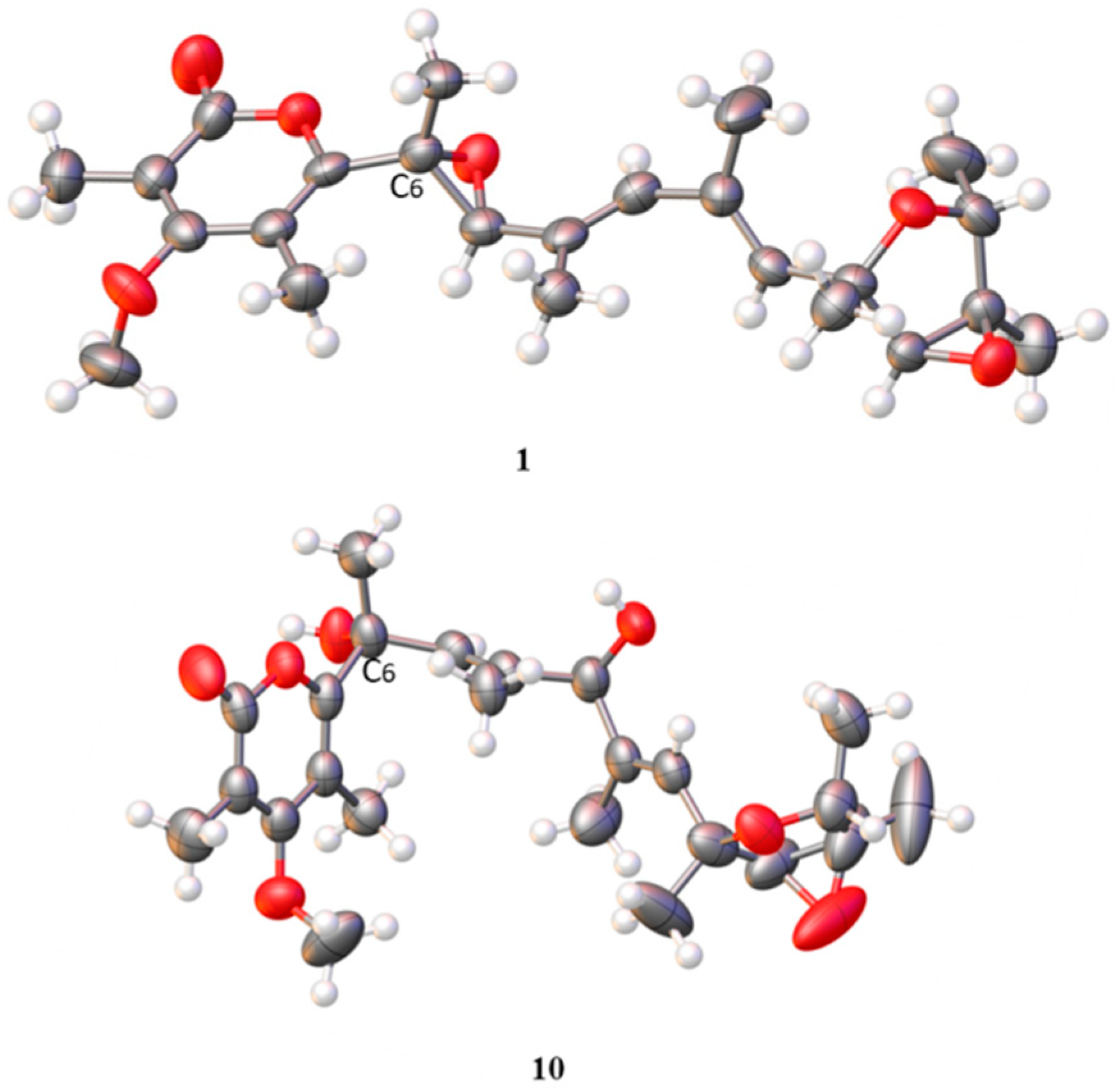
Compound
1 was obtained as white needle-like crystals and identified as verrucosidin by comparison of the NMR data reported in the literature [
4,
27] and the single-crystal X-ray crystallographic analysis (
Figure 3).
Penicicellarusin A (
3) was isolated as yellow oil with a molecular formula C
24H
32O
7 (indicating nine degrees of unsaturation) as deduced by HRESIMS data ([M+Na]
+ m/z 455.2041; calcd. 455.2040). The
1H-,
13C-NMR and HSQC spectra of
3 revealed the presence of eight methyls, including one oxygenated one [
δH/δC 1.23 (3H, d, 6.8 Hz) /19.2, 1.40 (3H, s)/22.1, 1.44 (3H, s)/15.7, 1.48 (3H, s)/13.8, 1.98 (3H, s)/18.7, 2.04 (3H, s)/10.4, 2.12 (3H, s)/9.7, and 3.91(3H, s)/61.3], one hydroxymethyl [
δH/
δC 4.36 (d,
J = 12.2 Hz), 4.41 (d,
J = 12.2 Hz)/59.6], five methines including three oxygenated [
δH/δC 3.60 (s)/68.7, 3.81 (s)/64.6, 4.09 (d, 6.8 Hz)/78.4] and two
sp2 methines [
δH/δC 5.60 (brs) /133.9, 5.98 (brs)/134.9]. The NMR data of compound
3 were similar with those of verrucosidin (
1) [
27], indicating the presence of a 3,6-dioxabicyclic[3.1.0]hexane moiety, a 2
H-pyran-2-one moiety, and a polyene chain in
3 (
Figure 1).
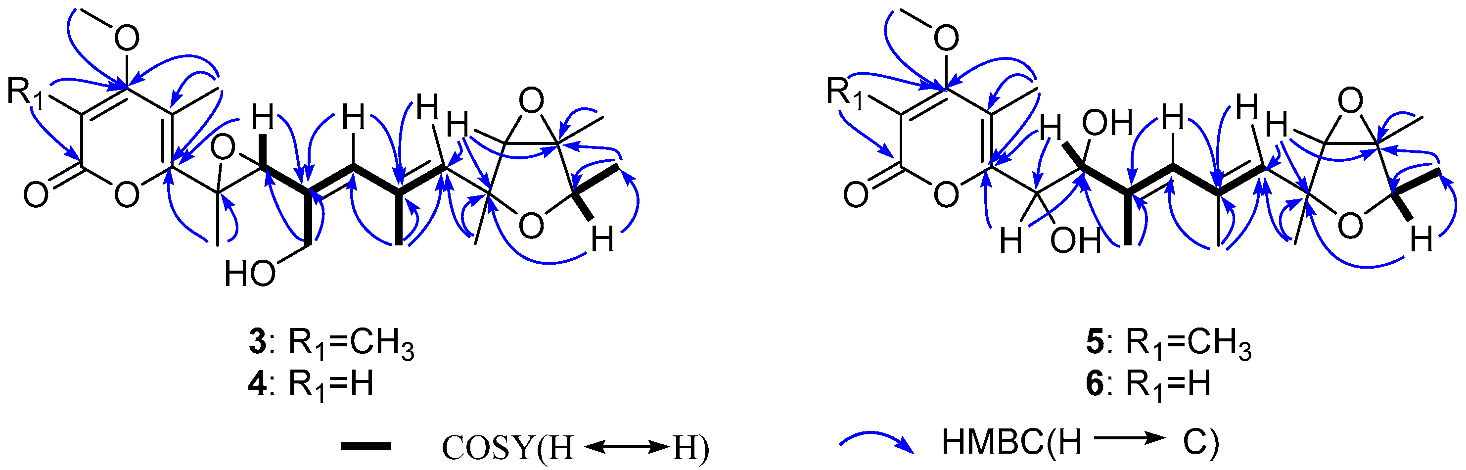
The HMBC correlations were detected from H
3-21 to C-11 and C-12, from H-13 to C-12 and C-14, from H
3-22 to C-14, from H
3-23 to C-14 and C-15, from H-15 to C-12, C-14 and C-23, which together with the
1H-
1H COSY correlations of H-15-H
3-23 confirmed the presence of the 3,6-dioxabicyclic[3.1.0]hexane moiety. The HMBC correlations of H
3-16 to C-1, C-2, and C-3, H
3-24 to C-3, H
3-17 to C-3, C-4, and C-5, and as well as the chemical shifts of C-1 (
δ 167.5), C-2 (
δ 111.6), C-3 (
δ 170.2), C-4 (
δ 112.2), and C-5 (
δ 157.3) completed the assignment of α-pyranone moiety. Furthermore, the
1H-
1H COSY correlations of H-7-H
3-19-H-9-H
3-20-H-11 together with the HMBC correlations from H
3-18 to C-5 and C-6, from H-7 to C-5, C-6, and C-8, from H
3-19 to C-7 and C-8, from H-9 to C-7, C-8, and C-10, from H
3-20 to C-9, C-10, and C-11, and from H-11 to C-10 and C-12 supported a heptadiene moiety which was connected with α-pyranone moiety through C-5 and linked with 3,6-dioxabicyclic[3.1.0]hexane moiety through C-12 (
Figure 4).
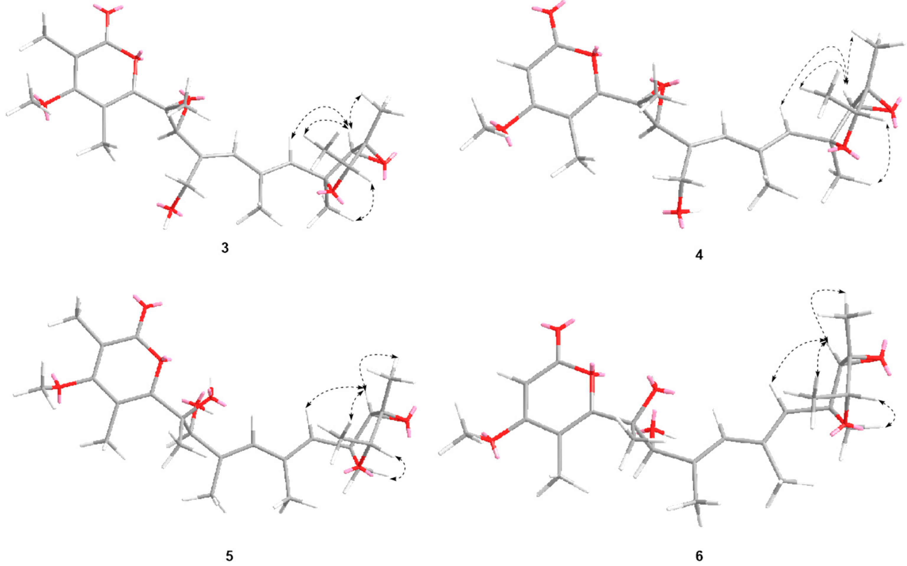
The relative configuration of
3 was confirmed by NOESY experiment (
Figure 5). The NOE correlations H-11 (
δ 5.60) to H
3-23 (
δ 1.23) and H-13 (
δ 3.60), H
3-22 (
δ 1.48) to H-13 and H
3-23, and H-15 (
δ 4.09) to H
3-21 (
δ 1.40), indicated that H
3-22, H
3-23, H-11, and H-13 were on the same face, while H
3-21 and H-15 were on the opposite face. The geometry of C
8=C
9 and C
10=C
11 were confirmed to be
E by the NOE correlations of H-7 (
δ 3.81) with H-9 (
δ 5.98) and H
3-20 with H
3-21.
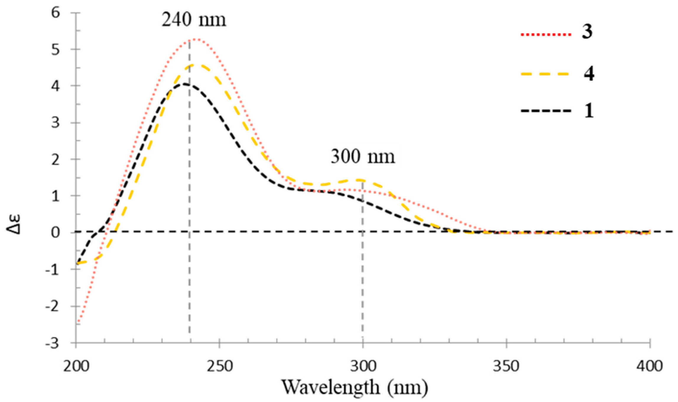
In the experimental ECD spectrum, compound
3 showed similar Cotton effects as verrucosidin (
1) (
Figure 6), supporting the same configuration at C-6 and C-7 between
1 and
3. Thus, compound
3 was assigned a 6
R, 7
S, 12
S, 13
S, 14
R, 15
R configuration and named penicicellarusin A.
Penicicellarusin B (
4) was obtained as yellow oil with the molecular formula C
23H
30O
7 and nine degrees of unsaturation, as deduced from HRESIMS data. The 1D NMR spectroscopic data of
4 (
Table 1) were similar with those of
3, except for the lack of a singlet methyl group and the presence of one additional olefinic proton at
δ 5.66 in
4. HMBC correlations of H-2 (
δ 5.66) to C-1, C-3 and C-4, H
3-17 to C-3 and C-5, H
3-24 to C-3 confirmed the structural changes on the 2
H-pyran-2-one moiety in
4. A further comprehensive analysis of its
1H-
1H COSY, HMQC, and HMBC spectra assigned the planar structure of
4 (
Figure 4).
The NOESY correlations of H-11 with H-13 and H
3-22, H
3-23 with H-13 and H
3-22, H-15 with H
3-21, H-7 with H-9 and H
3-17, H
3-20 with H
3-21 supported the same relative configurations for double bonds and 3,6-dioxabicyclic[3.1.0]hexane moiety between 4 and compound
3 (
Figure 5). Compound
4 showed similar Cotton effects in the experimental electronic circular dichroism (ECD) spectrum with those of
3 (
Figure 6), indicating it has the absolute configuration of 6
R, 7
S, 12
S, 13
S, 14
R, and 15
R, as described in
3.
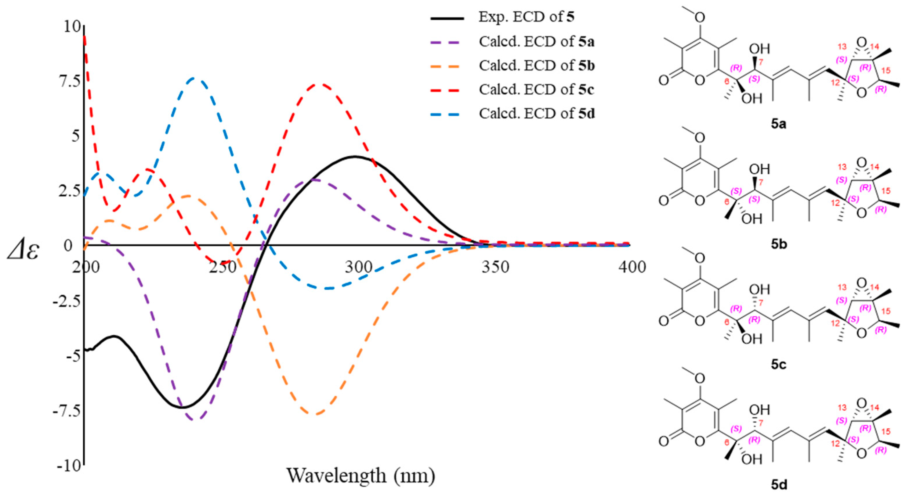
Compound
5 was assigned the molecular formula of C
24H
34O
7 (eight degree of unsaturation) on the basis of its HRESIMS at
m/z 457.2190 [M+Na]
+ and NMR data (
Table 2). The
1H-,
13C-NMR, and UV spectra of
5 were similar with those of verrucisidinol [
6], with the notable difference in the
1H-NMR data of C-3, C-4, C-5, and H-7 (
Table 2). A comprehensive analysis of its 2D NMR spectra including
1H-
1H COSY, HMQC, and HMBC experiments confirmed the planar structure of
5 (
Figure 4).
The partial relative configuration of
5 was confirmed by a NOESY experiment (
Figure 5). The geometry of C
8=C
9 and C
10=C
11 were confirmed to be
E by analysis of the NOESY observations. The key NOESY correlations of H-11 with H-13 and H
3-22, H
3-23 with H-13 and H
3-22, and H-15 with H
3-21 supported the same relative configurations on furan ring as verrucisidinol [
6]. Considering the same biosynthesis origin, compound
5 is deduced to share the same absolute configuration with those of
1–
4 in the furan ring. In addtion, the optical rotation data of
5 ([α
= +45.0,
c = 0.1, MeOH) were opposite to that of verrucisidinol ([α
= −10.0,
c = 0.1, MeOH), implying the enantiomeric relationship between them. To determine the absolute configurations at C-6 and C-7, ECD calculation method was applied. The four configurations (
5a,
5b,
5c and
5d,
Figure 7) were calculated using time-dependent density functional theory (TDDFT) at PBE1PBE/6-311 G* level with PCM model in methanol, and 60 exciting states were calculated. By comparison of the experimental and simulated ECD curves (
Figure 7), the experimental ECD was match better with
5a (6
R, 7
S, 12
S, 13
S, 14
R, and 15
R). Thus, the compound
5 was assigned as 6
R, 7
S, 12
S, 13
S, 14
R, and 15
R, and named as penicicellarusin C.
The molecular formula of penicicellarusin D (
6) was determined to be C
23H
32O
7 with the unsaturation degrees of eight on the basis of the HRESIMS data at
m/
z 443.2047 [M+Na]
+ (calcd. for C
23H
32O
7Na
m/
z 443.2040) and NMR data (
Table 2). The NMR data of
6 were similar to those of
5 except for the absence of one singlet methyl group. The key HMBC correlations from H-2 (
δ 5.60) to C-1, C-3 and C-4, as well as the upfield shift of C-2 (
δ 88.5) confirmed the disappearance of the methyl group on the C-2 position in
6. Furthermore, Compound
6 showed similar Cotton effects in the experimental CD spectrum with those of
5 (
Figure S5), which assigned the absolute configurations of
6 as 6
R, 7
S, 12
S, 13
S, 14
R, and 15
R. It was designated as penicicellarusin D.
Penicicellarusins E-G (compounds
7–9) were determined to be fatty acid esters of
5 by interpretation of the HRESIMS, 1D and 2D NMR data (
Table 3,
Figures S6 and S7), and ECD spectra (
Figure S8). The MS/MS data of
7–9 confirmed the presence of the fatty acid moiety in their structures. The pseudo molecular ion peaks [M+H]
+ at
m/
z 673.4669 in
7,
m/z 699.4819 in
8, and
m/z 697.4668 in
9 together with the fragment ion peaks [M+H-C
16H
32O
2]
+ at
m/
z 435.2381 in
7, [M+H-C
18H
32O
2]
+ at
m/
z 435.2375 in
8, [M+H-C
18H
30O
2]
+ at
m/
z 435.2374 in
9 due to the loss of the corresponding fatty acid moiety. To assign the structure of fatty acid moieties, compounds
7–9 was hydrolyzed with alkaline solution followed by methyl esterification. The fatty acid chain in
7–9 was determined to be the palmitic acid, the oleic acid, and the linoleic acid, respectively, by comparison of the retention time and MS spectrum with those of standards by GC-MS analysis (
Figure S9). Compounds
7–9 showed similar Cotton effects in the experimental CD spectrum with those of
5 (
Figure S8), which assigned their absolute configurations as 6
R, 7
S, 12
S, 13
S, 14
R, and 15
R.
With the help of single-crystal X-ray crystallographic analysis, the 6
R, 9
R, 12
S, 13
S, 14
R, and 15
R absolute configuration of penicyrone A (
10) was determined. The value of the Flack absolute structure parameter 0.03 (8) was obtained, and a perspective ORTEP plot was shown in
Figure 3 (CCDC 2039558). According to X-ray diffraction analysis, the configuration at the C-6 positions in
10 was 6
R, instead of 6
S reported in the literature [
28]. The structures of other known compounds were determined by comparing spectroscopic data with those in the literature.
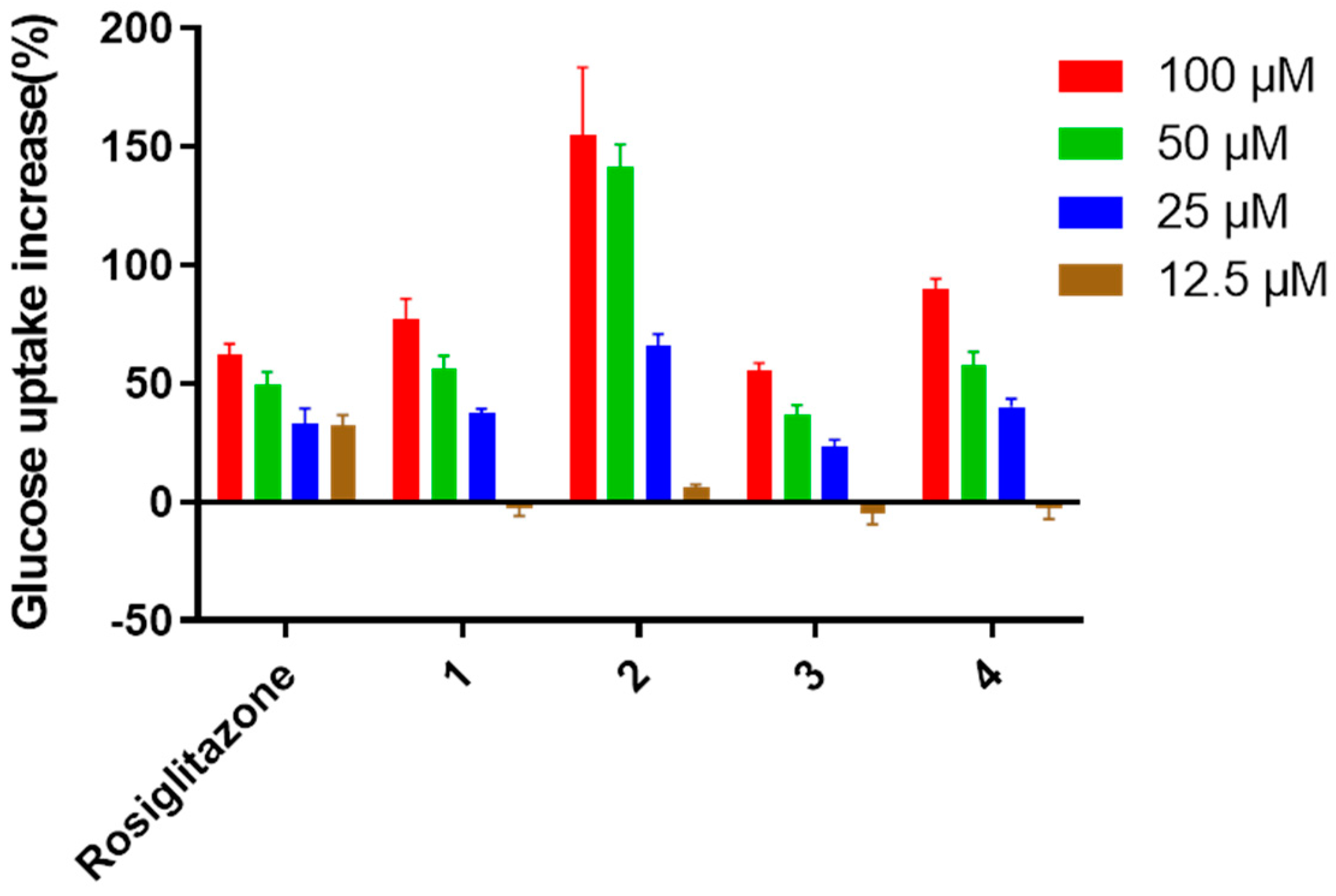
To explore the bioactivities of verrucosidins, compounds
1–12 were evaluated for the antimicrobial effect, cytotoxic activity, and hypoglycemic activity. As a result,
1–12 showed no significant bioactivity in the antimicrobial assays and cytotoxicity assays at the dose of 100 μM. However, Compounds
1–4 were found to enhance the insulin-stimulated uptake of 2-NBDG in insulin-resistant HepG2 cells with the EC
50 values at 47.2 ± 1.2, 9.9 ± 2.5, 93.2 ± 1.2 and 40.2 ± 1.3 μM, respectively, while the other compounds showed no significant activity (
Figure 8). In particular, compounds
1,
2, and
4 showed much stronger activity than the positive drug (rosiglitazone) in the range of 25–100 μM.
4. Discussion
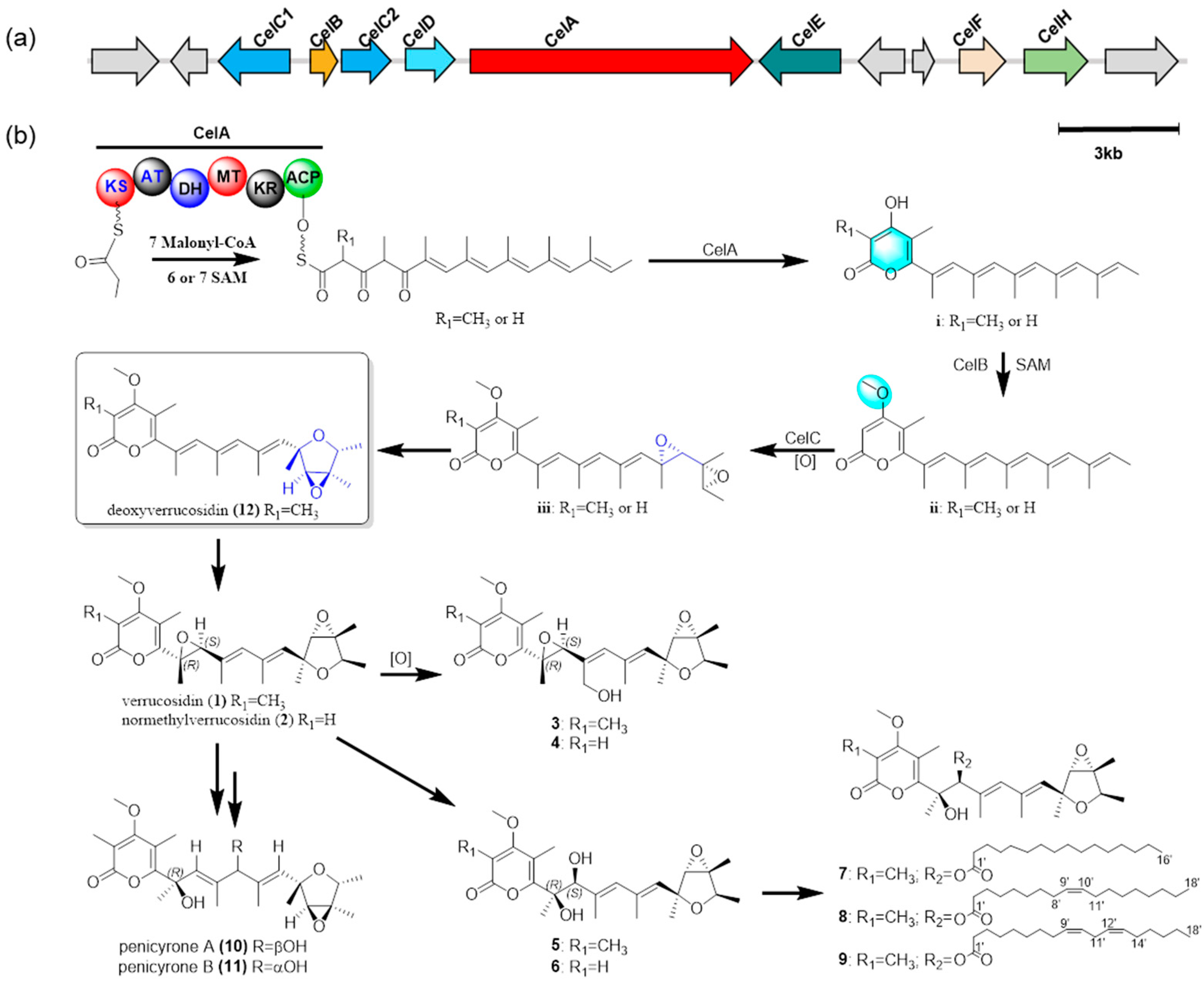
Up to now, less than 20 verrucosidins and structurally-related compounds have been found in fungi. In this study, seven new verrucosidin derivatives (compounds 3–9), were isolated together with five previously identified compounds from the fermentation products of fungus P. cellarum, suggesting that this fungus is an important producer of verrucosidins.
Verrucosidins share similar structural features with the citreoviridins, including a methylated α-pyrone, a polyene linker, and a tetrahydrofuran ring. The citreoviridin biosynthetic gene cluster containing a polyketide synthase (CtvA), a SAM-dependent methyltransferase (CtvB), a flavin-dependent monooxygenase (CtvC), and a hydrolase (CtvD) has been identified in
Aspergillus terreus var. aureus [
21]. CtvC is the only monooxygenase in the cluster, which can iteratively oxidize the terminal triene portion of the precursor into a bisepoxide moiety. As a regioselective hydrolase, CtvD can transform the bisepoxide moiety into a tetrahydrofuran ring moiety [
21,
30]. In addition, the verrucosidin bosynthetic gene cluster was confirmed by constructing deletion mutants for
verA gene coding for HR-PKS known to be the key enzyme of the biosynthesis. Different from citreoviridin, the bosynthetic gene cluster for verrucosidin in the genome of
P. polonicum contains two flavin-dependent monooxygenases, VerC1 and VerC2, which means that the cluster can synthesize compounds with higher oxidation degree [
22]. However, the enzymes involved in the biosynthesis of verrucosidin are largely uncharacterized.
In our work, with the genome of
P. cellarum YM1 sequenced in our group, we searched for the gene cluster for penicicellarusins. By similarity analysis with the polyketide synthase gene
ctvA and
verA, the putative gene cluster
celA was found in the genome of
P. cellarum (
Table 4 and
Figure S11). Further bioinformatic analysis revealed seven genes, the polyketide synthase gene (
celA), the SAM-dependent methyltransferase gene (
celB), the flavin-dependent monooxygenase (
celC1 and celC2), the cytochrome P450 gene (
celD), the acyl-acyltransferase gene (
celF), and the lyase gene (
celH) potentially involved in the biosynthesis of penicicellarusins in
P. cellarum. Based on above evidence, we propose the biosynthetic pathway of
1–12 (
Figure 9 and
Figure S10). Verrucosidin (
1) could be formed from
12 by oxidation of the olefinic bond, and further oxidation produces
3–6. Compounds
7–9 were biosynthesized by esterification of
5 with different fatty acids. Compounds
10 and
11 can be transformed from
5 or
6 by dehydration reaction.
Early studies have demonstrated that verrucosidins and structurally-related compounds are endowed with several interesting bioactivities, such as antibacterial activities [
3,
10], antitumor [
7,
8], antiviral [
9], and neurological activities [
11]. In this work, it was found that compounds
1–
4 show promising hypoglycemic activity, especially compounds
2 and
4. Preliminary structure-activity relationship showed that the formation of epoxy three-membered ring on C
6-C
7 in the structures contributes greatly for the glucose uptake-enhancing activity in insulin-resistant HepG2 cells. The promising hypoglycemic activity is an interesting new bioactivity for this class of compounds.


 ,
, 

