Article

Role of Two G-Protein α Subunits in Vegetative Growth, Cell Wall Integrity, and Virulence of the Entomopathogenic Fungus Metarhizium robertsii

1
Anhui Provincial Key Laboratory of Microbial Pest Control, Anhui Agricultural University, Hefei 230036, China
2
School of Life Sciences, University of Science and Technology of China, Hefei 230022, China
*
Authors to whom correspondence should be addressed.
J. Fungi 2022, 8(2), 132; https://doi.org/10.3390/jof8020132
Submission received: 13 January 2022 / Revised: 26 January 2022 / Accepted: 27 January 2022 / Published: 28 January 2022
(This article belongs to the Special Issue Interactions between Filamentous Fungal Pathogens and Hosts)

Abstract

:
Heterotrimeric G-proteins are crucial for fungal growth and differentiation. The α subunits of heterotrimeric G-proteins play an essential role in controlling signal transduction. However, the function of G-protein α subunits in entomopathogenic fungi remains poorly understood. Two group II Gα subunits (MrGPA2 and MrGPA4) were characterized in the entomopathogenic fungus, Metarhizium robertsii. Bioinformatics analysis showed that the relationship between MrGPA2 and MrGPA4 was closer than that of other MrGPAs. Both green fluorescent protein (GFP)-tagged MrGPA2 and MrGPA4 were localized at the cytoplasm. Furthermore, ∆MrGpa2MrGpa4 double mutants showed remarkably reduced vegetative growth compared to the wild-type and single-mutant strains, which was accompanied by the downregulation of several growth-related genes, such as ssk2, pbs2, stuA, hog1, and ac. Only the ∆MrGpa2MrGpa4 double mutant was sensitive to Congo red stress. The insect bioassay demonstrated significantly attenuated virulence for the ∆MrGpa2MrGpa4 double mutant compared to the wild-type and single-mutant strains. Further analysis indicated that double deletion of MrGpa2 and MrGpa4 had no effect on appressorium formation but suppressed the expression levels of several virulence-related genes in the insect hemocoel. These findings demonstrate that MrGpa2 and MrGpa4 exhibit functional redundancy and contribute to the vegetative growth, stress tolerance, and pest control potential in M. robertsii.

1. Introduction

Insect-pathogenic fungi, as a biological control agent that can cause the death of many insects, represent an environmentally friendly alternative to existing insecticides [1]. However, their use has been limited because of their slow killing speed, low sporulation rate, and sensitivity to high temperatures and UV stress. To kill pests, fungal spores first adhere to the pest cuticle. Then, the conidium germinates and penetrates the pest cuticle via mechanical pressure and cuticle-degrading enzymes. At that time, fungal pathogens must dodge acute immune responses to colonize the pest hemocoel. Finally, hyphae multiply within and emerge from the dead pest, and the newly created conidia disperse and infect additional insects if the conditions are favorable [2,3]. An understanding of the molecular mechanisms of its infection, disease development, and stress tolerance is necessary for the commercial application of fungal pathogens.
Heterotrimeric G-proteins, which are critical signaling proteins involved in receptor-mediated signal transduction, are present in all eukaryotic cells [4]. The G-proteins consisting of α, β, and γ subunits are coupled to the G-protein-coupled receptor (GPCR) for transferring environmental signals [5]. G-proteins are crucial for the regulation of cell function, division, and differentiation. In filamentous fungi, they contribute to hyphal growth, conidiation, conidial germination, stress resistance, and virulence [6,7,8,9].
G-protein α subunits can be classified into four groups (Gαi, Gαs, Gαq, and Gα12) in mammalian species, and only two kinds of Gα subunits (canonical Gα and Extra-large Gαs) exist in plants [4]. In yeast, two Gα subunits (GPA1 and GPA2) have been identified [10]. In particular, Gα subunits of the filamentous fungi are classified into three major groups: group I, group II, and group III [11]. The groups I and III Gα proteins contain a consensus myristylation site (MGXXXS) in the N terminus and a pertussis toxin-labeling site (CXXX) at the C termini of group I Gα proteins, but group II Gα proteins do not contain either site [11]. Based on their different structures, the functions of group II Gα proteins are diverse. The role of group II Gα proteins has been characterized in some fungi. For instance, the deletion of GNA-2 in Neurospora crassa intensified a reduction in mass accumulation on poor carbon sources in ∆gna-1 or ∆gna-3 strains [12]. In Magnaporthe grisea, disruption of magC resulted in a reduction in conidia [8]. ∆bcg-2 of Botrytis cinerea was only slightly reduced in pathogenicity [13]. Disruption of fga3 in Fusarium oxysporum f. sp. cucumerinum led to higher thermal resistance, possibly through the cAMP-dependent protein kinase A pathway [14]. However, the functions of the group II Gα proteins in entomopathogenic fungi are still unknown.
An entomopathogenic model fungus, Metarhizium robertsii, has been well developed as one of the most environmentally friendly insecticides [2]. Genes encoding four Gα subunits, MrGpa1 (EFZ00892.1), MrGpa2 (EFY98464.1), MrGpa3 (EFY99066.2), and MrGpa4 (EFZ00060.2), have been identified in M. robertsii [9]. We have previously shown that MrGPA1, belonging to group I, contributes to conidiation, stress resistance, virulence, and intracellular cAMP levels, while MrGpa3, which was identified as a group III Gα subunit, plays similar biological roles in the fungus [9]. MrGPA2 and MrGPA4 of M. robertsii are orthologs of N. crassa GNA-2; However, there is no report on the function of MrGPA2 and MrGPA4 in entomopathogenic fungi.

2. Materials and Methods

2.1. Fungal Strains and Culture

The wild-type (WT) strain M. robertsii ARSEF 23 (ATCC No.MYA-3075) was isolated from Conoderus sp. in North Carolina, USA, in 1961. The fungus and all of the mutant strains were maintained on potato dextrose agar (PDA, 20% potato, 2% glucose, and 2% agar, w/v) at 25 °C under dark conditions for 10 days [15]. After 10 days of incubation, conidia were harvested by vortex-mixing in 0.05% (v/v) Tween-80, filtrated through sterile non-woven fabric, and washed with sterile water.

2.2. Sequence Analysis

To determine the phylogenetic relationship among MrGPA2, MrGPA4, and their orthologues, the amino acid sequences of those functionally characterized GPA subunits of different fungi were downloaded from the National Center for Biotechnology Information (https://www.ncbi.nlm.nih.gov/, accessed on 20 September 2021). A neighbor-joining tree was constructed using the program MEGAX (https://www.megasoftware.net/, accessed on 20 September 2021).

2.3. Subcellular Localization of MrGPA2 and MrGPA4

To investigate the subcellular localization of MrGPA2 and MrGPA4, two genes were fused with a GFP tag and inserted into the plasmid pDHt-SK-bar (kindly provided by Dr. Chengshu Wang; the vector conferred resistance against glufosinateammonium) containing a strong promoter and a terminator, which were transformed into M. robertsii and screened as previously described [16]. The subcellular localization of MrGPA2-GFP and MrGPA4-GFP fusion proteins in conidia and mycelia were visualized using confocal microscopy (LSCM, Zeiss LSM980, Zeiss Gruppe, Oberkochen, Baden-Wurttemberg, German).

2.4. Gene Deletion and Complementation

Targeted gene disruption of MrGpa2 and MrGpa4 was performed by homologous recombination via Agrobacterium tumefaciens transformation as previously described [9]. In brief, the 5′ and 3′ flanking regions of MrGpa2 and MrGpa4 were inserted into the pDHt-SK-bar, and then the vectors pDHt-MrGpa2-bar and pDHt-MrGpa4-bar were obtained for fungal transformation. For double-gene deletion, the 5′ and 3′ flanking regions of MrGpa4 were inserted into the pDHt-SK-ben vector (containing benomyl resistance gene), and the product was transformed into ΔMrGpa2 strain and screened as previously mentioned.
For mutant complementation, the entire MrGpa2 and MrGpa4 genes were amplified together with their upstream and downstream regions and then inserted into the vector pDHt-SK-ben to produce the vectors pDHt-cpMrGpa2-ben and pDHt-cpMrGpa4-ben for fungal transformation. Next, all of transformants were verified by DNA sequencing. All the primers used in this study are listed in Supplementary Materials, Table S1.

2.5. Phenotype Assays

To assess conidiation capacity, 30 µL of conidial suspension (1 × 10⁶ conidia/mL) of WT, ∆MrGpa2, ∆MrGpa4, ∆MrGpa2MrGpa4, cp∆MrGpa2, and cp∆MrGpa4 strains was evenly spread on PDA medium and incubated for 14 days at 25 °C. These samples were then placed in 30 mL of 0.05% Tween 80 to release conidia by vortex-mixing, and these conidial suspensions were used for assessment of conidial concentration with a hemocytometer, followed by conversion of the number of conidia per square centimeter of the colony.
The vegetative growth assays of the WT, ∆MrGpa2, ∆MrGpa4, ∆MrGpa2MrGpa4, cp∆MrGpa2, and cp∆MrGpa4 strains were carried out on PDA, SDAY, and 1/4 SDAY (1/4 dilution of SDAY) media. For this, samples of conidial suspensions (1 µL; 1 × 10⁷ conidia/mL) were point-inoculated on each plate and incubated at 25 °C in the dark for 10 days. The colony diameters were then measured.
To determine the growth rates on Czapek agar (CZA, 3% sucrose, 0.3% NaNO3, 0.1% K2HPO4, 0.05% KCl, 0.05% MgSO4, 0.001% FeSO4,and 2% agar), amended CZAs containing different carbon or nitrogen sources were prepared by deleting 3% sucrose or 0.3% NaNO3; replacing the sole carbon source (3% sucrose) with 3% glucose, trehalose, lactose, fructose, maltose, mannitol, sorbitol, glycerol, ethanol, sodium acetate (NaAc), or oleic acid; and replacing the sole nitrogen source (0.3% NaNO3) with 0.3% NH4Cl or one of 20 amino acids, followed by incubation at 25 °C for 10 days. The colony diameters were then measured.
To analyze the chemical tolerance, conidial suspensions (1 μL; 1 × 10⁷ conidia/mL) of strains were cultured on PDA plates supplemented with NaCl (0.5 M), H2O2 (2 mM), or Congo red (2 mg/mL) in the dark at 25 °C for 10 days. The colony diameter was measured, and the relative inhibition rate was calculated.
To assay the heat tolerance, experiments were conducted on PDA plates. Conidial suspensions (5 × 10⁶ conidia/mL) of strains were incubated in a water bath at 42 °C or 28 °C (as control) for 1 h, and then 10 μL of the suspension was incubated at 25 °C in the presence of the medium. Germination was observed with Olympus BX 51(Tokyo, Japan)) at 12, 16, 20, and 24 h. Conidia were considered germinated when the length of the germ tube projecting from it reached the half length of the conidia. A total of 300 hundred conidia were counted per plate, and the relative germination rates were calculated to allow comparisons of the number of germinated conidia with and without heat stress.
To evaluate fungal tolerance to ultraviolet B (UV-B) light, heat tolerance assays were conducted as previously mentioned. These plates with conidia were exposed to an irradiance of 312 nm wavelength at 100 μJ cm−2 using HL-2000 Hybrilinker (UVP, Upland, CA, USA). Relative percent germination was assessed using the aforementioned methods.
For virulence assays, the tests were performed using Galleria mellonella larvae as previously described [9]. Briefly, conidial suspensions (1 × 10⁷ conidia/mL) harvested from a 10-day-old PDA culture were collected, and the larvae were separately immersed in each conidial suspension for 90 s, followed by incubation at 25 °C. All treated cohorts were performed in triplicate, with 18 larvae in each group, and monitored every 12 h to record survival, using the SPSS software to calculate the median lethal time (LT50) by probit analysis.
For appressorium formation assay, 1 mL of conidial suspension (1 × 10⁶ conidia/mL) in MMGly (minimal medium amended with 1% glycerol) was spread on a sterile plastic Petri dish (3.5 cm diameter), followed by 24 h incubation at 25 °C for 24 h. The appressorium formation rates were assessed as described previously [17].

2.6. Quantitative RT-PCR (RT-qPCR)

To analyze the transcriptional profiling of phenotype-related genes, WT, ∆MrGpa2MrGpa4, controls, and two single-mutant strains were cultured on PDA plates for 10 days, or G. mellonella larvae were infected by six strains for 48 h. Then, the mycelia or larval hemocoel were harvested and milled in liquid nitrogen for RNA extraction. Total RNA was isolated using the TRIzol reagent (Invitrogen, Foster City, CA, USA). cDNA was used as a template for RT-qPCR using a HiScript III 1st strand cDNA synthesis kit (Vazyme, Nanjing, China). Three biological replicates were analyzed for each treatment. qRT-PCR analysis was performed using the CFBR96TM Real-Time PCR System (Bio-Rad, Hercules, CA, USA) and AceQ qPCR SYBR Green Master Mix (Vazyme, Nanjing, China). The primers used are listed in Supplementary Material, Table S2. The gpd gene (MAA_07675, encoding glyceraldehyde 3-phosphate dehydrogenase) was used as an internal control, and relative gene expression was determined by normalizing the expression of each gene to GAPDH. The data were analyzed using the 2−ΔΔCt method [18].

2.7. Statistical Analysis

Each experiment was performed in triplicate. All data were analyzed using GraphPad Prism version 7.0 and SPSS v23.0 software (SPSS Inc., Chicago, IL, USA). Kolmogorov—Smirnov test and Levene’s test were used to confirm that the data followed normality and homoscedasticity, respectively. Then, data (mean  ±  SE) from different experimental groups were analyzed using Student’s t-test, one- or two-way analysis of variance (ANOVA) followed by a least significant difference (LSD) test. p < 0.05 and p < 0.01 were considered significant and extremely significant, respectively. * p < 0.05, ** p < 0.01.

3. Results

3.1. Characteristics of MrGPA2 and MrGPA4 from M. robertsii

We identified two N. crassa GNA-2 homologs, EFY98464.1 and EFZ00060.2, in M. robertsii, and named them MrGPA2 and MrGPA4, respectively. Bioinformatics analysis showed that MrGPA2- and MrGPA4-encoded proteins had 354 amino acids and 353 amino acids, respectively. Conserved domain analysis revealed that MrGpa2 and MrGpa4 contained the guanine nucleotide-binding site of heterotrimeric G-protein (34-347-aa protein and 34-348-aa protein) (Figure 1A).
The Gα sequences from the filamentous fungi were selected for phylogenetic analysis using Saccharomyces cerevisiae as an outgroup and were divided into three main clades including the group I (MrGPA1 M. robertsii), group II (MrGPA2 and MrGPA4 M. robertsii), and group III (MrGPA3 M. robertsii) clans (Figure 1B). Moreover, these MrGPA2 and MrGPA4 homologs were grouped together, and the relationship between MrGPA2 and MrGPA4 was closer than that of other MrGPAs. Intriguingly, phylogenetic analysis indicated that M. robertsii MrGPA4 homologs were only found in members of the genus Metarhizium and its sister genus Pochonia, whereas M. robertsii MrGPA2 homologs existed in filamentous fungi. Based on the relationship between the two GPAs and their distribution in filamentous fungi, MrGPA4 is likely an additional copy of MrGPA2.

3.2. MrGPA2 and MrGPA4 Are Located in Cytoplasm

To assess the subcellular localization of MrGPA2 and MrGPA4, we generated MrGpa2-gfp and MrGpa4-gfp strains for fluorescence observation (Supplementary Figure S1A–C). As shown in the LSCM images in Figure 2, the green fluorescence signals of MrGPA2 and MrGPA4 or 4,6-diamidino-2-phenylindole (DAPI)-stained nuclei were detected, but they were only observed in the cytoplasm, either in hyphae or in conidia, which both exhibited the cytoplasmic localization of MrGPA2 and MrGPA4.

3.3. Gene Knockout and Complementation

To further investigate the function of MrGpa2 and MrGpa4 in M. robertsii, ∆MrGpa4, ∆MrGpa4, double-knockout mutants ∆MrGpa2MrGpa4 were generated and detected by PCR amplification. (Supplementary Figure S1A,B). PCR analysis revealed that the one 286-bp fragment corresponding to the partial MrGpa2 gene sequence was not detected in the ∆MrGpa2 and ∆MrGpa2MrGpa4 strains, and the one 212-bp fragment corresponding to the partial MrGpa4 gene sequence was not found in the ∆MrGpa4 or ∆MrGpa2MrGpa4 strains. In addition, a partial 434-bp bar gene fragment was present in all transformants, and a partial 1993-bp ben gene fragment was detected in ∆MrGpa2MrGpa4, cp∆MrGpa2, and cp∆MrGpa4. These observations indicated the presence of deletion mutants (∆MrGpa4, ∆MrGpa2, and ∆MrGpa2MrGpa4) and complementation strains (cp∆MrGpa2 and cp∆MrGpa4).

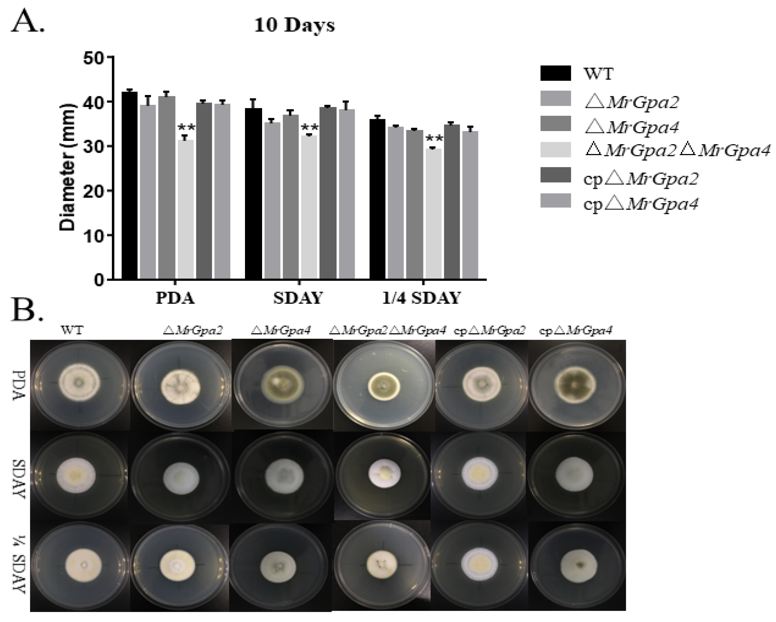
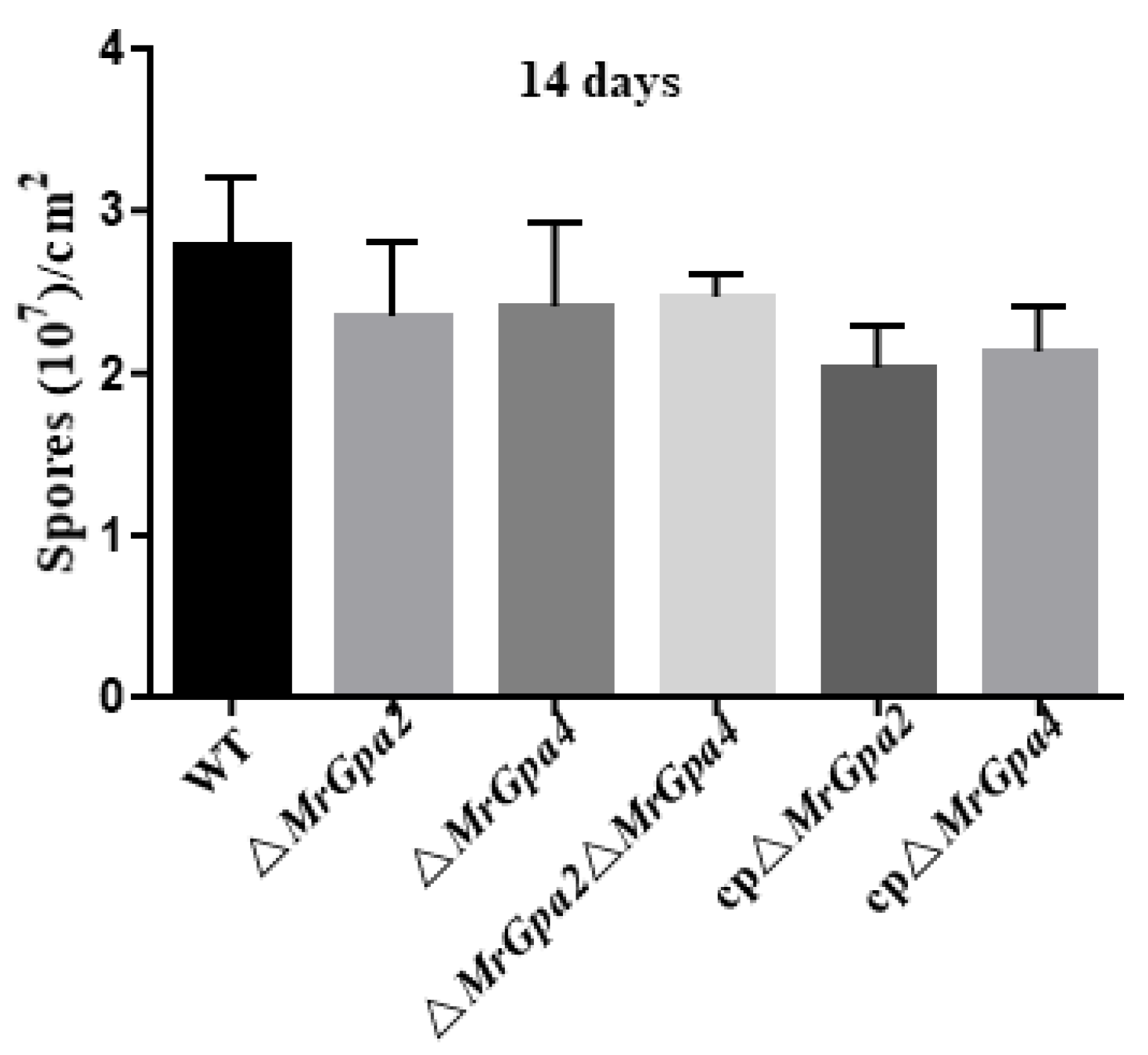
3.4. ∆MrGpa2∆MrGpa4 Strain Leads to a Decrease in Vegetative Growth but Had No Effect on Conidiation

We assessed the effect of MrGPAs’ disruption on vegetative growth. The single mutants of MrGpa2 or MrGpa4 had no influence in vegetative growth, but the double mutants affected vegetative growth. The growth rate of the ∆MrGpa2MrGpa4 mutant was significantly reduced by 26% (F5, 23 = 41.2, p < 0.01), 16% (F5, 23 = 15.5, p < 0.01), and 18% (F5, 23 = 36.9, p < 0.01) in PDA, SDAY, and 1/4 SDAY plates, respectively, compared to the WT strain (Figure 3A,B).
To further verify the effect of nutrient utilization on mutant growth in mixed-media CZAs amended with different carbon (sugars/non-sugars) or nitrogen (inorganic/organic) sources, colony diameters were assessed in the ∆MrGpa2MrGpa4 and WT strains. The ∆MrGpa2MrGpa4 mutant grew significantly slower than the WT strains on CZA media amended with sugar carbon sources such as glucose (t = 24.8, p < 0.01), trehalose (t = 23.5, p < 0.01), lactose (t = 13, p < 0.01), maltose (t = 19.9, p < 0.01), mannitol (t = 26.2, p < 0.01), or sorbitol (t = 12.9, p < 0.01), and CZAs media amended with the nitrogen sources with NH4Cl (t = 25.8, p < 0.01), or glycine (t = 15.2, p < 0.01) (Figure 4A,B). In contrast, colony size was not significantly different between ∆MrGpa2MrGpa4 and the control strain grown on CZA media supplemented with a non-sugar carbon source such as ethyl alcohol (t = 0.3, p = 0.75), glycerol (t = 0.9, p = 0.31), and NaAC (t = 0.7, p = 0.53). Moreover, the expression of six growth-related genes in M. robertsii was examined using RT-qPCR to explore the roles of the two genes in fungal vegetative growth. The ∆MrGpa2MrGpa4 strain demonstrated markedly reduced expression of ssk2 (MAA-04290, F5, 12 = 18.9, p < 0.01), pbs2 (MAA-00856, F5, 12 = 14.2, p < 0.01), stuA (MAA-02988, F5, 12 = 6.2, p < 0.01), hog1 (MAA-05126, F5, 12 = 12.0, p < 0.01), and ac (MAA-07239, F5, 12 = 11.7, p < 0.01) (Figure 4C). Collectively, these data indicate that MrGpa2 and MrGpa4 are involved in carbon/nitrogen metabolism and affect fungal vegetative growth.
In addition, the conidial yield was also assessed for the three mutants, which confirmed that the ∆MrGpa2, ∆MrGpa4, and ∆MrGpa2MrGpa4 strains were not required for conidial yield (Figure 5).

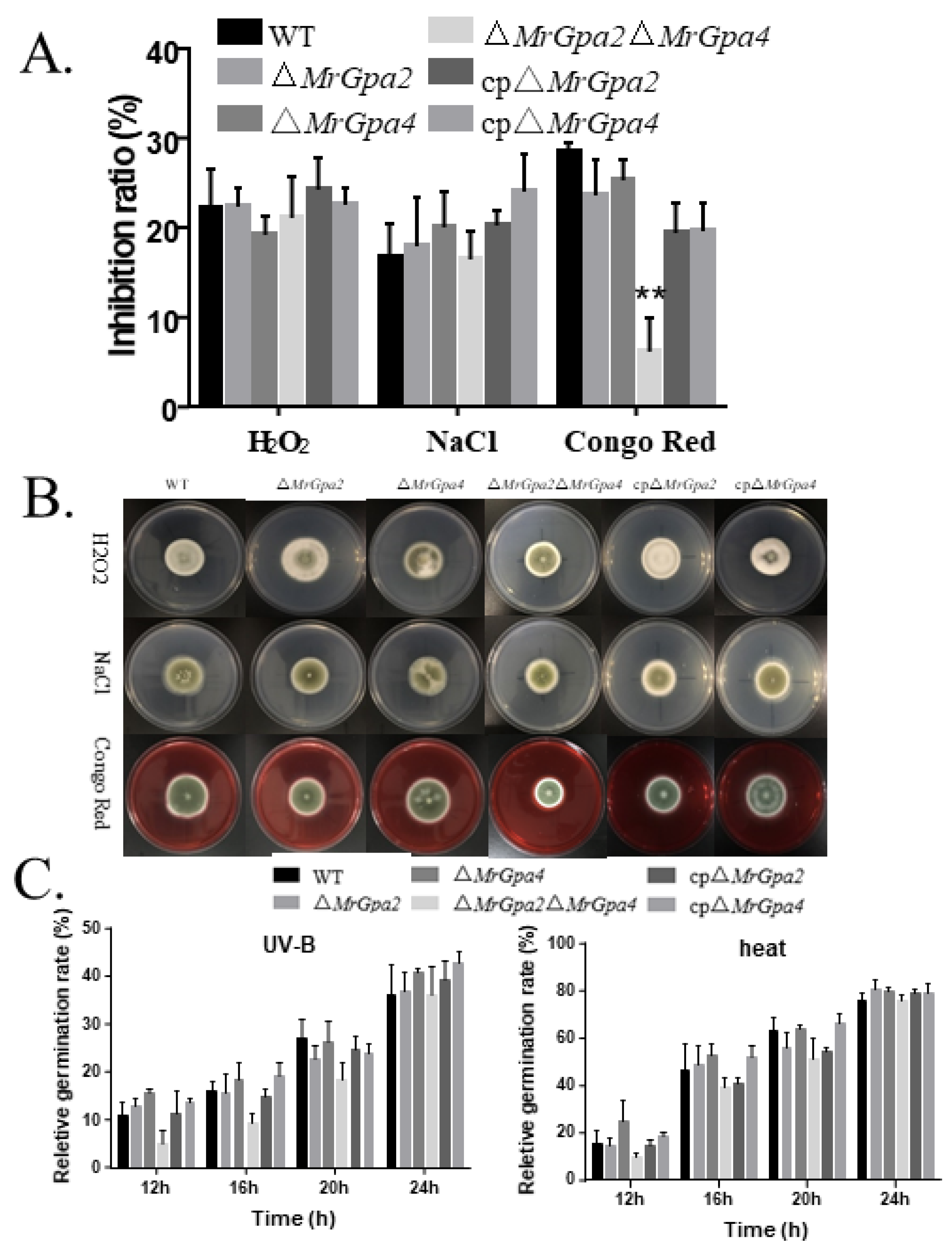
3.5. Deletion of Both MrGpa2 and MrGpa4 Enhanced Cell Wall Integrity

To examine whether the ∆MrGpa2, ∆MrGpa4, and ∆MrGpa2MrGpa4 strains showed an altered response to chemical stress, mycelial growth was observed on a PDA medium containing NaCl, H2O2, or Congo red (Figure 6A,B). On PDA plates containing Congo red, the ∆MrGpa2MrGpa4 strain showed altered mycelial diameter and a 78.3% reduction in the inhibition ratio compared to that of the WT strain (F5, 21 = 33.9, p < 0.01), which indicates the enhanced cell wall integrity of the double mutant. However, no effects on osmotic or antioxidant stress were observed in the ∆MrGpa2, ∆MrGpa4, or ∆MrGpa2MrGpa4 strains.
Furthermore, to confirm the role of MrGpa2 and MrGpa4 in heat and UV-B tolerance, conidial germination rates were calculated at 12, 16, 20, and 24 h after exposure to heat or UV-B irradiation stress (Figure 6C). Compared with the WT and CP strains, the conidial germination rates in the three mutants were not significantly different.

3.6. ∆MrGpa2∆MrGpa4 Strain Displayed an Increased Survival Rate of the Larvae in Bioassays

To investigate the role of MrGpa2 or MrGpa4 in virulence, the mortality rate of G. mellonella larvae caused by each mutant was assessed using bioassays (topical and injection). After topical application, the differences were significant between the WT (LT50 = 5.52 ± 0.06 days) and ∆MrGpa2MrGpa4 strains (LT50 = 6.24 ± 0.20 days) (F5, 21 = p < 0.05) (Figure 7A). However, no significant difference was observed for ∆MrGpa2 (LT50 = 5.45 ± 0.18 days) or ∆MrGpa4 (LT50 = 5.59 ± 0.06 days) compared with WT. Similarly, after the intrahemocoel injection, the insect groups infected with the ∆MrGpa2MrGpa4 strain exhibited an LT50 of 4.41 ± 0.05 days, with a significant attenuation of virulence, while the WT, control, and two single-mutant strains displayed LT50 values of 4.08 ± 0.10, 4.09 ± 0.06, 3.96 ± 0.11, 3.88 ± 0.07, and 3.76 ± 0.04 days, respectively (F5, 21 = 6.9, p < 0.05) (Figure 7B). The ∆MrGpa2MrGpa4 strain displayed an increase in LT50 and thus a significant difference in virulence, but the two single mutants demonstrated virulence comparable to the WT strain after both topical infection and injection of fungal spores into G. mellonella.
Furthermore, the appressorium formation rates were not remarkably different among the WT, ∆MrGpa2MrGpa4, control, or the two single-mutant strains, suggesting that MrGpa2 and MrGpa4 have no effect on fungal appressorium formation, which is required for cuticle penetration (Figure 7C).
Moreover, we investigated the transcriptional level of virulence-related genes in the insect hemocoel using RT-qPCR. Indeed, the transcriptional levels of mcl1 (a collagen-like protein, F5, 12 = 9.5, p < 0.01), pacC (a pH regulatory protein, F5, 12 = 5.2, p < 0.01), msn2 (a transcription factor, F5, 12 = 3.2, p < 0.05), dtxS1 (a nonribosomal peptide synthetase, F5, 12 = 14.2, p < 0.01), snf1 (encoding a sucrose-nonfermenting protein kinase, F5, 12 = 10.5, p < 0.01), and atm1 (acid trehalose, F5, 12 = 7.6, p < 0.01) were markedly downregulated in the ∆MrGpa2MrGpa4 strain, compared with the WT, control, and two single-mutant strains (Figure 7D) [19,20,21,22,23,24]. Therefore, MrGpa2 and MrGpa4 contribute to fungal virulence by affecting colonization and fungal cell growth in the insect hemocoel.

4. Discussion

As previously reported, G-proteins regulate a variety of fungal physiological processes, including conidiation, vegetative growth, stress tolerance, appressoria formation, and virulence [5,9,25]. In this study, we identified two group II Gα proteins, MrGPA2 and MrGPA4, in M. robertsii, both of which are involved in the vegetative growth, cell wall integrity, and virulence of M. robertsii, although the two single mutants did not display any defect in phenotypic characteristics.
Fungal Gα-proteins are classified into three major groups: group I, group II, and group III. In contrast to yeast, which contains two Gα proteins, most characterized filamentous fungi possess three Gα proteins that are members of distinct groups, including Magnaporthe grisea, Neurospora crassa, and Fusarium oxysporum f. sp. cucumerinum [4,7,8,12,14]. However, four Gα proteins have been identified in the genus Metarhizium and its sister genus Pochonia. Further analysis showed that group II consisted of MrGPA4 and MrGPA2. In this study, we found that MrGPA2 and MrGPA4 were functionally redundant in M. robertsii.
To transmit extracellular signals to intracellular effectors, G-proteins are localized to the plasma membrane [26]. However, in recent years, G-proteins have been identified in many other intracellular organelles, such as the mitochondria, Golgi apparatus, endoplasmic reticulum, and nucleus [27]. In our previous Metarhizium GPA study, MrGPA1 and MrGPA3 were found to be localized in the mitochondria and vacuoles, respectively [9]. However, in this study, both MrGPA2 and MrGPA4 were found to be localized in the cytoplasm, unlike the S. cerevisiae GPA2 embedded in the plasma membrane [10]. We speculate that the function of MrGPA2 and MrGPA4 differs from that of other fungal GPAs.
In this study, the ∆MrGpa2MrGpa4 double mutant showed inhibited growth in PDA, SDAY, 1/4 SDAY, and CZAs plates. Further growth analyses indicated that the double-deletion mutant was involved in carbon and nitrogen metabolism. In other filamentous fungi, the role of the group II Gα subunits is not related to growth, such as N. crassa, M. grisea, B. cinerea, and F. oxysporum f. sp. cucumerinum [11,13,14]. Thus, the relative importance of group II Gα subunits for colony growth may vary among fungal groups.
The LT50 values of the ∆MrGpa2MrGpa4 strain after topical application and injection were higher than those of the WT strain. However, appressorium formation in the ∆MrGpa2MrGpa4 double mutant was unaffected. In addition, the double-deletion mutant was found to be involved in carbon and nitrogen metabolism. Thus, the double deletion of MrGpa2 and MrGpa4 led to a statistically significant increase in the survival rate of G. mellonella larvae, which was probably due to the slower growth of the ∆MrGpa2MrGpa4 strain in G. mellonella hemocoel. This hypothesis was subsequently confirmed by the decrease in the expression of atm1 in the ∆MrGpa2MrGpa4 strain. In addition, the decrease in virulence of the ∆MrGpa2MrGpa4 strain might be partly due to defects in colonization, owing to the decrease in the expression of mcl1 (evading insect immune responses) [20,24]. Similarly, the group II Gα subunit of ∆bcg-2 in B. cinerea is slightly reduced in pathogenicity, and BCG2 is also necessary for a normal colonization rate [13].
Gα-proteins are extremely important in heterotrimeric G-protein signaling and regulate the growth and differentiation of pathogenic fungi. In Magnaporthe grisea, Gα subunits MAGA and MAGB are directly modulated by RGS1, which physically interacts with GPCR PTH11 in vivo to activate MAPK cascades in appressorium formation [28]. The loss of G-protein-coupled receptor K (GPRK) causes defects in radial growth in M. robertsii [29]. In M. robertsii, the deletion of GPCR MrGpr8 significantly downregulates two Gα subunit genes during appressorium formation [30]. In this study, we found that the deletion of two Gα subunits (MrGpa2 and MrGpa4) resulted in the defect of fungal growth rather than appressorium formation. We also found that relative to the WT, the MAPK components ssk2, pbs2, and hog1 were significantly downregulated in ∆MrGpa2MrGpa4. Thus, Gα subunits act as downstream GPCRs and upstream components of MAPK during appressorium differentiation and fungal growth.
Overall, this study revealed that both MrGPA2 and MrGPA4, belonging to group II Gα, are functionally redundant and required for vegetative growth, cell wall integrity, and virulence.
These findings broaden our understanding of the functions of G-proteins in entomopathogenic fungi and provide new insights into the genetic improvement of M. robertsii for pest control.

Supplementary Materials

The following are available online at https://www.mdpi.com/article/10.3390/jof8020132/s1. Figure S1: Gene deletion, complementation, and construction of GFP fusion vector of MrGpa2 and MrGpa4 in M. robertsii. Table S1: Primers of deletion, complementation and Construction of GFP fusion vector. Table S2: Gene expression analysis.

Author Contributions

Conceptualization, B.H.; methodology, Y.T. and H.W.; validation, L.H., J.Q., Z.L. and Y.W.; formal analysis, B.H. and M.C.; data curation, B.H. and M.C.; writing—review and editing, B.H. and M.C.; supervision, B.H. and M.C.; funding acquisition, B.H. All authors have read and agreed to the published version of the manuscript.

Funding

This work was supported by the National Natural Science Foundation of China (grant No. 31972332, 32172473 and 31772226).

Institutional Review Board Statement

Not applicable.

Informed Consent Statement

Not applicable.

Data Availability Statement

Not applicable.

Conflicts of Interest

The authors declare no conflict of interest.

References

  1. Zhu, X.G.; Tong, S.M.; Ying, S.H.; Feng, M.G. Antioxidant activities of four superoxide dismutases in Metarhizium robertsii and their contributions to pest control potential. Appl. Microbiol. Biotechnol. 2018, 102, 9221–9230. [Google Scholar] [CrossRef] [PubMed]
  2. Wang, C.; Wang, S. Insect Pathogenic Fungi: Genomics, Molecular Interactions, and Genetic Improvements. Annu. Rev. Entomol. 2017, 62, 73–90. [Google Scholar] [CrossRef] [PubMed]
  3. Zhang, A.X.; Mouhoumed, A.Z.; Tong, S.M.; Ying, S.H.; Feng, M.G. BrlA and abaA govern virulence-required dimorphic switch, conidiation, and pathogenicity in a fungal insect pathogen. mSystems 2019, 4, e00140-19. [Google Scholar] [CrossRef] [PubMed] [Green Version]
  4. Chakravorty, D.; Assmann, S.M. G protein subunit phosphorylation as a regulatory mechanism in heterotrimeric G protein signaling in mammals, yeast, and plants. Biochem. J. 2018, 475, 3331–3357. [Google Scholar] [CrossRef] [PubMed]
  5. Park, H.S.; Kim, M.J.; Yu, J.H.; Shin, K.S. Heterotrimeric G-protein signalers and RGSs in Aspergillus fumigatus. Pathogens 2020, 9, 902. [Google Scholar] [CrossRef]
  6. Liu, Y.; Yang, K.; Qin, Q.; Lin, G.; Hu, T.; Xu, Z.; Wang, S. G protein alpha subunit GpaB is required for asexual development, aflatoxin biosynthesis and pathogenicity by regulating cAMP signaling in Aspergillus flavus. Toxins 2018, 10, 117. [Google Scholar] [CrossRef] [Green Version]
  7. Guo, L.; Yang, L.; Liang, C.; Wang, J.; Liu, L.; Huang, J. The G-protein subunits FGA2 and FGB1 play distinct roles in development and pathogenicity in the banana fungal pathogen Fusarium oxysporum f. sp. cubense. Physiol. Mol. Plant Pathol. 2016, 93, 29–38. [Google Scholar] [CrossRef]
  8. Li, Y.; Que, Y.; Liu, Y.; Yue, X.; Meng, X.; Zhang, Z.; Wang, Z. The putative G gamma subunit gene MGG1 is required for conidiation, appressorium formation, mating and pathogenicity in Magnaporthe oryzae. Curr. Genet. 2015, 61, 641–651. [Google Scholar] [CrossRef]
  9. Tong, Y.; Wu, H.; Liu, Z.; Wang, Z.; Huang, B. G-protein subunit Gα(i) in mitochondria, MrGPA1, affects conidiation, stress resistance, and virulence of entomopathogenic fungus Metarhizium robertsii. Front. Microbiol. 2020, 11, 1251. [Google Scholar] [CrossRef]
  10. Harashima, T.; Heitman, J. G alpha subunit Gpa2 recruits kelch repeat subunits that inhibit receptor-G protein coupling during cAMP-induced dimorphic transitions in Saccharomyces cerevisiae. Mol. Biol. Cell 2005, 16, 4557–4571. [Google Scholar] [CrossRef] [Green Version]
  11. Li, L.; Wright, S.J.; Krystofova, S.; Park, G.; Borkovich, K.A. Heterotrimeric G protein signaling in filamentous fungi. Annu. Rev. Microbiol. 2007, 61, 423–452. [Google Scholar] [CrossRef] [PubMed]
  12. Won, S.; Michkov, A.V.; Krystofova, S.; Garud, A.V.; Borkovich, K.A. Genetic and physical interactions between G alpha subunits and components of the G beta gamma dimer of heterotrimeric G proteins in Neurospora crassa. Eukaryot. Cell 2012, 11, 1239–1248. [Google Scholar] [CrossRef] [PubMed] [Green Version]
  13. Gronover, C.S.; Kasulke, D.; Tudzynski, P.; Tudzynski, B. The role of G protein alpha subunits in the infection process of the gray mold fungus Botrytis cinerea. Mol. Plant Microbe Interact. 2001, 14, 1293–1302. [Google Scholar] [CrossRef] [PubMed] [Green Version]
  14. Guo, L.; Yang, Y.; Yang, L.; Wang, F.; Wang, G.; Huang, J. Functional analysis of the G-protein α subunits FGA1 and FGA3 in the banana pathogen Fusarium oxysporum f. sp. cubense. Physiol. Mol. Plant Pathol. 2016, 94, 75–82. [Google Scholar] [CrossRef]
  15. Gao, Q.; Jin, K.; Ying, S.H.; Zhang, Y.; Xiao, G.; Shang, Y.; Duan, Z.; Hu, X.; Xie, X.Q.; Zhou, G.; et al. Genome sequencing and comparative transcriptomics of the model entomopathogenic fungi Metarhizium anisopliae and M. acridum. PLoS Genet. 2011, 7, e1001264. [Google Scholar] [CrossRef] [PubMed] [Green Version]
  16. Fang, W.; Pei, Y.; Bidochka, M.J. Transformation of Metarhizium anisopliae mediated by Agrobacterium tumefaciens. Can. J. Microbiol. 2006, 52, 623–626. [Google Scholar] [CrossRef] [PubMed]
  17. Chen, Y.; Li, B.; Cen, K.; Lu, Y.; Zhang, S.; Wang, C. Diverse effect of phosphatidylcholine biosynthetic genes on phospholipid homeostasis, cell autophagy and fungal developments in Metarhizium robertsii. Environ. Microbiol. 2018, 20, 293–304. [Google Scholar] [CrossRef]
  18. Fang, W.; Bidochka, M.J. Expression of genes involved in germination, conidiogenesis and pathogenesis in Metarhizium anisopliae using quantitative real-time RT-PCR. Mycol. Res. 2006, 110 Pt 10, 1165–1171. [Google Scholar] [CrossRef]
  19. Huang, W.; Shang, Y.; Chen, P.; Gao, Q.; Wang, C. MrpacC regulates sporulation, insect cuticle penetration and immune evasion in Metarhizium robertsii. Environ. Microbiol. 2015, 17, 994–1008. [Google Scholar] [CrossRef]
  20. Jin, K.; Peng, G.; Liu, Y.; Xia, Y. The acid trehalase, ATM1, contributes to the in vivo growth and virulence of the entomopathogenic fungus, Metarhizium acridum. Fungal. Genet. Biol. 2015, 77, 61–67. [Google Scholar] [CrossRef]
  21. Liu, Q.; Ying, S.H.; Li, J.G.; Tian, C.G.; Feng, M.G. Insight into the transcriptional regulation of Msn2 required for conidiation, multi-stress responses and virulence of two entomopathogenic fungi. Fungal. Genet. Biol. 2013, 54, 42–51. [Google Scholar] [CrossRef] [PubMed] [Green Version]
  22. Ming, Y.; Wei, Q.; Jin, K.; Xia, Y. MaSnf1, a sucrose non-fermenting protein kinase gene, is involved in carbon source utilization, stress tolerance, and virulence in Metarhizium acridum. Appl. Microbiol. Biotechnol. 2014, 98, 10153–10164. [Google Scholar] [CrossRef] [PubMed]
  23. Wang, B.; Kang, Q.; Lu, Y.; Bai, L.; Wang, C. Unveiling the biosynthetic puzzle of destruxins in Metarhizium species. Proc. Natl. Acad. Sci. USA 2012, 109, 1287–1292. [Google Scholar] [CrossRef] [PubMed] [Green Version]
  24. Wang, C.; St Leger, R.J. A collagenous protective coat enables Metarhizium anisopliae to evade insect immune responses. Proc. Natl. Acad. Sci. USA 2006, 103, 6647–6652. [Google Scholar] [CrossRef] [PubMed] [Green Version]
  25. Patel, J.S.; Selvaraj, V.; Gunupuru, L.R.; Kharwar, R.N.; Sarma, B.K. Plant G-protein signaling cascade and host defense. 3 Biotech 2020, 10, 219. [Google Scholar] [CrossRef]
  26. Marrari, Y.; Crouthamel, M.; Irannejad, R.; Wedegaertner, P.B. Assembly and trafficking of heterotrimeric G proteins. Biochemistry 2007, 46, 7665–7677. [Google Scholar] [CrossRef] [Green Version]
  27. Nair, R.; Kiran, A.; Saini, D. G protein Signaling, Journeys Beyond the Plasma Membrane. J. Indian Inst. Sci. 2017, 97, 95–108. [Google Scholar] [CrossRef]
  28. Kou, Y.; Naqvi, N.I. Surface sensing and signaling networks in plant pathogenic fungi. Semin. Cell Dev. Biol. 2016, 57, 84–92. [Google Scholar] [CrossRef]
  29. Yu, D.; Xie, R.; Wang, Y.; Xie, T.; Xu, L.; Huang, B. The G-protein coupled receptor GPRK contributes to fungal development and full virulence in Metarhizium robertsii. J. Invertebr. Pathol. 2021, 183, 107627. [Google Scholar] [CrossRef]
  30. Shang, J.; Shang, Y.; Tang, G.; Wang, C. Identification of a key G-protein coupled receptor in mediating appressorium formation and fungal virulence against insects. Sci. China Life Sci. 2021, 64, 466–477. [Google Scholar] [CrossRef]
Figure 1. Phylogenetic analysis of four G-proteins in Metarhizium robertsii. (A) Conserved domain analysis of MrGPA2 and MrGPA4. The guanine nucleotide-binding domains of the GPAs are shown in blue. aa, amino acid. (B) Phylogenetic relationship analysis of four G-proteins in M. robertsii and their orthologs in different fungi by the neighbor-joining method. Four G-proteins of M. robertsii and their related proteins are given in brackets following each fungal name.
Jof 08 00132 g001
Figure 2. Localization of MrGPA2 and MrGPA4. The localization of MrGPA2 and MrGPA4 in hyphae and conidia, showing that MrGPA2 and MrGPA4 are located in the cytoplasm with the GFP fluorescence signal, scale bars: 10 μm.
Jof 08 00132 g002
Figure 3. MrGpa2 and MrGpa4 contribute to vegetative growth but are not involved in conidiation. (A) Colony diameters of six fungal strains on PDA, SDAY, and 1/4SDAY media. (B) Colony phenotype of six fungal strains. ** p < 0.01.
Jof 08 00132 g003
Figure 4. MrGpa2 and MrGpa4 affect hyphal growth in CZAs plates. (A) Colony diameters of WT and ∆MrGpa2MrGpa4 strains were estimated on the plates of CZA and modified CZAs and in the absence of carbon (C–), nitrogen (N–), or both ([CN]–). (B) Colony phenotype of WT and ∆MrGpa2MrGpa4 strains. (C) The expression of six growth-associated genes in WT and ∆MrGpa2MrGpa4 strains. ** p < 0.01.
Jof 08 00132 g004
Figure 5. The conidial yield of six strains on PDA plates at 25 °C for 14 days.
Jof 08 00132 g005
Figure 6. MrGpa2 and MrGpa4 affect cell wall integrity. (A) Inhibition ratio of six fungal strains on PDA medium containing NaCl (0.5 M), H2O2 (10 mM), or Congo red (2 mg/mL). (B) Colony phenotype of WT and ∆MrGpa2MrGpa4 strains. (C) Conidial relative germination of six strains on PDA medium 12, 16, 20, and 24 h after exposure to UV-B irradiation stress or heat stress at 42 °C. ** p < 0.01.
Jof 08 00132 g006
Figure 7. MrGpa2 and MrGpa4 impact fungal virulence. (A) Survival rate and LT50 (days) of G.mellonella after topical application of conidial suspensions in six fungal strains. The control insects were treated with sterile water. (B) Survival rate and LT50 (days) of G.mellonella after injection with conidial suspensions in six fungal strains. The control insects were treated with sterile water. (C) The appressorium formation rates of six fungal strains. (D) Expression levels of virulence-associated genes. * p < 0.05, ** p < 0.01.
Jof 08 00132 g007
Publisher’s Note: MDPI stays neutral with regard to jurisdictional claims in published maps and institutional affiliations.

