Article

Mycobiome Diversity of the Cave Church of Sts. Peter and Paul in Serbia—Risk Assessment Implication for the Conservation of Rare Cavern Habitat Housing a Peculiar Fresco Painting

by
Milica Ljaljević Grbić
1,
Ivica Dimkić
1,
Željko Savković
1,
Miloš Stupar
1,
Aleksandar Knežević
1,
Aleksa Jelikić
2 and
Nikola Unković
1,*
1
Faculty of Biology, University of Belgrade, Studentski trg 16, 11000 Belgrade, Serbia
2
Institute for the Protection of Cultural Monuments of Serbia—Belgrade, Radoslava Grujića 11, 11000 Belgrade, Serbia
*
Author to whom correspondence should be addressed.
J. Fungi 2022, 8(12), 1263; https://doi.org/10.3390/jof8121263
Submission received: 3 November 2022 / Revised: 23 November 2022 / Accepted: 23 November 2022 / Published: 30 November 2022
(This article belongs to the Section Environmental and Ecological Interactions of Fungi)

Abstract

:
The mycobiome of the cave Church of Sts. Peter and Paul, housing the peculiar fresco painting of “The Bald-headed Jesus”, was analyzed via culture-dependent and -independent methods. Salt efflorescence, colored patinas, and biofilm, as well as biopitting, discolorations, and fruiting bodies of wood-decay fungi were observed on surfaces within the church. Microscopic analyses showed an abundance of fungal structures, i.e., conidiophores, conidia, chlamydospores, and ascospores. The estimated values of the contamination classified all surfaces as the “Danger zone”. A total of 24 fungi from 17 genera were determined as part of the culturable mycobiome, with a dominance of Ascomycota of genera Penicillium. Biodegradative profiles analyzed via plate assays demonstrated positive reactions for 16 isolates: most commonly acid production (8), followed by pigment production and ligninolytic activity (6), protein degradation (5), cellulolytic activity (3) and carbonate dissolution (2). Metabarcoding analysis showed a dominance of Ascomycota in all samples (79.9–99.7%), with high relative abundance documented for Hypoxylon fuscopurpureum on the iconostasis and unclassified Mycosphaerellaceae family within order Capnodiales on fresco and stone, as well as moderate relative abundance for unclassified Dothideomycetes, Botryolepraria lesdainii, Verrucaria sp. and Cladosporium sp. on stone walls. The used set of integrative methods pointed out species of genus Neodevriesia and H. fuscopurpureum as the main deteriogenic agents of fresco and iconostasis surfaces, respectively.

1. Introduction

Since the dawn of humankind, traced back to the paleolithic ages, caves were used as primordial human shelters, as well as the first genuine places for artistic expression, as shrines, oracles, and places for magic and religious rituals. These subterranean sites were also known in Christian tradition to be repurposed as churches and sanctuaries by the construction of chapels and shrines at the cave entrances [1]. This was the case with the Cave Church of Üzümlü in Cappadocia, Turkey; the Crypt of the Original Sin in Matera; and the “Sant’Angelo in Criptis” cave church in Apulia, Italy; as well as Corbii de Piatră cave church in Argeș, Romania, to name the few [2,3,4,5]. In the last decades, due to remarkable geological features, usually earning them a status of a geoheritage site, and great cultural and spiritual significance, many cave churches have become tourist attractions. Because of their importance as witnesses of past communities and happenings, cave churches are also becoming milestones in landscape management, with local authorities aiming to preserve them to maintain both the local tradition and environment [5].
Due to specific environmental conditions, stone surfaces and fresco paintings decorating walls of lithic indoor environments are exposed to biodeterioration phenomena. It is characterized by a distinct phenomenology of alterations that can have spatial variations among the different areas of the walls [4]. Undesirable alterations are induced by different microbial communities that are part of cave biodiversity. Many of them coexist, interact with each other, and are embedded in extracellular polymeric substances, i.e., three-dimensional dynamic structures known as biofilm that establish on wall surfaces in contact with air [6]. Among various microorganisms (such as bacteria, cyanobacteria, algae, and fungi) that constitute a biofilm or are causative agents of various other alterations, microscopic fungi are usually considered the primary causes of deterioration of fresco paintings and other works of art found within (iconostasis, sacral relics, etc.) [7]. Within the caves, fungi can colonize a variety of substrata such as vermiculations on cave walls, cave sediments, carcasses, and bat guano, to name a few [8]. Cave stone walls made of limestone have low compressive strength and hardness and are highly bioreceptive and porose. Hence, they are prone to fungal colonization and later biodeterioration [9]. Recent studies confirmed that fungi can colonize limestone materials and cause deterioration via chromatic alterations, stone dissolution, and calcium oxalate formation [10,11]. Fungal propagules in caves could be dispersed by air and via visitors, especially in the caves of touristic and cultural heritage significance, such as cave churches, where they represent potential biodeterioration risk for objects within [12]. This is due to them being ubiquitous and cosmopolitan, with pronounced enzymatic activity and the ability to grow on substrates with low water activity [13]. As such, they can degrade all the organic and inorganic components of fresco paintings resulting in aesthetically unacceptable changes. In numerous cultivation-based studies conducted in the last few decades, as well as a limited number of studies based on metabarcoding analysis of unculturable samples, mycobiome of fresco paintings within historic buildings, churches and hypogea were mainly dominated with Ascomycota, while Zygomycota and Basidiomycota were less frequent on frescoes, but abundant in the surrounding environment [14,15,16,17,18]. In the case of cave churches, this diversity can be further increased by touristic activities that favor the dispersion and introduction of new fungi, enrich the environment with organic and inorganic leftovers, change pristine climate (e.g., temperature, the concentration of carbon dioxide, humidity, etc.), requiring additional and careful consideration and research [1]. Several studies have suggested combining culture-dependent and -independent approaches to obtain a sounder conclusion regarding the total microbial communities in the environment [18].
To the best of our knowledge, up to date, the literature on culturable and total mycobiome, as well as fungal-induced deterioration of unique geoheritage sites such as cave churches is still scarce. Having that in mind, the principal purpose of the study was to analyze fungal communities involved in the deterioration of limestone walls, fresco, and iconostasis surfaces within the rare subterranean habitat of Cave Church of Sts. Peter and Paul in village Rsovci (Serbia), evaluate their ecological features and deteriogenic ability, so adequate management and monitoring actions can take place, and a conservation plan and intervention can be formulated.

2. Materials and Methods

2.1. Study Site and Sampling Points

The complexity of the geological composition of the territory of Republic of Serbia is reflected in numerous and diverse caves and caverns, many of which are protected due to their scientific and cultural relevance and importance [19]. These vary from large caves of several hundred meters deep to small sites, around ten meters deep, or even simple rock shelters. Stara planina, a complex mountain both in geological and tectonic context, is known for its numerous sinkholes and uvalas, as well as for the explored caves of small size. One such limestone cave (approx. 4 m W × 8 m D × 4 m H) repurposed as the church dedicated to saints Peter and Paul in XIII century, is located in the village of Rsovci (43°10′32″ N 22°46′35″ E), in the rocky massif of the Kalika hill, 22 km from the town of Pirot. It is part of “Stara planina” nature park, a category I protected area of international, national, or exceptional importance. Since 1981, the cave church has been classified as an immovable cultural property, under the direction of the Institute for Protection of Cultural Monuments of Niš, and represents a unique example of sacral architecture of this type in the eastern part of Serbia. The entrance to the cave was closed, at the beginning of the XXI century, in the form of a wall with doors and windows, while the altar niche was added on the eastern inner side (Figure 1a,d). The entrance wall is made of stone with minor interventions in concrete, while the floor is partially polished using cement (concrete). A part of a fresco painting on the north side has been preserved, depicting Christ in a light mandorla. He has a youthful face, a halo around his head, a white cloak accented with shades of blue, and is in a state of blessing (Figure 1e). The preserved fresco was most likely part of the “Vision of Peter of Alexandria” scene. Due to the atypical representation with a little hair on the side and a high forehead, this representation of Christ is known to the people as “The Bald-headed Jesus”. According to stylistic features, this fresco painting dates back to the post-Byzantine period [20]. Detailed insight into the cave church interior and immediate surroundings of the cave can be seen via the link: https://www.srbija3d.rs/lokacija_4/index.html, accessed on 22 October 2022.
For analysis purposes, samples were divided into samples taken from the fresco and around the fresco. The four sampling points on a fresco painting distinguished by the presence of different mineral pigments or showing evident symptoms of deterioration were: (depiction of Jesus (sample 01); entire fresco except depiction of Jesus (sample 02); gray discoloration on fresco (sample 03); fresco surface damage (samples 04-05)), while six samples around the fresco included—one sampling point on the wooden iconostasis with symptoms of fungal infestation in form of pinkish gray powdery surface [iconostasis (sample 06)) and five sampling points located on interior cave walls characterized by the presence of differently colored patinas and formed biofilm (blue-green deposits on stone wall under the fresco (sample 07); green patina on stone wall (sample 08); black deposits on stone wall (sample 09); white deposits on stone wall (sample 10); and pink deposits on stone wall (sample 11)).

2.2. Indoor Microclimate and Moisture Content Measurements

The temperature (T °C) and relative humidity (RH %) of the cave church interior were measured every 30 min during the 8 months period (from October 2020 to May 2021). For this purpose, testo 176P1 data logger was installed on the southern stone wall at a height of 250 cm from the floor, and probe was fixed to the surface of the stone.
Moisture content (%) of cave stone walls, fresco, and wooden iconostasis surfaces was measured in October 2020 using testo 606-2 measuring instrument set to limestone and wood mode, respectively.

2.3. Determination of Surface Contamination

To in situ assess the degree of total contamination of deteriorated stone walls, fresco and iconostasis, with microorganisms and organic residues, rapid ATP bioluminescence method was used [21]. For this purpose, ATP swabs and Lightning MVP portable luminometer (BioControl Systems) were used on all sampling points. Results were compared to the manufacturer’s provided reference scale and placed in one of the three categories of contamination (zone of cleanliness): clean zone (0.0–2.5); caution zone (2.5–3.0); and danger zone (3.0–7.5).

2.4. In Situ Microscopy

Direct observation of microbial growth and structural micro-impairments of stone wall, fresco and iconostasis surfaces was performed using a portable Dino-Lite Edge digital microscope AM7915MZTL. Image processing and measurements were achieved via DinoCapture 2.0 v1.5.39.A software.

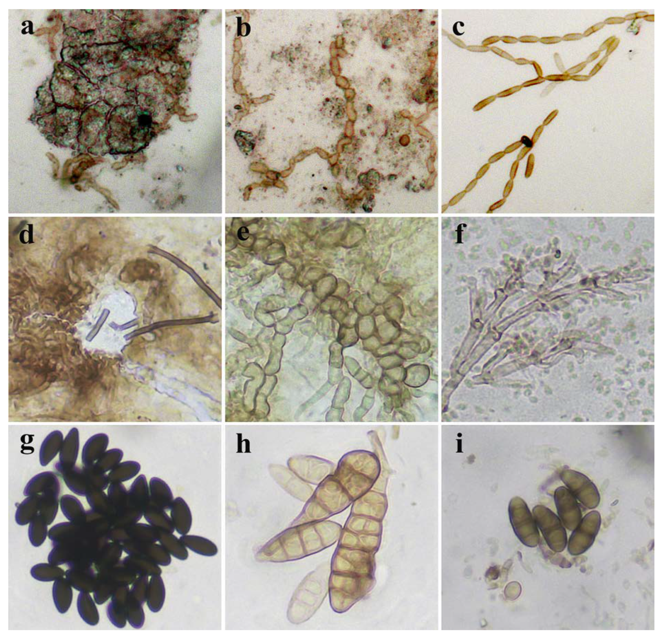
2.5. Optical Microscopy

To collect samples for optical microscopy a non-aggressive adhesive tape method was applied [22]. Prior to microscopy, samples were stained with several drops of Lactophenol Cotton Blue and analyzed with a Zeiss Axio Imager M1 microscope using AxioVision Release 4.6 software.

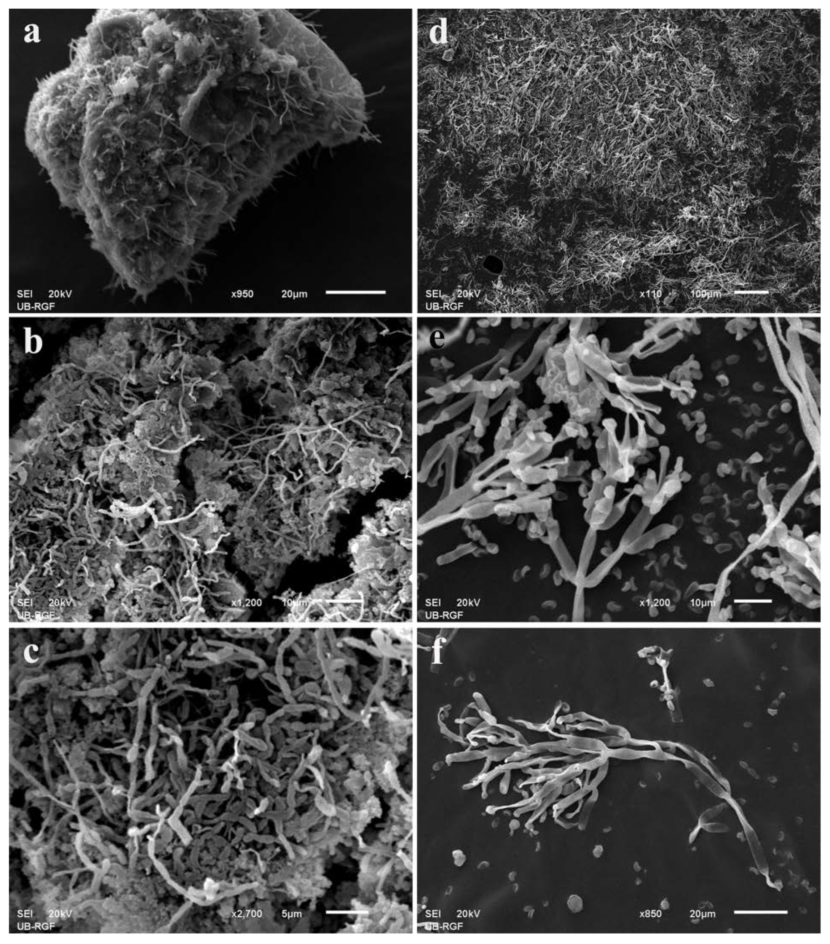
2.6. Scanning Electron Microscopy (SEM)

For SEM, fragments of analyzed substrata were sampled via adhesive carbon tape on aluminum cylinders. Images were obtained at the University of Belgrade—Faculty of Mining and Geology using a JEOL JSM–6610LV microscope. Samples were gold coated (d = 15 nm, ρ = 19.2 g/cm3) with a Leica EM SCD005 sputter coater. Secondary electron and backscattered electron images were obtained using a W-filament gun, at 20 kV acceleration voltage in high-vacuum mode (15–30 μPa of pressure in the sample chamber) and magnifications ranging from 150 to 30,000×.

2.7. Isolation and Identification of Culturable Fungi

Samples for the isolation of culturable mycobiome were collected with sterile swabs. After sampling, swab tips were immersed in 2 mL microtubes (Sarstedt Inc., Newton, NC, USA) containing 1 mL of 30% glycerol Mueller–Hinton broth medium, frozen, and transported to the laboratory under sterile conditions. From each microtube, after 1 min treatment in a vortex mixer, 100 μL was pipetted to malt extract agar (MEA), oatmeal agar (OA), and potato dextrose agar (PDA), supplemented with streptomycin (500 mg L−1; streptomycin sulfate salt, Sigma-Aldrich) to suppress bacterial growth. In order to minimize sampling errors and to increase the likelihood of obtaining the highest possible diversity of culturable mycobiota, inoculation was carried out in triplicates. Inoculated media were incubated in a thermostat (UE 500, Memmert) at 25 ± 2 °C for 7 days. After the incubation period, morphologically different colonies were reinoculated on MEA and incubated under the same conditions to obtain axenic cultures. Obtained isolates were initially identified based on colony morphology and the microscopic characteristics of reproductive structures observed with the optical microscope Zeiss Axio Imager M1 using AxioVision Release 4.6 software. Identification of fungi was performed using the identification keys [23,24,25].
For molecular characterization, 50–100 mg of dry marginal mycelia were collected and used for DNA extraction according to the manufacturer’s instructions for a DNA Mini Kit (Qiagen, Hilden, Germany). PCR amplification of the ITS region was conducted using the primers ITS1/ITS4 [26], and Bt2a/Bt2b primers [27] were used for amplification of the BenA gene (only for Aspergillus, Penicillium, and Fusarium species). PCR amplification of ITS and BenA was performed as described in Savković et al. [28]. The resulting PCR products were separated by electrophoresis and purified (Purification Kit, Qiagen) for sequencing (Eurofins Genomics, Vienna, Austria). Sequences were compared with other related sequences from the National Center for Biotechnology Information (NCBI) database using the BLASTn program. Phylogenetic analysis was conducted using the ClustalW algorithm integrated with MEGAX software and the tree was constructed using maximum likelihood joining phylogeny. The reliability of the constructed phylogenetic tree was evaluated with 1000 replications for branch stability.

2.8. Biodegradation Plate Assays

To assess biodeterioration potential in vitro, 30 fungal isolates were screened using qualitative assays. All isolates were screened for carbonate dissolution on CaCO3 enriched agar plates, according to Albertano and Urzì [29], while production of acidic metabolites was completed on creatine sucrose agar (CREA) and minimal Czapek broth (mCzB), as described in Savković et al. [30] and Borrego et al. [31]. Extracellular pigment production was evaluated on OA [30] and hue was determined per ISCC-NBS color palette (https://www.w3schools.com, accessed on 15 January 2022). Isolates from fresco were evaluated for proteolytic activity using casein agar (CN) medium which was prepared by the protocol given in Saran et al. [32] and Pangallo et al. [33]. Isolates from wooden iconostasis were screened for cellulolytic and hemicellulolytic activity on media enriched with carboxymethyl cellulose sodium salt or xylan from beech wood, respectively, as proposed by Teather and Wood [34] and Liang et al. [35]. Additionally, isolates were evaluated for ligninolytic activity, which was performed on PDA containing ABTS and CuSO4 to detect laccase activity, Mannitol agar medium (MAM) supplemented with azure B to detect lignin peroxidase activity, and Czapek agar (CzA) containing phenol red, MnCl2, and H2O2 for detection of Mn-dependent peroxidase activity [36,37,38].

2.9. Amplicon Sequencing

2.9.1. DNA Extraction, Library Preparation, and NGS Sequencing

Special sterile swabs were taken from all of the sampling points and protected by DNA/RNA shields (Zymo Research, Irvine, CA, USA) during transport. The extraction of ultra-pure DNA was completed using the Zymo BIOMICS DNA Mini Kit (Zymo Research) for several swabs from each sampling point, following the manufacturer’s protocol. The DNA yield was measured using Qubit Fluorometric Quantitation (Qubit 4 Fluorometer, Invitrogen™, Waltham, MA, USA). Library preparation, using Nextera XT Index Kit (FC-131-1096), and amplicon sequencing step was performed using a 2×300 bp paired-end run on a MiSeq Sequencer, according to manufacturer’s instructions (Illumina) in commercially available service (FISABIO, Valencia, Spain). To target the ITS II region, the primers [39] were as follows: forward primer ITS3_KYO2 (5′- GATGAAGAACGYAGYRAA-3′), and reverse primer ITS4_KYO1 (5′-TCCTCCGCTTWTTGWTWTGC-3′).

2.9.2. Sequence Data Process and Taxonomy Annotation

Quality-based filtering/trimming was performed using DaDa2 R package [40]. Primer trimming was performed with BBduk [41] using literal primer sequences as follows: ktrim = l—left trim; hdist = 1—hamming distance of 1—allow one mismatch to primer sequence; copy undefined—clone the reference primer sequences to represent every possibility for the degenerate bases; mm = f—disable mask middle option which is turned on by default and ignores the middle base of a kmer; rcomp = f—do not search reverse complement; and k = 15—use k-mer length 15. All sequences having more than 3 for forward and 2 for reverse strand expected errors (calculated as sum(10^(−Q/10))—where Q is the quality score), were discarded (argument:maxEE = c(3, 2)), as well as sequences shorter than 50 bp. Sequence merger was performed with a minimum overlap of 20 bases without mismatches. After merger, chimeric sequences were removed using default parameters in the DaDa2 R package. All sequences with a length shorter than 280 bp and longer than 367 bp were removed. Contaminant sequences were identified using the R package decontam [42] using the “combined” method which combines the frequency and prevalence probabilities by using Fisher’s method to identify contaminants (Table S1).
ITS taxonomy assignment was performed using the UNITE database which includes global and 97% singletons (https://dx.doi.org/10.15156/BIO/786369, accessed on 20 December 2021). The RDP naive Bayesian classifier [43] with default options as implemented in DaDa2 package was used to classify taxonomy up to the genus level. In addition, amplicon sequence variants, with high abundance and ambiguous taxonomy assignment, were annotated based on the BLAST best hit in the NCBI nucleotide database up to the species level annotation. Main diversity data reported have been obtained by considering the whole dataset, including singletons and/or under-represented taxa.

2.9.3. Statistical Analysis

Sequence diversity within samples (alpha diversity) was estimated using the phyloseq R package [44] at the amplicon sequence variant (ASV), genus, family, and phylum levels. Alpha diversity was shown through estimators Shannon and Gini–Simpson. Observed and estimated richness was determined according to the following estimators: number of observations (OBS), Chao1, and ACE. The Kruskal–Wallis test by ranks was used for testing significance among the samples obtained from fresco (samples 01-05) and from the cave interior including iconostasis (hereinafter: around fresco, samples 06-11). For visualization of beta diversity shared across sample communities, a double principal coordinate analysis—DPCoA [45] was used. This method requires phylogenetic distances of ASVs in addition to just abundances, and for the phylogenetic tree, ASVs were aligned using DECIPHER:AlignSeqs with default arguments [46]. Apart from this approach, a variance stabilizing transformation is implemented instead of rarefaction, and the multidimensional scaling transformation (MDS) was performed using Euclidean distances. For the visualization of the distances among samples, weighted UniFrac distances [47], which take into account phylogenetic distances between observed taxa in addition to taxa counts during the computation, were used. Node size corresponds to the number of connections that the node has, while the edge width corresponds to the distance among samples (closer distances equal wider edges). Nodes that were not connected to other nodes are not shown. For all analyses stated above, prior to computation of the diversities and distances, samples were rarefied to even depth (sample with the lowest number of reads—Figure S1).
The relationships among tax categories were visualized based on co-occurrence using package cooccur [48]. Only statistically significant co-occurrence patterns were shown. Prior to running the co-occurrence analysis all counts that were less than 5 were converted to 0 (categories with a count of less than 5 were considered absent). Additionally, a genus-level network based on weighted Jaccard distance (computed as 2B/(1 + B), where B is Bray–Curtis dissimilarity) was computed. Distances lower than 0.6 are shown as edges. Categories present in any sample with at least 0.5% were taken into account for both analyses.
Differential abundance analysis was performed using DESeq2 package [49]. Starting from the genus level aggregation the package metacoder [50] was used to generate an abundance matrix at all taxa levels. The matrix was filtered by removing taxa that did not have at least 5 reads in at least 3 samples. DESeq was performed using Wald-test statistic, log2-fold change threshold of 2, Benjamini and Holceberg [51] p-adjustment, while all other arguments were left at default. p-adjusted values < 0.05 were considered significant. Shrinkage of log2-fold changes was performed using the apeglm method [52].

3. Results

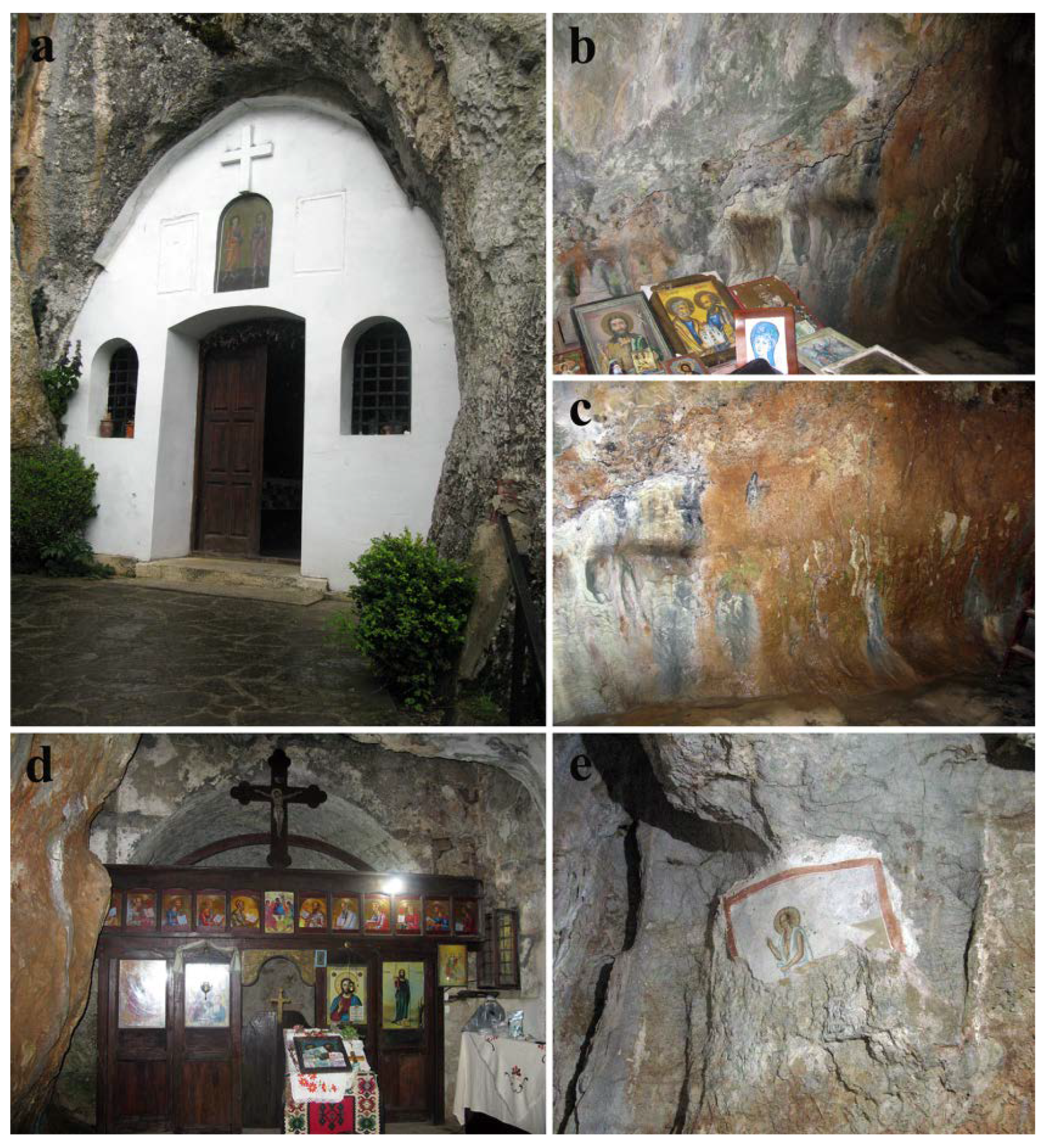
3.1. State of the Investigated Cave Church

There is an evident difference between the northern and southern walls within the cave. While the southern wall is mostly dry and lacking apparent alterations of the stone substrata, the northern wall is quite moist, with known occasional leakage of water, and with variously colored patinas and biofilm developed on the surface (Figure 2a–c). In several areas, surface salt efflorescence was observed (Figure 2d). On the upper right side of the northern stone wall, at about 3 m height, a preserved part of the “The Bald-headed Jesus” fresco painting is present. Only the upper half of the fresco remains, with both the painted layer and plaster of the bottom half completely missing. On the painted surface, numerous fungal-induced wet grayish discolorations of variable size and form are present (Figure 2e). Substantial damage of the painted layer, characterized by the loss of the painted layer uncovering the plaster below, is present in the upper right side of the preserved fresco painting (Figure 2f).
On the eastern side of the cave a wooden iconostasis, made of dark lacquered wood, is present separating the altar niche from the rest of the cave, for the most part unscathed. The bottom half of the iconostasis is, however, impaired and the presence of pinkish gray powdery mass covering substantial surfaces is clearly visible (Figure 2g,h). Furthermore, several wooden fragments are torn off from the iconostasis partition and covered with fully developed, albeit dry, fruiting bodies of Stereum gausapatum and other wood-decay fungi (Figure 2i,j).

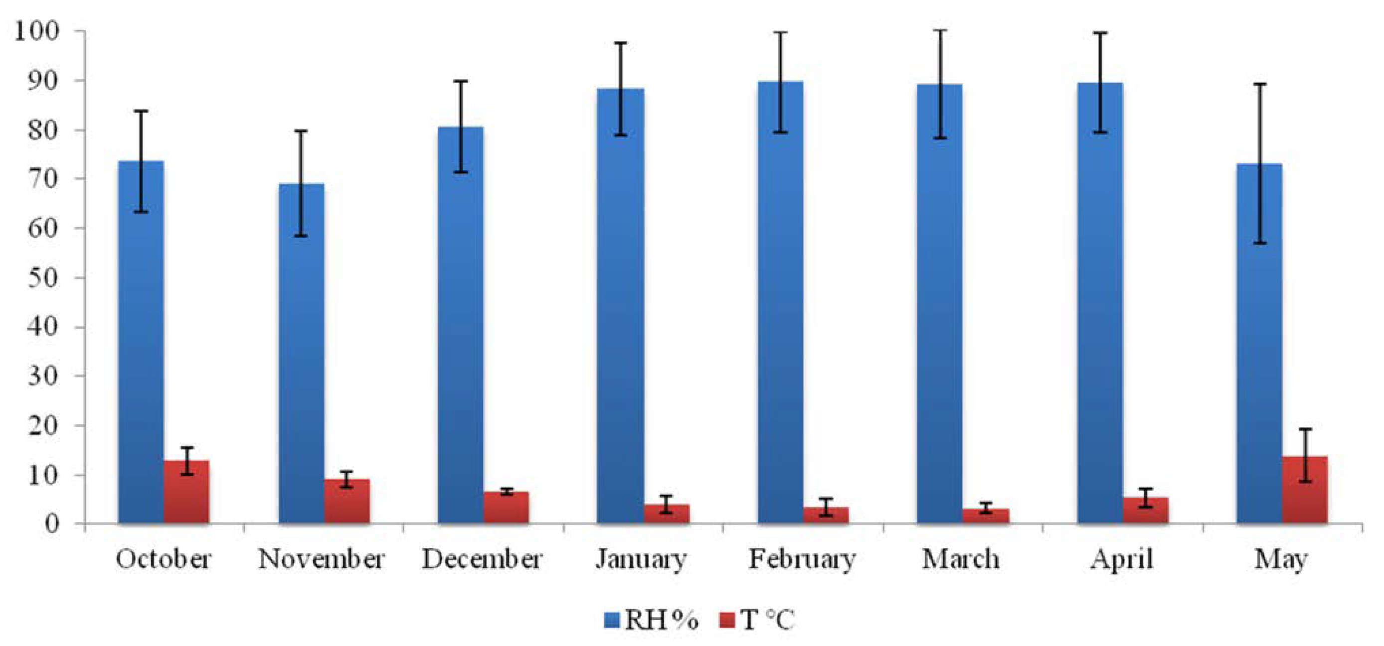
3.2. Indoor Church Microclimate and Material Surface Moisture Content

Based on 10,651 measured values during the 8 months period, documented T was in the range of 0.2 to 38.2 °C, with an average temperature of 6.7 °C; while RH was ranging from 26.6 to 99.9% (average of 82.4%) (Figure 3).
Measured values of surface moisture content for limestone cave walls, fresco, and wooden iconostasis are summarized in Table 1. Surface moisture content values were in the range of 20.1 to 23.8% and 1.6 to 3.0% for cave walls and frescos, respectively. The value of 17.1% was documented for wooden iconostasis.

3.3. Surface Microbial Contamination

Zone of cleanliness values, as indicators of the degree of total contamination of stone walls, fresco, and iconostasis, with microorganisms and organic residues, were in the range of 5.3 to 6.4 (Table 1). All documented values were according to the manufacturer’s provided reference scale placed in the “Danger zone” (3.0–7.5) category of contamination. Iconostasis surface was the most contaminated (6.4) as was expected due to visible fungal proliferation on the surface, followed by a stone wall (avg. 5.6) and fresco (avg. 5.4) surfaces.

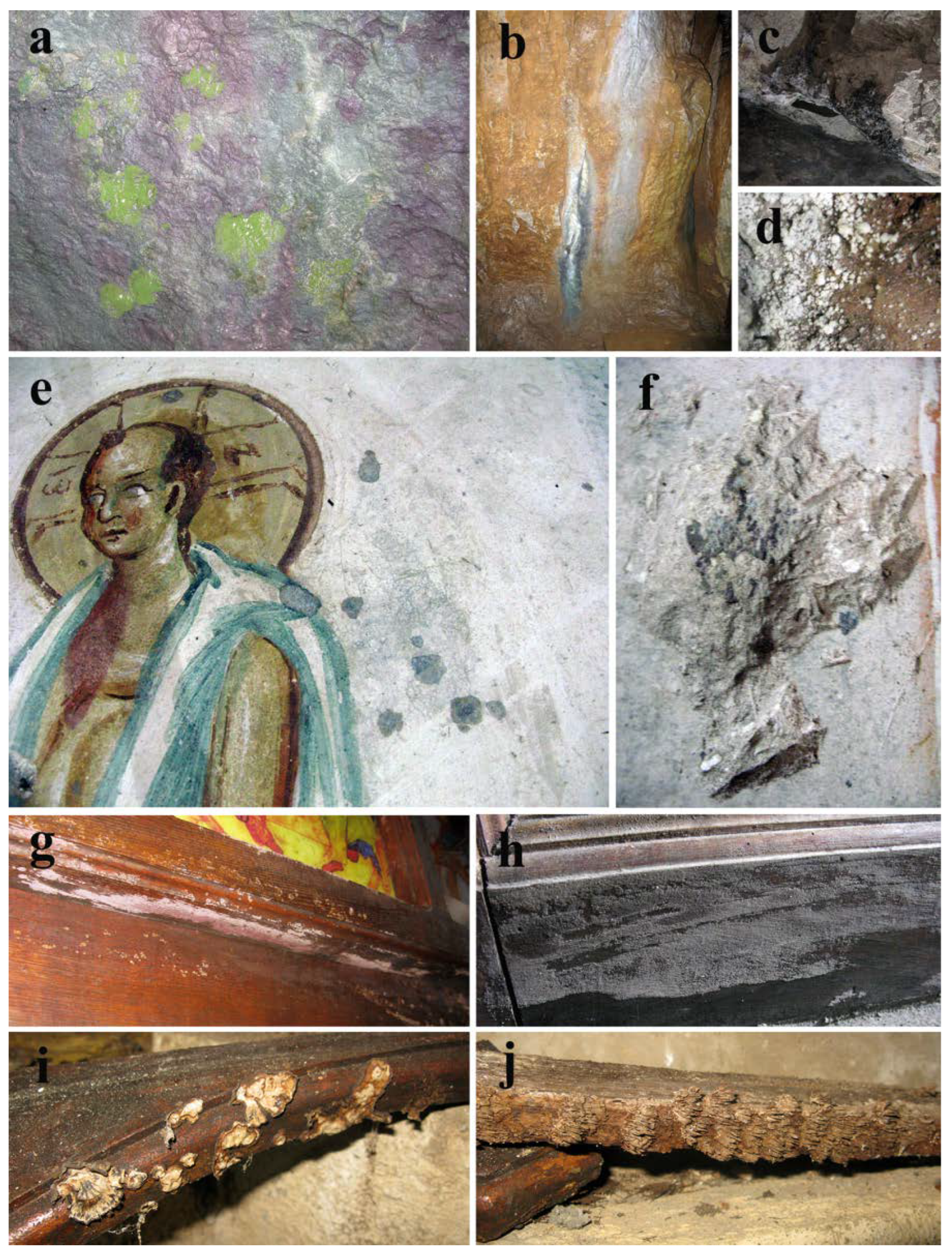
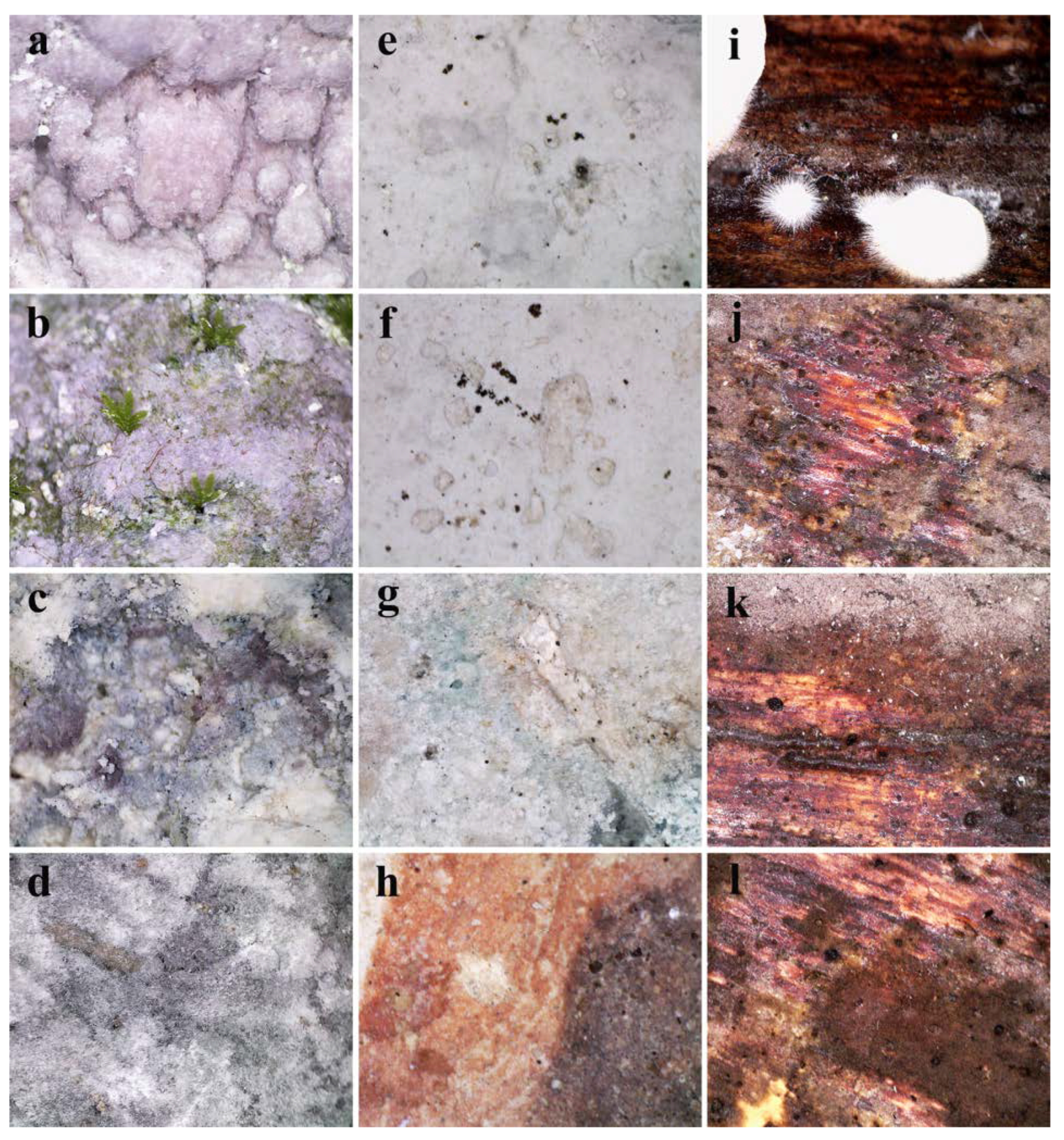
3.4. In Situ Assessment of Micro-Impairments and Surface Microbial Growth

Direct observation of the southern stone wall within the cave church, via portable in situ microscopes, revealed a rough surface covered with salt efflorescence resembling pink-hued snowballs (Figure 4a), with occasional moss gametophyte and visible thread-like protonema (Figure 4b). Furthermore, in general, observed stone surfaces highly deteriorated with visible structural impairments, i.e., variously colored granulated surfaces interspersed with conspicuous filamentous salts (Figure 4c,d). Predominant deterioration symptoms, observed on the surface of the investigated fresco, were loss of fragments of the painted layer along with clear signs of biopitting occasionally filled with melanized fungal structures of typical meristematic clusters characteristic of microcolonial (rock-inhabiting) fungi (Figure 4e–h). The surface of the wooden iconostasis was characterized by the presence of immature basidiomycetes fruiting bodies (Figure 4i) and was extensively covered with a pinkish-gray fungal mat with apparently present stromata with visible dark perithecia (Figure 4j–l).

3.5. Fungal Proliferation on Fresco and Iconostasis Surfaces

Adhesive tape samples, taken from wet grayish discolorations of the fresco-painted layer (Figure 1e and Figure 2e), have revealed numerous melanized Cladosporium-like conidial chains in the interspace between painted layer and the plaster below, possibly belonging to the fungus of Neodevriesia genus (Figure 5a,b). Furthermore, branched, fragile conidial chains, composed of golden-brown conidia with or without visible dark septum, are also observed proliferating on the surface of the painted layer (Figure 5c). On the other hand, a mass of melanized, fragmented hyphae with chlamydospores was observed permeating below the lacquered wood surface (Figure 5d,e). Optical microscopy of adhesive tape samples taken from a fungal mat resembling pinkish gray powdery surface (Figure 2g,h), a dominant deterioration symptom present on the wooden iconostasis, has shown a mass of branched, pale grayish-brown geniculate terminal conidiophores with numerous pale grayish-green conidia of the anamorphic state of Hypoxylon (Figure 5f) together with black-brown inequilaterally ellipsoid Hypoxylon ascospores (Figure 5f). The presence of Alternaria-like dictiospores and melanized ascospores of Pleosporales was also observed (Figure 5h,i).
Observations made with optical microscopy were additionally confirmed with SEM: Neodevriesia conidial chains were found permeating the plaster below the painted layer (Figure 6a–c), while on the iconostasis a mass of branched conidiophores of the anamorphic state of Hypoxylon (Figure 6d) and details of conidiogenous apparatus (Figure 6e,f) were observed.

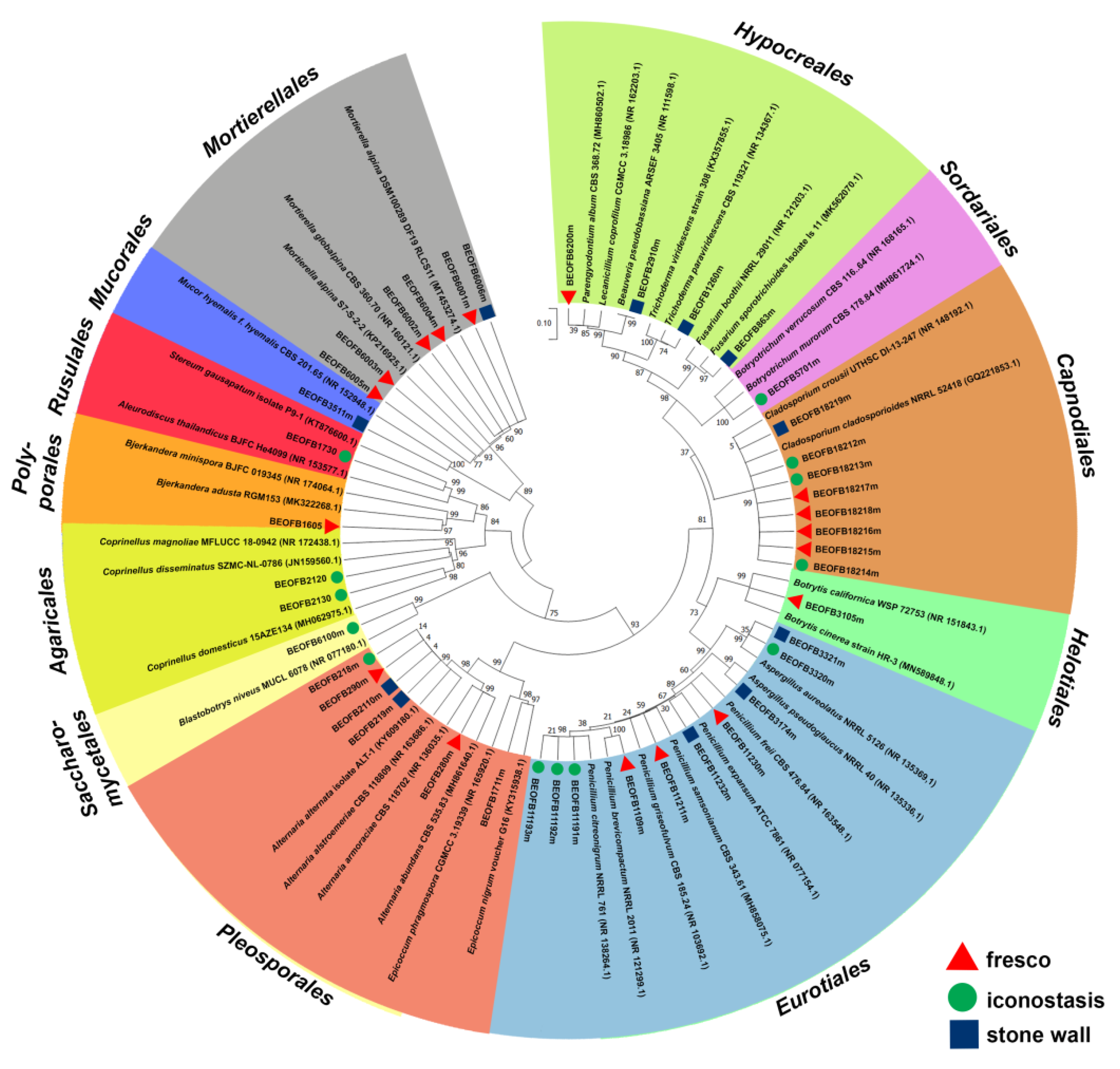
3.6. Culturable Mycobiome

A total of 24 fungi from 17 genera were determined as part of the culturable mycobiome of different substrata within the cave church (Figure 7, Table S2). The greatest diversity was documented on the stone walls (12 species), followed by fresco (10) and iconostasis (8). The majority of isolated fungi were Ascomycota (73.81%), while only 16.67% and 9.52% belonged to phyla Zygomycota and Basidiomycota, respectively. With five documented species, the genus Penicillium was the most diverse, while isolates of Cladosporium and Penicillium genera were the most frequently isolated.
Phylogenetic analyses showed that all Ascomycota members clustered together (bootstrap value (bv) = 75, Figure 7). Aspergillus and Penicillium species formed a well-supported clade (bv = 99) as members of the order Eurotiales. Likewise, Trichoderma harzianum, Fusarium sporotrichioides, Beauveria pseudobassiana, and Parengyodontium album clustered together as the members of Hypocreales clade (bv = 87). Cladosporium species clustered in a well-supported Capnodiales clade (bv = 99), as well as the members of Alternaria and Epicoccum species (Pleosporales clade, bv = 99). Among Basidiomycota, Coprinellus species clustered together with Bjerkandera and Stereum species in a well-supported Basidiomycota clade (bv = 84). Finally, Mortierella alpina isolates clustered with Mucor hyemalis in Zygomycota (bv = 89).

3.7. Biodegradative Profile of Isolated Fungi

Among 30 tested isolates, 16 (53.33%) demonstrated at least 1 positive reaction in biodegradation assays (Table 2). Acid production was observed in eight isolates (26.67%) with a reduction in pH of the liquid medium from 4.27 to 6.73, while carbonate dissolution was documented only for Penicillium samsonianum BEOFB11211m and Cladosporium cladosporioides BEOFB18216m, isolated form fresco surface. Likewise, coupled cellulolytic and hemicellulolytic activity is observed in Alternaria alternata BEOFB218m, Botryotrichum murorum BEOFB5701m, and Stereum gausapatum BEOFB1730 isolated from iconostasis, while Coprinellus disseminatus BEOFB2120 had only hemicellulolytic activity. Ligninolytic activity is observed in six fungal isolates from iconostasis (20.00%). Proteolytic activity was documented for five isolates (16.67%), while pigment production was demonstrated for six (20.00%). The colors of produced pigments were mostly in different hues of yellow: deep greenish yellow (#9b9400) in Aspergillus aureolatus BEOFB3320m and BEOFB3321m, vivid yellow–green (#8db600) in Penicillium freii BEOFB11230m, brilliant greenish yellow (#e9e450) in Beauveria pseudobassiana BEOFB2910m, strong orange yellow (#eaa221) in S. gausapatum BEOFB1730 and strong greenish yellow (#beb72e) in Bjerkandera adusta BEOFB1605.

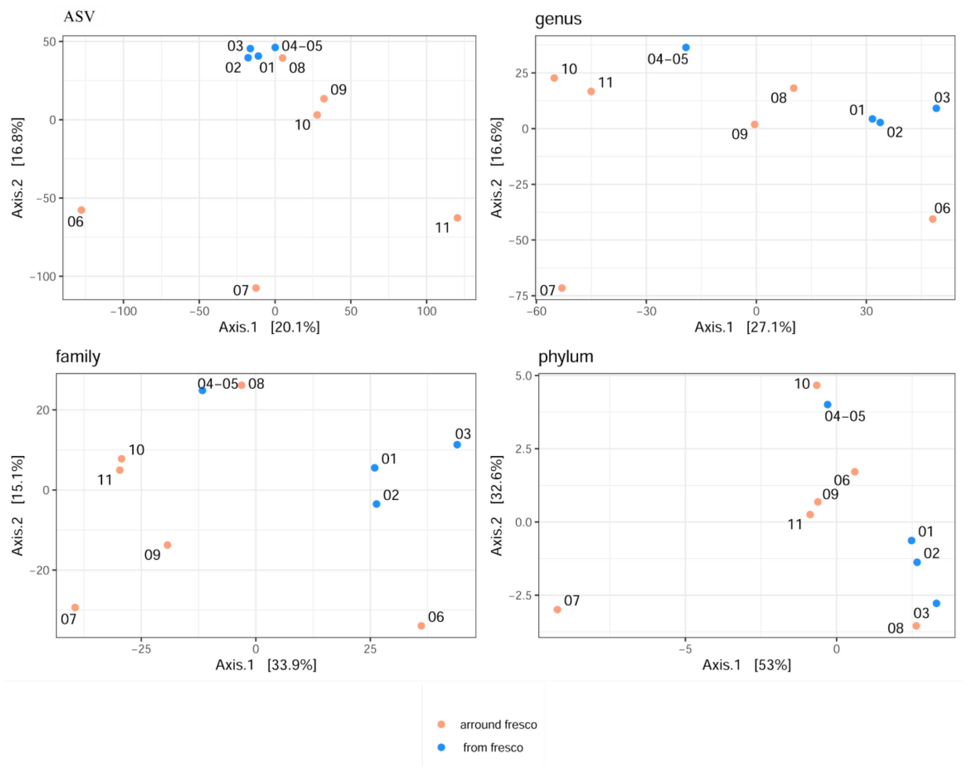
3.8. Diversity of Microbial Communities

A total of 1,505,219 raw sequences were obtained from the ITS libraries sequencing (from 106,494 to 222,761 per sample). After denoising, quality filtering, length trims, and sequence decontamination, the number of reads ranged from 76,734 up to 190,144 (Table S1). The mean length of obtained ASV sequences was 323. Based on alpha rarefaction curves (Figure S1), since there were evident differences in reads among samples, diversity indices were estimated after rarefaction to even depth according to the sample with the lowest number of reads (sample 07). Alpha diversity indices were calculated for OBS, Chao1, ACE, Shannon, and Gini–Simpson at phylum, family, genus, and ASV levels (Table 3). The diversity indices on average, Shannon and Gini–Simpson, indicated lower diversity in samples taken from the fresco itself, while richness estimators (Chao1 and ACE) reflected the higher fungal richness (at least double) in the samples taken around the fresco, especially at the family, genus, and AVS taxa levels. However, statistically significant differences, for the Shannon (p = 0.02) and Simpson (p = 0.02) indices of the fresco and around it, according to the Kruskal–Wallis test were observed only at the phylum level (p < 0.05). Reviewing the samples individually, it was noticed the highest fungal richness at the family, genus, and AVS taxa levels for the 04-05 sample taken from the fresco surface damage, while the same is obtained for the samples from the cave interior wall deposits in increasing order 11 > 10 > 07. Sample 03 collected from the gray discoloration on the fresco itself, showed the lowest richness and diversity according to the Shannon index, and a similar is scored for sample 06 from the iconostasis (Table S3). Sample 08 obtained from the green patina showed, among all samples from the wall deposit, the lowest richness and diversity.
For visualization of beta diversity, the initial analysis was performed with Bray–Curtis dissimilarities and samples were rarefied to even depth (variability between samples is shown with the DPCoA plot in Figure S2). Based on the results, it can be concluded that the samples from the fresco environment are jointly together, as well as the samples obtained from the side walls of the church in terms of microbial diversity at all taxonomic levels. The furthermost distance was achieved for samples 06 and 08, especially at the family, genus, and ASV taxa levels. Moreover, samples 09 and 11 were closely related to the samples from the fresco. Additionally, appropriate variance stabilization was performed later, and the results were presented by performing MDS (PCoA) analysis based on Euclidean distances (Figure 8).
From the PCoA plot, it can be observed that fresco’s samples 01, 02, and 03 were closely related to each other according to PCoA1 at phylum, family, and genus taxa levels (explaining 53%, 33.9%, and 27.4% of variability, respectively). Samples 06 and 11 were identified as outliers and more dissimilar to other samples, according to both axes at the ASV level, including sample 07 based on the second axis, as well.
Weighted UniFrac distances were used to visualize the distances among samples (Figure 9). Based on the results obtained at the family and genus taxa levels, samples 07 and 10 were quite far from the others and had a single connection but were sufficiently distant among themselves (proximity value of 0.2). Samples 01 and 04-05 at the ASV level had the highest number of connections with the other samples (the number of connections was 7) and were closest among themselves, according to the proximity value (0.061). The same proximity was noted for samples 02 and 03; 02 had the longest distance from samples 04-05 with 0.098; likewise, sample 03 from sample 01 with a 0.117 distance value. Additionally, samples 09 and 11 clustered together with the samples from the fresco, supporting the results of DPCoA analysis, among themselves were quite distant (0.212). At the genus and family taxa levels, the situation was similar for samples 04-05 and 11, along with five connections with the rest samples, and the last one (11) had a closer distance to the fresco’s samples. Sample 6, although somewhat distant, achieved five connections with samples 01, 02, 03, 04-05, and 11 only at the phylum level; in all other taxa comparisons, it was excluded from the analysis due to its excessive distance from the others.

3.9. Total Microbial Composition

The phylum Ascomycota dominated all samples (79.9–99.7%), while Basidiomycota was in higher abundance on the cave walls around the fresco. The phylum, which does not belong to fungi, Chlorophyta (with the algal species Pseudostichococcus monallantoides, according to BLAST results) was predominant for green patina on the stone wall (sample 08).
The fungal taxa that dominate the fresco’s samples communities in higher or moderate relative abundance (RA) are order Capnodiales with unclassified Mycosphaerellaceae family, unclassified class Dothideomycetes (more characteristic for the entire fresco except for depiction of Jesus and gray discoloration on fresco—samples 02 and 03), as well as taxa Lecanicillium, Acrodontium, and Hirsutella (relatively higher abundant in samples 01, 02 and 04-05; the highest in a sample from the depiction of Jesus) which according to NCBI Blast results could be associated with Gamszarea microspora, A. pigmentosum, and H. vermicola, respectively (Figure 10).
According to NCBI alignment of the sequence obtained for the order Capnodiales (which was the most abundant across all fresco samples), it was closest to the Mycosphaerellaceae and Teratosphaeriaceae families, i.e., the genera Annellosympodiella, Neodevriesia, and Catenulostroma. Additionally, order Hypocreales (NCBI-Thyronectria sp.) was characteristic for mentioned samples as well. Slightly more abundant in fresco surface damage (04-05) than in other fresco samples were Cladosporium, Phaeosphaeria, Alternaria, Mycosphaerella, Penicillium, and unclassified class Agaricomycetes. Contrary to that, in a sample taken from the iconostasis (06), we observed dominance of the genera Hypoxylon (H. fuscopurpureum), Sporothrix (Blastobotrys sp.), Acrodontium (A. pigmentosum), and unclassified family Exobasidiaceae (per NCBI could potentially annotate as Arcticomyces warmingii and Exobasidium sp.). It is interesting to note that the sample taken from gray discoloration on fresco (03) was relatively poorly abundant of detected taxa such as Beauveria (B. pseudobassiana), Phaeosphaeria (Phaeosphaeria sp.), and Alternaria (A. destruens) among many others.
The opposite situation occurred for the samples from the points located on interior cave walls. As long as the taxa Capnodiales with unclassified Mycosphaerellaceae family and unclassified class Dothideomycetes were ubiquitous for the samples 07, 09, and 11 (potentially indicating the origin of fresco’ colonization), other taxa such as Cladosporium (C. crousii/pini-ponderosae/colombiae), Beauveria (B. pseudobassiana), Mycosphaerella (Cladosporium subcinereum/antarcticum/phlei), Penicillium (P. brevicompactum/kongii), and class Agaricomycetes were attached for samples 07 and 10 with higher RA. Additionally, sample 07 was characterized with higher RA with class Sordariomycetes (Cryptendoxyla sp.), and genus Cyphellophora. For samples 09 and 11, genus Pectenia in higher abundance simultaneously co-occurred, while order Chaetothyriales, unclassified family Herpotrichiellaceae and genus Tausonia were present in higher abundance only in sample 09. Higher dominance of Chlorophyta (P. monallantoides—phycobiont of lichenized fungus Verrucaria sp.), and lichenized fungi Botryolepraria (Botryolepraria lesdainii), and Verrucaria (Verrucaria sp.) were noted only for sample 08 taken from an area of green patina. The most abundant taxa related to the composition within sample 10 were also Lecanicillium, Acrodontium, Alternaria, Simplicillium, and order Hypocreales.

3.10. Co-Occurrence and Relative Abundance Analyses

Relationships between taxonomic categories (positive, negative, and random interactions) were visualized based on the co-occurrence of taxa (“co-occurrence” analysis) as shown in Figure 11. Based on the statistically significant co-occurrence patterns, a positive correlation was observed among the genera Beauveria, Alternaria, Botrytis, Pseudogymnoascus, and Phaeosphaeria (with four positive connections for each of them). A positive correlation was also observed between Cephalotrichum (three connections) with Pectenia, Bradymyces, and an unknown taxon from the order Chaetothyriales. A negative correlation was observed only between the genera Verrucaria and Capronia. However, Verrucaria also has a simultaneous positive interaction with basidiomycetous Coprinellus. Additionally, a genus-level network based on weighted Jaccard distance computed on Bray–Curtis dissimilarity indicated numerous positive connections among different taxa (Figure 12). As can be noticed, the highest number of connections were achieved for Penicillium and Acremonium (each with seven positive connections), while the unknown taxa from the family Didymellaceae were tightly connected to Penicillium and Botrytis. Similar tight bonds are shown between Blastobotrys and Capronia, Bradymyces and Tausonia, as well as Tetracladium and Pyrenochaeta. Leptospora formed three positive connections with Alternaria, Simplicillium, and Parengyodontium, while unknown taxa from the order Hypocreales were positively connected with Acrodontium, Lecanicillium, and Mycosphaerella. Verrucaria has shown a slightly distant positive correlation with Botryolepraria, while Neodevriesia was closest to Acremonium.
Differential abundance analysis was used to generate a matrix of RA of statistically significant species at all taxonomic levels according to sample origin (Table 4). It was established that three known genera Pyrenochaeta, Phialophora, and Rhinocladiella were significantly present only in samples around the fresco, while only Hirsutella was exclusively important for the fresco environment itself and mightily be defined as “core” species.

4. Discussion

During the eight-month measuring period, the indoor temperature of the cave church was in the range of 0.2 to 38.2 °C, which is problematic having in mind the presence of fresco painting and the recommended range of temperatures for the preservation of frescoes which is from 6 to 25 °C [53]. On a daily basis, the indoor temperature did not change significantly and followed the seasonal trend of changes in outdoor temperature. This was expected since the church is part of the rock mass closed by a wall and therefore has high thermal inertia. The daily variation in the temperature usually did not exceed 1.5 °C; however, a frequent increase in daily temperature variation was noticeable after the middle of January and during the upcoming season, which correlates with the more frequent use and entry of visitors. Due to frequent liturgies during winter, it is sporadically heated to enable the comfortable presence of believers. Such sudden daily temperature variations, above recommended daily variation of 1.5 °C, contributed to the degradation of the material and originate from the human factor, and as such can be eliminated by corrective measures. Furthermore, during February and March, the temperature inside the church was constantly below 6 °C, sometimes reaching values close to 0 °C, creating a permanent danger of condensation processes and freezing of water present in the fresco plaster. This factor cannot be influenced by passive measures and was one of the main reasons for the substantial loss of fresco in the past. On the other hand, the documented minimum RH value was 26.6% and refers to the moment when the indoors of the church is extremely heated, while the recorded maximum of 99.9% is often repeated at longer intervals and for several days in January, February, March, and April. The recommended RH values for preserving fresco paintings are in the range of 45 to 60% [53]. RH values sometimes dropped to 56% and correspond to warmer periods and the daily use of the church. Although the RH values are mostly outside the recommended range, the daily variations are stable and do not change over 5% if there is no activity within the church. Most of the measurement intervals of the year RH values are above 70%, very often with long-term maximum saturation of water vapor, which leads to the appearance of moisture condensation in the fresco. The maximum values indicate the probable inflow of water into the walls by draining atmospheric water through porous limestone rocks. Aside from the issue of T and RH variation being physical agents of deterioration, for the most part of the measuring period both T and RH were in the range that favors fungal growth [54]. This growth is also certainly facilitated by the highly documented values of surface moisture content in cave walls and iconostasis. In the case of limestone walls, it is most likely due to drainage of water from the environment and the presence of soluble bicarbonate salts, while in the case of wood iconostasis increases compared to normal 10–12% of dry wood due to woods ability to quickly absorb excess water from the air and release it by acting as a buffer material, keeping the RH approximately constant in space. Having all that in mind, biofilm and patina development on stone, and fungal proliferation on fresco and iconostasis surfaces, as well as high values (5.3 to 6.4; danger zone) documented with rapid ATP bioluminescence method on all sampling points are to be expected.
Culturable mycobiome obtained from various substrata within Cave Church of Sts. Peter and Paul are in accordance with other mycological studies performed in caves and hypogean environments, with similar humidity and temperature conditions, indicating that these fungi have a particular affinity for environmental parameters prevailing in this type of cultural heritage site. The majority of documented culturable fungi have already been observed on these types of substrata (fresco, wood, and cave stone walls) in similar studies [55,56,57,58,59,60,61,62]. Several fungal species were isolated for the first time (e.g., Alternaria abundans, Penicillium freii, and P. samnosnianum from fresco; Aspergillus aureolatus and Blastobotrys niveus from iconostasis; As. pseudoglaucus, Beauveria pseudobassiana, Fusarium sporotrichioides, Mortierella alpina, and P. expansum from cave stone); however, fungi from these genera are known to colonize or be deposited on the surface of these cultural heritage substrata. In the case of two Basidiomycota isolated from wooden iconostasis (Coprinellus disseminatus and Stereum gausapatum), to the best of our knowledge, this is the first time they were documented on wooden substrata within cave churches. In the case of S. gausapatum, numerous fully developed, dry, fruiting bodies were observed on the basis of the iconostasis on torn-off wooden fragments. Fruiting bodies of representatives of these two genera were observed before on wood drainpipes of the Latvian Ethnographic open-air museum (S. sanguinolentum) and wooden chairs, benches, and barrel vaults that are part of the Republic of North Macedonia cultural heritage collection (S. hirsutum) [63,64] indicating that if environmental conditions are favorable and objects neglected wood-decay fungi will fructificate. Furthermore, the isolation of Botryotrichum murorum (syn. Chaetomium murorum), though previously unreported from iconostasis, is not unexpected due to its pronounced cellulolytic activities. It has been known to occur frequently in documentary heritage, frescos, and stone surfaces [65,66].
One of the more interesting fungal species, isolated from the studied deteriorated fresco, is hyphomycetes Parengyodontium album, commonly reported on fresco paintings in religious monuments such as churches, cathedrals, or monasteries, but also from stone walls and fresco paintings within various caves of cultural interest [67]. The potential of this fungus to colonize fresco paintings is attributed to its ability to utilize products used in the restoration of frescoes, as well as synthetic resins, calcium caseinate, and liquifying animal glue as nutrient sources. Parengyodontium album has been associated with several different deterioration symptoms: salt efflorescence is the most frequent, followed by discoloration and patinas of various colors, e.g., pink, ochre, dark/black, and green–black [67].
In vitro, biodeterioration plate assays demonstrated the potential of isolated fungi to degrade fresco, stone, and wood via the production of acids and enzymes, and to affect these substrata aesthetically, by the production of extracellular pigments. Acid metabolite production is regarded as one of the most important mechanisms of the chemical deterioration of cultural heritage and it has been claimed that most fungal species possess the ability to excrete organic acids [68]. One-quarter of tested fungi, especially those isolated from fresco surface, demonstrated positive reactions on CREA medium, indicating their potential to produce acidic metabolites. The fact that acid production is observed in a great number of isolates is consistent with the results reported earlier by the authors using the same biodegradation assays [16,30]. Namely, Savković et al. [30] demonstrated acid production for the majority of airborne Aspergillus and Penicillium isolates from cultural heritage conservation premises, while Unković et al. [16] demonstrated this ability for Cladosporium cladosporioides, and several Aspergillus and Penicillium isolates from sacral ambient.
Cladosporium cladosporioides and P. samnosnianum, both isolated from fresco surface, are the only two species for whom the carbonate dissolution, the primary cause of structural alterations of carbonate substrata, was documented. Various research conducted up to date, with fungi obtained from contaminated cultural heritage objects, demonstrated carbonate dissolution ability for only a small number of fungi, mainly of Aspergillus and Penicillium genera [16,33,69]. On the other hand, to the best of our knowledge, this is the first time that carbonate dissolution ability was observed for fungi of the genus Cladosporium, i.e., C. cladosporioides. In the case of P. samnosnianum, where a substantial reduction of pH values was documented in the acid production test (pH 4.75), carbonate dissolution probably occurred through the secretion of organic acids (with oxalic acid in most instances) that dissolve calcium carbonate, while C. cladosporioides could utilize one of several proposed mechanisms, such as enzymatic dissolution, ligand activity, and oxidation-reduction of redox-sensitive elements [16]. Trovão et al. [66] demonstrated carbonate dissolution in vitro for Pa. album and several Penicillium species isolated from walls of Coimbra cathedral in Portugal.
In proteolytic activity assay, the ability to degrade casein was documented in fungi of genera Penicillium, Parengyodontium, Cladosporium, and Bjerkandera, all isolated from the painted layer of studied fresco. The potential of C. cladosporioides and Bj. adusta to degrade proteinaceous substrate of cultural heritage objects and subsequently cause irreversible damages, as well as Parengyodontium album, to act as a causative agent of opportunistic mycoses, was previously reported by Macêdo et al. [70], Pangallo et al. [33] and Unković et al. [16]. However, this seems to be the first record of P. samsonianum and P. freii having proteolytic activity and posing threat to wall paintings where mineral pigments were bound with casein.
Extracellular pigment production was observed for one-fifth of tested fungi with the coloration of media mostly in some hue of yellow. Pigment production by Aspergillus and Penicillium species is well known and documented by other authors: Borrego et al. [71], Unković et al. [16], Savković et al. [30], to name just a few. To the best of our knowledge, the yellow pigment was not reported in Be. pseudobassiana, although Ávila-Hernández et al. [72] reported a yellow pigment in a closely related Be. bassiana. Conversely, yellowish pigments were documented for S. gausapatum by Boddy and Reiner [73]. Pigment production by fungal dwellers of cultural heritage is of great importance since it leads to, usually hard if not impossible to remove, discolorations and aesthetic changes in the original artwork. It should be noted that the growth of melanized fungi, such as Cladosporium and Alternaria species documented in our study with negative reactions in pigment production assay, can nonetheless lead to the appearance of observable dark colorations of cultural heritage objects which are notoriously hard to remove due to pigment melanin [17]. In the work of Caneva et al. [4] gray patina was characterized by the growth of C. sphaerospermum and meristematic fungi that paralleled the reduction in humidity levels and the increase in the amounts of organic substances, probably derived from the decay of pre-existing organisms.
The dominant presence of fungi from phylum Ascomycota, and to a lesser extent phylum Basidiomycota, observed in the study by metagenomic analysis, was also documented using culture-independent techniques on Palaeolithic representations in the Escoural Cave (Alentejo, Portugal) [74]. A high abundance of the lichen-forming and rock-inhabiting fungi from class Dothideomycetes (mainly of order Capnodiales), as well as the lower but almost consistent presence of class Agaricomycetes, across all samples, was shown to be consistent with the literature and was already reported on limestone walls of the Church of São Salvador da Trofa in the Portuguese district of Aveiro [9]. The abundant presence of entomopathogenic fungi, such as the one from order Lecanicillium, on fresco paintings and cave walls may suggest that insects act as vectors in fungal dispersion to and from the walls within the cave [75] thus contributing to the colonization of the painted layer of fresco paintings.
Moderately high RA of Pseudostichococcus monallantoides (Chlorophyta) was detected in a green patina observed on a stone wall near the fresco. This green alga, previously known as a colonizer of stone relief within the hypogean heritage site of Pommery Champagne cellar [76], is phycobiont of lichenized fungus Verrucaria sp., documented with moderately high RA as well, and is known to induce biopitting in limestone [77] such as the one observed on limestone walls of the investigated cave church. Furthermore, among different positive correlations observed between taxa in co-occurrence analysis, the most intriguing positive interaction is the one between the lichenized fungus Verrucaria and mushroom-forming fungus Coprinellus, indicating that the presence of one always entails the presence of the other. Positive interaction between these two distinct ecological groups is quite unexpected given that Verrucaria is stone-inhabiting lichen, while Coprinellus is most likely present in form of airborne basidiospores attached to stone and lichen surfaces with an unclear role in stone colonization [78]. The presence of Coprinellus was previously reported to always be associated with black discoloration of limestone, which we presume to be from crustose lichen thalli, though the origin of this alteration was not discerned [9]. A fungal mat resembling a pinkish-gray powdery surface, a dominant deterioration symptom on the wooden iconostasis, was proved to be formed by a member of the Hypoxylon genus which was expected as they are most commonly encountered ascomycetes group on wood in the temperate regions and usually one of the earliest fungi to colonize dead wood [79]. The dominant presence of Hypoxylon fuscopurpureum with a relative abundance of 78.5%, identified via metabarcoding analysis, was confirmed by morphological traits observed and analyzed using in situ, optical, and SEM analysis. Conclusively, this fungus can be regarded as the main biodeterioration agent of the studied wooden iconostasis, and members of this genus are known to induce soft rot of wood used architecturally often in contact with soil and in dump conditions as one that exists in churches and hypogean environments [80].
Microscopic analysis of samples taken from the dominant deterioration symptom on studied fresco, i.e., wet grayish discolorations of variable size and form, have revealed reproductive structures of Cladosporium-like fungus. Metabarcoding analysis of the same sample has revealed the predominant presence of Capnodiales most likely from genera Annellosympodiella, Neodevriesia, and Catenulostroma. The morphological characteristics of the observed reproductive structures showed the greatest similarity with the Neodevriesia genus. Fungi from this genus were previously reported using the culture-independent method, as the dominant group on 19th century mural paintings at Tha Kham temple in Thailand [18]. This genus, recently segregated from Cladosporium, encompasses species usually referred to as Cladosporium-like and of variable ecological preferences including extremophiles [81]. Members of the Neodevriesia genus include rock-inhabitant black fungi, characterized as highly melanized and slow-growing organisms exceptionally skilled at exploiting most kinds of extreme environments [9]. As was suggested by Suphaphimol et al. [18] fungi of the Neodevriesia genus probably originated from the surrounding stone walls which is most likely the case of fresco deterioration in our investigation as well since RA of this fungus, recognized as Capnodiales (unclassified Mycosphaerellaceae) in metabarcoding analysis, is up to 58.5% on stone walls around fresco.
The findings of this study have to be seen in light of some limitations. First, within the cave church, there are several substrata that have not been analyzed, such as concrete and stone as part of the entrance wall and altar area of the church, since the main focus of the research was to analyze substrata with visible symptoms of deterioration. Further research will include these substrata to obtain complete insight into the fungal diversity within the cave church. Additionally, some of the limitations might arise from the NGS sequencing short read results and could potentially lead to a lack of awareness of species levels, particularly those with the highest relative abundance. However, combining different approaches can improve and confirm our findings, as we presented in the current study. Moreover, in the future, some of the more sophisticated NGS techniques will be performed and precise information will be obtained.

5. Conclusions

This research gives insight into the problem of identifying fungi that are the main deterioration agents of cultural heritage objects. As a rule, extremophilic fungi that actively grow on frescoes and other works of art cannot be isolated. It is well known that the synergy of data obtained by applying both traditional and DNA-based methods gives the most precise identification of fungi and is of equal importance. The application of integrative methods, especially cross-referencing of total fungal diversity obtained by metabarcoding analysis with the morphological identification of the deterioration agents singled out and enabled the precise identification of fungi that grow actively on the “The Bald-headed Jesus’’ fresco (Neodevriesia sp.) and the wooden iconostasis (H. fuscopurpureum) within the Cave Church of Sts. Peter and Paul in Rsovci, Serbia. The metabarcoding analysis provides insight into the total mycobiome of the investigated substrate and directs the selection from the identified taxa to the few that possess the highest relative abundance and are simultaneously detected via microscopic methods as actively growing and causing deterioration. In this way, the results of the metabarcoding analysis allow us to focus on certain taxa and using appropriate identification keys and other available literature definitely identify them as the main agents of deterioration. Together, the traditional identification of fungi based on morphological features and modern DNA-based methods are powerful tools for accurate identification, especially of extremophiles as causative agents of deterioration of cultural heritage objects. This combined approach contributes to the development of strategies for conservation since it allows us to, based on the knowledge of the biology of the species, select appropriate nutrient media to isolate and cultivate the agents of deterioration, characterize their deterioration potential, and finally test their sensitivity to fungicides applicable in conservation, so conservators can select the appropriate procedures and biocides.

Supplementary Materials

The following supporting information can be downloaded at: https://www.mdpi.com/article/10.3390/jof8121263/s1, Figure S1: Rarefaction analysis at the ASV, genus, family, and phylum taxa levels; Figure S2: The DPCoA plot created with Bray–Curtis dissimilarities at the ASV, genus, family and phylum taxa levels, to present the variability within and between groups of samples; Table S1: Number of the read retention and sequence decontamination; Table S2: Molecular analyses of culturable fungi from the Cave Church of Sts. Peter and Paul; Table S3: Alpha diversity within the analyzed individual samples from fresco painting and from sampling points of the cave interior and presented at the phylum, family, genus, and ASV level.

Author Contributions

Conceptualization, M.L.G., I.D. and N.U.; methodology, I.D., M.S., Ž.S., A.K., A.J. and N.U.; software, I.D. and A.J.; validation, M.L.G. and N.U.; formal analysis, M.L.G., I.D. and N.U.; investigation, M.L.G., I.D., M.S., Ž.S., A.K., A.J. and N.U.; resources, N.U.; data curation, N.U.; writing—original draft preparation, M.L.G., I.D., M.S., Ž.S., A.K., A.J. and N.U.; writing—review and editing, M.L.G. and N.U.; visualization, M.L.G. and N.U.; supervision, M.L.G. and N.U.; project administration, N.U.; funding acquisition, N.U. All authors have read and agreed to the published version of the manuscript.

Funding

This research was supported by the Science Fund of the Republic of Serbia, PROMIS, #GRANT No. 6066210, PROTECTA.

Institutional Review Board Statement

Not applicable.

Informed Consent Statement

Not applicable.

Data Availability Statement

All data were deposited within the NCBI database as BioProject ID: PRJNA826742 (under the accessions from SAMN27582357 to SAMN27582366).

Acknowledgments

Not applicable.

Conflicts of Interest

The authors declare no conflict of interest.

References

  1. Taylor, E.L.S.; Resende-Stoianoff, M.A.A.; Lopes Ferreira, R. Mycological study for a management plan of a neotropical show cave (Brazil). Int. J. Speleol. 2013, 42, 267–277. [Google Scholar] [CrossRef] [Green Version]
  2. Gomoiu, I.; Mohanu, D.; Mohanu, I. Cercetări interdisciplinare privind conservarea picturilor murale din biserica rupestră de la Corbii de Piatră. Caiete ARA 2010, 1, 87–102. [Google Scholar] [CrossRef]
  3. Municchia, A.C.; Bartoli, F.; Taniguchi, Y.; Giordani, P.; Caneva, G. Evaluation of the biodeterioration activity of lichens in the Cave Church of Üzümlü (Cappadocia, Turkey). Int. Biodeterior. Biodegrad. 2018, 127, 160–169. [Google Scholar] [CrossRef]
  4. Caneva, G.; Bartoli, F.; Fontani, M.; Mazzeschi, D.; Visca, P. Changes in biodeterioration patterns of mural paintings: Multi-temporal mapping for a preventive conservation strategy in the Crypt of the Original Sin (Matera, Italy). J. Cult. Herit. 2019, 40, 59–68. [Google Scholar] [CrossRef]
  5. Garavelli, A.; Andriani, G.F.; Fioretti, G.; Iurilli, V.; Marsico, A.; Pinto, D. The “Sant’Angelo in Criptis” cave church in Santeramo in Colle (Apulia, South Italy): A multidisciplinary study for the evaluation of conservation state and stability assessment. Geosciences 2021, 11, 382. [Google Scholar] [CrossRef]
  6. Albertano, P. Cyanobacterial biofilms in monuments and caves. In Ecology of Cyanobacteria II: Their Diversity in Space and Time; Whitton, B.A., Ed.; Springer: Amsterda, The Netherlands, 2012; pp. 317–343. [Google Scholar]
  7. Sterflinger, K.; Piñar, G. Microbial deterioration of cultural heritage and works of art—Tilting at windmills? Appl. Microbiol. Biotechnol. 2013, 97, 9637–9646. [Google Scholar] [CrossRef] [PubMed] [Green Version]
  8. Dimkić, I.; Fira, D.; Janakiev, T.; Kabić, J.; Stupar, M.; Nenadić, M.; Unković, N.; Ljaljević Grbić, M. The microbiome of bat guano: For what is this knowledge important? Appl. Microbiol. Biotechnol. 2021, 5, 1407–1419. [Google Scholar] [CrossRef] [PubMed]
  9. Paiva, D.S.; Fernandes, L.; Trovão, J.; Mesquita, N.; Tiago, I.; Portugal, A. Uncovering the Fungal Diversity Colonizing Limestone Walls of a Forgotten Monument in the Central Region of Portugal by High-Throughput Sequencing and Culture-Based Methods. Appl. Sci. 2022, 12, 10650. [Google Scholar] [CrossRef]
  10. Nováková, A.; Hubka, V.; Valinová, Š.; Kolařík, M.; Hillebrand-Voiculescu, A.M. Cultivable microscopic fungi from an underground chemosynthesis-based ecosystem: A preliminary study. Folia Microbiol. 2018, 63, 43–55. [Google Scholar] [CrossRef]
  11. Trovão, J.; Gil, F.; Catarino, L.; Soares, F.; Tiago, I.; Portugal, A. Analysis of fungal deterioration phenomena in the first Portuguese King tomb using a multi-analytical approach. Int. Biodeter. Biodegr 2020, 149, 104933. [Google Scholar] [CrossRef]
  12. Wang, W.; Ma, X.; Ma, Y.; Mao, L.; Wu, F.; Ma, X.; An, L.; Feng, H. Molecular characterization of airborne fungi in caves of the Mogao Grottoes, Dunhuang, China. Int. Biodeterior. Biodegrad. 2011, 65, 726–731. [Google Scholar] [CrossRef]
  13. Sterflinger, K. Fungi: Their role in deterioration of cultural heritage. Fungal Biol. Rev. 2010, 24, 47–55. [Google Scholar] [CrossRef]
  14. Rosado, T.; Mirão, J.; Candeias, A.; Caldeira, A.T. Microbial communities analysis assessed by pyrosequencing—A new approach applied to conservation state studies of mural paintings. Anal. Bioanal. Chem. 2014, 406, 887–895. [Google Scholar] [CrossRef] [Green Version]
  15. Ma, Y.; Zhang, H.; Du, Y.; Tian, T.; Xiang, T.; Liu, X.; Wu, F.; An, L.; Wang, W.; Gu, J.; et al. The community distribution of bacteria and fungi on ancient wall paintings of the Mogao Grottoes. Sci. Rep. 2015, 5, 7752. [Google Scholar] [CrossRef] [PubMed] [Green Version]
  16. Unković, N.; Dimkić, I.; Stupar, M.; Stanković, S.; Vukojević, J.; Ljaljević Grbić, M. Biodegradative potential of fungal isolates from sacral ambient: In vitro study as risk assessment implication for the conservation of wall paintings. PLoS ONE 2018, 13, e0190922. [Google Scholar] [CrossRef]
  17. Savković, Ž.; Stupar, M.; Unković, N.; Knežević, A.; Vukojević, J.; Ljaljević Grbić, M. Fungal Deterioration of Cultural Heritage Objects. In Biodegradation Technology of Organic and Inorganic Pollutants; Mendes, K.F., de Sousa, R.N., Mielke, K.C., Eds.; IntechOpen: London, UK, 2021; pp. 267–288. [Google Scholar]
  18. Suphaphimol, N.; Suwannarach, N.; Purahong, W.; Jaikang, C.; Pengpat, K.; Semakul, N.; Yimklan, S.; Jongjitngam, S.; Jindasu, S.; Thiangtham, S.; et al. Identification of microorganisms dwelling on the 19th Century Lanna mural paintings from Northern Thailand using culture-dependent and -independent approaches. Biology 2022, 11, 228. [Google Scholar] [CrossRef] [PubMed]
  19. Popović, S.; Subakov-Simić, G.; Stupar, M.; Unković, N.; Krunić, O.; Savić, N.; Ljaljević Grbić, M. Cave biofilms: Characterization of phototrophic cyanobacteria and algae and chemotrophic fungi from three caves in Serbia. J. Cave Karst Stud. 2017, 79, 10–23. [Google Scholar] [CrossRef]
  20. Rakocija, M. Manastiri i Crkve Južne i Istočne Srbije; Zavod za Zaštitu Spomenika Kulture Niš: Niš, Srbija, 2013. [Google Scholar]
  21. Unković, N.; Ljaljević Grbić, M.; Stupar, M.; Vukojević, J.; Subakov-Simić, G.; Jelikić, A.; Stanojević, D. ATP bioluminescence method: Tool for rapid screening of organic and microbial contaminants on deteriorated mural paintings. Nat. Prod. Res. 2019, 33, 1061–1069. [Google Scholar] [CrossRef] [PubMed]
  22. Urzì, C.; De Leo, F. Sampling with adhesive tape strips: An easy and rapid method to monitor microbial colonization on monument surfaces. J. Microbiol. Methods 2001, 44, 1–11. [Google Scholar] [CrossRef]
  23. Bensch, K.; Braun, U.; Groenewald, J.Z.; Crous, P.W. The genus Cladosporium. Stud. Mycol. 2012, 72, 1–401. [Google Scholar] [CrossRef]
  24. Woudenberg, J.H.C.; Groenewald, J.Z.; Binder, M.; Crous, P.W. Alternaria redefined. Stud. Mycol. 2013, 75, 171–212. [Google Scholar] [CrossRef] [Green Version]
  25. Sampson, R.A.; Houbraken, J.; Thrane, U.; Frisvad, J.C.; Andersen, B. Food and Indoor Fungi; Westerdijk Fungal Biodiversity Institute: Utrecht, Netherlands, 2019. [Google Scholar]
  26. White, T.J.; Bruns, T.; Lee, S.; Taylor, J.W. Amplification and direct sequencing of fungal ribosomal RNA genes for phylogenetics. In PCR Protocols: A Guide to Methods and Applications; Innis, M.A., Gelfand, D.H., Sninsky, J.J., White, T.J., Eds.; Academic Press: New York, NY, USA, 1990; pp. 315–322. [Google Scholar]
  27. Glass, N.L.; Donaldson, G.C. Development of primer sets designed for use with the PCR to amplify conserved genes from filamentous ascomycetes. Appl. Environ. Microbiol. 1995, 61, 1323–1330. [Google Scholar] [CrossRef] [PubMed] [Green Version]
  28. Savković, Ž.; Stupar, M.; Unković, N.; Ivanović, Ž.; Blagojević, J.; Popović, S.; Vukojević, J.; Ljaljević Grbić, M. Diversity and seasonal dynamics of culturable airborne fungi in a cultural heritage conservation facility. Int Biodeterior Biodegrad 2021, 157, 105163. [Google Scholar] [CrossRef]
  29. Albertano, P.; Urzì, C. Structural interactions among epilithic cyanobacteria and heterotrophic microorganisms in Roman hypogea. Microb. Ecol. 1999, 38, 244–252. [Google Scholar] [CrossRef]
  30. Savković, Ž.; Stupar, M.; Unković, N.; Ivanović, Ž.; Blagojević, J.; Vukojević, J.; Ljaljević Grbić, M. In vitro biodegradation potential of airborne Aspergilli and Penicillia. Sci. Nat. 2019, 106, 8. [Google Scholar] [CrossRef]
  31. Borrego, S.; Guiamet, P.; de Saravia, S.G.; Batistini, P.; Garcia, M.; Lavin, P.; Perdomo, I. The quality of air at archives and the biodeterioration of photographs. Int. Biodeterior. Biodegrad. 2010, 64, 139–145. [Google Scholar] [CrossRef]
  32. Saran, S.; Isar, J.; Saxena, R.K. A modified method for the detection of microbial proteases on agar plates using tannic acid. J. Biochem. Biophys. Methods 2007, 70, 697–699. [Google Scholar] [CrossRef] [PubMed]
  33. Pangallo, D.; Kraková, L.; Chovanová, K.; Šimonovičová, A.; De Leo, F.; Urzì, C. Analysis and comparison of the microflora isolated from fresco surface and from surrounding air environment through molecular and biodegradative assays. World J. Microbiol. Biotechnol. 2012, 28, 2015–2027. [Google Scholar] [CrossRef]
  34. Teather, R.M.; Wood, P.J. Use of Congo red-polysaccharide interactions in enumeration and characterization of cellulolytic bacteria from the bovine rumen. Appl. Environ. Microbiol. 1982, 43, 777–780. [Google Scholar] [CrossRef] [PubMed] [Green Version]
  35. Liang, Y.L.; Zhang, Z.; Wu, M.; Wu, Y.; Feng, J.X. Isolation, screening, and identification of cellulolytic bacteria from natural reserves in the subtropical region of China and optimization of cellulase production by Paenibacillus terrae ME27-1. BioMed Res. Int. 2014, 2014, 512497. [Google Scholar] [CrossRef]
  36. Kuwahara, M.; Glenn, J.K.; Morgan, M.A.; Gold, M.H. Separation and characterization of two extracellular H2O2-dependent oxidases from ligninolytic cultures of Phanerochaete chrysosporium. FEBS Lett. 1984, 169, 247–250. [Google Scholar] [CrossRef] [Green Version]
  37. Casciello, C.; Tonin, F.; Berini, F.; Fasoli, E.; Marinelli, F.; Pollegioni, L.; Rosini, E. A valuable peroxidase activity from the novel species Nonomuraea gerenzanensis growing on alkali lignin. Biotechnol. Rep. 2017, 13, 9–57. [Google Scholar] [CrossRef]
  38. Senthivelan, T.; Kanagaraj, J.; Panda, R.C.; Narayani, T. Screening and production of a potential extracellular fungal laccase from Penicillium chrysogenum: Media optimization by response surface methodology (RSM) and central composite rotatable design (CCRD). Biotechnol. Rep. 2019, 23, e00344. [Google Scholar] [CrossRef] [PubMed]
  39. Toju, H.; Tanabe, A.S.; Yamamoto, S.; Sato, H. High-coverage ITS primers for the DNA-based identification of ascomycetes and basidiomycetes in environmental samples. PLoS ONE 2012, 7, e40863. [Google Scholar] [CrossRef] [PubMed] [Green Version]
  40. Callahan, B.J.; McMurdie, P.J.; Rosen, M.J.; Han, A.W.; Johnson, A.J.A.; Holmes, S.P. DADA2: High-resolution sample inference from Illumina amplicon data. Nat. Methods 2016, 13, 581–583. [Google Scholar] [CrossRef] [PubMed] [Green Version]
  41. Bushnell, B.; Rood, J.; Singer, E. BBMerge–accurate paired shotgun read merging via overlap. PLoS ONE 2017, 12, e0185056. [Google Scholar] [CrossRef] [PubMed]
  42. Davis, N.M.; Proctor, D.M.; Holmes, S.P.; Relman, D.A.; Callahan, B.J. Simple statistical identification and removal of contaminant sequences in marker-gene and metagenomics data. Microbiome 2018, 6, 226. [Google Scholar] [CrossRef] [Green Version]
  43. Wang, Q.; Garrity, G.M.; Tiedje, J.M.; Cole, J.R. Naive Bayesian classifier for rapid assignment of rRNA sequences into the new bacterial taxonomy. Appl. Environ. Microbiol. 2007, 73, 5261–5267. [Google Scholar] [CrossRef] [Green Version]
  44. McMurdie, P.J.; Holmes, S. Phyloseq: An R package for reproducible interactive analysis and graphics of microbiome census data. PLoS ONE 2013, 8, e61217. [Google Scholar] [CrossRef] [Green Version]
  45. Pavoine, S.; Dufour, A.B.; Chessel, D. From dissimilarities among species to dissimilarities among communities: A double principal coordinate analysis. J. Theor. Biol. 2004, 228, 523–537. [Google Scholar] [CrossRef]
  46. Wright, E.S. DECIPHER: Harnessing local sequence context to improve protein multiple sequence alignment. BMC Bioinform. 2015, 16, 322. [Google Scholar] [CrossRef] [PubMed] [Green Version]
  47. Lozupone, C.A.; Hamady, M.; Kelley, S.T.; Knight, R. Quantitative and qualitative β diversity measures lead to different insights into factors that structure microbial communities. Appl. Environ. Microbiol. 2007, 73, 1576–1585. [Google Scholar] [CrossRef] [Green Version]
  48. Griffith, D.M.; Veech, J.A.; Marsh, C.J. Cooccur: Probabilistic species co-occurrence analysis in R. J. Stat. Softw. 2016, 69, 1–17. [Google Scholar] [CrossRef]
  49. Love, M.I.; Huber, W.; Anders, S. Moderated estimation of fold change and dispersion for RNA-seq data with DESeq2. Genome Biol. 2014, 15, 550. [Google Scholar] [CrossRef] [Green Version]
  50. Foster, Z.S.; Sharpton, T.J.; Grünwald, N.J. Metacoder: An R package for visualization and manipulation of community taxonomic diversity data. PLoS Comput. Biol. 2017, 13, e1005404. [Google Scholar] [CrossRef] [PubMed] [Green Version]
  51. Benjamini, Y.; Hochberg, Y. Controlling the false discovery rate: A practical and powerful approach to multiple testing. J. R. Stat. Soc. Ser. B 1995, 57, 289–300. [Google Scholar] [CrossRef]
  52. Zhu, A.; Ibrahim, J.G.; Love, M.I. Heavy-tailed prior distributions for sequence count data: Removing the noise and preserving large differences. Bioinformatics 2019, 35, 2084–2092. [Google Scholar] [CrossRef] [Green Version]
  53. Zarzo, M.; Fernández-Navajas, A.; García-Diego, F.J. Long-term monitoring of fresco paintings in the Cathedral of Valencia (Spain) through humidity and temperature sensors in various locations for preventive conservation. Sensors 2011, 11, 8685–8710. [Google Scholar] [CrossRef]
  54. Florian, M.L.E. Fungal Facts: Solving Fungal Problems in Heritage Collections; Archetype Publications: London, UK, 2002. [Google Scholar]
  55. Chlebicki, A.; Zielenkiewicz, U.; Wilczek, A.M. Fungi are not involved in biofilm formation on rock wall in subterranean arsenic mine in Poland. Nova Hedwig. 2014, 99, 255–269. [Google Scholar] [CrossRef]
  56. Lupan, I.; Ianc, M.B.; Kelemen, B.S.; Carpa, R.; Rosca-Casian, O.; Chiriac, M.T.; Popescu, O. New and old microbial communities colonizing a seventeenth-century wooden church. Folia Microbiol. 2014, 59, 45–51. [Google Scholar] [CrossRef]
  57. Popović, S.; Subakov Simić, G.; Stupar, M.; Unković, N.; Predojević, D.; Jovanović, J.; Ljaljević Grbić, M. Cyanobacteria, algae and microfungi present in biofilm from Božana Cave (Serbia). Int. J. Speleol. 2015, 44, 141–149. [Google Scholar] [CrossRef] [Green Version]
  58. Unković, N.; Erić, S.; Šarić, K.; Stupar, M.; Savković, Ž.; Stanković, S.; Stanojević, O.; Dimkić, I.; Vukojević, J.; Ljaljević Grbić, M. Biogenesis of secondary mycogenic minerals related to wall paintings deterioration process. Micron 2017, 100, 1–9. [Google Scholar] [CrossRef]
  59. Madariaga, J.M.; Costantini, I.; Castro, K. Analysis of the Degradation of Medieval Mural Paintings in the Open Air Abandoned Church of Ribera, North of Spain. In Raman Spectroscopy in Archeology and Art History; Vandenabeele, P., Edwards, H., Eds.; The Royal Society of Chemistry: London, UK, 2018; Volume 2, pp. 213–233. [Google Scholar]
  60. Ogórek, R. Fungal Communities on Rock Surfaces in Demänovská Ice Cave and Demänovská Cave of Liberty (Slovakia). Geomicrobiol. J. 2018, 35, 266–276. [Google Scholar] [CrossRef]
  61. Jurado, V.; Gonzalez-Pimentel, J.L.; Hermosin, B.; Saiz-Jimenez, C. Biodeterioration of Salón de Reinos, Museo Nacional del Prado, Madrid, Spain. Appl. Sci. 2021, 11, 8858. [Google Scholar] [CrossRef]
  62. Zucconi, L.; Canini, F.; Isola, D.; Caneva, G. Fungi affecting wall paintings of historical value: A worldwide meta-analysis of their detected diversity. Appl. Sci. 2022, 12, 2988. [Google Scholar] [CrossRef]
  63. Irbe, I.; Karadelev, M.; Andersone, I.; Andersons, B. Biodeterioration of external wooden structures of the Latvian cultural heritage. J. Cult. Herit. 2012, 13, S79–S84. [Google Scholar] [CrossRef]
  64. Kovachovska, L.; Rusevska, K.; Karadelev, M. Diversity of wood-inhabiting fungi (Basidiomycota) in Macedonian cultural heritage. Maced. J. Ecol. Environ. 2020, 22, 5–16. [Google Scholar]
  65. Sequeira, S.O.; De Carvalho, H.P.; Mesquita, N.; Portugal, A.; Macedo, M.F. Fungal stains on paper: Is what you see what you get? Conserv. Património 2019, 32, 18–27. [Google Scholar] [CrossRef] [Green Version]
  66. Trovão, J.; Tiago, I.; Catarino, L.; Gil, F.; Portugal, A. In vitro analyses of fungi and dolomitic limestone interactions: Bioreceptivity and biodeterioration assessment. Int. Biodeterior. Biodegrad. 2020, 155, 105107. [Google Scholar] [CrossRef]
  67. Leplat, J.; François, A.; Bousta, F. Parengyodontium album, a frequently reported fungal species in the cultural heritage environment. Fungal Biol. Rev. 2020, 34, 126–135. [Google Scholar] [CrossRef]
  68. Dakal, T.C.; Cameotra, S.S. Microbially induced deterioration of architectural heritages: Routes and mechanisms involved. Environ. Sci. Eur. 2012, 24, 36. [Google Scholar] [CrossRef]
  69. Pangallo, D.; Chovanova, K.; Šimonovičová, A.; Ferianc, P. Investigation of microbial community isolated from indoor artworks and air environment: Identification, biodegradative abilities, and DNA typing. Can. J. Microbiol. 2009, 55, 277–287. [Google Scholar] [CrossRef] [PubMed]
  70. Macêdo, D.P.C.; Neves, R.P.; Souza-Motta, C.M.D.; Magalhães, O.M.C. Engyodontium album fungaemia: The first reported case. Braz. J. Microbiol. 2007, 38, 110–112. [Google Scholar] [CrossRef]
  71. Borrego, S.; Molina, A.; Santana, A. Fungi in archive repositories environments and the deterioration of the graphics documents. EC Microbiol. 2017, 11, 205–226. [Google Scholar]
  72. Ávila-Hernández, J.G.; Carrillo-Inungaray, M.L.; De, R.; La Cruz-Quiroz, J.E.; Paz, D.B.; Parra, R.; Aguilar, C.M.; del Campo, R. Beauveria bassiana secondary metabolites: A review inside their production systems, biosynthesis, and bioactivities. Mex. J. Biotechnol. 2020, 5, 1–33. [Google Scholar] [CrossRef]
  73. Boddy, L.; Rayner, A.D.M. Population structure, inter-mycelial interactions and infection biology of Stereum gausapatum. Trans. Br. Mycol. Soc. 1982, 78, 337–351. [Google Scholar] [CrossRef]
  74. Caldeira, A.T.; Schiavon, N.; Mauran, G.; Salvador, C.; Rosado, T.; Mirão, J.; Candeias, A. On the biodiversity and biodeteriogenic activity of microbial communities present in the hypogenic Environment of the Escoural Cave, Alentejo, Portugal. Coatings 2021, 11, 209. [Google Scholar] [CrossRef]
  75. Trovão, J.; Mesquita, N.; Paiva, D.S.; Paiva de Carvalho, H.; Avelar, L.; Portugal, A. Can arthropods act as vectors of fungal dispersion in heritage collections? A case study on the archive of the University of Coimbra, Portugal. Int. Biodeterior. Biodegrad. 2013, 79, 49–55. [Google Scholar] [CrossRef]
  76. Franco-Castillo, I.; Misra, A.; Laratte, S.; Gommeaux, M.; Perarnau, R.; Vaillant-Gaveau, N.; Pierlot, C.; Mitchell, S.G.; Streb, C.; Eyssautier-Chuine, S. New protective coatings against lampenflora growing in the Pommery Champagne cellar. Int. Biodeterior. Biodegrad. 2022, 173, 105459. [Google Scholar] [CrossRef]
  77. McIlroy de La Rosa, J.P.; Warke, P.A.; Smith, B.J. Microscale biopitting by the endolithic lichen Verrucaria baldensis and its proposed role in mesoscale solution basin development on limestone. Earth Surf. Process. Landf. 2012, 37, 374–384. [Google Scholar] [CrossRef]
  78. Santo, A.P.; Cuzman, O.A.; Petrocchi, D.; Pinna, D.; Salvatici, T.; Perito, B. Black on white: Microbial growth darkens the external marble of Florence cathedral. Appl. Sci. 2021, 11, 6163. [Google Scholar] [CrossRef]
  79. Tang, A.M.; Jeewon, R.; Hyde, K.D. A re-evaluation of the evolutionary relationships within the Xylariaceae based on ribosomal and protein-coding gene sequences. Fungal Divers. 2009, 34, 127–155. [Google Scholar]
  80. Caneva, G.; Nugari, M.P.; Nugari, M.P.; Salvadori, O. Plant Biology for Cultural Heritage: Biodeterioration and Conservation; Getty Publications: Los Angeles, CA, USA, 2008. [Google Scholar]
  81. Wang, M.M.; Shenoy, B.D.; Li, W.; Cai, L. Molecular phylogeny of Neodevriesia, with two new species and several new combinations. Mycologia 2017, 109, 965–974. [Google Scholar] [CrossRef] [PubMed]
Figure 1. Cave Church of Sts. Peter and Paul: (a) exterior of the church; (b,c) southern cave walls; (d) wooden iconostasis; (e) fresco painting with a depiction of “The Bald-headed Jesus” on the northern wall.
Jof 08 01263 g001
Figure 2. Alterations observed on the various substrata within the Cave Church of Sts. Peter and Paul: (a) green biofilm and pink patina on northern stone wall; (b,c) differently pigmented patinas; (d) surface salt efflorescence; (e) wet grayish surface discolorations; (f) loss of fresco painted layer and plaster; (g,h) fungal infestation on wooden iconostasis; (i,j) fruiting bodies of Stereum gausapatum and other wood-decay fungi on iconostasis fragments.
Jof 08 01263 g002
Figure 3. Relative humidity (RH%) and temperature (°C) fluctuations within Cave Church of Sts. Peter and Paul.
Jof 08 01263 g003
Figure 4. In situ microscopy of deteriorated surfaces in the ambient of Cave Church of Sts. Peter and Paul: (ad) visible biofilm with salt efflorescence on stone walls; (eh) biopitting symptoms on fresco painting surface; (il) pronounced fungal growth on wooden iconostasis.
Jof 08 01263 g004
Figure 5. Optical micrographs of fungal structures in adhesive tape samples from fresco painting (ac) and wooden iconostasis (di): (ac) Neodevriesia conidial chains on deteriorated fresco surface; (d,e) fungal structures permeating lacquered wood surface; (f) branched geniculate terminal conidiophore of the anamorphic state of Hypoxylon; (g) inequilaterally ellipsoid Hypoxylon ascospores in mass; (h) Alternaria-like dictiospores; (i) Dematiaceous hyphomycetes fragmospores, (400×).
Jof 08 01263 g005
Figure 6. Scanning electron micrographs of fragments from deteriorated fresco painting and wooden iconostasis: (ac) Neodevriesia conidial chains from wet grayish fresco discoloration; (df) branched conidiophores with conidia of the anamorphic state of Hypoxylon.
Jof 08 01263 g006
Figure 7. Maximum likelihood tree of ITS region of culturable fungi isolated from the Cave Church of Sts. Peter and Paul.
Jof 08 01263 g007
Figure 8. The MDS (PCoA) plot created with Euclidean distances at the phylum, family, genus, and ASV taxa levels, showing the variability between samples.
Jof 08 01263 g008
Figure 9. Network analysis of samples with weighted UniFrac distances at the: (a) ASV; (b) genus; (c) family; and (d) phylum taxa levels. Node size corresponds to the number of connections, while edge width corresponds to the distance among samples (closer distances equal wider edges). Red nodes are from the samples around the fresco, while blue nodes are the fresco’s samples.
Jof 08 01263 g009
Figure 10. Relative abundance of fungal taxa at the last taxonomic rank according to UNITE database and NCBI assignments at the species level. Relative abundances of fungal taxa are present at all proportion levels, and different colors indicate low or high abundance. (p)—phylum; (c)—class; (o)—order; (f)—family; (g)—genus; (-)—not determined. The gray color indicates differences between UNITE and NCBI results. Only taxa with a total percentage abundance above 0.5% for all samples were included. Blue, yellow, and red colors—lowest, medium, and highest RA.
Jof 08 01263 g010
Figure 11. Co-occurrence analysis between detected taxa with positive, negative, and random interactions shown. Only those taxa that were present in any sample with at least 0.5% of occurrence were analyzed.
Jof 08 01263 g011
Figure 12. A genus-level network based on weighted Jaccard distance. Distances lower than 0.6 are shown as edges. Only those taxa that were present in any sample with at least 0.5% of occurrence were analyzed.
Jof 08 01263 g012
Table 1. Zone of cleanliness and material moisture content (%) values of various sampling points on cave walls, fresco, and iconostasis within Cave Church of Sts. Peter and Paul.
Sampling PointZone of CleanlinessMaterial Moisture Content (%)
Sample 01—depiction of Jesus5.4—Danger zone2.2
Sample 02—entire fresco except depiction of Jesus5.6—Danger zone3.0
Sample 03—gray discoloration on fresco5.4—Danger zone1.6
Samples 04–05—fresco surface damage 5.3 – Danger zone1.7
Sample 06—pinkish gray powdery surface of iconostasis6.4—Danger zone17.1
Sample 07—blue-green deposits on stone wall under the fresco6.1—Danger zone20.1
Sample 08—green patina on stone wall5.7—Danger zone23.6
Sample 09—black deposits on stone wall5.5—Danger zone23.8
Sample 10—white deposits on stone wall5.3—Danger zone22.4
Sample 11—pink deposits on stone wall5.5—Danger zone22.0
Table 2. Biodegradative profile of culturable fungi from the Cave Church of Sts. Peter and Paul.
No.StrainIsolateOriginBiodegradation Activity
1BEOFB218mAlternaria alternataIconostasisCA, HA, LA (Lac, LiP, MnP)
2BEOFB3320mAspergillus aureolatusLA (Lac, MnP), PP (#9b9400)
3BEOFB6100mBlastobotrys niveus-
4BEOFB5701mBotryotrichum murorumCA, HA, LA (Lac, LiP, MnP)
5BEOFB18212mCladosporium cladosporioides-
6BEOFB2120Coprinellus disseminatusAP (6.73), HA, LA (Lac, LiP, MnP)
7BEOFB11191mPenicillium citreonigrumLA (Lac, MnP)
8BEOFB1730Stereum gausapatumCA, HA, LA (Lac, MnP), PP (#eaa221)
9BEOFB280mAlternaria abundansFresco-
10BEOFB290mAlternaria brassicae-
11BEOFB1605Bjerkandera adustaAP (6.65), PA, PP (#beb72e)
12BEOFB3105mBotrytis cinereaAP (4.80)
13BEOFB18216mCladosporium cladosporioidesAP (6.72), CD, PA
14BEOFB6001mMortierella alpina-
15BEOFB6200mParengyodontium albumPA
16BEOFB1109mPenicillium brevicompactum-
17BEOFB11230mPenicillium freiAP (4.27), PA, PP (#8db600)
18BEOFB11211mPenicillium samsonianumAP (4.75), CD, PA
19BEOFB219mAlternaria alternataStone-
20BEOFB3321mAspergillus aureolatusPP (#9b9400)
21BEOFB3174mAspergillus pseudoglaucus-
22BEOFB2910mBeauveria pseudobassianaPP (#e9e450)
23BEOFB18219mCladosporium cladosporioides-
24BEOFB2130Coprinellus domesticus-
25BEOFB1711mEpicoccum nigrum-
26BEOFB863mFusarium sporotrichioides-
27BEOFB6006mMortierella alpinaAP (4.90)
28BEOFB3511mMucor hiemalis-
29BEOFB11132mPenicillium expansumAP (6.72)
30BEOFB1260mTrichoderma viridescens-
AP—acid production (documented pH values); PA—proteolytic activity; CA—cellulolytic activity; HA—hemicellulolytic activity; LA—ligninolytic activity (Lac—laccase, LiP—lignin peroxidase, MnP—Mn-oxidizing peroxidases); CD—carbonate dissolution; PP—pigment production (NBS-ISCC color hex code).
Table 3. Alpha diversity within the analyzed samples from fresco and around it (average values) and presented at the phylum, family, genus, and ASV taxa levels.
IndicesDescriptionMeanSt. Dev.Min.Max.Kruskal–Wallis Sign. (p < 0.05)
Phylum
OBSFresco3.000.003.003.000.224
Around Fresco3.671.213.006.00
Chao1Fresco3.000.003.003.000.224
Around Fresco3.671.213.006.00
ACEFresco3.000.003.003.000.224
Around Fresco3.671.213.006.00
ShannonFresco0.110.090.020.220.019
Around Fresco0.340.130.160.52
Gini–SimpsonFresco0.050.040.010.110.019
Around Fresco0.170.090.060.32
Family
OBSFresco56.7529.8822.0095.000.136
Around Fresco109.5043.1645.00153.00
Chao1Fresco57.6530.1422.0095.750.136
Around Fresco110.7744.0945.20157.09
ACEFresco57.8830.1322.2395.960.136
Around Fresco111.4543.7746.08157.69
ShannonFresco1.170.330.781.580.088
Around Fresco2.110.861.083.24
Gini–SimpsonFresco0.490.060.420.560.088
Around Fresco0.720.210.380.92
Genus
OBSFresco90.0061.0726.00173.000.136
Around Fresco181.1791.3760.00276.00
Chao1Fresco93.4361.4426.00175.100.136
Around Fresco184.9291.9462.50285.05
ACEFresco92.8661.5326.19175.250.136
Around Fresco185.0790.7964.38283.09
ShannonFresco1.220.370.781.690.088
Around Fresco2.281.021.103.55
Gini–SimpsonFresco0.490.060.420.560.088
Around Fresco0.730.210.380.93
ASV
OBSFresco155.50108.8247.00306.000.088
Around Fresco340.00169.06141.00526.00
Chao1Fresco158.40110.2947.00310.500.088
Around Fresco349.57171.66141.86551.60
ACEFresco157.79109.5847.00308.760.088
Around Fresco348.55171.91142.27551.26
ShannonFresco2.080.401.582.540.201
Around Fresco3.091.121.704.32
Gini–SimpsonFresco0.730.040.670.780.286
Around Fresco 0.850.140.640.97
Table 4. Differential abundance analysis of statistically significant species according to the origin of the samples.
Taxon NamebaseMeanlog2 FoldChangelfcSEStatp Value
Hirsutella711.943552−10.868228461.941390071−4.5679786814.9245 × 10−6
Pyrenochaeta37.811351721.526686962.6443916387.3841887411.53386 × 10−13
Phialophora101.74220222.768085042.776105417.4810145767.3751 × 10−14
Rhinocladiella166.7391359.3862908222.1565763093.4250078670.000614782
Taxon Namep adjlog2 FoldChange_apeglmlfcSE_apeglmp value_apeglmp adj_apeglm
Hirsutella0.000428432−10.24026881.9412377782.16628 × 10−81.69692 × 10⁻⁶
Pyrenochaeta2.00168 × 10−110.468972681.1869744313.93628 × 10−164.62513 × 10⁻¹⁴
Phialophora1.9249 × 10−110.42007581.1540116352.37508 × 10−164.62513 × 10⁻¹⁴
Rhinocladiella0.04011450110.28645024.0845143561.34653 × 10−50.000791086
p-adjusted values < 0.05 according to Benjamini and Holceberg method were considered significant.
Publisher’s Note: MDPI stays neutral with regard to jurisdictional claims in published maps and institutional affiliations.

