Article

Multiple F-Box Proteins Collectively Regulate Cell Development and Pathogenesis in the Human Pathogen Cryptococcus neoformans

1
Public Health Research Institute, New Jersey Medical School, Rutgers University, Newark, NJ 07103, USA
2
Department of Microbiology, Biochemistry and Molecular Genetics, New Jersey Medical School, Rutgers University, Newark, NJ 07103, USA
3
Rutgers Center for Lipid Research, Rutgers University, New Brunswick, NJ 08901, USA
*
Author to whom correspondence should be addressed.
J. Fungi 2022, 8(12), 1259; https://doi.org/10.3390/jof8121259
Submission received: 16 November 2022 / Revised: 27 November 2022 / Accepted: 28 November 2022 / Published: 29 November 2022
(This article belongs to the Special Issue Cryptococcus and Cryptococcosis 2.0)

Abstract

:
The ubiquitin–proteasome system (UPS) mediates intracellular proteins degradation that influences various cellular functions in eukaryotic cells. The UPS is also involved in the development and virulence of pathogenic fungi. F-box proteins, which are part of the SCF (Skp1-Cullin-F-box protein) ligase, are a key component of UPS and are essential for the recognition of specific substrates. In this study, we identified 20 F-box proteins in C. neoformans and obtained deletion mutants for 19 of them. A comprehensive phenotypic analysis of these mutants revealed the diverse function of F-box proteins in stress response, cell size regulation, sexual reproduction, antifungal drug resistance, and fungal virulence in C. neoformans. The importance of three F-box proteins: Fbp4, Fbp8, and Fbp11, in these cellular functions were characterized in detail. This study provides an overall view of the F-box gene family in C. neoformans, which will lead to a better understanding of the function of fungal SCF E3 ligase-mediated UPS in fungal development and pathogenesis.

1. Introduction

Invasive fungal infections kill ~1.5 million people each year and are a significant global public health burden [1]. Due to the limited drug options for treating fungal infections and increased drug resistance, elucidating the regulatory mechanisms of important cellular functions and developing novel drug targets to combat fungal infections are critical. Cryptococcus neoformans is a yeast pathogen and the most common cause of fungal meningitis in the immunocompromised population which includes individuals with HIV infection, organ transplant recipients, and patients undergoing immunosuppressive therapy [2,3]. The global burden of cryptococcal meningitis accounts for over 180,000 deaths each year, representing ~15% of AIDS-related deaths [4]. The systemic infection of Cryptococcus first occurs through inhalation of spores or small yeast cells from the environment into the lung. Subsequently, the yeast disseminates to the central nervous system to cause life-threatening meningoencephalitis [5]. C. neoformans produces several virulence factors, including polysaccharide capsule, melanin, the ability to grow at 37 °C, and cell enlargement to adapt to host conditions [6,7]. Thus, understanding how fungal cells regulate these virulence factors in the context of fungal pathogenesis is critical for discovering novel drug targets and developing specialized therapeutic approaches [8,9,10].
The ubiquitin–proteasome system (UPS) is a key regulatory mechanism for cellular processes in eukaryotes, and has been well studied in several model organisms, including the model yeast Saccharomyces cerevisiae [11,12]. The UPS-mediated protein turnover includes a series of enzymatic reactions, including the activation of ubiquitin by the ubiquitin-activating enzyme E1, the transfer of activated ubiquitin to the ubiquitin-conjugating enzyme E2, and then ubiquitin binding to target substrates by interacting with ubiquitin ligase E3. Repeating this process leads to polyubiquitination of the targeted proteins, which are then degraded into small peptides and reusable ubiquitin in the 26S proteasome complex [11,12]. The UPS-mediated rapid protein turnover plays multiple key roles in fungal virulence and host adaptation [13,14,15,16,17,18,19,20]. The Skp1-Cullin-F-box protein (SCF) ligase complex is the largest E3 ligase family in which the exchangeable F-box proteins directly bind to target proteins and are responsible for substrate specificity [21]. Studies in medically and agriculturally important fungal pathogens (e.g., Species of Aspergillus, Candida, Cryptococcus, Fusarium and Magnaporthe, etc.) have revealed that many F-box proteins, including Cdc4 and Grr1 homologs, are required for fungal virulence [13,14,19,22,23,24,25,26].
C. neoformans has at least 20 proteins containing the F-box domain. Our previous studies have characterized Fbp1, which contains an F-box domain and 12 leucine rich repeats [13,27,28,29]. Fbp1 is essential for fungal virulence, as the fbp1∆ mutant infection induced a superior Th1 protective immunity that prevents fungal dissemination to cause disease [13,28]. Interestingly, mice vaccinated with heat-killed fbp1Δ cells develop a robust Th1 response and acquire protection against infection with Cryptococcus species and Aspergillus fumigatus [28,29]. The function of many other F-box proteins in this fungus remains unknown.
In this study, we set out to investigate the function of other F-box proteins identified in C. neoformans by mutagenesis. Characterizing all 19 mutants, we found that three F-box proteins regulate virulence factor development and are required for fungal virulence. These proteins include two Cdc4 homologs (Fbp4 and Fbp11) and one novel protein Fbp8. In a recent study, Fbp4 (CNAG_00693), named Cdc4 due to its sequence homology with Cdc4 in S. cerevisiae and C. albicans, was identified as a regulator of cell membrane integrity, stress response, sexual reproduction, and fungal virulence [22]. We identified a second Cdc4 homolog, Fbp11 (CNAG_05294) and found redundant yet distinct roles of Fbp4 and Fbp11 in regulating cell size and fungal pathogenicity in C. neoformans. Deletion of FBP4 or FBP11 increased cell body size and titan cell production. While the fbp4∆ mutant showed enlarged capsule size, cell membrane defects, and failed to produce spores, fbp11∆ mutant had melanin production defect. Both the fbp4∆ mutant and the fbp11∆ mutant show attenuated virulence in a murine inhalation model of cryptococcosis. Additionally, we identified that Fbp8 (CNAG_04341) is required for cell membrane integrity and fungal virulence. Our work illustrates the overall function of F-box proteins in C. neoformans.

2. Results

2.1. Identification of F-Box Proteins in C. neoformans

To understand the role of F-box proteins in fungal pathogenicity in C. neoformans, we scanned the genome sequence of C. neoformans strain H99 and identified 20 proteins containing an F-box domain in the H99 genome database (https://fungidb.org/fungidb, accessed on 20 March 2020) (Table 1). Among them, only Fbp1 contains Leucine Rich Repeats (LRRs) and 7 proteins containing WD40 repeats. We numbered the 20 F-box proteins from Fbp1 to Fbp20 based on the Gene ID, with the previously published Fbp1 (CNAG_05280) used as a control. We obtained 14 mutants from a genome-wide Cryptococcus deletion library [30] and confirmed them using colony-PCR (Figure S1A). We also generated deletion mutants for five of the remaining six genes in wild type strain KN99α (Table 1) (Fbp6-CNAG_03157, Fbp7-CNAG_03421, Fbp12-CNAG_05450, Fbp13-CNAG_05454, Fbp19-CNAG_07551) using electroporation (Figure S1B) and colony-PCR screening (Figure S1C). We found Fbp14 (CNAG_05773) may be an essential gene in C. neoformans as its homolog Met30 is essential in S. cerevisiae [31] and were unable to generate a deletion strain after several attempts.

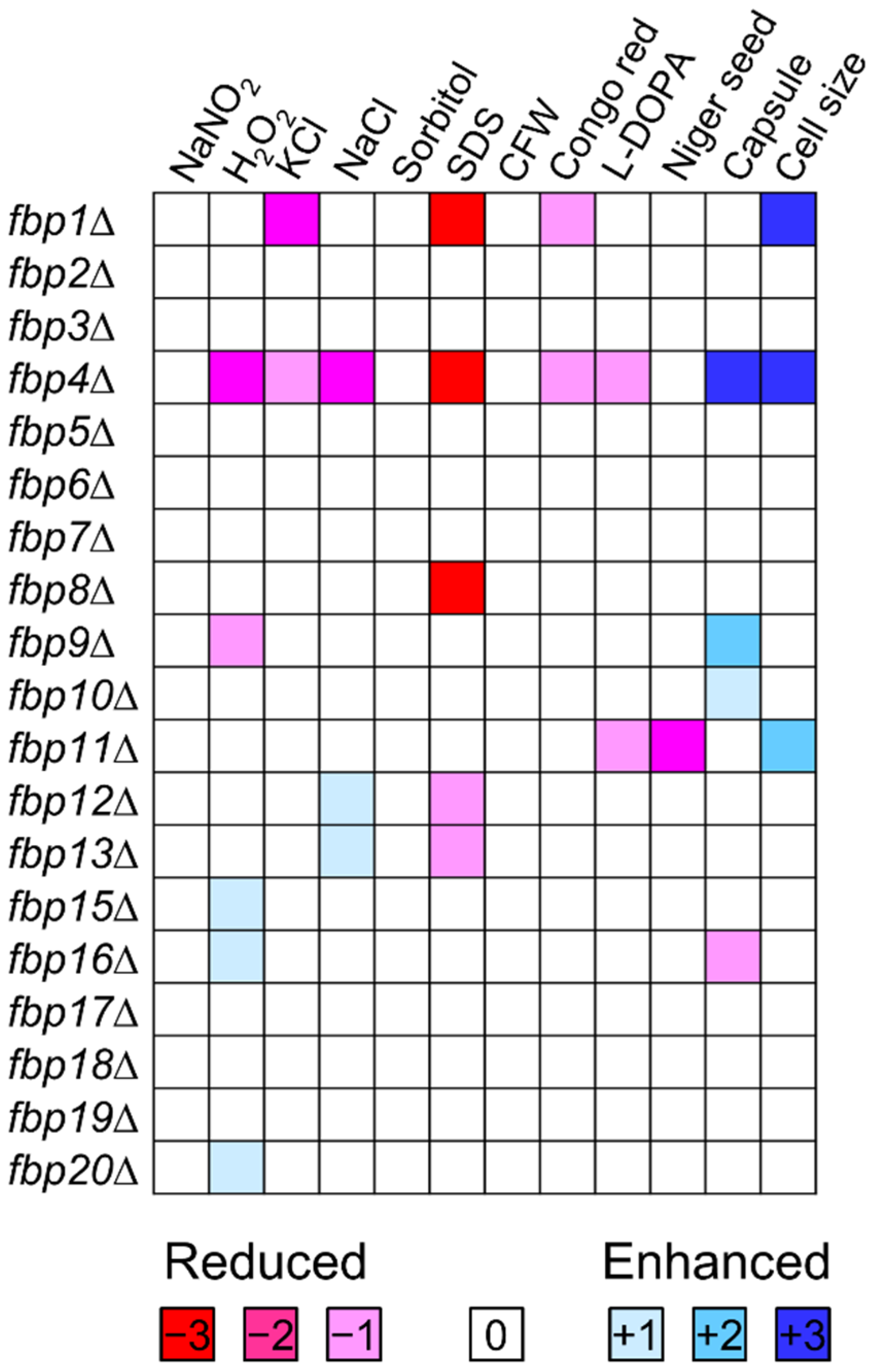
2.2. Phenotypes of F-Box Protein Encoding Genes Deletion Mutants under In Vitro Conditions

To elucidate functions of F-box proteins in adaptation and virulence of C. neoformans, we examined phenotypic traits under distinct in vitro conditions: nitrosative and oxidative stress (NaNO2, H2O2), osmotic stress (KCl, NaCl, Sorbitol), cell membrane and cell wall integrity (SDS, CFW, Congo red), and virulence factors (melanin, capsule, and cell size) (Figure S2 and Figure 1). The phenotypic data set of all 19 mutants were qualitatively illustrated by color scale, and the phenotype of KN99α was performed as a control (Figure 1). We found that 11 mutants have at least one phenotype and 8 mutants with modest phenotypic change compared to wild type strain. The fbp8∆ mutant (CNAG_04341) possesses an SDS sensitivity phenotype in the conditions we tested. fbp4∆ (CNAG_00693) and fbp11∆ mutants (CNAG_05294) showed multiple phenotypic changes in the virulence and stress response to environment including cell membrane integrity, cell size development, and melanin production. Fbp4 and Fbp11 are two F-box and WD-40 domain-containing proteins that share protein sequence similarity with Cdc4 in S. cerevisiae (35% and 32%, respectively), suggesting that Fbp4 and Fbp11 are two Cdc4 homologs in C. neoformans. To identify the functional correlation between Fbp4 and Fbp11, we generated a fbp4fbp11∆ double mutant and investigated the function of Fbp4, Fbp11, and Fbp8 as their deletion mutants demonstrate clear phenotypes.

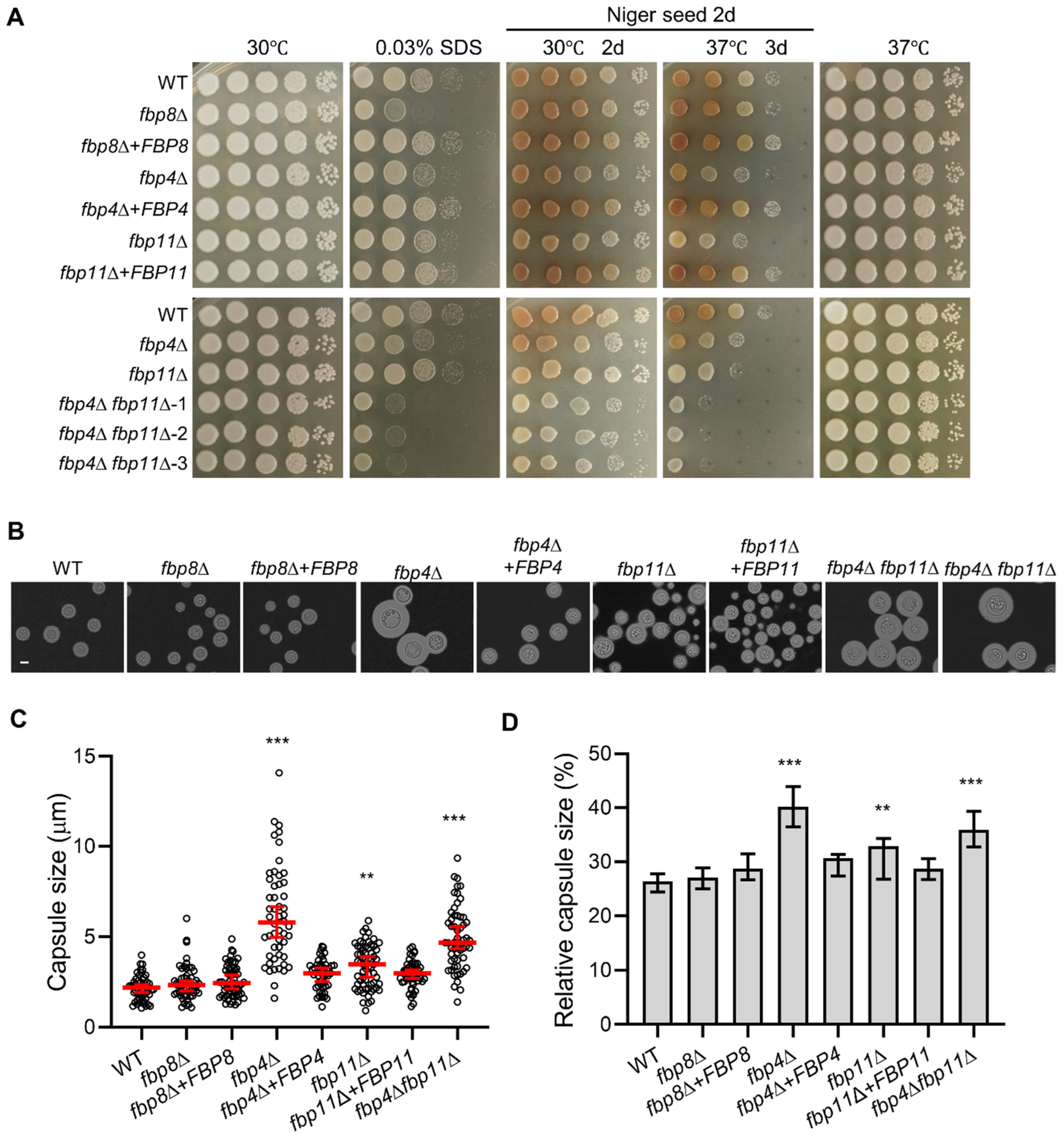
2.3. F-Box Proteins Are Important for Cell Membrane Integrity and Virulence Factor Production

To assay for cell membrane integrity defects, we treated mutants with 0.03% SDS as previously described [13]. We found that both the fbp4∆ and fbp4fbp11∆ mutants were hypersensitive to SDS, whereas the fbp11∆ single mutant showed no sensitivity difference compared to the wild type strain. These observations suggest a specific role of Fbp4 in cell membrane integrity (Figure 2A). Additionally, the fbp8∆ mutant showed increased sensitivity to SDS compared to wild type (Figure 2A). To demonstrate that this loss of function in cell membrane integrity was due to F-box protein deletion, we tested complemented strains of these mutants (fbp4∆ + FBP4, fbp11∆ + FBP11, and fbp8∆ + FBP8). Indeed, complemented strains of the F-box protein deletion mutants restored cell membrane integrity indicated by normal growth in SDS condition (Figure 2A).
Several virulence factors, such as the polysaccharide capsule and the polyphenol pigment melanin, are essential for C. neoformans virulence [6,7,37,38]. We examined the melanin production of these F-box protein mutants on both L-DOPA (L-3,4-dihydroxyphenylalanine) and Niger seed agar media. The fbp11∆ and fbp4fbp11∆ mutants exhibited a melanin production defect (Figure 2A and Figure S2), whereas the fbp4∆ and fbp8∆ mutants produced normal melanin on both media (Figure 2A and Figure S2). We investigated capsule development of F-box protein mutants on DME agar medium at 30 °C (Figure 2B and Figure S3). Quantitative measurement of capsule thickness (Figure 2C) and relative capsule size (Figure 2D) showed that deletion of either FBP4 or FBP11 increased capsule production and the fbp4∆ + FBP4 or fbp11∆ + FBP11 reconstituted strain reverted to normal capsule size. These results indicate an important role of Fbp4 in capsule size regulation. Additionally, we observed the capsule enlargement of the fbp9∆ mutant on DME agar medium at 37 °C (Figure S3).

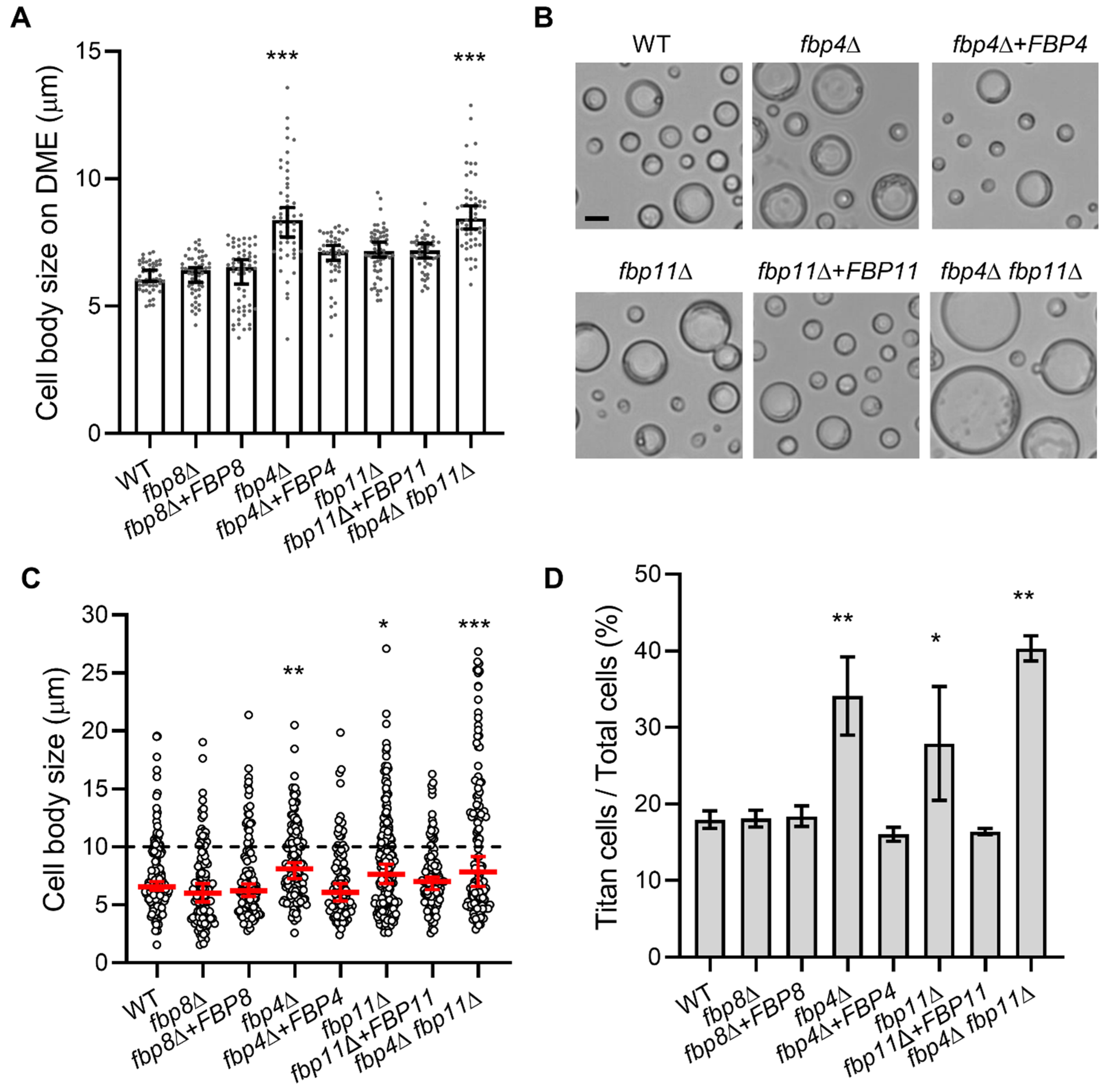
2.4. Fbp4 and Fbp11 Negatively Regulate Cell Size

Titan cell formation has been identified as an important virulence factor in C. neoformans [39,40,41]. Our previous study demonstrated that Fbp1 negatively regulates yeast cell size and the fbp1∆ strain significantly induces titan cells compared to wild type in both in vitro conditions and in vivo from infected lungs [42]. Similarly, the fbp4∆ mutant cells were significantly larger than the wild type cells when grown on capsule-inducible medium DME (Figure 2B, Figure 3A and Figure S3). The fbp4fbp11∆ double mutant also showed increased cell size similar to the fbp4∆ single mutant (Figure 3A). We then measured the cell body size and calculated the large cell percentage of these mutants under in vitro titan cell inducing conditions as previously reported [43] (Figure 3C,D). Here, fbp4∆ had a larger cell body size (8.1 µm in fbp4∆ vs. 6.6 µm in WT, p = 0.003) and a higher percentage of titan cells formation than wild type (34.1% in fbp4∆ vs. 18.0% in WT, p < 0.01). Additionally, fbp11Δ mutant increased the median cell body size and titan cell production (Figure 3C,D). This phenotype was fully rescued in the reconstituted compliment strains. Other mutants, including fbp8∆, had no significant cell size changes. Interestingly, the fbp4Δ fbp11Δ double mutant produced more titan cells compared to the fbp4Δ or the fbp11Δ single mutant (Figure 3D). These results indicate that Fbp4 and Fbp11 have overlapping yet additive roles in titan cell production.

2.5. Fbp8 and Fbp4 Are Important for Fluconazole Resistance

To illustrate the potential role of F-box proteins on regulation of antifungal sensitivity, we performed an in vitro susceptibility assay for all the F-box protein mutants to measure the MIC90 of three antifungal drugs: fluconazole, caspofungin, and amphotericin B. MIC90 testing showed that both the fbp8Δ and fbp4Δ mutants are both highly susceptible to fluconazole with a MIC90 at 2 µg/mL, compared to the wild type MIC90 of 8 µg/mL (Table 2). The fbp4Δ fbp11Δ double mutant had the same fluconazole sensitivity as the fbp4Δ mutant. Meanwhile, all mutants had same MIC90 value for caspofungin and amphotericin B as wild type (Table 2). To test the hypothesis that Fbp4 and Fbp8 regulates the ergosterol biosynthesis as a mean of fungal resistance to triazole drugs, we performed qRT-PCR to measure the expression of ERG11 in wild type and the fbp4∆ and fbp8∆ mutants. Interestingly, fbp4Δ and fbp8Δ did not show a significant difference in ERG11 expression level compared to wild type when grown at half MIC90 fluconazole conditions. These findings suggest the regulation of ergosterol biosynthesis may not be at the transcription level or involves other mechanisms which target fluconazole susceptibility in these mutants (Figure S4).

2.6. Fbp4 Is Essential for Sexual Reproduction

To evaluate the function of F-box proteins in sexual reproduction, we performed genetic cross between KN99a and mutants of F-box genes in KN99α background to generate mutants in opposite mating type and obtained all F-box protein mutants in both α and a mating types. We examined the development of dikaryotic hyphae and basidiospores in both unilateral crosses and bilateral crosses for all mutants of the F-box genes (Figure S5). A mating cross between KN99α and KN99a was performed as the control (Figure S5). We did not observe a clear defect in mating hyphae production or sporulation in unilateral crosses of all mutants. The bilateral mating between fbp4Δ mutants produced normal mating hyphae but failed to produce basidiospores, which is consistent with a previous finding [22].These data suggest that Fbp4 and Fbp11 play distinct roles in sexual reproduction (Figure S5).

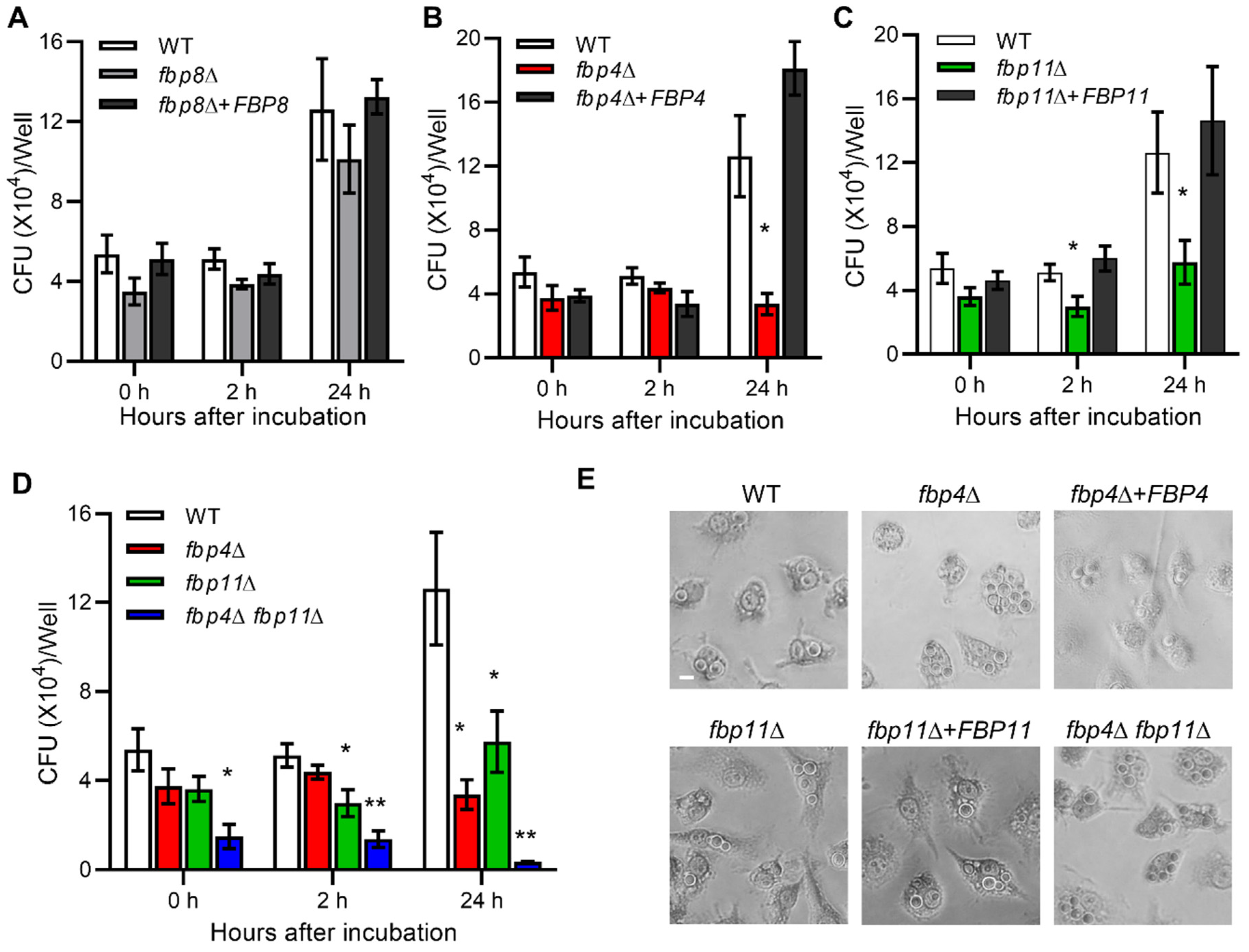
2.7. F-Box Proteins Are Involved in Cryptococcus-Macrophage Interaction

As a facultative pathogen, C. neoformans can survive and proliferate inside macrophages, which is important for Cryptococcus dissemination [44,45,46]. We investigated the potential role of F-box proteins in Cryptococcus-macrophage interaction with macrophage cell line RAW264.7 following a previously described protocol [27] (Figure 4 and Figure S6). Non-adherent extracellular yeast cells were removed after 2 h of Cryptococcus-macrophage coincubation. The phagocytosis assay and macrophage killing assay were performed after incubation for an additional 0, 2, and 24 h. We found that Fbp8 was not required for Cryptococcus-macrophage interaction (Figure 4A), while deletion of FBP4 or FBP11 significantly reduced intracellular growth in macrophages after 24 h coincubation compared to the wild type strain. The complement strains completely restored the survival in macrophages (Figure 4B,C). The CFUs (Colony forming units) recovered from the fbp4Δ fbp11Δ double mutant engulfed by macrophages were significantly less than either the fbp4Δ or fbp11Δ single mutants (Figure 4D). These results demonstrate that Fbp4 and Fbp11 are important for fungal survival in macrophages. In addition, we observed that the fbp7Δ mutant had proliferation defect inside macrophages while deletion of FBP9 induced C. neoformans proliferation in macrophages, both are interesting observations that warrant further investigation (Figure S6A).

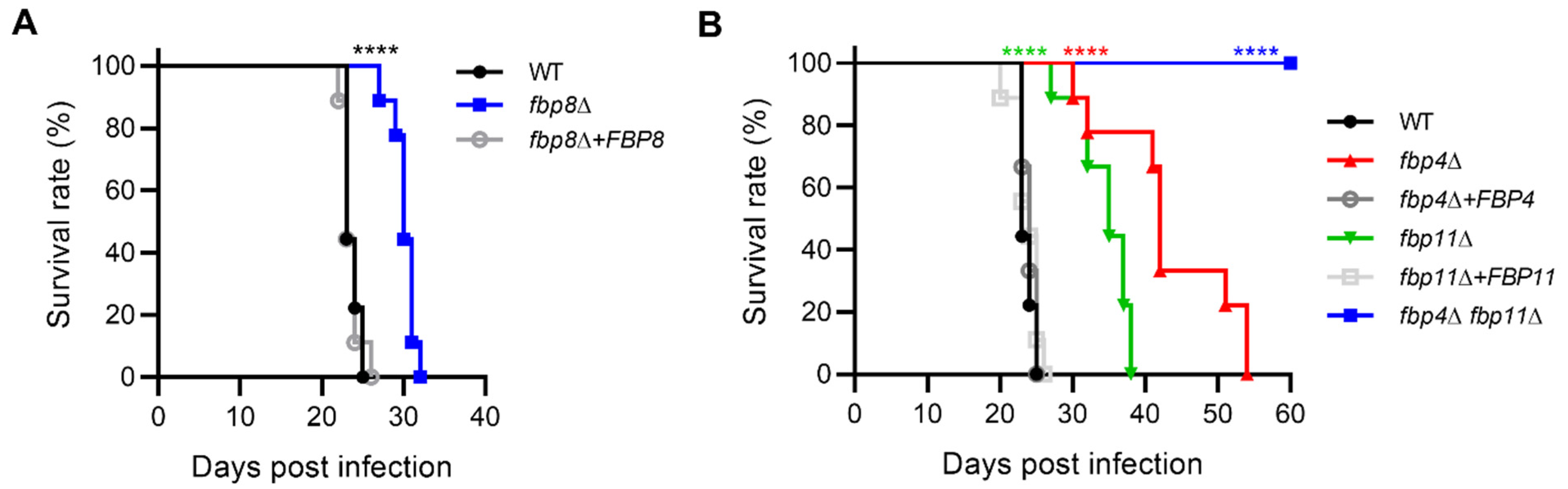
2.8. Roles of F-Box Proteins in Fungal Virulence

Because of the importance of Fbp4, Fbp11, and Fbp8 in development of Cryptococcus virulence factors, we assessed the virulence of the fbp4Δ, fbp11∆, fbp8Δ, and fbp4Δ fbp11Δ mutants in a murine inhalation model of systemic cryptococcosis. Mice were infected with 105 cells of each strain and surveilled for survival following infection. Mice infected with fbp8Δ and fbp11Δ mutants succumbed to infection within 32 and 38 days post-inoculation, respectively, while those infected with wild type strain died within 25 days as expected (Figure 5). These results indicated that fbp8Δ and fbp11Δ have significant virulence attenuation. The fbp4Δ-infected mice survival to 54 dpi (days post infection) also indicated significant virulence attenuation (Figure 5B). Excitingly, mice infected with the fbp4Δ fbp11Δ double mutant survived to the termination point of the experiment (60 dpi) with no visible symptoms (Figure 5B), suggesting Fbp4 and Fbp11 share an additive function in regulating fungal virulence.

3. Discussion

F-box proteins determine the substrate specificity of the UPS and play multiple important roles in fungal development and virulence [14,47]. C. neoformans possesses at least 20 F-box proteins including Fbp1 [13,28,29]. In this study, we obtained mutants of 19 F-box protein encoding genes, including 14 mutants from the Cryptococcus deletion library generated by Dr. Madhani group [30] and five new mutants generated in this study. We could not generate the mutant for FBP14 (homolog of MET30) and suspected it to be an essential gene in C. neoformans. Fbp1 has been studied extensively [13,27,28,29,42], and thus used as a control in this study. We studied functions of the remaining 18 non-essential F-box proteins by analyzing their respective mutants under a range of stress conditions. Here, we identified three F-box proteins (Fbp4, Fbp8, and Fbp11) with important cellular functions based on clear phenotypes of their mutants, including sexual reproduction, cell size regulation, drug resistance, and fungal virulence. Fbp4 and Fbp11 share high protein structure and sequence similarity with Cdc4 in S. cerevisiae, suggesting that both are Cdc4 homologs in C. neoformans. We also found Fbp4 and Fbp11 share overlapping and distinct cellular functions, including cell integrity, stress response, cell size regulation, capsule and melanin production, and fungal–host interactions. Fbp8 is required for fungal cell membrane integrity as its mutant is hypersensitive to SDS treatment, but not cell wall stressing agents calcofluor white and congo red.
Cryptococcal adaptation to the hostile host environment is critical for its virulence. The fbp4Δ mutant exhibited increased susceptibility to oxidative stress H2O2, as well as cell membrane stressor SDS, which may interfere with the ability of the fungal proliferation inside macrophages. The UPS regulation of oxidative stress responses has been identified across multiple eukaryotic species [48,49]. For example, deletion of UBI4 shows hypersensitivity to H2O2 in S. cerevisiae [50]. Two ubiquitin-conjugating enzymes Ubc6-2 and Ubc8 and a deubiquitinating enzyme Ubp5 are involved in diverse stress responses in C. neoformans [49,51]. We found that both fbp4Δ and fbp8Δ mutants are sensitive to SDS, but not the cell wall stressors such as calcofluor white and congo red. These findings indicate that Fbp4 (Cdc4 in a separated report [22]) and Fbp8 are involved in cell membrane integrity similar to Fbp1 [13,22]. It is possible that some substrates of these F-box proteins are important for membrane integrity. Indeed, homolog of Fbp8 in S. cerevisiae (Rcy1) is involved in recycling plasma membrane proteins. Rcy1 directly interacts with C-terminal region of phospholipid flippase Drs2 and plays an important role in the function of Drs2 in S. cerevisiae [52].
Consistent with the SDS sensitivity phenotype of the fbp4Δ and fbp8Δ mutants, we also observed that both Fbp4 and Fbp8 are required for fungal resistance to azole drug fluconazole, but not for other classes of antifungal agents including caspofungin and amphotericin B. This suggested that Fbp4 and Fbp8 may be involved in regulation of the ergosterol biosynthesis pathway. As overexpression of ERG11 gene leads to overproduction of the target enzyme of fluconazole and increase drug resistance [53], we tested the role of Fbp4 and Fbp8 in the regulation of ERG11 expression. However, we observed no expression change of ERG11 in the fbp4Δ and fbp8Δ mutants compared to their parental wild type and complement strains. It is possible that these F-box proteins regulate Erg11 through post-translational modifications (e.g., ubiquitin-mediated proteolysis) rather than transcriptional regulation. In addition, alterations in efflux pumps may play a role in azole susceptibility in the forementioned F-box protein mutants [54]. Alteration of drug accessibility to the target enzyme by regulation of efflux pumps (including CDR and MDR genes) has been shown to be an important mechanism of azole resistance in Candida species [53,54]. Although previous studies have indicated that efflux pumps (Afr1 and Afr2) only play a minor role in C. neoformans resistance to azoles [55,56], it remains possible that they contribute to F-box protein-mediated azole resistance. Overall, the mechanism of increased azole sensitivity in these F-box protein mutants remains to be further investigated. In S. cerevisiae, Rcy1 is required for the Drs2 function in the early endosome to the trans-Golgi network (TGN) transport [52]. Cdc50-Drs2 function as lipid translocase (flippase) generates membrane phospholipid asymmetry and regulates ergosterol distribution [57,58,59]. In Cryptococcus, Cdc50 is the only homolog of the ScCdc50 gene family and the cdc50Δ mutant sensitive to fluconazole [60]. As there is a similar sensitivity with the fbp8Δ mutant, these findings suggest Fbp8 may also regulate lipid flippase function in C. neoformans.
Among the three well studied virulence factors in C. neoformans, none of these mutants exhibit significant growth defect at a host body temperature of 37 °C. A melanin defect in the fbp11∆ mutant, but not in the fbp4∆ mutant, suggests that although both Fbp4 and Fbp11 are homologs of Cdc4 in S. cerevisiae, they have distinct functions. Yet, it is interesting that both the fbp4∆ mutant and the fbp11∆ mutant showed increased capsule sizes. Further study is warranted to understand how these two F-box proteins regulate capsule growth, which is beyond the scope of this study.
Titan cell production (cell body size over 10 µm) can be observed during cryptococcal infection and has been identified as a virulence factor [39,40,61]. The Gpa1-cAMP/PKA signal pathway is critical in regulating cell size change during Cryptococcus infection [41,62]. We found that both Fbp4 and Fbp11 are involved in cell size regulation, and that the deletion of FBP4 or FBP11 induces titan cell production. The fbp4Δ fbp11Δ double mutant exhibits greater titan cell percentage in a population than either single mutant. In S. cerevisiae, Cdc4 is required for the G1/S and G2/M phase transitions [63,64]. Because dysregulation of the cell cycle has been shown to be involved in titan cell production in C. neoformans [65], it is not surprising that Fbp4 and Fbp11 are involved in cell size regulation. It has been reported that the meiosis machinery is involved in ploidy reduction during titan cell proliferation [66]. Our previous work found that the CDK-dependent protein kinase Crk1, a meiosis regulator, positively regulates titan cell production in C. neoformans [42]. Deletion of Fbp4 blocks sporulation but has no impact on other events in bilateral mating, such as cell fusion, dikaryotic hyphae formation, and nuclear fusion in C. neoformans [67]. These findings similar to the fbp1Δ mutant [13] and the published data on Cdc4 [22] thus indicate Fbp4 may play a key role in meiosis regulation. Since both Saccharomyces Cdc4 and Cryptococcus Fbp4 are involved in sexual reproduction [68,69], it is possible that Fbp4 may also regulate titan cell production through the meiosis machinery.
Despite producing larger capsule sizes in the fbp4Δ and fbp11Δ mutants, both mutants and the fbp4Δ fbp11Δ double mutant showed virulence attenuation in a murine inhalation model of cryptococcosis. Together with our previous findings on Fbp1 function, our studies further demonstrate the importance of F-box proteins in pathogenicity of C. neoformans. We found that fungal virulence of these three mutants were consistent with their ability of intracellular proliferation in macrophages. Wu et al. reported that Cdc4 (Fbp4) was involved in the virulence in C. neoformans through similar mechanisms as Fbp1 and that the loss of CDC4 blocked C. neoformans dissemination from lung to other organs and infected mice survived for over two months [22]. In our study, albeit virulence attenuation, fbp4Δ-infected mice still succumb to lethal infection between 30 and 54 dpi. The difference may be due to different Cryptococcus strain and mouse strain backgrounds used in these studies. Specifically, our fbp4Δ mutant was generated from the KN99α background strain and tested for virulence in the BALB/c mouse strain, while Wu and colleagues utilized the C57BL/6 mouse strain and C. neoformans H99 background strain in their studies. The fbp4fbp11∆ double mutant showed significantly more severe virulence attenuation than either of the single mutants, indicating these two Cdc4 homologs are both required for fungal virulence and have additive functions. In aggregate, our systemic characterization of the entire F-box gene family in C. neoformans reveals a diverse role of F-box proteins in Cryptococcus development and virulence. Further study on the molecular detail of SCF E3 ligase-mediated proteolysis may lead to novel drug targets in fungal pathogens.

4. Materials and Methods

Strains and media: Cryptococcus neoformans gene deletion libraries were generated by Hiten Madhani at UCSF and purchased from the Fungal Genetic Stock Center (FGSC). Other strains used in this study are listed in Table S1. YPD medium (2% peptone, 1% yeast extract, 2% glucose) was used for routine culture of C. neoformans strains. Stress media were created by adding different stress-inducing agents into YPD agar medium. L-DOPA (L-3,4-dihydroxyphenylalanine) and Niger seed media were used to test melanin production. Dulbecco’s modified Eagle’s (DME) medium for assessing capsule production was prepared as previously described [70]. V8 juice agar medium (50 mL V8 original juice, 0.05% KH2PO4, 2% agar, pH 5.0) and modified Murashige and Skoog (MS) medium minus sucrose (Sigma-Aldrich, Steinhelm, Germany) were used for mating and sporulation assays [71]. Minimal medium (MM, 15 mM D-glucose, 10 mM MgSO4, 29.4 mM KH2PO4, 13 mM Glycine, 3.0 μM Thiamine) was used for titan cell induction [43]. Macrophage murine cell line RAW264.7 was grown in liquid Dulbecco Modified Eagles (DME) medium with 10% heat inactivated fetal calf serum (FBS) (ATL Biologicals) and prepared as previously described [71].
Electroporation: Electroporation was performed as previously described [72]. Briefly, overnight cultures were transferred into 100 mL YPD at an initial OD600 around 0.2 and grown for 4–5 h at 30 °C until the OD600 reach 0.6–0.8. All samples were kept cold in the following steps. Cells were collected, washed twice, resuspended in electroporation buffer (10 mM Tris-HCl pH 7.5, 1 mM MgCl2, 270 mM sucrose), and incubated with 1 mM DTT on ice for one hour. Cells were pelleted and resuspended in 250 μL electroporation buffer. 45 μL cells were mixed with 5 μL DNA and transferred to a precooled 2 mm gap cuvette. An Eppendorf multiporator was used for electroporation with the following settings: V = 2 kv. Cells were resuspended in 1 mL YPD, transferred to 1.5 mL Eppendorf tubes, and incubated at 30 °C for 1.5 h before plating onto a selective agar plate.
Colony PCR: Colony PCR was conducted on Cryptococcus cultures grown on YPD agar plate. Small colonies were picked and transferred to PCR tubes as direct PCR template. Colony PCR bands were checked by gel electrophoresis in 1% agarose gel.
RNA extraction and qRT-PCR: Total RNA was extracted from C. neoformans strains with Trizol (Invitrogen). Extracted RNA was treated with rDNAse kit and purified using the Nucleospin RNA cleanup kit (Macherey–Nagel). Relative mRNA levels were determined by quantitative reverse transcription (qRT) PCR. Cryptococcus strains were grown at a 1.0 OD for 4 h at 30 °C with 1μg/mL of fluconazole. An amount of 2 μg of purified RNA were converted to cDNA using SMARTScribe Reverse Transcriptase cDNA synthesis kit (Takara Bio). The SYBR Green PCR Master mix (Applied Biosystems) and constructed primers for each gene were used and normalized to the gene encoding glyceraldehyde-3-phosphate dehydrogenase (GAPDH). Gene expression relative to parental KN99α grown in the same conditions was calculated by the ΔΔCT method.
Phenotypic assays. (i) Cell wall and membrane integrity: Overnight cultures with serial dilutions were placed on YPD agar plates containing 0.03% SDS to test cell membrane integrity. YPD with the supplement of 0.5% congo red, or 250 μg/mL calcofluor white (CFW) were used to test cell wall integrity. The agar plates were incubated at 30 or 37 °C for 3 days before being photographed.
(ii) Melanin and capsule production: Overnight cultures with serial dilutions were placed on (L-DOPA) agar medium or overnight cultures were inoculated on Niger seed agar medium to measure the melanin production. The agar plates were incubated at 30 or 37 °C for 3 days before being photographed. Overnight cultures were inoculated on Dulbecco modified Eagle (DME) medium and incubated at 37 °C for 7 days to examine capsule production. The capsule size was visualized by adding a drop of India ink to the cell suspensions and observed on an Olympus AX70 microscope (Melville, NY, USA). The relative capsule size was calculated by dividing capsule size with the whole-cell size (capsule and cell size). The average and standard deviation from at least 50 cells were calculated for each condition tested.
(iii) Assays of stress responses: Yeast cells from overnight cultures were washed, resuspended, and serially diluted (1:10) in ddH2O and spotted (5 µL) on YPD agar plates containing 1.0 M KCl, 1.5 M NaCl, or 2.5 M sorbitol for osmotic shock, 5 mM H2O2 for oxidative stress, and 1 mM NaNO2 for nitrosative stress as previously described [60].
(iv) In vitro titan cell induction and measurement: Titan cell inductions were performed as reported previously [43]. Strains were grown overnight at 30 °C in YPD liquid medium. Cells were collected, washed twice with minimal medium, and suspended in minimal medium with the final concentration of 106 cells/mL. A 1 mL suspension was incubated at 30 °C for 3 days in a 1.5 mL Eppendorf tube with the cap closed. Body sizes of more than 100 cells were measured and the titan cell (cell body size over 10 µm) proportions were calculated.
(v) Assays of mating: C. neoformans cells of opposite mating types were mixed and cultured on V8 or MS agar medium at 25 °C in the dark. Mating filaments and basidiospores formation were examined and recorded by photography after incubation for 7 or 14 days.
Cryptococcus-macrophage interaction assay: Macrophage cell line RAW264.7 cells were cultured in a DME medium with 10% heat inactivated FBS at 37 °C with 5% CO2. A total of 5 × 10⁴ macrophage cells in 0.5 mL fresh DME was put into each well of a 48-well culture plate and incubated at 37 °C in 5% CO2 overnight. To activate macrophage cells, 50 unit/mL gamma interferon (IFNγ, Invitrogen) and 1 μg/mL lipopolysaccharide (LPS, Sigma) were added into each well. C. neoformans overnight cultures were washed with PBS twice and opsonized with 20% mouse complement. A total of 2 × 10⁵ Cryptococcus cells were added into each well (yeast/macrophage ratio, 4:1). To assess intracellular proliferation of C. neoformans, non-adherent extracellular yeast cells were removed by washing with fresh DME medium after 2 h co-incubation and cultures were incubated for another 0, 2, and 24 h. At indicated time points, the medium in each well was replaced by ddH2O to lyse the macrophage cells for 30 min at room temperature. The lysate was spread on YPD plates and CFU was counted to determine intracellular proliferation.
Virulence studies: Yeast strains were grown at 30 °C overnight and washed twice with PBS buffer and resuspended to a final concentration of 2 × 10⁶ cells/mL. Groups of 10 female BALB/c mice were intranasally infected with 105 yeast cells of each strain as previously described [73]. Over the course of the experiments, animals that appeared moribund or in pain were sacrificed by CO2 inhalation. Survival data from the murine experiments were statistically analyzed between paired groups using the long-rank test using the PRISM program 8.0 (GraphPad Software) (p values of ≤0.05 were considered significant). All animal studies were conducted following biosafety level 2 (BSL-2) protocols and procedures approved by the Institutional Animal Care and Use Committee (IACUC) and Institutional Biosafety Committee of Rutgers University, respectively. The studies were conducted in facilities accredited by the Association for Assessment and Accreditation of Laboratory Animal Care (AAALAC).

Supplementary Materials

The following supporting information can be downloaded at: https://www.mdpi.com/article/10.3390/jof8121259/s1, Figure S1. Deletion of FBP6 (CNAG_03157), FBP7 (CNAG_03421), FBP12 (CNAG_05450), FBP13 (CNAG_05454), and FBP19 (CNAG_07551) in C. neoformans. (A) Colony PCR to confirm the mutants from the Cryptococcus deletion library. (B) Strategy for gene knockout by fusion PCR assays. Allele was replaced with the nourseothricin resistance marker NAT1. (C) Colony PCR screening and confirmation of the positive transformants. Primers used in this study are listed in Table S2. Figure S2. In vitro stress responses of F-box proteins in C. neoformans. Phenotypic assays were examined under different conditions as indicated. Overnight cultures were collected and resuspended to an optical density at 600 nm (OD600) of 10.0. Tenfold serial dilutions were made in ddH2O, and 3 μL of each was plated. The plates were incubated at 37 °C for 3 days before being photographed. Figure S3. Representative images to show capsule formation at 30 °C (A) or 37 °C (B) on DME medium. Capsule production was visualized by India ink staining after cells were grown on DME medium for 7 days. Figure S4. ERG11 expression in F box protein deletion mutants and complimented strains analyzed by qRT-PCR. Samples were grown in YPD with 1 μg/mL Fluconazole for 4 h at 30 °C. Differential gene expression relative to GAPDH was calculated by the ΔΔCT method. Figure S5. Mating filament production and sporulation of the wild type and mutants of F-box protein encoding genes in C. neoformans. Bilateral were performed on MS medium. Mating structures at ×40 magnification (left) and spores at ×200 magnification (right) were photographed after 2 weeks of incubation in the dark at 25 °C. Figure S6. Killing assay and phagocytosis of the wild type and mutants of F-box protein encoding genes in C. neoformans. Phagocytosis was allowed to occur for 2 h to measure both fungicidal and fungistatic activity. After each time interval, macrophages were lysed by adding sterile ddH2O. Cells were counted and plated on YPD agar plates to determine CFU. (B) Typical field views of phagocytosis for each sample. Table S1. Strains and plasmids used in this study. References [13,30,74,75] are cited in the Table S1. Table S2. Primers used in this study.

Author Contributions

Conceptualization, C.C. and C.X.; methodology, C.C., Y.W., S.L.A. and J.W.; software, C.C., Y.W., S.L.A. and C.X.; validation, C.C., Y.W., S.L.A. and C.X.; formal analysis, C.C., Y.W. and S.L.A.; investigation, C.C., Y.W. and S.L.A.; resources, C.X.; data curation, C.C., Y.W. and S.L.A.; writing—original draft preparation, C.C.; writing—review and editing, C.C., Y.W., S.L.A., J.W. and C.X.; visualization, C.C. and C.X.; supervision, C.X.; project administration, C.X.; funding acquisition, C.X. All authors have read and agreed to the published version of the manuscript.

Funding

This research was funded by NIH grants R01AI123315, R01AI141368, and the Rutgers Health Advance Fund (partially supported through NIH U01HL150852) to C.X. S.L.A. is supported by a NIH Research Supplement R01AI141368-03S1.

Institutional Review Board Statement

The animal study protocol was approved by the Institutional Animal Care and Use Committee (IACUC) of Rutgers University (protocol code: 999901066) on 1 March 2021.

Informed Consent Statement

Not applicable.

Data Availability Statement

Not applicable.

Acknowledgments

We acknowledge the use of the Cryptococcus gene deletion collection that was generated by the Madhani Laboratory at University of San Francisco and funded by the NIH funding (R01AI100272).

Conflicts of Interest

The authors declare no conflict of interest.

References

  1. Bongomin, F.; Gago, S.; Oladele, R.O.; Denning, D.W. Global and multi-national prevalence of fungal diseases-estimate precision. J. Fungi 2017, 3, 57. [Google Scholar] [CrossRef] [PubMed]
  2. Pappas, P.G. Cryptococcal infections in non-HIV-infected patients. Trans. Am. Clin. Climatol. Assoc. 2013, 124, 61–79. [Google Scholar] [PubMed]
  3. Beardsley, J.; Sorrell, T.C.; Chen, S.C. Central nervous system cryptococcal infections in Non-HIV infected patients. J. Fungi 2019, 5, 71. [Google Scholar] [CrossRef] [PubMed] [Green Version]
  4. Rajasingham, R.; Smith, R.M.; Park, B.J.; Jarvis, J.N.; Govender, N.P.; Chiller, T.M.; Denning, D.W.; Loyse, A.; Boulware, D.R. Global burden of disease of HIV-associated cryptococcal meningitis: An updated analysis. Lancet Infect. Dis. 2017, 17, 873–881. [Google Scholar] [CrossRef] [Green Version]
  5. Velagapudi, R.; Hsueh, Y.P.; Geunes-Boyer, S.; Wright, J.R.; Heitman, J. Spores as infectious propagules of Cryptococcus neoformans. Infect. Immun. 2009, 77, 4345–4355. [Google Scholar] [CrossRef] [Green Version]
  6. Kronstad, J.; Jung, W.H.; Hu, G. Beyond the big three: Systematic analysis of virulence factors in Cryptococcus neoformans. Cell Host. Microbe 2008, 4, 308–310. [Google Scholar] [CrossRef] [Green Version]
  7. Zaragoza, O. Basic principles of the virulence of Cryptococcus. Virulence 2019, 10, 490–501. [Google Scholar] [CrossRef] [Green Version]
  8. Nielsen, K.; Heitman, J. Sex and virulence of human pathogenic fungi. Adv. Genet. 2007, 57, 143–173. [Google Scholar]
  9. Idnurm, A.; Bahn, Y.S.; Nielsen, K.; Lin, X.; Fraser, J.A.; Heitman, J. Deciphering the model pathogenic fungus Cryptococcus neoformans. Nat. Rev. Microbiol. 2005, 3, 753–764. [Google Scholar] [CrossRef]
  10. Altamirano, S.; Jackson, K.M.; Nielsen, K. The interplay of phenotype and genotype in Cryptococcus neoformans disease. Biosci. Rep. 2020, 40, BSR20190337. [Google Scholar] [CrossRef]
  11. Glickman, M.H.; Ciechanover, A. The ubiquitin-proteasome proteolytic pathway: Destruction for the sake of construction. Physiol. Rev. 2002, 82, 373–428. [Google Scholar] [CrossRef] [PubMed]
  12. Finley, D.; Ulrich, H.D.; Sommer, T.; Kaiser, P. The ubiquitin-proteasome system of Saccharomyces cerevisiae. Genetics 2012, 192, 319–360. [Google Scholar] [PubMed] [Green Version]
  13. Liu, T.B.; Wang, Y.; Stukes, S.; Chen, Q.; Casadevall, A.; Xue, C. The F-Box protein Fbp1 regulates sexual reproduction and virulence in Cryptococcus neoformans. Eukaryot. Cell 2011, 10, 791–802. [Google Scholar] [CrossRef] [Green Version]
  14. Cao, C.; Xue, C. More than tust cleaning: Ubiquitin-mediated proteolysis in fungal pathogenesis. Front. Cell. Infect. Microbiol. 2021, 11, 774613. [Google Scholar] [CrossRef]
  15. Atir-Lande, A.; Gildor, T.; Kornitzer, D. Role for the SCFCDC4 ubiquitin ligase in Candida albicans morphogenesis. Mol. Biol. Cell 2005, 16, 2772–2785. [Google Scholar] [CrossRef] [PubMed] [Green Version]
  16. Shieh, J.C.; White, A.; Cheng, Y.C.; Rosamond, J. Identification and functional characterization of Candida albicans CDC4. J. Biomed. Sci. 2005, 12, 913–924. [Google Scholar] [CrossRef] [PubMed]
  17. Butler, D.K.; All, O.; Goffena, J.; Loveless, T.; Wilson, T.; Toenjes, K.A. The GRR1 gene of Candida albicans is involved in the negative control of pseudohyphal morphogenesis. Fungal Genet. Biol. 2006, 43, 573–582. [Google Scholar] [CrossRef]
  18. Li, W.J.; Wang, Y.M.; Zheng, X.D.; Shi, Q.M.; Zhang, T.T.; Bai, C.; Li, D.; Sang, J.L.; Wang, Y. The F-box protein Grr1 regulates the stability of Ccn1, Cln3 and Hof1 and cell morphogenesis in Candida albicans. Mol. Microbiol. 2006, 62, 212–226. [Google Scholar] [CrossRef]
  19. Jonkers, W.; van Kan, J.; Tijm, P.; Lee, Y.-W.; Tudzynski, P.; Rep, M.; Michielse, C.B. The FRP1 F-box gene has different functions in sexuality, pathogenicity and metabolism in three fungal pathogens. Mol. Plant Pathol. 2011, 12, 548–563. [Google Scholar] [CrossRef]
  20. Leach, M.D.; Stead, D.A.; Argo, E.; MacCallum, D.M.; Brown, A.J. Molecular and proteomic analyses highlight the importance of ubiquitination for the stress resistance, metabolic adaptation, morphogenetic regulation and virulence of Candida albicans. Mol. Microbiol. 2011, 79, 1574–1593. [Google Scholar] [CrossRef] [Green Version]
  21. Skowyra, D.; Craig, K.L.; Tyers, M.; Elledge, S.J.; Harper, J.W. F-box proteins are receptors that recruit phosphorylated substrates to the SCF ubiquitin-ligase complex. Cell 1997, 91, 209–219. [Google Scholar] [CrossRef] [PubMed]
  22. Wu, T.; Fan, C.L.; Han, L.T.; Guo, Y.B.; Liu, T.B. Role of F-box protein Cdc4 in fungal virulence and sexual reproduction of Cryptococcus neoformans. Front. Cell. Infect. Microbiol. 2021, 11, 806465. [Google Scholar] [CrossRef] [PubMed]
  23. Duyvesteijn, R.G.; van Wijk, R.; Boer, Y.; Rep, M.; Cornelissen, B.J.; Haring, M.A. Frp1 is a Fusarium oxysporum F-box protein required for pathogenicity on tomato. Mol. Microbiol. 2005, 57, 1051–1063. [Google Scholar] [CrossRef] [PubMed]
  24. Johnk, B.; Bayram, O.; Abelmann, A.; Heinekamp, T.; Mattern, D.J.; Brakhage, A.A.; Jacobsen, I.D.; Valerius, O.; Braus, G.H. SCF ubiquitin ligase F-box protein Fbx15 controls nuclear co-repressor localization, stress response and virulence of the human pathogen Aspergillus fumigatus. PLoS Pathog. 2016, 12, e1005899. [Google Scholar] [CrossRef] [PubMed] [Green Version]
  25. Frawley, D.; Bayram, O. Identification of SkpA-CulA-F-box E3 ligase complexes in pathogenic Aspergilli. Fungal Genet. Biol. 2020, 140, 103396. [Google Scholar] [CrossRef]
  26. Shi, H.B.; Chen, N.; Zhu, X.M.; Liang, S.; Li, L.; Wang, J.Y.; Lu, J.P.; Lin, F.C.; Liu, X.H. F-box proteins MoFwd1, MoCdc4 and MoFbx15 regulate development and pathogenicity in the rice blast fungus Magnaporthe oryzae. Environ. Microbiol. 2019, 21, 3027–3045. [Google Scholar] [CrossRef]
  27. Liu, T.B.; Xue, C. Fbp1-mediated ubiquitin-proteasome pathway controls Cryptococcus neoformans virulence by regulating fungal intracellular growth in macrophages. Infect. Immun. 2014, 82, 557–568. [Google Scholar] [CrossRef] [PubMed] [Green Version]
  28. Masso-Silva, J.; Espinosa, V.; Liu, T.B.; Wang, Y.; Xue, C.; Rivera, A. The F-Box protein Fbp1 shapes the immunogenic potential of Cryptococcus neoformans. Mbio 2018, 9, e01828-17. [Google Scholar] [CrossRef] [PubMed] [Green Version]
  29. Wang, Y.; Wang, K.; Masso-Silva, J.A.; Rivera, A.; Xue, C. A heat-killed Cryptococcus mutant strain induces host protection against multiple invasive mycoses in a murine vaccine model. Mbio 2019, 10, e02145-19. [Google Scholar] [CrossRef] [Green Version]
  30. Chun, C.D.; Madhani, H.D. Applying genetics and molecular biology to the study of the human pathogen Cryptococcus neoformans. Methods Enzym. 2010, 470, 797–831. [Google Scholar]
  31. Thomas, D.; Kuras, L.; Barbey, R.; Cherest, H.; Blaiseau, P.L.; Surdin-Kerjan, Y. Met30p, a yeast transcriptional inhibitor that responds to S-adenosylmethionine, is an essential protein with WD40 repeats. Mol. Cell. Biol. 1995, 15, 6526–6534. [Google Scholar] [CrossRef] [PubMed] [Green Version]
  32. Bailey, R.B.; Woodword, A. Isolation and characterization of a pleiotropic glucose repression resistant mutant of Saccharomyces cerevisiae. Mol. Gen Genet. 1984, 193, 507–512. [Google Scholar] [CrossRef] [PubMed]
  33. Feldman, R.M.; Correll, C.C.; Kaplan, K.B.; Deshaies, R.J. A complex of Cdc4p, Skp1p, and Cdc53p/cullin catalyzes ubiquitination of the phosphorylated CDK inhibitor Sic1p. Cell 1997, 91, 221–230. [Google Scholar] [CrossRef] [PubMed] [Green Version]
  34. Wiederkehr, A.; Avaro, S.; Prescianotto-Baschong, C.; Haguenauer-Tsapis, R.; Riezman, H. The F-box protein Rcy1p is involved in endocytic membrane traffic and recycling out of an early endosome in Saccharomyces cerevisiae. J. Cell Biol. 2000, 149, 397–410. [Google Scholar] [CrossRef] [PubMed] [Green Version]
  35. Samanta, M.P.; Liang, S. Predicting protein functions from redundancies in large-scale protein interaction networks. Proc. Natl. Acad. Sci. USA 2003, 100, 12579–12583. [Google Scholar] [CrossRef] [Green Version]
  36. Escusa, S.; Camblong, J.; Galan, J.M.; Pinson, B.; Daignan-Fornier, B. Proteasome- and SCF-dependent degradation of yeast adenine deaminase upon transition from proliferation to quiescence requires a new F-box protein named Saf1p. Mol. Microbiol. 2006, 60, 1014–1025. [Google Scholar] [CrossRef]
  37. Kozel, T.R.; Gotschlich, E.C. The capsule of Cryptococcus neoformans passively inhibits phagocytosis of the yeast by macrophages. J. Immunol. 1982, 129, 1675–1680. [Google Scholar]
  38. Wang, Y.; Aisen, P.; Casadevall, A. Cryptococcus neoformans melanin and virulence: Mechanism of action. Infect. Immun. 1995, 63, 3131–3136. [Google Scholar] [CrossRef] [Green Version]
  39. Okagaki, L.H.; Strain, A.K.; Nielsen, J.N.; Charlier, C.; Baltes, N.J.; Chretien, F.; Heitman, J.; Dromer, F.; Nielsen, K. Cryptococcal cell morphology affects host cell interactions and pathogenicity. PLoS Pathog. 2010, 6, e1000953. [Google Scholar] [CrossRef]
  40. Zaragoza, O.; Garcia-Rodas, R.; Nosanchuk, J.D.; Cuenca-Estrella, M.; Rodriguez-Tudela, J.L.; Casadevall, A. Fungal cell gigantism during mammalian infection. PLoS Pathog. 2010, 6, e1000945. [Google Scholar] [CrossRef]
  41. Garcia-Rodas, R.; de Oliveira, H.C.; Trevijano-Contador, N.; Zaragoza, O. Cryptococcal titan cells: When yeast cells are all grown up. Curr. Top Microbiol. Immunol. 2019, 422, 101–120. [Google Scholar] [PubMed]
  42. Cao, C.; Wang, K.; Wang, Y.; Liu, T.B.; Rivera, A.; Xue, C. Ubiquitin proteolysis of a CDK-related kinase regulates titan cell formation and virulence in the fungal pathogen Cryptococcus neoformans. Nat. Commun. 2022, 13, 6397. [Google Scholar] [CrossRef] [PubMed]
  43. Hommel, B.; Mukaremera, L.; Cordero, R.J.B.; Coelho, C.; Desjardins, C.A.; Sturny-Leclere, A.; Janbon, G.; Perfect, J.R.; Fraser, J.A.; Casadevall, A.; et al. Titan cells formation in Cryptococcus neoformans is finely tuned by environmental conditions and modulated by positive and negative genetic regulators. PLoS Pathog. 2018, 14, e1006982. [Google Scholar] [CrossRef] [PubMed]
  44. Alvarez, M.; Casadevall, A. Phagosome extrusion and host-cell survival after Cryptococcus neoformans phagocytosis by macrophages. Curr. Biol. 2006, 16, 2161–2165. [Google Scholar] [CrossRef] [Green Version]
  45. Ma, H.; Croudace, J.E.; Lammas, D.A.; May, R.C. Expulsion of live pathogenic yeast by macrophages. Curr. Biol. 2006, 16, 2156–2160. [Google Scholar] [CrossRef] [Green Version]
  46. Kim, K.S. Mechanisms of microbial traversal of the blood-brain barrier. Nat. Rev. Microbiol. 2008, 6, 625–634. [Google Scholar] [CrossRef] [PubMed] [Green Version]
  47. Liu, T.B.; Xue, C. The ubiquitin-proteasome system and F-box proteins in pathogenic fungi. Mycobiology 2011, 39, 243–248. [Google Scholar] [CrossRef] [PubMed] [Green Version]
  48. Vandenbroucke, K.; Robbens, S.; Vandepoele, K.; Inze, D.; Van de Peer, Y.; Van Breusegem, F. Hydrogen peroxide-induced gene expression across kingdoms: A comparative analysis. Mol. Biol. Evol. 2008, 25, 507–516. [Google Scholar] [CrossRef] [PubMed]
  49. Ko, Y.J.; Yu, Y.M.; Kim, G.B.; Lee, G.W.; Maeng, P.J.; Kim, S.; Floyd, A.; Heitman, J.; Bahn, Y.S. Remodeling of global transcription patterns of Cryptococcus neoformans genes mediated by the stress-activated HOG signaling pathways. Eukaryot Cell 2009, 8, 1197–1217. [Google Scholar] [CrossRef] [Green Version]
  50. Cheng, L.; Watt, R.; Piper, P.W. Polyubiquitin gene expression contributes to oxidative stress resistance in respiratory yeast (Saccharomyces cerevisiae). Mol. Gen. Genet. 1994, 243, 358–362. [Google Scholar] [CrossRef]
  51. Fang, W.; Price, M.S.; Toffaletti, D.L.; Tenor, J.; Betancourt-Quiroz, M.; Price, J.L.; Pan, W.H.; Liao, W.Q.; Perfect, J.R. Pleiotropic effects of deubiquitinating enzyme Ubp5 on growth and pathogenesis of Cryptococcus neoformans. PLoS ONE 2012, 7, e38326. [Google Scholar] [CrossRef] [PubMed] [Green Version]
  52. Hanamatsu, H.; Fujimura-Kamada, K.; Yamamoto, T.; Furuta, N.; Tanaka, K. Interaction of the phospholipid flippase Drs2p with the F-box protein Rcy1p plays an important role in early endosome to trans-Golgi network vesicle transport in yeast. J. Biochem. 2014, 155, 51–62. [Google Scholar] [CrossRef] [PubMed] [Green Version]
  53. Spampinato, C.; Leonardi, D. Candida infections, causes, targets, and resistance mechanisms: Traditional and alternative antifungal agents. Biomed. Res. Int. 2013, 2013, 204237. [Google Scholar] [CrossRef]
  54. Khosravi Rad, K.; Falahati, M.; Roudbary, M.; Farahyar, S.; Nami, S. Overexpression of MDR-1 and CDR-2 genes in fluconazole resistance of Candida albicans isolated from patients with vulvovaginal candidiasis. Curr. Med. Mycol. 2016, 2, 24–29. [Google Scholar] [CrossRef] [PubMed] [Green Version]
  55. Sionov, E.; Chang, Y.C.; Kwon-Chung, K.J. Azole heteroresistance in Cryptococcus neoformans: Emergence of resistant clones with chromosomal disomy in the mouse brain during fluconazole treatment. Antimicrob. Agents Chemother. 2013, 57, 5127–5130. [Google Scholar] [CrossRef] [PubMed] [Green Version]
  56. Chang, M.; Sionov, E.; Khanal Lamichhane, A.; Kwon-Chung, K.J.; Chang, Y.C. Roles of three Cryptococcus neoformans and Cryptococcus gattii efflux pump-coding genes in response to drug treatment. Antimicrob. Agents Chemother. 2018, 62, e01751-17. [Google Scholar] [CrossRef] [PubMed] [Green Version]
  57. Lenoir, G.; Williamson, P.; Puts, C.F.; Holthuis, J.C. Cdc50p plays a vital role in the ATPase reaction cycle of the putative aminophospholipid transporter Drs2p. J. Biol. Chem. 2009, 284, 17956–17967. [Google Scholar] [CrossRef] [Green Version]
  58. Furuta, N.; Fujimura-Kamada, K.; Saito, K.; Yamamoto, T.; Tanaka, K. Endocytic recycling in yeast is regulated by putative phospholipid translocases and the Ypt31p/32p-Rcy1p pathway. Mol. Biol. Cell 2007, 18, 295–312. [Google Scholar] [CrossRef]
  59. Hankins, H.M.; Sere, Y.Y.; Diab, N.S.; Menon, A.K.; Graham, T.R. Phosphatidylserine translocation at the yeast trans-Golgi network regulates protein sorting into exocytic vesicles. Mol. Biol. Cell 2015, 26, 4674–4685. [Google Scholar] [CrossRef]
  60. Huang, W.; Liao, G.; Baker, G.M.; Wang, Y.; Lau, R.; Paderu, P.; Perlin, D.S.; Xue, C. Lipid flippase subunit Cdc50 mediates drug resistance and virulence in Cryptococcus neoformans. Mbio 2016, 7, e00478-16. [Google Scholar] [CrossRef] [Green Version]
  61. Love, G.L.; Boyd, G.D.; Greer, D.L. Large Cryptococcus neoformans isolated from brain abscess. J. Clin. Microbiol. 1985, 22, 1068–1070. [Google Scholar] [CrossRef] [PubMed] [Green Version]
  62. Okagaki, L.H.; Wang, Y.; Ballou, E.R.; O’Meara, T.R.; Bahn, Y.S.; Alspaugh, J.A.; Xue, C.; Nielsen, K. Cryptococcal titan cell formation is regulated by G-protein signaling in response to multiple stimuli. Eukaryot. Cell 2011, 10, 1306–1316. [Google Scholar] [CrossRef] [PubMed] [Green Version]
  63. Hartwell, L.H.; Mortimer, R.K.; Culotti, J.; Culotti, M. Genetic control of the cell division cycle in yeast: V. Genetic analysis of cdc mutants. Genetics 1973, 74, 267–286. [Google Scholar] [CrossRef] [PubMed]
  64. Goh, P.Y.; Surana, U. Cdc4, a protein required for the onset of S phase, serves an essential function during G(2)/M transition in Saccharomyces cerevisiae. Mol. Cell. Biol. 1999, 19, 5512–5522. [Google Scholar] [CrossRef] [Green Version]
  65. Altamirano, S.; Li, Z.; Fu, M.S.; Ding, M.; Fulton, S.R.; Yoder, J.M.; Tran, V.; Nielsen, K. The cyclin Cln1 controls polyploid titan cell formation following a stress-Induced G2 arrest in Cryptococcus. Mbio 2021, 12, e0250921. [Google Scholar] [CrossRef]
  66. Zhao, Y.; Wang, Y.; Upadhyay, S.; Xue, C.; Lin, X. Activation of meiotic genes mediates ploidy reduction during Cryptococcal infection. Curr. Biol. 2020, 30, 1387–1396 e1385. [Google Scholar] [CrossRef]
  67. Lin, X. Cryptococcus neoformans: Morphogenesis, infection, and evolution. Infect. Genet. Evol. 2009, 9, 401–416. [Google Scholar] [CrossRef]
  68. Simchen, G.; Hirschberg, J. Effects of the mitotic cell-cycle mutation cdc4 on yeast meiosis. Genetics 1977, 86, 57–72. [Google Scholar] [CrossRef]
  69. Zhu, Z.; Bani Ismail, M.; Shinohara, M.; Shinohara, A. SCF(Cdc4) ubiquitin ligase regulates synaptonemal complex formation during meiosis. Life Sci. Alliance 2021, 4, e202000933. [Google Scholar] [CrossRef]
  70. Bahn, Y.S.; Hicks, J.K.; Giles, S.S.; Cox, G.M.; Heitman, J. Adenylyl cyclase-associated protein Aca1 regulates virulence and differentiation of Cryptococcus neoformans via the cyclic AMP-protein kinase A cascade. Eukaryot. Cell 2004, 3, 1476–1491. [Google Scholar] [CrossRef] [Green Version]
  71. Xue, C.; Tada, Y.; Dong, X.; Heitman, J. The human fungal pathogen Cryptococcus can complete its sexual cycle during a pathogenic association with plants. Cell Host Microbe 2007, 1, 263–273. [Google Scholar] [CrossRef] [PubMed] [Green Version]
  72. Fan, Y.; Lin, X. Multiple applications of a transient CRISPR-Cas9 coupled with electroporation (TRACE) system in the Cryptococcus neoformans species complex. Genetics 2018, 208, 1357–1372. [Google Scholar] [CrossRef] [PubMed] [Green Version]
  73. Cox, G.M.; Mukherjee, J.; Cole, G.T.; Casadevall, A.; Perfect, J.R. Urease as a virulence factor in experimental cryptococcosis. Infect. Immun. 2000, 68, 443–448. [Google Scholar] [CrossRef] [PubMed]
  74. Perfect, J.R.; Schell, W.A.; Rinaldi, M.G. Uncommon invasive fungal pathogens in the acquired immunodeficiency syndrome. J. Med. Vet. Mycol. 1993, 31, 175–179. [Google Scholar] [CrossRef]
  75. Nielsen, K.; Cox, G.M.; Wang, P.; Toffaletti, D.L.; Perfect, J.R.; Heitman, J. Sexual cycle of Cryptococcus neoformans var. grubii and virulence of congenic a and alpha isolates. Infect. Immun. 2003, 71, 4831–4841. [Google Scholar] [CrossRef]
Figure 1. Phenotypic clustering of F-box proteins in C. neoformans. Phenotypic assays were examined under different conditions and scored on a 7-point scale. −, reduced. +, enhanced.
Jof 08 01259 g001
Figure 2. F-box proteins are required for cell integrity and development of virulence factors. (A) Cell membrane stress and melanin production assays. Overnight cultures were collected and resuspended to an optical density at 600 nm (OD600) of 10.0. Tenfold serial dilutions were made in ddH2O, and 5 μL of each were plated on 0.03% SDS or Niger seed agar. Plates were incubated at 37 °C or 30 °C for 2 or 3 days. (B) Representative images to show capsule formation at 37 °C on DME medium. Capsule production was visualized by India ink staining after cells were grown on DME medium for 7 days. Bar, 5 µm. (C) Capsule size was measured by India ink staining. (D) Relative capsule size was calculated by dividing observed capsule size by whole-cell size. The average and standard deviation from at least 50 cells were calculated for each condition tested. WT, wild type. Statistical analysis for all measurements in this figure was performed with the Kruskal–Wallis nonparametric test for multiple comparisons. **, p ≤ 0.01. ***, p ≤ 0.001.
Jof 08 01259 g002
Figure 3. Fbp4 and Fbp11 are required for cell size control. (A) Quantitative measurement of cell body size grown on DME medium for 7 days. Error bar indicates the standard deviation of the mean for more than 50 cells. (B) Representative images of WT, fbp4Δ, fbp4Δ + FBP4, fbp11Δ, fbp11Δ + FBP11, and fbp4Δ fbp11Δ strains under in vitro titan cell inducible condition. Bar, 10 µm. (C,D) Quantitative measurement of cell size (C) and titan cell percentage (D) of strains cultured under in vitro titan cell inducing conditions. Error bar indicates the standard deviation of the mean for more than 100 cells. Statistical analysis for all measurements in this figure was performed with the Kruskal–Wallis nonparametric test for multiple comparisons. *, p ≤ 0.05. **, p ≤ 0.01. ***, p ≤ 0.001.
Jof 08 01259 g003
Figure 4. Fbp4 and Fbp11 are important for Cryptococcus-macrophage interactions. Statistical analysis for all measurements in this figure was performed with the Kruskal–Wallis nonparametric test for multiple comparisons. *, p ≤ 0.05. **, p ≤ 0.01. (AC) Killing assay of fbp8Δ (A), fbp4Δ (B), fbp11Δ (C). Phagocytosis was allowed to occur for 2 h to measure both fungicidal and fungistatic activity. After each time interval, macrophages were lysed by adding sterile ddH2O. Cells were plated on YPD agar plates and counted to determine CFU. (D) Killing assay of fbp4Δ, fbp11Δ, and fbp4Δ fbp11Δ strains. (E) Typical field views of phagocytosis for each sample.
Jof 08 01259 g004
Figure 5. F-box proteins regulate virulence in a murine inhalation model. Survival curves for mice after intranasal infection with fbp8Δ (A), or fbp4Δ, fbp11Δ, or the fbp4Δ fbp11Δ double mutant (B). 6–8-week-old Female BALB/C mice (n = 10/group) were inoculated intranasally with 105 cells of each strain and monitored for survival. Statistical analysis was performed based on Log-rank (Mantel–Cox) test. ****, p ≤ 0.0001.
Jof 08 01259 g005
Table 1. The F-box proteins in Cryptococcus neoformans.
NameGene IDHomolog in S. cerevisiaeDeletion LocationFunctionReferences
Fbp1CNAG_05280Grr115C11F-box and leucine-rich repeat protein GRR1[13,32]
Fbp2CNAG_00134-44B9hypothetical protein
Fbp3CNAG_00416-21C12F-box/WD-repeat protein lin-23
Fbp4CNAG_00693Cdc434G11F-box and WD-40 domain-containing protein CDC4[22,33]
Fbp5CNAG_02349-16H8hypothetical protein
Fbp6CNAG_03157-KOhypothetical protein
Fbp7CNAG_03421-KOhypothetical protein
Fbp8CNAG_04341Rcy131E5recyclin-1[34]
Fbp9CNAG_04462Hrt331F6F-box protein 9[35]
Fbp10CNAG_04606-31H11hypothetical protein
Fbp11CNAG_05294Cdc433B3F-box and WD-40 domain-containing protein CDC4 [22,33]
Fbp12CNAG_05450-KOhypothetical protein
Fbp13CNAG_05454-KOhypothetical protein
Fbp14CNAG_05773Met30-F-box and WD-40 domain-containing protein MET30[31]
Fbp15CNAG_05874-37A5hypothetical protein
Fbp16CNAG_06382-38C8beta-transducin repeat containing protein
Fbp17CNAG_06722-43F9hypothetical protein
Fbp18CNAG_07482Saf140C3SCF-associated factor 1[36]
Fbp19CNAG_07551-KOhypothetical protein
Fbp20CNAG_07702-11F3hypothetical protein
The gene functions and domains were searched in fungiDB (https://fungidb.org/fungidb, accessed on 20 March 2020). Abbreviation: KO: Gene knockout. Mutants can be found in Cryptococcus deletion collection generated by Madhani laboratory [30].
Table 2. In vitro susceptibility of strains to antifungal drugs.
StrainsMIC90 (µg/mL)
FluconazoleCaspofunginAmphotericin B
WT8161
fbp82161
fbp8∆ + FBP88161
fbp42161
fbp4∆ + FBP48161
fbp118161
fbp11∆ + FBP118161
fbp4fbp112161
Publisher’s Note: MDPI stays neutral with regard to jurisdictional claims in published maps and institutional affiliations.

