Review

Genetic Manipulation as a Tool to Unravel Candida parapsilosis Species Complex Virulence and Drug Resistance: State of the Art

1
Department of Environmental Toxicology, Eawag, Swiss Federal Institute of Aquatic Science and Technology, 8600 Dübendorf, Switzerland
2
Department of Chemistry and Industrial Chemistry, University of Pisa, 56127 Pisa, Italy
3
Department of Biology, University of Pisa, 56127 Pisa, Italy
*
Author to whom correspondence should be addressed.
J. Fungi 2021, 7(6), 459; https://doi.org/10.3390/jof7060459
Submission received: 29 April 2021 / Revised: 3 June 2021 / Accepted: 3 June 2021 / Published: 7 June 2021

Abstract

:
An increase in the rate of isolation of Candida parapsilosis in the past decade, as well as increased identification of azole-resistant strains are concerning, and require better understanding of virulence-like factors and drug-resistant traits of these species. In this regard, the present review “draws a line” on the information acquired, thus far, on virulence determinants and molecular mechanisms of antifungal resistance in these opportunistic pathogens, mainly derived from genetic manipulation studies. This will provide better focus on where we stand in our understanding of the C. parapsilosis species complex–host interaction, and how far we are from defining potential novel targets or therapeutic strategies—key factors to pave the way for a more tailored management of fungal infections caused by these fungal pathogens.

1. Introduction

In recent years, we witnessed an increase in the frequency of invasive fungal infections worldwide and a significant change in the epidemiological scenario: if Candida albicans dominated the clinical setting over the last century, now non-albicans Candida (NAC) species are on the rise [1,2].
Among these, the Candida species, belonging to the C. parapsilosis complex, namely Candida parapsilosis, C. orthopsilosis and C. metapsilosis [3], are isolating in increasing rates, with C. parapsilosis currently representing the second most commonly isolated Candida species from blood cultures worldwide [4,5,6]. C. parapsilosis is considered a commensal yeast commonly found on human skin, but it is also known for nosocomial spread by hand carriage and because of its propensity to form biofilms on catheters and other medical or prosthetic devices. These aspects make C. parapsilosis of special concern in hospitalized patients and in immunocompromised individuals. In addition, C. parapsilosis is recognized as affecting newborns in neonatal intensive care units, accounting for 33% of all invasive Candida diseases, and for approximately 80% of NAC-associated infections in premature newborns [7,8,9]. Despite Candida orthopsilosis being less frequently isolated, evidence derived from in vitro and in vivo studies reveal that this species shares similar virulence traits and the ability to sustain serious infection to that of the most successful species of the complex [10]. C. metapsilosis is the least pathogenic species of the three, although it circulates in the nosocomial environment with various frequencies, according to different geographical areas [11].
Further complication in the management of invasive infections, due to the C. parapsilosis species complex, involves reduced susceptibility to echinocandins [12,13], considered as a first choice in the treatment of disseminated candidiasis, and the increasing isolation frequency of azole-resistant strains [14,15,16,17,18,19].
Comparative genomic analyses in these species have identified the presence of virulence-related gene families, such as aspartyl proteinases, lipases, or agglutinin-like sequence genes, whose role in virulence and pathogenicity is only starting to be elucidated [2,20,21,22,23]. The need to shed light on the molecular aspects underlying drug resistance phenotypes is derived from the clinical setting, where azole-resistant C. parapsilosis or C. orthopsilosis isolates are now being collected with increasing frequencies [19,24,25,26,27,28].
Given the increasing clinical relevance of the C. parapsilosis species complex, the substantial gap existing in our knowledge on their virulence properties or drug resistance determinants is concerning. To address this, the present review draws a line on the information acquired, thus far, on virulence and drug resistance in these opportunistic pathogens, mainly derived from gene disruption or gene editing studies, and to pave the way for better understanding and more tailored management of fungal infections caused by the C. parapsilosis species complex.

2. History of Genetic Manipulation in C. parapsilosis

Before the development of gene manipulation platforms, genetic analysis of C. parapsilosis mainly relied on the use of parasexual genetic techniques [29], consisting in the generation of auxotrophic strains following ultraviolet (UV) irradiation, or on DNA fingerprinting approaches, such as pulsed-field gel electrophoresis (PFGE) [30], randomly amplified polymorphic DNA (RAPD) methods [31,32], DNA sequencing of internally transcribed spacer (ITS) regions [31], and restriction fragment length polymorphisms (RFLPs) analysis [33,34]. Genetic studies in C. parapsilosis have been hampered due to the absence of suitable genetic manipulation tools coupled with the lack of a sexual cycle and the diploid nature of its genome. Further steps towards a deeper understanding of C. parapsilosis biology and pathogenesis were achieved with the development of a genetic manipulation system based on the jointly usage of an autonomous replicating plasmid carrying the CpGAL1 nutritional marker and a galactose auxotrophic strain of C. parapsilosis [35]. As reported by the same authors, introduction of the cloning vector inside C. parapsilosis was achieved through the establishment of a lithium acetate electroporation protocol, which was further optimized in the following years by other research groups to enable increasing transformation efficiencies [36,37]. Three years later, a dominant transformation system based on the C. albicans IMH3 (CaIMH3) drug-resistant marker, conferring resistance to mycophenolic acid (MPA), was optimized by Gacser and colleagues, making gene manipulation of clinical isolates of C. parapsilosis finally possible [36]. Amplification of the MPA resistance gene was detected in all the C. parapsilosis transformants, while southern blot analysis indicated single and multiple integrations of the drug resistance gene either randomly or in a hot spot of the C. parapsilosis genome [36]. A further set of vectors based on auxotrophic (CpGAL1, CpURA3, CpMET2, CpLYS4) and dominant (CaIMH3) selection was developed in 2007 for genetic manipulation of C. parapsilosis [38]. Heterologous expression of Candida proteins in model yeasts such as Saccharomyces cerevisiae or Pichia pastoris has represented a powerful tool for protein function studies, aimed, for example, at investigating the catalytic activity associated with C. parapsilosis lipases [39,40]. The usage of yeast models as a surrogate host to express Candida proteins, however, has its own limitations, first and foremost, the mistranslation of the CUG codon into leucine instead of serine that occur when the recombinant proteins of the C. parapsilosis species complex are expressed in a model organisms outside the CTG clade [41]. A turning point for targeted gene disruption studies is represented by the work of Ding and Butler, with the adaptation for C. parapsilosis of the SAT1-flipper cassette system, a gene disruption cassette originally developed for C. albicans by Morschhäuser and colleagues [42,43] (Figure 1). In the original form, the SAT1-flipper cassette relies on the use of a dominant selection marker (CaSAT1), conferring resistance to the antimicrobic nourseothricin, under the control of the constitutive C. albicans ACT1 promoter. The cassette contains a C. albicans-adapted flippase gene (FLP), which allows the recycle of the cassette and the inactivation of the remaining wild type allele through the recognition of two short flippase recognition target (FRT) sites located at both sides of the SAT-flipper cassette. The presence of the C. albicans MAL2 promoter placed in front of the FLP gene ensures temporal control of gene expression and, therefore, the excision of the disruption cassette under inducible conditions. At both sites of the disruption cassette, two homology regions are cloned in order to allow the cassette integration in the locus of interest. Adaptation of the SAT1-flipper cassette for C. parapsilosis gene disruption involved the replacement of the CaACT1 and CaMAL2 promoter regions with the equivalent species-specific sequences. As a proof of principle, the CpURA3 gene, encoding for an enzyme taking part to the synthesis of pyrimidine ribonucleotides, and CpBRC1 gene, encoding for a transcription factor involved in biofilm formation, were knocked-out [42]. Since then, the SAT1-flipper cassette has been intensively used to interrogate C. parapsilosis gene function through the creation of null mutant and complemented strains, respectively through the sequential inactivation of both alleles of the target gene and the reintegration of a wild type copy in the resulting knockout strain. Restoration of the gene of interest can be performed through its integration in the original genomic location or in a neutral locus, such as the CpNEUT5L of C. parapsilosis [44], the ortholog of a large intergenic region of C. albicans (NEUT5L) commonly used for knock-in experiments [45,46]. Gene disruption using the SAT1-flipper cassette was also employed for the efficient generation of mutant strains in the closely related species, C. orthopsilosis [47]. Despite its widespread use, the SAT1-flipper also suffers from several drawbacks, which mainly depend on the laborious work for the cloning of the targeted gene homology regions and on the time-consuming process required for the generation of mutants due to the consecutive targeting of two alleles and the consequent recycle of the cassette. These impediments become insurmountable when interested in studying a large set of genes or when dealing with gene families. A compromise for such necessity was met with the development of gene disruption cassettes generated by fusion PCR. This technique, firstly developed in S. cerevisiae [48] and later adapted by Noble and colleagues in C. albicans [49], makes use of auxotrophic strains and disruption cassettes, bearing nutritional markers, rapidly assembled by fusion PCR. Generation of null mutants is achieved by auxotrophic complementation obtained via the sequential targeting of each allele with a different nutritional marker. This approach was also used in C. parapsilosis for the construction of a large-scale gene deletion collection carrying mutations in 100 genes encoding for transcription factors, protein kinases, and species-specific proteins [50]. Despite the high-throughput gene deletion analysis that this system offers, gene manipulation is still hampered by several hurdles. First, as for the SAT1-flipper cassette, PCR-synthesis of marker cassettes relies on two transformation steps for the inactivation of both alleles. Second, auxotrophic parental strains need to be used for the construction of null mutants, thus preventing gene function studies in clinical isolates. To conclude, generation of multiple knockouts in the same strain is unfeasible as the nutritional markers are not recycled. The development of the Clustered Regularly Interspaced Short Palindromic Repeats (CRISPR)/CRISPR-associated protein 9 (Cas9) technology as gene editing tool marked a revolutionary breakthrough in genetic manipulation, making the generation of mutants no longer an experimental bottleneck [51]. In the type II CRISPR/Cas9 system, 20 nucleotides at the 5′ end of the guide RNA (gRNA) directs an RNA-dependent endonuclease, called Cas9, to the genomic region of interest, using DNA-RNA complementary rules. The perfect complementarity and the presence of a protospacer adjacent motif (PAM) (5′-NGG) immediately after the target sequence, triggers Cas9 to operate a double strand break (DSB). The break is repaired either by the non-homologous end-joining (NHEJ), or homology-directed repair (HDR), resulting in the insertion/deletions (indels) of random DNA bases or in the precise insertion of specific sequences, respectively [52]. The simplicity and versatility of the CRISPR/Cas9 technology has drawn considerable attention over the years, resulting in the publication of countless works on gene editing platforms. The first application of CRISPR/Cas9 gene editing in C. albicans was reported by Vyas and colleagues in 2015 through the establishment of an integrative strategy where both a CTG codon-optimized version of Cas9 and a gRNA were integrated in the C. albicans genome, allowing the generation of single and multiple knockouts in a single transformation experiment [53]. Following this first report, the CRISPR/Cas9 method was further improved and adapted in other non-albicans Candida species (reviewed in [54]). For example, in 2017, the CRISPR/Cas9 system was adapted and further implemented for the first time in C. parapsilosis by Lombardi and co-workers through the establishment of a plasmid-based gene editing approach [55]. In this system, both Cas9 and the gRNA are maintained on a episomal vector, pRIBO, encompassing the dominant selection marker SAT1 and an autonomously replicating sequence from C. parapsilosis (ARS7, [35]), which eliminates the need for genome integration of the CRISPR/Cas9 elements. Higher expression levels of the gRNA are ensured by the usage of an RNA polymerase II promoter together with the hammerhead (HH) and hepatitis delta virus (HDV) ribozymes [56,57] respectively cloned upstream and downstream the gRNA sequence. The co-transformation of pRIBO plasmid, along with a repair template flanked by 40-bp homology regions, allowed to efficiently generate gene deletions or incorporate unique tags in both reference and clinical isolates [55]. Moreover, in the absence of selection, the CRISPR vector is easily lost, allowing to sequentially target more genes in the same strain of interest. The episomal CRISPR/Cas9 strategy also proved to work for the generation of mutant strains in C. orthopsilosis [47]. A further improvement of the episomal CRISPR/Cas9 strategy described for C. parapsilosis was published by the same authors in 2019 with the replacement of the HH ribozyme with the C. parapsilosis tRNAAla sequence. This substitution allowed not only to accelerate the cloning procedure and speed up the generation of mutant strains [58], but also to achieve a more efficient gRNA maturation thanks to the endogenous ribonuclease Z-mediated cleavage of the tRNA [57,58]. In the same study, C. parapsilosis heterozygous strains were obtained either by varying the distance between the Cas9 DSB site and the inserted DNA or via transformation with two different repair templates. Both the pRIBO plasmid approach and the tRNA strategy, were used for the efficient generation of mutant strains in the closely related species, C. orthopsilosis and C. metapsilosis [28,58] demonstrating, once again, the versatile applicability of genetic manipulation systems among members of the same complex.

3. Characterization of C. parapsilosis Species Complex Virulence Factors

The shift from non-pathogenic commensal microorganism to pathogen is the result of a delicate misbalance in the interplay between the yeast and the host, which is facilitated by the expression of a variety of virulence factors such as hydrolytic enzymes, adhesins, and biofilm formation, contributing to the pathogenesis of the Candida parapsilosis species complex. The availability of sequenced genomes [21,59,60,61], together with the development of diverse and more optimized genetic manipulation techniques, has allowed the publication of several studies aimed at dissecting the role of virulence factors in these three closely related species. In the following paragraphs, a description of functional studies to interrogate gene function will be provided. Figure 1 and Table 1 provides an overview of the genetic manipulation techniques used for the C. parapsilosis species complex and the targeted virulence and drug resistance genes.

3.1. Lipases

Fungal lipases are secreted hydrolytic enzymes able to catalyze the hydrolysis or synthesis of triacylglycerol [76]. In addition to their important role in nutrient acquisition, lipases are believed to participate to a wide range of additional crucial activities involved in Candida infection, such as adhesion to host cells, damage of host tissues, interaction with immune cells and lysis of competing microflora [77,78,79]. Two lipase genes, named CpLIP1 and CpLIP2, and containing a GXSXG motif, usually present in the active sites of most lipolytic enzymes, were identified in C. parapsilosis by Neugnot and colleagues in 2002 [40]. Amino acid sequence alignment revealed a high degree of homology with the C. albicans lipase gene family, which, in contrast, encompasses 10 members (LIP1-10). Different gene manipulation approaches were employed over the years with the aim of characterizing lipase function (summarized in Figure 1a). First attempts of gene function studies of C. parapsilosis lipases involved the CpLIP1-2 heterologous expression in S. cerevisiae [40]. Each CpLIP gene was cloned inside an expressing vector and placed under the control of the constitutive alcohol dehydrogenase promoter of S. cerevisiae. Transformation and phenotypic characterization of the resulting overexpression S. cerevisiae strains indicated lipolytic activity only for CpLIP2 but not for CpLIP1. CpLIP2 catalytic behavior was additionally observed two years later with its overexpression in the yeast P. pastoris [39]. As already mentioned in the previous section, studies performed in S. cerevisiae or in any phylogenetically distant yeast have also to take into account the serine/leucine translation change occurring between these organisms and Candida species belonging to the CUG clade. In this respect, differently from CpLIP2, sequence analysis of CpLIP1 revealed two CUG codons whose mistranslation may perhaps account for the missing lipolytic activity. In 2007, Gacser and colleagues presented one of the first evidence of C. parapsilosis lipases as fungal virulence factors. In this work, the authors demonstrated that ebelactone B, a specific inhibitor of fungal lipases, significantly protected both the human reconstituted oral and epidermal tissues from C. parapsilosis infection [80]. In a follow-up study, C. parapsilosis lipase function was investigated in the GA1 clinical isolate via targeted gene deletion using the SAT1-flipper cassette [62]. By exploiting the consecutive localization of CpLIP1 and CpLIP2 on chromosome 7, the authors targeted the entire lipase locus resulting in the generation of a double mutant strain. A reconstituted strain, obtained by reintroducing a copy of CpLIP2 in its original genomic location, was generated and included in the analysis. A comprehensive phenotypical characterization of the mutant collection brought to light the key role of lipases in C. parapsilosis virulence and pathogenesis. First, deletion of CpLIP genes resulted in abolished lipolytic activity, as demonstrated by the absence of a red-stained region around colonies grown on plates containing rhodamine B and the lack of catalytic activity retrieved in supernatant from homozygous lipase-negative cells grown in lipid-containing media. Compared to the wild type, heterozygous and reconstituted strains, the null mutant strain showed reduced growth in lipid-rich media, impaired biofilm formation, and decreased cell injury when tested in a human endothelial cell line. Attenuation of cell damage and reduced levels of released lactate dehydrogenase (LDH) were observed following infection of the human reconstituted epithelium with the lipase-negative mutant cells. To conclude, deletion of C. parapsilosis lipase genes resulted in a more efficient killing by murine macrophages-like cells and reduced pathogenic potential when tested in a murine intraperitoneal infection model. As revealed by two follow-up studies published by the Gacser group, the lipase negative strain was more efficiently phagocytized by both human monocyte derived dendritic cells and primary human macrophages and induced higher gene expression of inflammatory mediators than wild type yeasts [23,63]. Following whole-genome sequencing, two additional CpLIP-like genes, whose roles to date remain uninvestigated, were suggested to be present in C. parapsilosis, [79,81]. Altogether, evidence gathered so far indicates that the role of C. parapsilosis lipases is pleiotropic, ranging from enzymes involved in nutrient acquisition, to effector molecules required for biofilm formation, damage of host tissues, and modulation of the immune response.
In contrast, very little is known about lipase production in C. orthopsilosis and C. metapsilosis and no gene manipulation studies dissecting their role have been published yet. Notably, in 2013 a study aimed at comparing the virulence properties of 93 clinical isolates belonging to the C. parapsilosis species complex, most of C. orthopsilosis and all C. metapsilosis strains failed in producing lipolytic activity, which may partially explain the reduced virulence of the latter member of the complex compared to C. parapsilosis [82]. However, sequencing of the C. orthopsilosis strain MCO456 indicated a duplication of the lipases locus, resulting in four genes rather than two, as observed in the reference strain 90–125 [83]. Similarly, sequencing of a panel of eleven C. metapsilosis clinical isolates from different geographical locations revealed the presence of similar number (5) of putative lipase encoding genes [60]. Despite previous studies reporting poor or absent lipolytic activity in C. orthopsilosis and C. metapsilosis [82], the presence of additional lipase genes retrieved from both genomes suggests that a tuned regulation, possibly depending on the stage of the infection, or the extracellular environment, may affect the expression of lipase genes in these two species, as already observed for C. albicans lipases [78]. As a result, gene manipulation studies coupled with specific experimental conditions that can activate the lipase gene expression are required to shed light on the role of C. metapsilosis secreted hydrolytic enzymes.

3.2. Secreted Aspartyl Proteinases

Secreted aspartyl proteinases (Sap) play a critical role in the pathogenicity of Candida. The number of Sap encoding genes varies among Candida species, with C. albicans possessing 10 genes (SAP1-10), and C. parapsilosis encompassing only three genes, named SAPP1-3 [84,85], where SAPP1 consists of an internal duplication, referred to as SAPP1a and SAPP1b [65]. Only the role of the latter genes has been investigated, despite genome mining analysis performed by Parra-Ortega in 2009 revealing the presence of an additional 11 putative SAPP genes of C. parapsilosis, for a total of 14 members [86]. First synthetized as pre-pro-enzymes, C. parapsilosis aspartyl proteinases undergo auto-activation or proteolytic processing operated by Kex2-like proteins before being released as mature protein [87]. Among Candida species, mature aspartyl proteinases share a common basic structure encompassing an amino terminal signal peptide, four cysteine residues involved in the maintenance of the tridimensional structure and two aspartyl proteases active sites responsible for the catalytic activity [86]. In addition to the simple role of providing nutrients, Sap proteins are considered key enzymes that contribute to the fungal infection by mediating the interaction with the host during the infection and promoting the evasion from the host immune response. C. albicans Sap proteins are extremely versatile: among them we find enzymes that cleave complement proteins like C3b, C4b, and C5 [88], degrade host cell surface structures, enhance fungal attachment to mucosal surfaces [89], inhibit antimicrobial peptides such as histatin-5 [88], or hydrolyze host defense molecules [90,91,92,93]. Similarly, substantial efforts have been made to characterize C. parapsilosis SAPP genes. The importance of Sapp proteins in the pathogenicity of C. parapsilosis was already evident when the use of the proteinase inhibitor, pepstatin A, dramatically reduced the ability of C. parapsilosis strains to cause damage in reconstituted human tissues models [80]. The proteolytic activity of SAPP2 was initially investigated via heterologous expression in Escherichia coli, as demonstrated by hemoglobin cleavage test [64]. Figure 1b summarizes all the manipulation techniques employed to dissect the role of Sapp. The first gene manipulation study aimed at dissecting the role of SAPP genes was published by Horvath and colleagues with the generation of SAPP1a and SAPP1b single and double mutant strains [65]. Two different SAT1-flipper disruption cassettes, targeting SAPP1a and SAPP1b, respectively, were generated and transformed in C. parapsilosis GA1 clinical isolate. Despite previous reports indicating higher production of SAPP1 compared to SAPP2 under inducing conditions [64,94], increased transcripts and protein levels of SAPP2 were detected when the double knockout strain (ΔΔsapp1a-ΔΔsapp1b) was tested under the same experimental conditions. These data suggested a fine regulation of the SAPP gene family and a compensation attempt operated by the double mutant strain. Characterization of the mutant collection revealed a significant reduction of the ΔΔsapp1a-ΔΔsapp1b mutant growth in intact human serum, suggesting a potential role for SAPP1 in inactivating the host defense mechanisms during C. parapsilosis infection, similar to that observed with C. albicans Saps [88]. This observation was further confirmed by the increased killing efficiency of peripheral blood mononuclear cells (PBMCs) and PBMC-derived macrophages (PBMC-DM), and in the increased incidence phagolysosomal fusion PBMC-DMs when challenged with the SAPP1 defective strain compared to the wild type strain [65].
A comprehensive study aimed at functionally characterizing the role of SAPP gene family was published in 2019 [22]. Using the SAT1-flipper cassette, a triple SAPP mutant strain was generated in the background of the ΔΔsapp1a-ΔΔsapp1b mutant strain by sequentially deleting SAPP2 and SAPP3 genes, respectively. The contribution of individual SAPP genes was evaluated through the generation of single reconstituted strains, obtained by reintegrating in the CpNEUT5L locus the respective SAPP open reading frame under the control of a constitutive promoter (CaTDH3). The generation of all reintegrated mutant strains was established in the background of the triple mutant strain in order to avoid any possible cross-interference caused by the remaining members of the SAPP family. Characterization of the mutant strains indicated that all SAPP genes are involved in the adhesion of C. parapsilosis to polystyrene surfaces, while only SAPP1-2 are required for the adhesion to human oral epithelial cell line, TR146. Similarly, SAPP1 and SAPP2, but not SAPP3, were found to contribute to the host cell damage, cytokines production and phagosome-lysosome maturation when their respective reconstituted strains were co-incubated with PBMC-DMs. Finally, purified Sapp1p and Sapp2p proteins were also shown to efficiently cleave human complement proteins, C3b, C4b, complement regulator factor H (FH) and FHR5. Although reintegration in the CpNEUT5L locus has been demonstrated to be compatible with knock-in experiments [44], we can’t exclude that the ectopic expression of SAPP genes under the control of a constitutive promoter may have an effect on the phenotype of the resulting reconstituted strain, which does not reflect the normal cellular environment. Circumvention of this problem can be achieved through the integration of the desired open reading frame in the original locus, which in turn, however, is more laborious and time-consuming.
When C. orthopsilosis and C. metapsilosis Sap production was analyzed, a variable proportion of isolates exhibited proteinase activity, which ranged from high protease activity in all C. metapsilosis and most of the C. orthopsilosis strains, as observed by Németh and colleagues [82], to low, as reported by Sabino [95] and Trevino-Range [96]. In a study performed on 22 strains of C. metapsilosis (20 clinical isolates and 2 ATCC reference strains), only five clinical isolates showed secreted proteolytic activity [97]. Interestingly, as reported by the authors, a significant correlation was found between the protease producer strains and their geographic origin [97]. Sequencing of C. orthopsilosis and C. metapsilosis genomes revealed the presence of 11 and 14 secreted aspartyl proteinases encoding genes [60], respectively, which is more than other Candida spp. species, C. albicans included. Despite that, the role of SAPP genes and their contribution to the virulence of these two species remains to be elucidated.

3.3. Phospholipases

Phospholipases are a class of enzymes capable of hydrolyzing one or more ester linkage in glycerophospholipids [98]. These secreted enzymes play an active role during fungal infection, helping the disruption of cell membranes and facilitating host invasion. Although the contribution of C. albicans phospholipases were already suggested through the generation of null mutant strains and their subsequent test in various murine models of infections [98,99], the role of this important class of secreted enzymes in the pathogenicity of the C. parapsilosis species complex remains to be elucidated. Several studies published throughout the years have reported prolific phospholipase production in C. parapsilosis and C. metapsilosis from various clinical sources, e.g., blood clinical isolates, and oral and cutaneous samples [100,101,102]. In contrast, no C. orthopsilosis strains were included in the analysis and therefore could not be tested [103]. In a work focused on the evaluation of the enzymatic activity of the C. parapsilosis species complex, a higher phospholipase activity was displayed by C. orthopsilosis strains compared to the other two members of the complex, which in addition statistically correlated with the hematogenous origin of the isolates [96]. Syntenic sequences of the C. albicans phospholipase genes, such as PLB2, PLC1, and PLD1, were found in all members of the C. parapsilosis species complex, while PLB1 syntenic gene was retrieved in C. parapsilosis only. No genetic manipulation studies have ever been performed even though they will be necessary to clarify the relationship between phospholipases and virulence of the C. parapsilosis species complex.

3.4. Adhesion and Biofilm

Adhesion of Candida spp. to host surfaces, such as endothelial and epithelial cells, represents a prerequisite for the invasion and the establishment of fungal infection. As well as adhering on biotic surfaces, Candida spp. can bind inert materials, such as catheters and prosthetic devices, or interact with the human microflora or other Candida species, leading to the formation of surface-associated microbial communities, known as biofilms, which are characterized by a reduced susceptibility to antifungal drugs [104]. Because of the above-mentioned reasons, adhesion has been recognized as a major virulence factor. Compared to C. parapsilosis and C. orthopsilosis, virulence studies available so far have identified C. metapsilosis as the least virulent member, either in terms of colonization/invasion of the reconstituted epithelium [80] or with regard to the susceptibility to phagocytosis and anticandidal activity in an in vitro infection model of microglial cells [105]. Despite C. parapsilosis strains produced quantitatively less biofilm with reduced structural complexity compared to C. albicans [81,106], a positive correlation between biofilm-forming isolates and increased mortality was observed, as shown in a retrospective 5-year period cohort study performed in an Italian hospital [107]. When biofilm formation is analyzed, C. parapsilosis clinical isolates tend to be more prolific biofilm producers than C. orthopsilosis and C. metapsilosis [3,97,108,109], a characteristic that seems to be reflected on their adhesive ability. In fact, assessment of the adhesive properties of reference and clinical isolates belonging to the three species, indicated similar adhesion abilities for C. parapsilosis and C. orthopsilosis, while a significantly reduced adhesion to human buccal epithelial cells (HBECs) was displayed by C. metapsilosis [10]. A large repertoire of adhesins, localized at the cell wall level mostly via a GPI anchor, have been identified in all three species of the C. parapsilosis species complex, potential contributing to fungal adhesion and biofilm formation. These adhesins are mainly organized in three gene families: (i) the agglutinin-like sequence (ALS) gene family; (ii) the IPF family F/hyphal-upregulated protein (IFF/HYR); (iii) the hyphal wall protein (HWP) gene family [21,59,104,110]. A recent comprehensive study on the cell wall proteome of C. parapsilosis identified members of all three gene families in the cell wall extracts of clinical isolates [109].
The ALS gene family, originally discovered in C. albicans more than two decades ago [108,111], and later identified in all of the three members of the C. parapsilosis species complex [20,21,59], encodes for cell wall glycoproteins involved in the adhesion to both biotic and abiotic surfaces. Cross hybridization studies between C. albicans ALS genes and the genome of the C. parapsilosis CDC317 sequenced strain revealed the presence of 5 ALS genes (CpALS4770, CpALS4780, CpALS4790, CpALS4800, and CpALS0660) [21,59]. By contrast, 3 ALS genes (CoALS4210, CoALS4220, CoALS800) were identified in the sequenced strain of C. orthopsilosis 90–125 [61], whose sequences, however, were initially incomplete due to the presence of extensive tracts of repeated sequences which are known to be difficult to assemble from short-read sequence data. A new assembly obtained with the joint use of Illumina (short-read) and Oxford Nanopore (long-read) methods recently allowed the sequencing of misassembled repetitive regions and confirmed the number of ALS genes [20]. The use of the same sequencing approach revealed 4 ALS genes (CmALS4210, CmALS4220, CmALS800, and CmALS2265) in C. metapsilosis [21]. Structure analysis of the Als proteins indicated that just a small portion of the C. parapsilosis species complex adhesins fits in the “NT/T/TR/CT” consensus definition proposed for C. albicans adhesins [108]. This model envisioned Als proteins as organized in an amino terminal (NT) domain, mainly involved in the adhesion process, followed by a threonine rich (T) region, a tandem repeat and a Ser/Thr rich C-terminal (CT) domain. In contrast, the majority of the Als proteins of the C. parapsilosis species complex lack a TR region, which is instead replaced with imperfect repeats (SSSEPP motif and/or a GSGN+ motif) [21].
Figure 1c recapitulates the gene disruption and gene editing techniques used to dissect the role of adhesins and transcription factors in adhesion and biofilm formation in the C. parapsilosis species complex. The first evidence of a direct role of ALS genes in the ability of C. parapsilosis to adhere to host surfaces was performed by Bertini and colleagues in 2016, with the generation of a CpALS4800 mutant collection encompassing heterozygous, null mutant and complemented strains using the SAT1-flipper cassette [66]. Characterization of the CpALS4800 null mutant strain indicated a marked reduction in the adhesion ability to HBECs and in the pathogenic potential if tested in a murine model of urinary infection. The introduction of a wild type allele in the native genomic locus restored the wild type phenotype, confirming the direct role of CpALS4800 in the adhesion process of C. parapsilosis. Both the wild type and null mutant strains obtained in the previous study were tested by Neale and coworkers for their ability to bind immobilized host extracellular matrix proteins under physiological fluid shear forces, which are typically encountered when Candida reaches the bloodstream. Results obtained further supported the previously reported role of CpALS4800 in C. parapsilosis virulence [67]. Moreover, heterologous expression studies performed by the same authors indicated increased adhesion of S. cerevisiae under the same shear forces [67]. Similar findings were reported with the SAT1-flipper mediated disruption of CpALS4790 and CpALS0660, showing the contribution of only CpALS4790 in the adhesion to HBECs. The adhesion assay on either HBECs or human umbilical vein endothelial cells (HUVECs) (data not shown), did not show any reduced adhesion of the CpALS0660 mutant strain, suggesting that further investigation aimed at understanding the adhesion role of this gene is required. Interestingly, both CpALS4790 and CpALS0660 contributed to C. parapsilosis pathogenesis when tested in a murine model of vaginal infection [68]. Difficulties in the deletion of the remaining wild type allele of CpALS0660 were overcome with the generation of an alternative disruption cassette targeting a more internal upstream homology region. Similar difficulties in generating homozygous mutants using the conventional gene disruption approaches were already described by Zhao and colleagues when ALS2 of C. albicans was selected as target for mutagenesis [112]. The high efficiency of the CRISPR/Cas9 technology in generating homozygous mutations with unprecedented ease and accuracy, was harnessed by our group to investigate the role of the remaining two uncharacterized C. parapsilosis ALS genes, CpALS4770 and CpALS4780 [69]. Single and double mutant strains lacking functional Als proteins were readily generated using the episomal CRISPR/Cas9 technology, described by Lombardi and colleagues [55], and a repair template carrying two stop codons and a restriction site. The mutant collection was generated in the background of CP50, a clinical isolate of C. parapsilosis previously characterized as highly adherent to HBECs [10]. Phenotypical characterization of the mutant strains unveiled the key role of CpALS4770 in biofilm formation on polystyrene surfaces and adhesion to HBECs. Notably, the double mutant strain showed an increased ability to adhere to HBECs, a peculiar characteristic that was also observed when biofilm formation was assessed. The exposure of different adhesive moieties may account for the hyper-adhesive phenotype of the double mutant strain, as already hypothesized for other C. albicans ALS mutant strains [113]. Conversely, both single and contextual deletion of CpALS4770 and CpALS4780 genes negatively affected the ability of C. parapsilosis to colonize and persist in the host environment, as highlighted from the murine vaginal infection experiment [69].
While to date no genetic manipulation studies have been performed to decipher the role of ALS genes in C. metapsilosis, substantial efforts have been made in the closely related species, C. orthopsilosis. A gene disruption approach based on the SAT1-flipper cassette was used by our group to dissect the role played by the C. orthopsilosis CoALS4210 gene [47]. Heterozygous and null mutant strains were obtained in the background of the sequenced strain, 90–125. In the same study, two independent CRISPR/Cas9 edited strains, lacking a functional CoAls4210 adhesin, were generated to validate the phenotype observed in the homozygous gene disrupted strain, demonstrating at same time the adaptability in C. orthopsilosis of the episomal CRISPR/Cas9 technology described by the Butler group [47]. Both the knockout and gene edited CoALS4210 strains showed a marked reduction in the adhesion to HBECs, attesting for the first time the involvement of an ALS gene in the adhesion process of C. orthopsilosis. As previously mentioned, one of the most attractive features of the CRISPR/Cas9 technology is the possibility of targeting, with unprecedented ease, an entire gene family, or at least several members of the same gene family, in a single experiment, thus speeding up enormously the creation of multiple mutant strains. By taking advantage of the high degree of homology shared among the N-terminal domains of ALS genes, we designed a unique gRNA able to target each member of the ALS gene family of C. orthopsilosis [71]. A common repair template was co-transformed and integrated at the Cas9 DSB cutting site, allowing the simultaneous interruption of the open reading frame of CoALS4210, CoALS4220, CoALS800 genes with the insertion of premature stop codons. Triple edited strains were obtained in the background of both the sequenced strain, 90–125, and in the highly adhesive clinical isolate, CP124. Both mutant strains showed a dramatic impairment in the adhesion to HBECs, which was even more pronounced for the clinical isolate. Compared to traditional gene disruption techniques, which would have hampered the study of the ALS gene family by requiring laborious cloning steps and several transformation and recycling experiments, the CRISPR/Cas9 system allowed us to generate multi-gene edited strains of C. orthopsilosis cost-efficiently and in an extremely short amount of time. Moreover, a fungal model in which to investigate the role of single ALS genes of C. orthopsilosis was generated, also thus allowing the elimination of the cross-interference that other members of the same family may cause.
A plethora of different proteins localized at the cell-wall level is involved in the adhesion of Candida spp. to biotic or abiotic surfaces. Another gene family that has been discovered to be enriched in the genome of pathogenic Candida species is the Iff/Hyr gene family, which in C. parapsilosis encodes for 17 putative genes [21,59]. Studies in C. albicans have revealed a role for the Iff/Hyr proteins that goes beyond fungal adhesion to host surfaces [114] and that also involves their participation in the cell wall structural support, as proved by the significant structural alteration displayed by the IFF11 mutant strain [115]. Interestingly, the C. parapsilosis genome shows duplications of members of this gene family on three chromosomes, suggesting a potential role for the Iff/Hyr proteins in the pathogenicity of this species [59]. This is especially true if considered that the closely related species C. orthopsilosis, which has been associated with decreased pathogenic potential, does not show any duplication and encompasses only three members [61]. In contrast, 13 IFF/HYR genes have been identified in C. metapsilosis [21,60] and evidences of recombination events between this gene family and the ALS genes are supported by the structural analysis of the CmALS2265 gene, which shows repeated sequences and a C-terminal region resembling the C. albicans IFF/HYR genes [21]. Unfortunately, no genetic manipulation studies have been performed to date to dissect the role of this gene family in any of the members of the C. parapsilosis species complex. This would be of extreme interest in order to explain the apparent incongruence between the reduced pathogenicity of C. metapsilosis and its expansion of the IFF/HYR gene family compared to C. orthopsilosis. Interestingly, C. metapsilosis is the only member of the C. parapsilosis species complex unable to form pseudohyphae. This defect in morphogenesis appears to correlate with a lower number of ALS gene members but not with the IFF/HYR gene family [21]. Nevertheless, we believe that the availability of efficient gene editing tools, such as the CRISPR/Cas9 system, will stimulate the Candida scientific community to shed light on the role played by IFF/HYR gene family.
Despite evidence reporting a role for HWP genes in the C. albicans adhesion to host cell surfaces and biofilm formation, genetic manipulation studies dissecting the role of this gene family in the C. parapsilosis species complex is still lacking. Putative homologs of C. albicans HWP1 and RBT1, both belonging to the hypha specific HWP adhesin family [116], have been identified in C. parapsilosis and C. orthopsilosis [104,110]. A syntenic sequence of both HWP1 and RBT1 can be retrieved in C. metapsilosis in the Candida Gene Order Browser (http://cgob.ucd.ie/ (accessed on 20 April 2021) [117,118]), unlike HWP2, which is apparently missing in all three members of the C. parapsilosis species complex. The presence of the Rbt1 protein at the cell-wall level has been recently shown by the cell wall proteome studies performed on both C. parapsilosis CDC317 strain and clinical isolates [109].
In addition to the role of the adhesion molecules discussed above, other virulence factors, such as transcription factors and other proteins participate to the formation of C. parapsilosis biofilms. The C. albicans BCR1 gene encode for a zing finger protein strictly involved in biofilm formation, as demonstrated by the defective biofilm formed in vitro by C. albicans knockout strains [119]. This transcription factor regulates the expression of cell surface proteins, adhesins, such as ALS3 and HWP1 genes, as well as the chitinase CHT2 gene [119,120,121]. The first C. parapsilosis CpBCR1 deletion collection encompassing heterozygous, knockout and reconstituted strains were generated with the SAT1-flipper cassette using the ATCC 22019 reference strain as parental strain [42]. Unlike the wild type, heterozygous and reconstituted strains, the CpBCR1 null mutant failed to produce biofilm on silicone surfaces. Despite the fact that deletion of CpBCR1 did not result in altered ALS genes expression profiles, downregulation of CpRBT1, a close relative of C. albicans HWP1, was shown in the knockout strain [42], which may correlate with the defective biofilm phenotype observed. Interestingly, the CpBCR1 role as major regulator of C. parapsilosis biofilm formation was shown to be strain dependent, as demonstrated by the work of Pannanusorn and colleagues [70]. In this study, CpBCR1 deletion mutants were obtained in the background of clinical isolates previously characterized as low or high biofilm producers. Surprisingly, the transcription factor CpBcr1 showed to play an essential role in C. parapsilosis biofilm formation only for those strains with low capacity for biofilm formation [70]. The phenotypical screen of the collection of C. parapsilosis mutant strains created by Holland and colleagues [50] allowed the identification of seven transcription factors (CpEFG1, CpCZF1, CpGZF3, CpUME6, CpCPH2, CpBCR1, and CpACE2) and one protein kinase (CpMKC1) that are required for in vitro biofilm formation. Interestingly, when biofilm development was tested in a rat central venous catheter model of infection, only some of these genes were found to be required, suggesting a context dependent regulation of C. parapsilosis biofilms. As shown by the same authors, the generation of C. albicans null mutant strains lacking the orthologous transcription factors and protein kinase found to be major biofilm regulators in C. parapsilosis, indicated that only BCR1, EFG1 and ACE2 show a conserved role in the biofilm regulation between the two species. Thus, CpCZF1, CpGZF3, CpUME6, CpCPH2, and CpMKC1 appear to be unique regulators of C. parapsilosis biofilm development pathways.

4. Drug Susceptibility in Candida parapsilosis Species Complex

The emergence and continuous rise of antifungal resistance and multi-resistance represents an important threat to the patient management and clinical success, as this drastically reduces the treatment options [122]. The incidence of antifungal resistance among C. parapsilosis species complex has been kept at low and at non-alarming numbers for many years as shown by surveillance studies [11,123]; however, late reports outlined an increasing isolation rate of isolates resistant to antifungal drugs whose occurrence varies among different geographic regions [18,124,125].
Four classes of antifungal drugs: azoles, echinocandins, polyenes, and nucleoside analogs are available for candidemia treatment [126]. Fluconazole was first introduced in general practice and it is still widely used in prophylaxis [126,127]. Nowadays, depending on the clinical presentation, echinocandins, or amphotericin-B are recommended as first-line treatment for invasive and disseminated candidiasis; however, fluconazole is still suitable for non-critically ill patients [126]. It is believed that this extensive use of fluconazole is associated with increased incidence in azole resistance among non-albicans Candida species [128].
Molecular mechanisms of antifungal resistance have been widely characterized for C. albicans, but little is known for the members of the C. parapsilosis species complex [129,130]. Recognized azole resistance mechanisms in C. albicans include (a) overexpression of ATP-binding cassette (ABC) superfamily drug transporters CDR1, CDR2 and major facilitator superfamily (MFS) MDR1 transporter; (b) gain of function mutations in the TAC1 and MRR1 gene transcription factors associated to the overexpression of the mentioned efflux pumps of the ABC and MFS superfamily, respectively; (c) overexpression of ERG11 associated to the presence of gain of function mutations in the transcription factor Upc2; (d) drug target alteration associated with the presence of amino acid substitutions in Erg11, resulting in a lower drug binding affinity; and (e) inactivation of Erg3 [129,131]. Echinocandin resistance in C. albicans is associated with the presence of point mutations in the FKS1 gene that encodes a subunit of the β-(1,3)-glucan synthase [132].
Studies to elucidate the resistance mechanisms in these species have been mainly assessed using orthologous of C. albicans genes known to be associated with antifungal resistance. Strategies such as whole genome sequencing (WGS), sequencing, and expression analysis have been commonly used to characterize the resistant phenotype. However, a complete validation of the ascribed mechanisms in the development of resistance by means of gene manipulation strategies has been afforded by few authors. In the following sections, an overview of the study of antifungal resistance using different gene manipulation strategies among the members of the C. parapsilosis species complex will be presented (Figure 1c).

5. Genetic Manipulation Approaches to Investigate C. parapsilosis Species Complex Drug Resistance

5.1. Azole Resistance in Candida parapsilosis Species Complex

Azole drugs, among them fluconazole, voriconazole, posaconazole, itraconazole, and isavuconazole are heterocyclic synthetic compounds that target the cytochrome P450 enzyme lanosterol-14α-demethylase involved in the biosynthesis of ergosterol, a major component of the fungal cell membrane [133]. Lanosterol-14α-demethylase is encoded by the ERG11 gene in Candida species and its inhibition exerts a fungistatic effect as this leads to the disruption of the cell membrane stability and permeability, irregular function of membrane-associated proteins and the accumulation of toxic sterol intermediates like 14-α-methyl-3,6-diol [133,134].
Azole resistance among C. parapsilosis species complex has been described worldwide [135]. Assessment on the underlying molecular mechanism associated with antifungal resistance has unveiled the resemblance to some of the already known drug resistance mechanisms in C. albicans, as well as the potential role of different mechanisms not previously described in this species. Although the number of works describing the association of these different mechanisms to a resistant phenotype are consistent, the validation and characterization using gene manipulation strategies were less explored.
Association of overexpression of CpMRR1 to fluconazole resistance in C. parapsilosis clinical isolates was first suggested by Grossman and collaborators [14]. Similarly, Silva and colleagues demonstrated that overexpression of CpMDR1 and CpMRR1 correlated with point mutations in CpMRR1 in laboratory-induced, azole-resistant C. parapsilosis isolates derived from fluconazole and voriconazole in vitro exposure. In addition, the same authors also showed upregulation of two transcription factors CpUPC2, CpNDT80, and 13 different CpERG genes including CpERG11 in resistant strains exposed in vitro to posaconazole [136]. Validation of these resistance mechanisms was undertaken by the same research group using the SAT1-flipper cassette (Table 1). Branco and coworkers assessed the contribution of the different CpMRR1 non-synonymous mutations in the development of resistance using in vitro induced fluconazole and voriconazole C. parapsilosis resistant isolates [136]. CpMRR1 alleles containing G1747A, A2619C and A3191C point mutations, leading to the G583R, K873N, and Q1064P amino acid substitutions, respectively, were integrated into a C. parapsilosis mutant strain [50] where both copies of the CpMRR1 had been deleted. Upon acquisition of the gene harboring the point mutations, it was observed that G583R, K873N, but not Q1064P conferred resistance to both fluconazole and voriconazole. Furthermore, expression levels of CpMRR1 and CpMDR1 among the mutant strains were assessed and compared to the wild type ones: data obtained indicated that both mutants had higher levels of expression. In particular, CpMDR1 transcripts showed a 70-fold higher expression level, which further confirmed the role of G583R, K873N amino acid substitutions in CpMRR1 and CpMDR1 overexpression as a mechanism of azole resistance [72].
Figure 1. Overview of the genetic manipulation techniques employed to investigate the role of virulence and drug resistance genes in the C. parapsilosis species complex. The figure is divided into four panels describing the gene manipulation experiments performed to dissect the role of (a) lipases; (b) secreted aspartyl proteinases; (c) adhesion and biofilm; and (d) drug resistance genes, respectively. Each strategy illustrated in each color-coded panel is linked to its respective reference. A detailed description of each study is provided in the text of the review. Color-coding was applied to distinguish C. parapsilosis (green), C. orthopsilosis (violet), and S. cerevisiae/P. pastoris (brown). ScADH: S. cerevisiae alcohol dehydrogenase promoter; PpAOX1 P. pastoris methanol inducible promoter of the alcohol oxidase 1 gene; ScGAL1: galactose-inducible promoter; CmLEU2: C. maltosa LEU2; CdHIS1: C. dubliniensis HIS1. Figure created with BioRender (https://biorender.com/ (accessed on 7 June 2021)).
Jof 07 00459 g001
Furthermore, the contribution of overexpression of the transcription factors CpUPC2 and CpNDT80 to azole resistance in C. parapsilosis was evaluated. As previously mentioned, in vitro exposure to posaconazole resulted in the development of fluconazole, voriconazole, and posaconazole resistance, and was linked to overexpression of CpUPC2, CpNDT80, and a number of ERG genes [136]. Deletion of CpUPC2 and CpNDT80 individually or in combination using the SAT1-flipper cassette strategy showed a restoration of azole susceptibility and a decrease in the expression levels of overexpressed ERG genes in the resistant isolate, even though in both cases the deletion of CpUPC2 had a more significant effect. For instance, fluconazole MIC value (<0.125 µg/mL) in the CpUPC2 knockout mutant strain was lower than the parental susceptible strain (1 µg/mL) [73].
Besides MDR1, CDR1 and ERG11 were also found overexpressed in fluconazole-resistant C. parapsilosis clinical isolates [74]. Assessment of the individual role of the two drug transporters was performed using the SAT1-flipper cassette gene disruption strategy. Deletion of both alleles led to a reduction in the MIC value (1-dilution decrease susceptibility), but it did not restore the susceptibility. Interestingly, despite CpMDR1 deletion, one of the mutant strains did not show any susceptibility modification. Although the role of different amino acid substitutions (G650E, L978W) in CpTAC1 and (A854V, R479K, I283R) in CpMRR1 was not evaluated in this study, their association to the overexpression of CpCDR1 and CpMDR1 respectively, and the regulation of different proteins contributing to azole resistance was suggested. These results indicated that CpCDR1 and CpMDR1 overexpression partially contributes to the azole resistance in C. parapsilosis. However, other mechanisms different from the ones commonly found in C. albicans are likely to be associated to the development of a drug resistant phenotype in C. parapsilosis. It was also determined that activating mutations in the CpUPC2 transcription factor may not account for the level of overexpression of CpERG11 [74].
Alteration in the gene target CpERG11, such as the A395T missense mutation leading to the amino acid substitution Y132F already described in C. albicans was also reported in C. parapsilosis fluconazole-resistant isolates [14,74]. Indeed, it is the one most commonly associated with the development of resistance in C. parapsilosis [14]. In a recent study, Singh and collaborators described the Y132F amino acid substitution and novel A428G non-synonymous mutation leading to the K143R amino acid substitution, widely distributed among resistant and susceptible-dose dependent (SDD) isolates with a frequency of about 92%. The role of both mutations in the development of resistance was assessed by gap-repair cloning introduced into S. cerevisiae (Table 1). It was observed that the presence of the vector containing the mutation led to an increase of resistance compared to the WT CpERG11 or the empty vector (4- to 16-fold greater MIC values) [16].
Azole resistance in C. albicans was also suggested to be associated with the inactivation ERG3 [131]. In C. parapsilosis, two amino acid substitutions R135I [73] and G111R [75] are thought to have a potential role in azole resistance associated with impaired activity of sterol C-5 desaturase encoded by CpERG3. Rybak and collaborators assessed the role of Erg3 and an amino acid substitution in the development of antifungal drug resistance in two genetically related C. parapsilosis clinical isolates. A strain resistant to azole drugs presenting echinocandin intermediate resistance, and a susceptible strain were investigated. The SAT1-flipper cassette strategy was used first to delete the CpERG3 in both the azole-resistant and susceptible isolates; interestingly, ERG3 deletion led to a resistant phenotype to fluconazole and to an intermediate/resistant phenotype to echinocandin drugs. In addition, evaluation of the role of CpErg3 G111R amino acid substitution by the same cassette strategy showed that replacement of both alleles containing the point mutation in the resistant isolate with the WT gene from the susceptible isolate restored the susceptible phenotype. Notably, echinocandin susceptibility was also restored. Moreover, gas chromatography (GC)-mass spectrometry (MS) complementary analysis showed that the presence of the amino acid substitution was associated with a reduced activity of the protein a reduction of the sterol desaturase activity was observed as the accumulation of ergosta-7,22-dienol, and ergosta-7-enol [75].
Regarding C. orthopsilosis, few data are currently available to address this point. We characterized a panel of C. orthopsilosis clinical isolates from two different geographical regions to assess the frequency and molecular mechanism(s) underlying azole-resistant phenotypes: 16 of 40 isolates were found to be resistant to fluconazole and at least one other azole. Sequencing of CoERG11 demonstrated the presence of six non-synonymous mutations, five of which were present in both resistant and susceptible isolates, while only the A395T mutation Y132F amino acid substitution occurred among resistant strains (10 out of 16 resistant isolates). The effect of the A395T mutation on C. orthopsilosis susceptibility to azoles was evaluated using the SAT-1 flipper cassette strategy. A WT copy of CoERG11 was replaced by a gene copy containing the A395T mutation, and both homozygous and heterozygous mutants were successfully obtained. The presence of the heterozygosis mutation was sufficient alone to induce azole multi-resistance, with the homozygous mutant showing the highest MIC values [19].
In a recent study, CRISPR-Cas9 technology was applied to evaluate the role of two amino acid substitutions in CoERG11 gene as a mechanism of azole resistance in C. orthopsilosis. From a fluconazole-resistant clinical strain in which six different heterozygotic polymorphisms were found, two mutations (T1126A and G1372A, leading to the L376I and G458S amino acid substitutions, respectively) were further investigated as likely to explain the resistant phenotype. Single base editing was performed to introduce the desired mutation in a fluconazole susceptible C. orthopsilosis isolate. It was demonstrated that G458S, but not L376I, had a role in the development of fluconazole and voriconazole resistance, while no effect in posaconazole susceptibility was observed. Heterozygous and homozygous mutants were obtained for G458S. The fluconazole MIC in heterozygous mutants showed a four-fold increase from 0.5 to 2 µg/mL, while in homozygous mutants an eight-fold increase (4 µg/mL) was observed [28]. No studies addressing the molecular basis of isavuconazole resistance have been performed, thus preventing a full comprehension on azole resistance in these species.
While azole resistance is generally uncommon in C. metapsilosis, some studies have reported the isolation of resistant or dose dependent susceptible isolates [137,138]. In this species, no gene manipulation studies to unveil the antifungal resistance mechanisms are currently available.

5.2. Echinocandins Resistance in Candida parapsilosis Species Complex

Echinocandins drugs are natural product derivatives composed of a cyclic hexapeptide core with an N-linked fatty-acyl side chain: caspofungin, anidulafungin, and micafungin are fungicidal drugs that bind Fks1, one of the catalytic subunits of (1,3)-β-D-glucan synthase involved in cell wall biosynthesis [139]. The inhibition of (1,3)-β-D-glucan synthesis leads to a disruption of the cell wall integrity and the imbalance in osmotic pressure [129]. A unique feature among C. parapsilosis species complex is the intrinsic reduced in vitro susceptibility to echinocandins as a result of a naturally occurring polymorphism non-synonymous mutation P660A in FKS1 [140]. Echinocandin resistance in C. parapsilosis is not common, indeed a 0.1% frequency has been reported in a 20-year surveillance study including 135 medical centers in different countries of North America, South America, North America, Latin America, Europe, and the Asia-Pacific region [125]. Only one gene manipulation study has assessed echinocandin resistance in C. parapsilosis, with the Erg3 G111R amino acid substitution observed [75] as described above. To the best of our knowledge, no studies regarding C. orthopsilosis or C. metapsilosis have been performed yet.

6. Conclusions

Current knowledge on the complex interplay between the fungal species C. parapsilosis, C. orthopsilosis, C. metapsilosis and the host is still incomplete. As emerged from this review, most evidence was collected on C. parapsilosis, while C. metapsilosis, the least virulent member of the complex, remains a poorly characterized yeast, despite being capable of causing serious infections in the clinical setting. Identification of virulence factors and molecular mechanisms associated with antifungal resistance have been afforded, mainly thanks to functional studies, where gene disruption and more recently gene editing strategies have allowed characterization of new genes and their association with virulence or antifungal resistance. Moreover, in light of the emergence of multidrug resistance strains, the selective targeting of virulence mechanisms represents a valid alternative to the overuse of conventional antifungal drugs. Anti-virulence drugs were not only shown to be less toxic, but also less prone to induce resistance, compared to conventional antifungal therapies [141]. Among the virulence factors, adhesion of Candida spp. to biotic surfaces is a necessary prerequisite for host colonization and infection. As a result, the development of new molecules inhibiting or preventing the adhesion process holds great promise as a novel therapeutic strategy, with some compounds targeting C. albicans Als currently undergoing preclinical studies and clinical trials [141,142,143,144].
We need to expand upon these novel therapeutic strategies to other fungal pathogens, but to do so, much groundwork still needs to be performed on NAC species. Effectiveness and flexibility of CRISPR/Cas based toolbox has the potential to significantly speed up genetic manipulation of pathogenic fungi. Indeed, similar to the deletion libraries generated in C. albicans [145,146], the unprecedented ease and precision of the CRISPR technology in gene targeting has the potential to be further exploited and upgraded for high-throughput functional studies in the C. parapsilosis species complex. This will consequently allow the large-scale generation of mutant strains and the broadening of the current knowledge on virulence and drug resistance traits of these species.

Author Contributions

M.Z. Conceptualization, writing—original draft preparation, data curation; N.P. writing—original draft preparation, data curation; M.D.L. writing—review and editing; D.B. writing—review and editing; A.T. Supervision, writing—review and editing. All authors have read and agreed to the published version of the manuscript.

Funding

This research received no external funding.

Institutional Review Board Statement

Not applicable.

Informed Consent Statement

Not applicable.

Data Availability Statement

Not applicable.

Conflicts of Interest

The authors declare no conflict of interest.

References

  1. Paramythiotou, E.; Frantzeskaki, F.; Flevari, A.; Armaganidis, A.; Dimopoulos, G. Invasive fungal infections in the ICU: How to approach, how to treat. Molecules 2014, 19, 1085–1119. [Google Scholar] [CrossRef] [PubMed] [Green Version]
  2. Toth, R.; Nosek, J.; Mora-Montes, H.M.; Gabaldon, T.; Bliss, J.M.; Nosanchuk, J.D.; Turner, S.A.; Butler, G.; Vagvolgyi, C.; Gacser, A. Candida parapsilosis: From Genes to the Bedside. Clin. Microbiol. Rev. 2019, 32. [Google Scholar] [CrossRef] [Green Version]
  3. Tavanti, A.; Davidson, A.D.; Gow, N.A.; Maiden, M.C.; Odds, F.C. Candida orthopsilosis and Candida metapsilosis spp. nov. to replace Candida parapsilosis groups II and III. J. Clin. Microbiol. 2005, 43, 284–292. [Google Scholar] [CrossRef] [Green Version]
  4. Guinea, J. Global trends in the distribution of Candida species causing candidemia. Clin. Microbiol. Infect. 2014, 20 (Suppl. 6), 5–10. [Google Scholar] [CrossRef] [Green Version]
  5. Diaz-Garcia, J.; Mesquida, A.; Sanchez-Carrillo, C.; Reigadas, E.; Munoz, P.; Escribano, P.; Guinea, J. Monitoring the Epidemiology and Antifungal Resistance of Yeasts Causing Fungemia in a Tertiary Care Hospital in Madrid, Spain: Any Relevant Changes in the Last 13 Years? Antimicrob. Agents Chemother. 2021, 65. [Google Scholar] [CrossRef] [PubMed]
  6. Choi, H.J.; Shin, J.H.; Park, K.H.; Shin, M.G.; Suh, S.P.; Ryang, D.W. A fatal case of Candida orthopsilosis fungemia. Korean J. Clin. Microbiol. 2010, 13, 140–143. [Google Scholar] [CrossRef] [Green Version]
  7. Harrington, R.; Kindermann, S.L.; Hou, Q.; Taylor, R.J.; Azie, N.; Horn, D.L. Candidemia and invasive candidiasis among hospitalized neonates and pediatric patients. Curr. Med. Res. Opin. 2017, 33, 1803–1812. [Google Scholar] [CrossRef]
  8. Lupetti, A.; Tavanti, A.; Davini, P.; Ghelardi, E.; Corsini, V.; Merusi, I.; Boldrini, A.; Campa, M.; Senesi, S. Horizontal transmission of Candida parapsilosis candidemia in a neonatal intensive care unit. J. Clin. Microbiol. 2002, 40, 2363–2369. [Google Scholar] [CrossRef] [Green Version]
  9. Pammi, M.; Holland, L.; Butler, G.; Gacser, A.; Bliss, J.M. Candida parapsilosis is a significant neonatal pathogen: A systematic review and meta-analysis. Pediatric Infect. Dis. J. 2013, 32, e206–e216. [Google Scholar] [CrossRef] [Green Version]
  10. Bertini, A.; de Bernardis, F.; Hensgens, L.A.; Sandini, S.; Senesi, S.; Tavanti, A. Comparison of Candida parapsilosis, Candida orthopsilosis, and Candida metapsilosis adhesive properties and pathogenicity. Int. J. Med. Microbiol. 2013, 303, 98–103. [Google Scholar] [CrossRef]
  11. Lockhart, S.R.; Messer, S.A.; Pfaller, M.A.; Diekema, D.J. Geographic distribution and antifungal susceptibility of the newly described species Candida orthopsilosis and Candida metapsilosis in comparison to the closely related species Candida parapsilosis. J. Clin. Microbiol. 2008, 46, 2659–2664. [Google Scholar] [CrossRef] [PubMed] [Green Version]
  12. Arastehfar, A.; Daneshnia, F.; Hilmioglu-Polat, S.; Ilkit, M.; Yasar, M.; Polat, F.; Metin, D.Y.; Dokumcu, U.Z.; Pan, W.; Hagen, F.; et al. Genetically related micafungin-resistant Candida parapsilosis blood isolates harbouring novel mutation R658G in hotspot 1 of Fks1p: A new challenge? J. Antimicrob. Chemother. 2021, 76, 418–422. [Google Scholar] [CrossRef]
  13. Davari, A.; Haghani, I.; Hassanmoghadam, F.; Nabili, M.; Shokohi, T.; Hedayati, M.T.; Shabanzadeh, S.; Moazeni, M. Echinocandin resistance in Candida parapsilosis sensu stricto: Role of alterations in CHS3, FKS1 and Rho gene expression. J. Glob. Antimicrob. Resist 2020, 22, 685–688. [Google Scholar] [CrossRef] [PubMed]
  14. Grossman, N.T.; Pham, C.D.; Cleveland, A.A.; Lockhart, S.R. Molecular mechanisms of fluconazole resistance in Candida parapsilosis isolates from a U.S. surveillance system. Antimicrob. Agents Chemother. 2015, 59, 1030–1037. [Google Scholar] [CrossRef] [Green Version]
  15. Asadzadeh, M.; Ahmad, S.; Al-Sweih, N.; Khan, Z. Epidemiology and Molecular Basis of Resistance to Fluconazole Among Clinical Candida parapsilosis Isolates in Kuwait. Microb. Drug Resist. 2017, 23, 966–972. [Google Scholar] [CrossRef] [PubMed]
  16. Singh, A.; Singh, P.K.; de Groot, T.; Kumar, A.; Mathur, P.; Tarai, B.; Sachdeva, N.; Upadhyaya, G.; Sarma, S.; Meis, J.F.; et al. Emergence of clonal fluconazole-resistant Candida parapsilosis clinical isolates in a multicentre laboratory-based surveillance study in India. J. Antimicrob. Chemother. 2019, 74, 1260–1268. [Google Scholar] [CrossRef] [PubMed] [Green Version]
  17. Thomaz, D.Y.; de Almeida, J.N., Jr.; Lima, G.M.E.; Nunes, M.O.; Camargo, C.H.; Grenfell, R.C.; Benard, G.; Del Negro, G.M.B. An Azole-Resistant Candida parapsilosis Outbreak: Clonal Persistence in the Intensive Care Unit of a Brazilian Teaching Hospital. Front Microbiol. 2018, 9, 2997. [Google Scholar] [CrossRef] [PubMed]
  18. Mesini, A.; Mikulska, M.; Giacobbe, D.R.; del Puente, F.; Gandolfo, N.; Codda, G.; Orsi, A.; Tassinari, F.; Beltramini, S.; Marchese, A.; et al. Changing epidemiology of candidaemia: Increase in fluconazole-resistant Candida parapsilosis. Mycoses 2020, 63, 361–368. [Google Scholar] [CrossRef]
  19. Rizzato, C.; Poma, N.; Zoppo, M.; Posteraro, B.; Mello, E.; Bottai, D.; Lupetti, A.; Sanguinetti, M.; Tavanti, A. CoERG11 A395T mutation confers azole resistance in Candida orthopsilosis clinical isolates. J. Antimicrob. Chemother. 2018, 73, 1815–1822. [Google Scholar] [CrossRef]
  20. Lombardi, L.; Zoppo, M.; Rizzato, C.; Bottai, D.; Hernandez, A.G.; Hoyer, L.L.; Tavanti, A. Characterization of the Candida orthopsilosis agglutinin-like sequence (ALS) genes. PLoS ONE 2019, 14, e0215912. [Google Scholar] [CrossRef]
  21. Oh, S.H.; Smith, B.; Miller, A.N.; Staker, B.; Fields, C.; Hernandez, A.; Hoyer, L.L. Agglutinin-Like Sequence (ALS) Genes in the Candida parapsilosis Species Complex: Blurring the Boundaries Between Gene Families That Encode Cell-Wall Proteins. Front Microbiol. 2019, 10, 781. [Google Scholar] [CrossRef] [PubMed] [Green Version]
  22. Singh, D.K.; Nemeth, T.; Papp, A.; Toth, R.; Lukacsi, S.; Heidingsfeld, O.; Dostal, J.; Vagvolgyi, C.; Bajtay, Z.; Jozsi, M.; et al. Functional Characterization of Secreted Aspartyl Proteases in Candida parapsilosis. mSphere 2019, 4. [Google Scholar] [CrossRef] [PubMed] [Green Version]
  23. Toth, A.; Nemeth, T.; Csonka, K.; Horvath, P.; Vagvolgyi, C.; Vizler, C.; Nosanchuk, J.D.; Gacser, A. Secreted Candida parapsilosis lipase modulates the immune response of primary human macrophages. Virulence 2014, 5, 555–562. [Google Scholar] [CrossRef] [Green Version]
  24. Thomaz, D.Y.; de Almeida, J.N., Jr.; Sejas, O.N.E.; del Negro, G.M.B.; Carvalho, G.; Gimenes, V.M.F.; de Souza, M.E.B.; Arastehfar, A.; Camargo, C.H.; Motta, A.L.; et al. Environmental Clonal Spread of Azole-Resistant Candida parapsilosis with Erg11-Y132F Mutation Causing a Large Candidemia Outbreak in a Brazilian Cancer Referral Center. J. Fungi 2021, 7, 259. [Google Scholar] [CrossRef]
  25. Guo, J.; Zhang, M.; Qiao, D.; Shen, H.; Wang, L.; Wang, D.; Li, L.; Liu, Y.; Lu, H.; Wang, C.; et al. Prevalence and Antifungal Susceptibility of Candida parapsilosis Species Complex in Eastern China: A 15-Year Retrospective Study by ECIFIG. Front. Microbiol. 2021, 12, 644000. [Google Scholar] [CrossRef] [PubMed]
  26. Corzo-Leon, D.E.; Peacock, M.; Rodriguez-Zulueta, P.; Salazar-Tamayo, G.J.; MacCallum, D.M. General hospital outbreak of invasive candidiasis due to azole-resistant Candida parapsilosis associated with an Erg11 Y132F mutation. Med. Mycol. 2020, 1–8. [Google Scholar] [CrossRef]
  27. Papp, C.; Bohner, F.; Kocsis, K.; Varga, M.; Szekeres, A.; Bodai, L.; Willis, J.R.; Gabaldon, T.; Toth, R.; Nosanchuk, J.D.; et al. Triazole Evolution of Candida parapsilosis Results in Cross-Resistance to Other Antifungal Drugs, Influences Stress Responses, and Alters Virulence in an Antifungal Drug-Dependent Manner. mSphere 2020, 5. [Google Scholar] [CrossRef] [PubMed]
  28. Morio, F.; Lombardi, L.; Binder, U.; Loge, C.; Robert, E.; Graessle, D.; Bodin, M.; Lass-Florl, C.; Butler, G.; Le Pape, P. Precise genome editing using a CRISPR-Cas9 method highlights the role of CoERG11 amino acid substitutions in azole resistance in Candida orthopsilosis. J. Antimicrob. Chemother. 2019, 74, 2230–2238. [Google Scholar] [CrossRef] [PubMed]
  29. Whelan, W.L.; Kwon-Chung, K.J. Auxotrophic heterozygosities and the ploidy of Candida parapsilosis and Candida krusei. J. Med. Vet. Mycol. 1988, 26, 163–171. [Google Scholar] [CrossRef]
  30. Doi, M.; Homma, M.; Chindamporn, A.; Tanaka, K. Estimation of chromosome number and size by pulsed-field gel electrophoresis (PFGE) in medically important Candida species. J. Gen. Microbiol. 1992, 138, 2243–2251. [Google Scholar] [CrossRef] [Green Version]
  31. Lin, D.; Wu, L.C.; Rinaldi, M.G.; Lehmann, P.F. Three distinct genotypes within Candida parapsilosis from clinical sources. J. Clin. Microbiol. 1995, 33, 1815–1821. [Google Scholar] [CrossRef] [Green Version]
  32. Lott, T.J.; Kuykendall, R.J.; Welbel, S.F.; Pramanik, A.; Lasker, B.A. Genomic heterogeneity in the yeast Candida parapsilosis. Curr. Genet. 1993, 23, 463–467. [Google Scholar] [CrossRef] [PubMed]
  33. Scherer, S.; Stevens, D.A. Application of DNA typing methods to epidemiology and taxonomy of Candida species. J. Clin. Microbiol. 1987, 25, 675–679. [Google Scholar] [CrossRef] [Green Version]
  34. Roy, B.; Meyer, S.A. Confirmation of the distinct genotype groups within the form species Candida parapsilosis. J. Clin. Microbiol. 1998, 36, 216–218. [Google Scholar] [CrossRef] [PubMed] [Green Version]
  35. Nosek, J.; Adamikova, L.; Zemanova, J.; Tomaska, L.; Zufferey, R.; Mamoun, C.B. Genetic manipulation of the pathogenic yeast Candida parapsilosis. Curr. Genet. 2002, 42, 27–35. [Google Scholar] [CrossRef] [PubMed]
  36. Gacser, A.; Salomon, S.; Schafer, W. Direct transformation of a clinical isolate of Candida parapsilosis using a dominant selection marker. FEMS Microbiol. Lett. 2005, 245, 117–121. [Google Scholar] [CrossRef] [PubMed] [Green Version]
  37. Zemanova, J.; Nosek, J.; Tomaska, L. High-efficiency transformation of the pathogenic yeast Candida parapsilosis. Curr. Genet. 2004, 45, 183–186. [Google Scholar] [CrossRef] [PubMed]
  38. Kosa, P.; Gavenciakova, B.; Nosek, J. Development of a set of plasmid vectors for genetic manipulations of the pathogenic yeast Candida parapsilosis. Gene 2007, 396, 338–345. [Google Scholar] [CrossRef] [Green Version]
  39. Brunel, L.; Neugnot, V.; Landucci, L.; Boze, H.; Moulin, G.; Bigey, F.; Dubreucq, E. High-level expression of Candida parapsilosis lipase/acyltransferase in Pichia pastoris. J. Biotechnol. 2004, 111, 41–50. [Google Scholar] [CrossRef]
  40. Neugnot, V.; Moulin, G.; Dubreucq, E.; Bigey, F. The lipase/acyltransferase from Candida parapsilosis: Molecular cloning and characterization of purified recombinant enzymes. Eur. J. Biochem. 2002, 269, 1734–1745. [Google Scholar] [CrossRef]
  41. Fitzpatrick, D.A.; Logue, M.E.; Stajich, J.E.; Butler, G. A fungal phylogeny based on 42 complete genomes derived from supertree and combined gene analysis. BMC Evol. Biol. 2006, 6, 99. [Google Scholar] [CrossRef] [Green Version]
  42. Ding, C.; Butler, G. Development of a gene knockout system in Candida parapsilosis reveals a conserved role for BCR1 in biofilm formation. Eukaryot. Cell 2007, 6, 1310–1319. [Google Scholar] [CrossRef] [Green Version]
  43. Reuss, O.; Vik, A.; Kolter, R.; Morschhauser, J. The SAT1 flipper, an optimized tool for gene disruption in Candida albicans. Gene 2004, 341, 119–127. [Google Scholar] [CrossRef] [PubMed]
  44. Nemeth, T.; Papp, C.; Vagvolgyi, C.; Chakraborty, T.; Gacser, A. Identification and Characterization of a Neutral Locus for Knock-in Purposes in C. parapsilosis. Front. Microbiol. 2020, 11, 1194. [Google Scholar] [CrossRef] [PubMed]
  45. Gerami-Nejad, M.; Zacchi, L.F.; McClellan, M.; Matter, K.; Berman, J. Shuttle vectors for facile gap repair cloning and integration into a neutral locus in Candida albicans. Microbiology 2013, 159, 565–579. [Google Scholar] [CrossRef] [Green Version]
  46. Legrand, M.; Bachellier-Bassi, S.; Lee, K.K.; Chaudhari, Y.; Tournu, H.; Arbogast, L.; Boyer, H.; Chauvel, M.; Cabral, V.; Maufrais, C.; et al. Generating genomic platforms to study Candida albicans pathogenesis. Nucleic Acids Res. 2018, 46, 6935–6949. [Google Scholar] [CrossRef] [PubMed] [Green Version]
  47. Zoppo, M.; Lombardi, L.; Rizzato, C.; Lupetti, A.; Bottai, D.; Papp, C.; Gacser, A.; Tavanti, A. CORT0C04210 is required for Candida orthopsilosis adhesion to human buccal cells. Fungal Genet. Biol. 2018, 120, 19–29. [Google Scholar] [CrossRef] [PubMed]
  48. Wach, A. PCR-synthesis of marker cassettes with long flanking homology regions for gene disruptions in S. cerevisiae. Yeast 1996, 12, 259–265. [Google Scholar] [CrossRef]
  49. Noble, S.M.; Johnson, A.D. Strains and strategies for large-scale gene deletion studies of the diploid human fungal pathogen Candida albicans. Eukaryot. Cell 2005, 4, 298–309. [Google Scholar] [CrossRef] [Green Version]
  50. Holland, L.M.; Schroder, M.S.; Turner, S.A.; Taff, H.; Andes, D.; Grozer, Z.; Gacser, A.; Ames, L.; Haynes, K.; Higgins, D.G.; et al. Comparative phenotypic analysis of the major fungal pathogens Candida parapsilosis and Candida albicans. PLoS Pathog. 2014, 10, e1004365. [Google Scholar] [CrossRef] [Green Version]
  51. Jinek, M.; Chylinski, K.; Fonfara, I.; Hauer, M.; Doudna, J.A.; Charpentier, E. A programmable dual-RNA-guided DNA endonuclease in adaptive bacterial immunity. Science 2012, 337, 816–821. [Google Scholar] [CrossRef] [PubMed]
  52. Sander, J.D.; Joung, J.K. CRISPR-Cas systems for editing, regulating and targeting genomes. Nat. Biotechnol. 2014, 32, 347–355. [Google Scholar] [CrossRef]
  53. Vyas, V.K.; Barrasa, M.I.; Fink, G.R. A Candida albicans CRISPR system permits genetic engineering of essential genes and gene families. Sci. Adv. 2015, 1, e1500248. [Google Scholar] [CrossRef] [Green Version]
  54. Morio, F.; Lombardi, L.; Butler, G. The CRISPR toolbox in medical mycology: State of the art and perspectives. PLoS Pathog. 2020, 16, e1008201. [Google Scholar] [CrossRef] [PubMed] [Green Version]
  55. Lombardi, L.; Turner, S.A.; Zhao, F.; Butler, G. Gene editing in clinical isolates of Candida parapsilosis using CRISPR/Cas9. Sci. Rep. 2017, 7, 8051. [Google Scholar] [CrossRef] [Green Version]
  56. Gao, Y.; Zhao, Y. Self-processing of ribozyme-flanked RNAs into guide RNAs in vitro and in vivo for CRISPR-mediated genome editing. J. Integr. Plant Biol. 2014, 56, 343–349. [Google Scholar] [CrossRef]
  57. Ng, H.; Dean, N. Dramatic Improvement of CRISPR/Cas9 Editing in Candida albicans by Increased Single Guide RNA Expression. mSphere 2017, 2. [Google Scholar] [CrossRef] [PubMed] [Green Version]
  58. Lombardi, L.; Oliveira-Pacheco, J.; Butler, G. Plasmid-Based CRISPR-Cas9 Gene Editing in Multiple Candida Species. mSphere 2019, 4. [Google Scholar] [CrossRef] [Green Version]
  59. Butler, G.; Rasmussen, M.D.; Lin, M.F.; Santos, M.A.; Sakthikumar, S.; Munro, C.A.; Rheinbay, E.; Grabherr, M.; Forche, A.; Reedy, J.L.; et al. Evolution of pathogenicity and sexual reproduction in eight Candida genomes. Nature 2009, 459, 657–662. [Google Scholar] [CrossRef] [Green Version]
  60. Pryszcz, L.P.; Nemeth, T.; Saus, E.; Ksiezopolska, E.; Hegedusova, E.; Nosek, J.; Wolfe, K.H.; Gacser, A.; Gabaldon, T. The Genomic Aftermath of Hybridization in the Opportunistic Pathogen Candida metapsilosis. PLoS Genet. 2015, 11, e1005626. [Google Scholar] [CrossRef]
  61. Riccombeni, A.; Vidanes, G.; Proux-Wera, E.; Wolfe, K.H.; Butler, G. Sequence and analysis of the genome of the pathogenic yeast Candida orthopsilosis. PLoS ONE 2012, 7, e35750. [Google Scholar] [CrossRef] [Green Version]
  62. Gacser, A.; Trofa, D.; Schafer, W.; Nosanchuk, J.D. Targeted gene deletion in Candida parapsilosis demonstrates the role of secreted lipase in virulence. J. Clin. Investig. 2007, 117, 3049–3058. [Google Scholar] [CrossRef] [Green Version]
  63. Nagy, I.; Filkor, K.; Nemeth, T.; Hamari, Z.; Vagvolgyi, C.; Gacser, A. In vitro interactions of Candida parapsilosis wild type and lipase deficient mutants with human monocyte derived dendritic cells. BMC Microbiol. 2011, 11, 122. [Google Scholar] [CrossRef] [PubMed] [Green Version]
  64. Merkerova, M.; Dostal, J.; Hradilek, M.; Pichova, I.; Hruskova-Heidingsfeldova, O. Cloning and characterization of Sapp2p, the second aspartic proteinase isoenzyme from Candida parapsilosis. FEMS Yeast Res. 2006, 6, 1018–1026. [Google Scholar] [CrossRef]
  65. Horvath, P.; Nosanchuk, J.D.; Hamari, Z.; Vagvolgyi, C.; Gacser, A. The identification of gene duplication and the role of secreted aspartyl proteinase 1 in Candida parapsilosis virulence. J. Infect. Dis. 2012, 205, 923–933. [Google Scholar] [CrossRef]
  66. Bertini, A.; Zoppo, M.; Lombardi, L.; Rizzato, C.; de Carolis, E.; Vella, A.; Torelli, R.; Sanguinetti, M.; Tavanti, A. Targeted gene disruption in Candida parapsilosis demonstrates a role for CPAR2_404800 in adhesion to a biotic surface and in a murine model of ascending urinary tract infection. Virulence 2016, 7, 85–97. [Google Scholar] [CrossRef] [Green Version]
  67. Neale, M.N.; Glass, K.A.; Longley, S.J.; Kim, D.J.; Laforce-Nesbitt, S.S.; Wortzel, J.D.; Shaw, S.K.; Bliss, J.M. Role of the Inducible Adhesin CpAls7 in Binding of Candida parapsilosis to the Extracellular Matrix under Fluid Shear. Infect Immun. 2018, 86. [Google Scholar] [CrossRef] [Green Version]
  68. Zoppo, M.; Fiorentini, F.; Rizzato, C.; Di Luca, M.; Lupetti, A.; Bottai, D.; Colone, M.; Stringaro, A.; de Bernardis, F.; Tavanti, A. Role of CpALS4790 and CpALS0660 in Candida parapsilosis Virulence: Evidence from a Murine Model of Vaginal Candidiasis. J. Fungi 2020, 6, 86. [Google Scholar] [CrossRef]
  69. Zoppo, M.; Di Luca, M.; Franco, M.; Rizzato, C.; Lupetti, A.; Stringaro, A.; de Bernardis, F.; Schaudinn, C.; Barrasa, M.I.; Bottai, D.; et al. CpALS4770 and CpALS4780 contribution to the virulence of Candida parapsilosis. Microbiol. Res. 2020, 231, 126351. [Google Scholar] [CrossRef] [PubMed]
  70. Pannanusorn, S.; Ramirez-Zavala, B.; Lunsdorf, H.; Agerberth, B.; Morschhauser, J.; Romling, U. Characterization of biofilm formation and the role of BCR1 in clinical isolates of Candida parapsilosis. Eukaryot. Cell 2014, 13, 438–451. [Google Scholar] [CrossRef] [Green Version]
  71. Zoppo, M.; Luca, M.D.; Villarreal, S.N.; Poma, N.; Barrasa, M.I.; Bottai, D.; Vyas, V.K.; Tavanti, A. A CRISPR/Cas9-based strategy to simultaneously inactivate the entire ALS gene family in Candida orthopsilosis. Future Microbiol. 2019, 14, 1383–1396. [Google Scholar] [CrossRef]
  72. Branco, J.; Silva, A.P.; Silva, R.M.; Silva-Dias, A.; Pina-Vaz, C.; Butler, G.; Rodrigues, A.G.; Miranda, I.M. Fluconazole and Voriconazole Resistance in Candida parapsilosis Is Conferred by Gain-of-Function Mutations in MRR1 Transcription Factor Gene. Antimicrob. Agents Chemother. 2015, 59, 6629–6633. [Google Scholar] [CrossRef] [Green Version]
  73. Branco, J.; Ola, M.; Silva, R.M.; Fonseca, E.; Gomes, N.C.; Martins-Cruz, C.; Silva, A.P.; Silva-Dias, A.; Pina-Vaz, C.; Erraught, C.; et al. Impact of ERG3 mutations and expression of ergosterol genes controlled by UPC2 and NDT80 in Candida parapsilosis azole resistance. Clin. Microbiol. Infect. 2017, 23, 575.e1–575.e8. [Google Scholar] [CrossRef]
  74. Berkow, E.L.; Manigaba, K.; Parker, J.E.; Barker, K.S.; Kelly, S.L.; Rogers, P.D. Multidrug Transporters and Alterations in Sterol Biosynthesis Contribute to Azole Antifungal Resistance in Candida parapsilosis. Antimicrob. Agents Chemother. 2015, 59, 5942–5950. [Google Scholar] [CrossRef] [Green Version]
  75. Rybak, J.M.; Dickens, C.M.; Parker, J.E.; Caudle, K.E.; Manigaba, K.; Whaley, S.G.; Nishimoto, A.T.; Luna-Tapia, A.; Roy, S.; Zhang, Q.; et al. Loss of C-5 Sterol Desaturase Activity Results in Increased Resistance to Azole and Echinocandin Antifungals in a Clinical Isolate of Candida parapsilosis. Antimicrob. Agents Chemother. 2017, 61. [Google Scholar] [CrossRef] [Green Version]
  76. Brockerhoff, H. Model of interaction of polar lipids, cholesterol, and proteins in biological membranes. Lipids 1974, 9, 645–650. [Google Scholar] [CrossRef]
  77. Schaller, M.; Borelli, C.; Korting, H.C.; Hube, B. Hydrolytic enzymes as virulence factors of Candida albicans. Mycoses 2005, 48, 365–377. [Google Scholar] [CrossRef]
  78. Stehr, F.; Felk, A.; Gacser, A.; Kretschmar, M.; Mahnss, B.; Neuber, K.; Hube, B.; Schafer, W. Expression analysis of the Candida albicans lipase gene family during experimental infections and in patient samples. FEMS Yeast Res 2004, 4, 401–408. [Google Scholar] [CrossRef] [Green Version]
  79. Toth, R.; Toth, A.; Vagvolgyi, C.; Gacser, A. Candida parapsilosis Secreted Lipase as an Important Virulence Factor. Curr. Protein Pept. Sci. 2017, 18, 1043–1049. [Google Scholar] [CrossRef] [Green Version]
  80. Gacser, A.; Schafer, W.; Nosanchuk, J.S.; Salomon, S.; Nosanchuk, J.D. Virulence of Candida parapsilosis, Candida orthopsilosis, and Candida metapsilosis in reconstituted human tissue models. Fungal Genet. Biol. 2007, 44, 1336–1341. [Google Scholar] [CrossRef]
  81. Trofa, D.; Gacser, A.; Nosanchuk, J.D. Candida parapsilosis, an emerging fungal pathogen. Clin. Microbiol. Rev. 2008, 21, 606–625. [Google Scholar] [CrossRef] [PubMed] [Green Version]
  82. Nemeth, T.; Toth, A.; Szenzenstein, J.; Horvath, P.; Nosanchuk, J.D.; Grozer, Z.; Toth, R.; Papp, C.; Hamari, Z.; Vagvolgyi, C.; et al. Characterization of virulence properties in the C. parapsilosis sensu lato species. PLoS ONE 2013, 8, e68704. [Google Scholar] [CrossRef] [Green Version]
  83. Pryszcz, L.P.; Nemeth, T.; Gacser, A.; Gabaldon, T. Genome comparison of Candida orthopsilosis clinical strains reveals the existence of hybrids between two distinct subspecies. Genome Biol. Evol. 2014, 6, 1069–1078. [Google Scholar] [CrossRef] [PubMed] [Green Version]
  84. Pichova, I.; Pavlickova, L.; Dostal, J.; Dolejsi, E.; Hruskova-Heidingsfeldova, O.; Weber, J.; Ruml, T.; Soucek, M. Secreted aspartic proteases of Candida albicans, Candida tropicalis, Candida parapsilosis and Candida lusitaniae. Inhibition with peptidomimetic inhibitors. Eur. J. Biochem. 2001, 268, 2669–2677. [Google Scholar] [CrossRef]
  85. Monod, M.; Togni, G.; Hube, B.; Sanglard, D. Multiplicity of genes encoding secreted aspartic proteinases in Candida species. Mol. Microbiol. 1994, 13, 357–368. [Google Scholar] [CrossRef]
  86. Parra-Ortega, B.; Cruz-Torres, H.; Villa-Tanaca, L.; Hernandez-Rodriguez, C. Phylogeny and evolution of the aspartyl protease family from clinically relevant Candida species. Mem. Inst. Oswaldo Cruz 2009, 104, 505–512. [Google Scholar] [CrossRef] [Green Version]
  87. Dostal, J.; Dlouha, H.; Malon, P.; Pichova, I.; Hruskova-Heidingsfeldova, O. The precursor of secreted aspartic proteinase Sapp1p from Candida parapsilosis can be activated both autocatalytically and by a membrane-bound processing proteinase. Biol. Chem. 2005, 386, 791–799. [Google Scholar] [CrossRef]
  88. Gropp, K.; Schild, L.; Schindler, S.; Hube, B.; Zipfel, P.F.; Skerka, C. The yeast Candida albicans evades human complement attack by secretion of aspartic proteases. Mol. Immunol. 2009, 47, 465–475. [Google Scholar] [CrossRef]
  89. Borg, M.; Ruchel, R. Expression of extracellular acid proteinase by proteolytic Candida spp. during experimental infection of oral mucosa. Infect. Immun. 1988, 56, 626–631. [Google Scholar] [CrossRef] [Green Version]
  90. Kaminishi, H.; Tanaka, M.; Cho, T.; Maeda, H.; Hagihara, Y. Activation of the plasma kallikrein-kinin system by Candida albicans proteinase. Infect. Immun. 1990, 58, 2139–2143. [Google Scholar] [CrossRef] [PubMed] [Green Version]
  91. Naglik, J.R.; Challacombe, S.J.; Hube, B. Candida albicans secreted aspartyl proteinases in virulence and pathogenesis. Microbiol. Mol. Biol. Rev. 2003, 67, 400–428, table of contents. [Google Scholar] [CrossRef] [Green Version]
  92. Ruchel, R. A variety of Candida proteinases and their possible targets of proteolytic attack in the host. Zentralbl. Bakteriol. Mikrobiol. Hyg. A 1984, 257, 266–274. [Google Scholar] [CrossRef]
  93. Ruchel, R.; de Bernardis, F.; Ray, T.L.; Sullivan, P.A.; Cole, G.T. Candida acid proteinases. J. Med. Vet. Mycol. 1992, 30 (Suppl. 1), 123–132. [Google Scholar] [CrossRef] [PubMed]
  94. Hruskova-Heidingsfeldova, O.; Dostal, J.; Majer, F.; Havlikova, J.; Hradilek, M.; Pichova, I. Two aspartic proteinases secreted by the pathogenic yeast Candida parapsilosis differ in expression pattern and catalytic properties. Biol. Chem. 2009, 390, 259–268. [Google Scholar] [CrossRef] [PubMed]
  95. Sabino, R.; Sampaio, P.; Carneiro, C.; Rosado, L.; Pais, C. Isolates from hospital environments are the most virulent of the Candida parapsilosis complex. BMC Microbiol. 2011, 11, 180. [Google Scholar] [CrossRef] [PubMed] [Green Version]
  96. Trevino-Rangel Rde, J.; Gonzalez, J.G.; Gonzalez, G.M. Aspartyl proteinase, phospholipase, esterase and hemolysin activities of clinical isolates of the Candida parapsilosis species complex. Med. Mycol. 2013, 51, 331–335. [Google Scholar] [CrossRef] [Green Version]
  97. Hensgens, L.A.; Tavanti, A.; Mogavero, S.; Ghelardi, E.; Senesi, S. AFLP genotyping of Candida metapsilosis clinical isolates: Evidence for recombination. Fungal Genet. Biol. 2009, 46, 750–758. [Google Scholar] [CrossRef]
  98. Ghannoum, M.A. Potential role of phospholipases in virulence and fungal pathogenesis. Clin. Microbiol. Rev. 2000, 13, 122–143. [Google Scholar] [CrossRef] [PubMed]
  99. Leidich, S.D.; Ibrahim, A.S.; Fu, Y.; Koul, A.; Jessup, C.; Vitullo, J.; Fonzi, W.; Mirbod, F.; Nakashima, S.; Nozawa, Y.; et al. Cloning and disruption of caPLB1, a phospholipase B gene involved in the pathogenicity of Candida albicans. J. Biol. Chem. 1998, 273, 26078–26086. [Google Scholar] [CrossRef] [Green Version]
  100. Dagdeviren, M.; Cerikcioglu, N.; Karavus, M. Acid proteinase, phospholipase and adherence properties of Candida parapsilosis strains isolated from clinical specimens of hospitalised patients. Mycoses 2005, 48, 321–326. [Google Scholar] [CrossRef]
  101. Pereira, C.A.; da Costa, A.C.; Machado, A.K.; Beltrame Junior, M.; Zollner, M.S.; Junqueira, J.C.; Jorge, A.O. Enzymatic activity, sensitivity to antifungal drugs and Baccharis dracunculifolia essential oil by Candida strains isolated from the oral cavities of breastfeeding infants and in their mothers’ mouths and nipples. Mycopathologia 2011, 171, 103–109. [Google Scholar] [CrossRef]
  102. Fernanado, P.H.; Panagoda, G.J.; Samaranayake, L.P. The relationship between the acid and alkaline phosphatase activity and the adherence of clinical isolates of Candida parapsilosis to human buccal epithelial cells. APMIS Acta Pathol. Microbiol. Immunol. Scand. 1999, 107, 1034–1042. [Google Scholar] [CrossRef] [PubMed]
  103. Ge, Y.P.; Lu, G.X.; Shen, Y.N.; Liu, W.D. In vitro evaluation of phospholipase, proteinase, and esterase activities of Candida parapsilosis and Candida metapsilosis. Mycopathologia 2011, 172, 429–438. [Google Scholar] [CrossRef] [PubMed]
  104. De Groot, P.W.; Bader, O.; de Boer, A.D.; Weig, M.; Chauhan, N. Adhesins in human fungal pathogens: Glue with plenty of stick. Eukaryot. Cell 2013, 12, 470–481. [Google Scholar] [CrossRef] [PubMed] [Green Version]
  105. Orsi, C.F.; Colombari, B.; Blasi, E. Candida metapsilosis as the least virulent member of the ’C. parapsilosis’ complex. Med. Mycol. 2010, 48, 1024–1033. [Google Scholar] [CrossRef] [PubMed] [Green Version]
  106. Kuhn, D.M.; Chandra, J.; Mukherjee, P.K.; Ghannoum, M.A. Comparison of biofilms formed by Candida albicans and Candida parapsilosis on bioprosthetic surfaces. Infect. Immun. 2002, 70, 878–888. [Google Scholar] [CrossRef] [PubMed] [Green Version]
  107. Tumbarello, M.; Posteraro, B.; Trecarichi, E.M.; Fiori, B.; Rossi, M.; Porta, R.; de Gaetano Donati, K.; La Sorda, M.; Spanu, T.; Fadda, G.; et al. Biofilm production by Candida species and inadequate antifungal therapy as predictors of mortality for patients with candidemia. J. Clin. Microbiol. 2007, 45, 1843–1850. [Google Scholar] [CrossRef] [PubMed] [Green Version]
  108. Hoyer, L.L.; Cota, E. Candida albicans Agglutinin-Like Sequence (Als) Family Vignettes: A Review of Als Protein Structure and Function. Front. Microbiol. 2016, 7, 280. [Google Scholar] [CrossRef] [PubMed] [Green Version]
  109. Moreno-Martínez, A.E.; Gómez-Molero, E.; Sánchez-Virosta, P.; Dekker, H.L.; de Boer, A.; Eraso, E.; Bader, O.; de Groot, P.W.J. High Biofilm Formation of Non-Smooth Candida parapsilosis Correlates with Increased Incorporation of GPI-Modified Wall Adhesins. Pathogens 2021, 10, 493. [Google Scholar] [CrossRef]
  110. Abastabar, M.; Hosseinpoor, S.; Hedayati, M.T.; Shokohi, T.; Valadan, R.; Mirhendi, H.; Mohammadi, R.; Aghili, S.R.; Rahimi, N.; Aslani, N.; et al. Hyphal wall protein 1 gene: A potential marker for the identification of different Candida species and phylogenetic analysis. Curr. Med. Mycol. 2016, 2, 1–8. [Google Scholar] [CrossRef] [Green Version]
  111. Hoyer, L.L.; Scherer, S.; Shatzman, A.R.; Livi, G.P. Candida albicans ALS1: Domains related to a Saccharomyces cerevisiae sexual agglutinin separated by a repeating motif. Mol. Microbiol. 1995, 15, 39–54. [Google Scholar] [CrossRef] [PubMed]
  112. Zhao, X.; Oh, S.H.; Yeater, K.M.; Hoyer, L.L. Analysis of the Candida albicans Als2p and Als4p adhesins suggests the potential for compensatory function within the Als family. Microbiology 2005, 151, 1619–1630. [Google Scholar] [CrossRef] [PubMed] [Green Version]
  113. Zhao, X.; Oh, S.H.; Hoyer, L.L. Deletion of ALS5, ALS6 or ALS7 increases adhesion of Candida albicans to human vascular endothelial and buccal epithelial cells. Med. Mycol. 2007, 45, 429–434. [Google Scholar] [CrossRef] [PubMed] [Green Version]
  114. Fu, Y.; Luo, G.; Spellberg, B.J.; Edwards, J.E., Jr.; Ibrahim, A.S. Gene overexpression/suppression analysis of candidate virulence factors of Candida albicans. Eukaryot. Cell 2008, 7, 483–492. [Google Scholar] [CrossRef] [PubMed] [Green Version]
  115. Bates, S.; de la Rosa, J.M.; MacCallum, D.M.; Brown, A.J.; Gow, N.A.; Odds, F.C. Candida albicansIff11, a secreted protein required for cell wall structure and virulence. Infect Immun 2007, 75, 2922–2928. [Google Scholar] [CrossRef] [Green Version]
  116. Ene, I.V.; Bennett, R.J. Hwp1 and related adhesins contribute to both mating and biofilm formation in Candida albicans. Eukaryot. Cell 2009, 8, 1909–1913. [Google Scholar] [CrossRef] [Green Version]
  117. Maguire, S.L.; OhEigeartaigh, S.S.; Byrne, K.P.; Schroder, M.S.; O’Gaora, P.; Wolfe, K.H.; Butler, G. Comparative genome analysis and gene finding in Candida species using CGOB. Mol. Biol. Evol. 2013, 30, 1281–1291. [Google Scholar] [CrossRef] [Green Version]
  118. Candida Gene Order Browser. Available online: http://cgob.ucd.ie/ (accessed on 18 April 2021).
  119. Nobile, C.J.; Mitchell, A.P. Regulation of cell-surface genes and biofilm formation by the C. albicans transcription factor Bcr1p. Curr. Biol. 2005, 15, 1150–1155. [Google Scholar] [CrossRef] [Green Version]
  120. Nobile, C.J.; Mitchell, A.P. Genetics and genomics of Candida albicans biofilm formation. Cell Microbiol. 2006, 8, 1382–1391. [Google Scholar] [CrossRef]
  121. Nobile, C.J.; Andes, D.R.; Nett, J.E.; Smith, F.J.; Yue, F.; Phan, Q.T.; Edwards, J.E.; Filler, S.G.; Mitchell, A.P. Critical role of Bcr1-dependent adhesins in C. albicans biofilm formation in vitro and in vivo. PLoS Pathog. 2006, 2, e63. [Google Scholar] [CrossRef]
  122. Perlin, D.S.; Rautemaa-Richardson, R.; Alastruey-Izquierdo, A. The global problem of antifungal resistance: Prevalence, mechanisms, and management. Lancet Infect. Dis. 2017, 17, e383–e392. [Google Scholar] [CrossRef]
  123. Pfaller, M.A.; Diekema, D.J.; International Fungal Surveillance Participant, G. Twelve years of fluconazole in clinical practice: Global trends in species distribution and fluconazole susceptibility of bloodstream isolates of Candida. Clin. Microbiol. Infect. 2004, 10 (Suppl. 1), 11–23. [Google Scholar] [CrossRef] [PubMed] [Green Version]
  124. Govender, N.P.; Patel, J.; Magobo, R.E.; Naicker, S.; Wadula, J.; Whitelaw, A.; Coovadia, Y.; Kularatne, R.; Govind, C.; Lockhart, S.R.; et al. Emergence of azole-resistant Candida parapsilosis causing bloodstream infection: Results from laboratory-based sentinel surveillance in South Africa. J. Antimicrob. Chemother. 2016, 71, 1994–2004. [Google Scholar] [CrossRef] [PubMed] [Green Version]
  125. Pfaller, M.A.; Diekema, D.J.; Turnidge, J.D.; Castanheira, M.; Jones, R.N. Twenty Years of the SENTRY Antifungal Surveillance Program: Results for Candida Species From 1997–2016. Open Forum Infect. Dis. 2019, 6, S79–S94. [Google Scholar] [CrossRef] [PubMed] [Green Version]
  126. Pappas, P.G.; Kauffman, C.A.; Andes, D.R.; Clancy, C.J.; Marr, K.A.; Ostrosky-Zeichner, L.; Reboli, A.C.; Schuster, M.G.; Vazquez, J.A.; Walsh, T.J.; et al. Clinical Practice Guideline for the Management of Candidiasis: 2016 Update by the Infectious Diseases Society of America. Clin. Infect. Dis. 2016, 62, e1–e50. [Google Scholar] [CrossRef] [PubMed]
  127. Allen, D.; Wilson, D.; Drew, R.; Perfect, J. Azole antifungals: 35 years of invasive fungal infection management. Expert Rev. Anti Infect. Ther. 2015, 13, 787–798. [Google Scholar] [CrossRef]
  128. Nguyen, M.H.; Peacock, J.E., Jr.; Morris, A.J.; Tanner, D.C.; Nguyen, M.L.; Snydman, D.R.; Wagener, M.M.; Rinaldi, M.G.; Yu, V.L. The changing face of candidemia: Emergence of non-Candida albicans species and antifungal resistance. Am. J. Med. 1996, 100, 617–623. [Google Scholar] [CrossRef]
  129. Lee, Y.; Puumala, E.; Robbins, N.; Cowen, L.E. Antifungal Drug Resistance: Molecular Mechanisms in Candida albicans and Beyond. Chem. Rev. 2021, 121, 3390–3411. [Google Scholar] [CrossRef]
  130. Kołaczkowska, A.; Kołaczkowski, M. Drug resistance mechanisms and their regulation in non-albicans Candida species. J. Antimicrob. Chemother. 2016, 71, 1438–1450. [Google Scholar] [CrossRef] [Green Version]
  131. Whaley, S.G.; Berkow, E.L.; Rybak, J.M.; Nishimoto, A.T.; Barker, K.S.; Rogers, P.D. Azole Antifungal Resistance in Candida albicans and Emerging Non-albicans Candida Species. Front. Microbiol. 2016, 7, 2173. [Google Scholar] [CrossRef] [Green Version]
  132. Perlin, D.S. Resistance to echinocandin-class antifungal drugs. Drug Resist Updat. 2007, 10, 121–130. [Google Scholar] [CrossRef] [PubMed] [Green Version]
  133. Berman, J.; Krysan, D.J. Drug resistance and tolerance in fungi. Nat. Rev. Microbiol. 2020, 18, 319–331. [Google Scholar] [CrossRef] [PubMed]
  134. Lupetti, A.; Danesi, R.; Campa, M.; del Tacca, M.; Kelly, S. Molecular basis of resistance to azole antifungals. Trends Mol. Med. 2002, 8, 76–81. [Google Scholar] [CrossRef]
  135. Goncalves, S.S.; Souza, A.C.R.; Chowdhary, A.; Meis, J.F.; Colombo, A.L. Epidemiology and molecular mechanisms of antifungal resistance in Candida and Aspergillus. Mycoses 2016, 59, 198–219. [Google Scholar] [CrossRef]
  136. Silva, A.P.; Miranda, I.M.; Guida, A.; Synnott, J.; Rocha, R.; Silva, R.; Amorim, A.; Pina-Vaz, C.; Butler, G.; Rodrigues, A.G. Transcriptional profiling of azole-resistant Candida parapsilosis strains. Antimicrob. Agents Chemother. 2011, 55, 3546–3556. [Google Scholar] [CrossRef] [PubMed] [Green Version]
  137. Chen, Y.C.; Lin, Y.H.; Chen, K.W.; Lii, J.; Teng, H.J.; Li, S.Y. Molecular epidemiology and antifungal susceptibility of Candida parapsilosis sensu stricto, Candida orthopsilosis, and Candida metapsilosis in Taiwan. Diagn. Microbiol. Infect. Dis. 2010, 68, 284–292. [Google Scholar] [CrossRef]
  138. Brilhante, R.S.N.; Sales, J.A.; da Silva, M.L.Q.; de Oliveira, J.S.; Pereira, L.A.; Pereira-Neto, W.A.; Cordeiro, R.A.; Sidrim, J.J.C.; Castelo-Branco, D.; Rocha, M.F.G. Antifungal susceptibility and virulence of Candida parapsilosis species complex: An overview of their pathogenic potential. J. Med. Microbiol. 2018, 67, 903–914. [Google Scholar] [CrossRef]
  139. Denning, D.W. Echinocandin antifungal drugs. Lancet 2003, 362, 1142–1151. [Google Scholar] [CrossRef]
  140. Garcia-Effron, G.; Katiyar, S.K.; Park, S.; Edlind, T.D.; Perlin, D.S. A naturally occurring proline-to-alanine amino acid change in Fks1p in Candida parapsilosis, Candida orthopsilosis, and Candida metapsilosis accounts for reduced echinocandin susceptibility. Antimicrob. Agents Chemother. 2008, 52, 2305–2312. [Google Scholar] [CrossRef] [Green Version]
  141. Martin, H.; Kavanagh, K.; Velasco-Torrijos, T. Targeting adhesion in fungal pathogen Candida albicans. Future Med. Chem. 2021, 13, 313–334. [Google Scholar] [CrossRef]
  142. Edwards, J.E., Jr.; Schwartz, M.M.; Schmidt, C.S.; Sobel, J.D.; Nyirjesy, P.; Schodel, F.; Marchus, E.; Lizakowski, M.; DeMontigny, E.A.; Hoeg, J.; et al. A Fungal Immunotherapeutic Vaccine (NDV-3A) for Treatment of Recurrent Vulvovaginal Candidiasis-A Phase 2 Randomized, Double-Blind, Placebo-Controlled Trial. Clin. Infect. Dis. 2018, 66, 1928–1936. [Google Scholar] [CrossRef] [PubMed] [Green Version]
  143. Alqarihi, A.; Singh, S.; Edwards, J.E., Jr.; Ibrahim, A.S.; Uppuluri, P. NDV-3A vaccination prevents C. albicans colonization of jugular vein catheters in mice. Sci. Rep. 2019, 9, 6194. [Google Scholar] [CrossRef] [PubMed] [Green Version]
  144. Singh, S.; Uppuluri, P.; Mamouei, Z.; Alqarihi, A.; Elhassan, H.; French, S.; Lockhart, S.R.; Chiller, T.; Edwards, J.E., Jr.; Ibrahim, A.S. The NDV-3A vaccine protects mice from multidrug resistant Candida auris infection. PLoS Pathog. 2019, 15, e1007460. [Google Scholar] [CrossRef] [PubMed] [Green Version]
  145. Costa, A.; Omran, R.P.; Correia-Mesquita, T.O.; Dumeaux, V.; Whiteway, M. Screening of Candida albicans GRACE library revealed a unique pattern of biofilm formation under repression of the essential gene ILS1. Sci. Rep. 2019, 9, 9187. [Google Scholar] [CrossRef]
  146. Shapiro, R.S.; Chavez, A.; Porter, C.B.M.; Hamblin, M.; Kaas, C.S.; DiCarlo, J.E.; Zeng, G.; Xu, X.; Revtovich, A.V.; Kirienko, N.V.; et al. A CRISPR-Cas9-based gene drive platform for genetic interaction analysis in Candida albicans. Nat. Microbiol. 2018, 3, 73–82. [Google Scholar] [CrossRef] [PubMed] [Green Version]
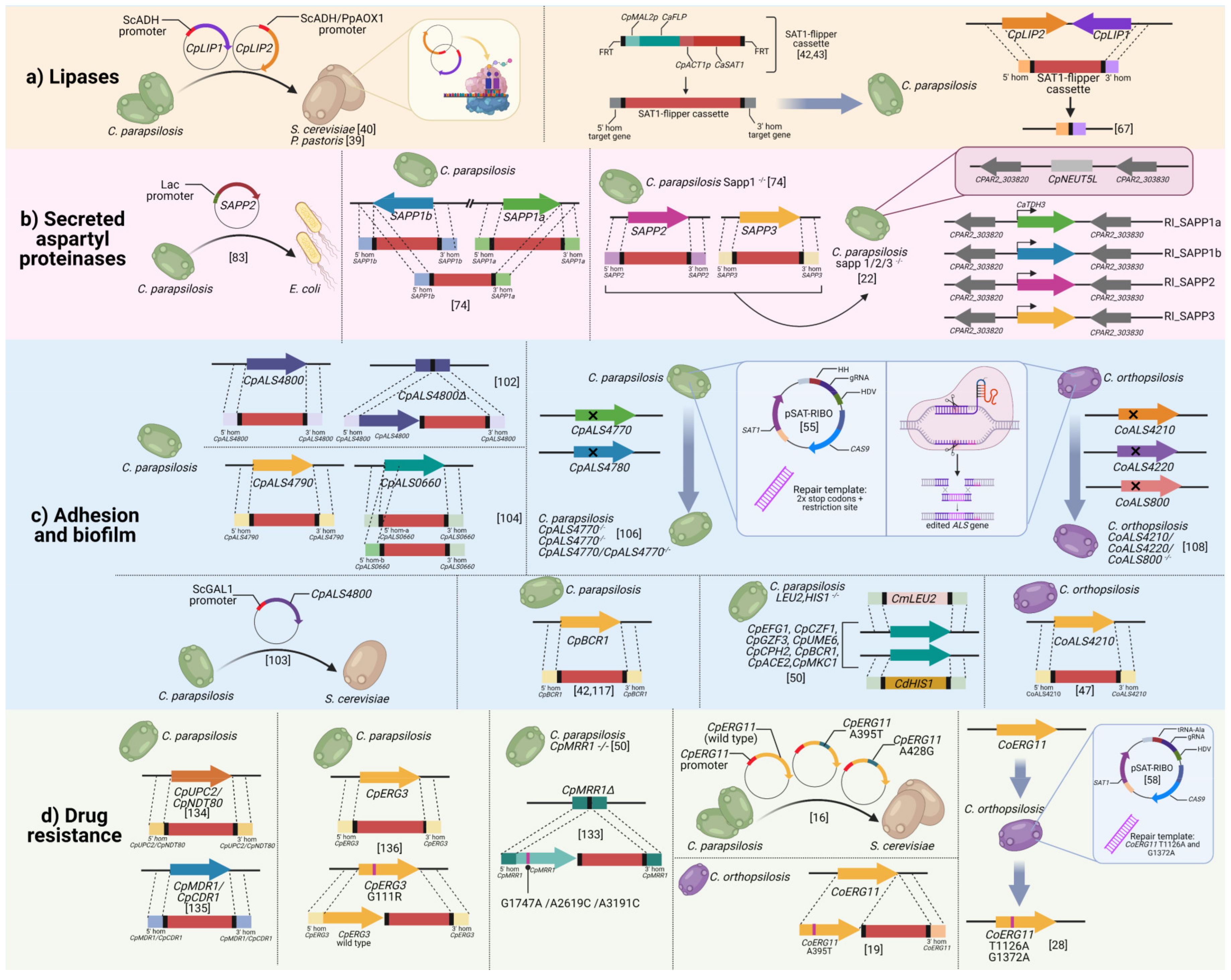
Table 1. Overview of the genetic manipulation studies dissecting C. parapsilosis species complex virulence factor and summarized outcome of the phenotypical characterization.
Virulence FactorSpeciesGenesGenetic Manipulation Technique UsedPhenotypical Characterization
LipasesC. parapsilosisCpLIP1-2Heterologous expression in S. cerevisiaeLipolytic activity detected only for CpLIP2 but not for CpLIP1 [40].
CpLIP2Heterologous expression in P. pastorisCpLIP2 lipolytic activity detected [39].
CpLIP1-2SAT1-flipper cassetteCpLip1/2−/− mutant strain exhibit abolished catalytic activity, reduced growth in lipid-rich media, impaired biofilm formation, more efficient killing by macrophages-like cells and monocyte derived dendritic cells, and reduced pathogenic potential in a murine intraperitoneal infection model [23,62,63].
Secreted aspartyl proteinasesC. parapsilosisSAPP2Heterologous expression in E. coliSAPP2 proteolytic activity was demonstrated through the hemoglobin cleavage test [64].
SAPP1a-bSAT-1 flipper cassetteSapp1a/1b −/− mutant strains showed Sapp2 overexpression under induced conditions, growth reduction in human serum, increased killing by PBMCs and PBMC-DM and increased phagolysosomal fusion in PBMC-DMs [65].
SAPP1a-b, SAPP2,
SAPP3
SAT-1 flipper cassetteA sapp1/2/3/− defective strain was generated as well as reintegrated mutants. All SAPP genes are involved in the adhesion to polystyrene surfaces; SAPP1 and SAPP2 are required for the adhesion on TR146 cells and host cell damage, phagocytosis, phagosome-lysosome maturation, killing, and cleavage of human complement proteins [22].
Adhesion and biofilmC. parapsilosisCpALS4800SAT1-flipper cassetteMarked reduction of the CpALS4800 null mutant strain in the adhesion to HBECs and in the pathogenic potential if tested in a murine model of urinary infection. Reintegration of CpALS4800 in the original locus restored the adhesive ability [66].
Heterologous expression in S. cerevisiaeCpALS4800 expression resulted in the increased adhesion of S. cerevisiae [67].
CpALS4790-
CpALS0660
SAT1-flipper cassetteCpALS4790 is required for the adhesion to HBECs. Deletion of either CpALS4790 or CpALS0660 resulted in a reduced pathogenic potential when tested in a murine model of vaginal candidiasis [68].
CpALS4770
CpALS4780
CRISPR/Cas9 systemCpALS4770 edited strain showed impaired ability to form biofilm on polystyrene surfaces and to adhere on HBECs. The contextual deletion of CpALS4770 and CpALS4780 resulted in an increased tendency of the double mutant strain to form cellular aggregates, adhere on HBECs, and form biofilm on plastic surfaces. Both single and double mutant strains showed a reduced ability to colonize and persist in the murine vaginal mucosa [69].
CpBCR1SAT1-flipperCpBCR1 is required for C. parapsilosis biofilm formation on silicone surfaces and for the expression of the cell wall protein CpRBT1 [42]. Clinical isolates prolific in biofilm production are not dependent on CpBcr1 transcription factor [70].
Transcription factors: CpEFG1, CpCZF1, CpGZF3, CpUME6, CpCPH2, CpBCR1, CpACE2,
Protein kinase: CpMKC1
Gene disruption cassette generated by fusion PCRGene disruption resulted in impaired biofilm formation in vitro and in vivo [50].
C. orthopsilosisCoALS4210SAT1-flipper cassette and CRISPR/Cas9 systemCoALS4210 knockout and CRISPR edited strains showed reduced adhesion to HBECs [47].
CoALS410
CoALS4120
CoALS800
CRISPR/Cas9 systemTriple edited strains lacking the entire ALS gene family showed dramatic reduction in the adhesion to HBECs [71].
Drug susceptibilityC. parapsilosisCpMRR1SAT1-flipper cassetteAcquisition of point mutations G1747A, A2619C leading to G583R, K873N amino acid substitutions, respectively were involved in the development of fluconazole and voriconazole resistance, in addition to CpMDR1 and CpMRR1 overexpression [72].
CpUPC2
CpNDT80
SAT1-flipper cassetteDeletion of overexpressed CpUPC2 and CpNDT80 alone or in combination in fluconazole, voriconazole, and posaconazole-resistant isolates led to the restoration of in vitro susceptibility. CpUPC2 deletion had a more significant effect [73].
CpMDR1
CpCDR1
SAT1-flipper cassetteDeletion of overexpressed CpMDR1 and CpCDR1 in azole-resistant isolates led to the partial restoration of in vitro susceptibility [74].
CpERG11Heterologous expression in S. cerevisiaeAcquisition of A395T and A428G point mutations leading to Y132F and K143F amino acid substitutions, respectively, were involved in the development of in vitro azole resistance in S. cerevisiae [16].
CpERG3SAT1-flipper cassetteCpERG3 knockout led to in vitro azole resistance and intermediate resistance to echinocandins.
CpErg3 G111R amino acid substitution was involved in in vitro azole and echinocandin resistance [75].
C. orthopsilosisCoERG11SAT1-flipper cassetteAcquisition of A395T leading to Y132F amino acid substitution, was involved in the development of in vitro azole resistance. Highest MIC values in homozygous mutant [19].
CRISPR/Cas9 systemG1372A point mutations leading G458S amino acid substitution was involved in in vitro fluconazole and voriconazole resistance. Highest MIC values in homozygous mutants. No effect in posaconazole resistance was observed [28]
Publisher’s Note: MDPI stays neutral with regard to jurisdictional claims in published maps and institutional affiliations.

