Case Report

Breakthrough Invasive Fungal Infections in Allogeneic Hematopoietic Stem Cell Transplantation

1
Hematology and Bone Marrow Transplant Unit, IRCCS San Raffaele Scientific Institute, Via Olgettina 60, 20132 Milan, Italy
2
Faculty of Medicine and Surgery, Vita-Salute San Raffaele University, IRCCS San Raffaele Scientific Institute, 20132 Milan, Italy
3
Clinic of Infectious Diseases, Division of Immunology, Transplantation and Infectious Diseases, IRCCS San Raffaele Scientific Institute, 20132 Milan, Italy
4
Pediatric Immunohematology and Bone Marrow Transplantation Unit, IRCCS San Raffaele Scientific Institute, 20132 Milan, Italy
5
San Raffaele Telethon Institute for Gene Therapy (SR-Tiget), IRCCS San Raffaele Scientific Institute, 20132 Milan, Italy
6
Neuroradiology Department, IRCCS San Raffaele Scientific Institute, 20132 Milan, Italy
7
Department of General and Emergency Surgery, IRCCS San Raffaele Scientific Institute, 20132 Milan, Italy
*
Author to whom correspondence should be addressed.
J. Fungi 2021, 7(5), 347; https://doi.org/10.3390/jof7050347
Submission received: 6 April 2021 / Revised: 24 April 2021 / Accepted: 27 April 2021 / Published: 28 April 2021
(This article belongs to the Special Issue Chemotherapy and Invasive Fungal Infection)

Abstract

:
Despite the recent introduction of mold-active antifungal prophylaxis (MAP), breakthrough invasive fungal infections (b-IFI) still represent a possible complication and a cause of morbidity and mortality in hematological patients and allogeneic hematopoietic stem-cell transplantation recipients (HSCT). Data on incidence and type of b-IFI are limited, although they are mainly caused by non-fumigatus Aspergillus and non-Aspergillus molds and seem to depend on specific antifungal prophylaxis and patients’ characteristics. Herein, we described the clinical presentation and management of two cases of rare b-IFI which recently occurred at our institution in patients undergoing HSCT and receiving MAP. The management of b-IFI is challenging due to the lack of data from prospective trials and high mortality rates. A thorough analysis of risk factors, ongoing antifungal prophylaxis, predisposing conditions and local epidemiology should drive the choice of antifungal treatments. Early broad-spectrum preemptive therapy with a lipid formulation of amphotericin-B, in combination with a different mold-active azole plus/minus terbinafine, is advisable. The therapy would cover against rare azole-susceptible and -resistant fungal strains, as well as atypical sites of infections. An aggressive diagnostic work-up is recommended for species identification and subsequent targeted therapy.

1. Introduction

In the era of mold-active antifungal prophylaxis (MAP), invasive fungal infections (IFI) still represent one of the major cause of morbidity and mortality in hematological patients and allogeneic hematopoietic stem-cell transplantation (HSCT) recipients. Following the decrease of invasive candidiasis due to fluconazole prophylaxis, an epidemiological shift occurred toward mold infections, mainly invasive aspergillosis (IA). Depending on risk factors, the 12-months IFI incidence approaches 8%, reaching 17% in haploidentical and cord blood transplantations [1,2]. Recently, ECIL-6 guidelines recommended primary MAP in high-risk HSCT recipients [3]. Nonetheless, MAP exposes the patient to development of breakthrough IFI (b-IFI), mainly by rare non-Aspergillus molds. To date, data on incidence and type of b-IFI are limited, although they seem to depend on specific prophylaxis and patients’ characteristics [4,5,6].
We described two cases of rare b-IFI which recently occurred at our institution in patients receiving MAP.

2. Case Descriptions, Methods and Results

Case 1: a 52-year-old woman diagnosed with acute myeloid leukemia (AML) was referred to our institution for HSCT. Four years before, she underwent HSCT from a matched unrelated donor (MUD), which was complicated by acute and chronic graft-versus-host-disease (GvHD). After a second haploidentical HSCT for disease reoccurrence, the FLT3-ITD mutated AML relapsed again. She received Gilteritinib for 6 months without MAP, obtaining complete remission with incomplete hematological recovery.
At admission, the patient was in good clinical condition. Baseline evaluations were unremarkable, except for persistent severe neutropenia and cellulitis in left leg. Gilteritinib was stopped and cellulitis treated with daptomycin. Then, she received a reduced-intensity conditioning regimen followed by haploidentical HSCT. GvHD prophylaxis relied upon post-transplantation cyclophosphamide, mycophenolate-mofetil and sirolimus. Posaconazole was given as MAP from day -7 by therapeutic drug monitoring (TDM). On day +2 piperacillin/tazobactam was given for febrile neutropenia. Blood cultures and chest X-Ray were negative. A week later, fever reappeared, along with productive cough. Piperacillin/tazobactam was switched to meropenem, while posaconazole was continued. On day +11 she developed headache and diplopia, then dysarthria, drowsiness and ocular nystagmus. A brain magnetic resonance imaging scan showed multiple rounded lesions, diagnostic for septic embolism (Figure 1). Soon after, the patient developed hemorrhagic skin lesions and multi-organ failure. As galactomannan (GM) monitoring in serum was negative and preliminary results of both sputum and blood culture suggested a yeast-like fungus, posaconazole was switched to liposomal amphotericin-B (L-AMB) (5 mg/kg/day) and anidulafungin. Two days later, definitive results showed Lomentospora prolificans (Table 1); however, the same day, she went into a coma and died.
Case 2: a 34-year-old man diagnosed with immune-mediated thrombocytopenia (ITP) and primary immunodeficiency was referred to our institution for HSCT. ITP was refractory to several treatments, including steroids, immunoglobulins, rituximab, danazol, azathioprine and romiplostim. Investigations of pre-existent lymphopenia suggested a primary immunodeficiency. Although next-generation sequencing did not identify any pathogenetic variants, HSCT was indicated based on clinical phenotype.
At admission, the patient was in good clinical condition. He received a myeloablative conditioning regimen followed by HSCT from MUD. GvHD prophylaxis relied upon ATG, cyclosporine-A and methotrexate. Voriconazole was given as MAP from day −7 and by TDM. Complications were febrile neutropenia and severe mucositis, treated with piperacillin/tazobactam, then meropenem and tigecycline for ESBL-producer Klebsiella pneumoniae blood-stream infection. Neutrophils engraftment was achieved on day +15. Two weeks later the patient presented acute peritonitis. Abdomen CT scan showed ischemic jejunal perforation and he underwent emergency open surgery with intestinal resection and anastomosis. Considering critical condition, MAP was modified in favor of intravenous isavuconazole to contain toxicities. GM monitoring in serum was negative. Histopathological examination of jejunum revealed tissue necrosis and angioinvasion by fungal hyphae consistent with mucormycosis. Further foci of IFI were excluded and a combinational therapy adding L-AMB 5 mg/kg/day to isavuconazole was preferred while waiting for microbiological results. Subsequently, the patient presented cutaneous-subcutaneous necrosis around the surgical site, requiring multiple debridements and vacuum-assisted closure therapy. Histopathological examination of soft tissue confirmed mucormycosis and culture of tissue biopsy detected Rhizopus microsporus (Table 1). Fortunately, the patient had a slow but continuous improvement. L-AMB was administered for 45 days while oral isavuconazole was continued for a further 8 months during immunosuppression therapy (IST). At last follow-up (18 months after HSCT), he persisted in good clinical condition.

3. Discussion

The management of b-IFI during MAP is challenging, as no standardized prospective data are available to guide therapeutic decisions. Consensus statements suggest aggressive diagnostic work-up, multidisciplinary management and early broad-spectrum preemptive therapy with L-AMB, with or without a different mold-active azole, thus covering against azole-susceptible and -resistant fungal strains [7,8].
In patients receiving posaconazole, prospective trials reported b-IFI up to 3.7%: mainly IA and a few cases of lomentosporiosis [9,10]. Retrospective studies also reported incidence of b-IFI between 0–10.9%: A. fumigatus was the most common, followed by non-fumigatus IA, mucormycosis, fusariosis and, rarely, candidiasis [4,6,7]. Lomentospora prolificans, previously termed as Scedosporium prolificans, is a rare opportunist mainly affecting immunocompromised and hematological patients [11]. Lomentosporiosis accounts for 25% of all non-IA in solid organ and HSCT recipients [12]. Clinical manifestations range from localized to severe invasive mycoses. Risk factors for dissemination include: underlying disease (mainly acute leukemia), type of transplantation/IST, duration and degree of neutropenia, antifungal prophylaxis and environmental exposure [11,13]. Disseminated disease occurs more often in HSCT recipients, mainly in the pre-engraftment period, and frequently presents as fungemia, with a high percentage of isolation from blood cultures. It is characterized by mortality rates >80% due to intrinsically pan-antifungal resistance [11,13,14]. Results of our susceptibility test were consistent with those reported in literature. In a recent review of 41 cases of lomentosporiosis, combinational therapy with voriconazole plus terbinafine resulted in increased survival [15]. The ECCM-ASM guidelines actually recommend a first-line voriconazole-based combinational therapy, particularly with terbinafine, alongside surgical debridement and recovery of immunosuppression [8,16]. The novel compounds E1210/APX001 and F901318 showed preliminary in vitro activity, and in vivo clinical trials are ongoing [17,18]. In case 1, clinical presentation was suggestive for b-IFI and the patient had multiple risk factors, e.g., advanced stage heavily pretreated AML, long-lasting severe neutropenia, previous HSCT and IST for acute and chronic GvHD. Before the confirmation of lomentosporiosis, considering epidemiology, posaconazole prophylaxis, GM negativity and initial suggestion for yeast-like fungi, we chose a combinational therapy with L-AMB and anidulafungin. Although often detectable in aspergillosis, GM is typically negative in cases of mucormycosis, lomentosporiosis and scedosporiosis, because it does not constitute a component of the cell wall of these fungi. The role of GM monitoring in the diagnostic work-up of patients receiving MAP is controversial, due to lower sensitivity and specificity [19]. However, these findings were mainly confirmed by studies with Posaconazole as MAP, rather than all studies [20]. Therefore, GM monitoring is still routinely performed in many centers when a b-IFI is clinically suspected in the setting of high-risk hematological patients [7,21]. Unfortunately, in our experience, when definitive microbiological results became available, the rapid clinical worsening did not allow administration of an adequate antifungal therapy.
In case 2, suspicion of b-IFI was initially less consistent, as clinical scenarios could lead to other pathogens and noninfectious complications. Nonetheless, the patient had multiple risk factors for b-IFI, including prolonged steroid therapy, primary immunodeficiency, myeloablative conditioning, ATG administration and neutropenia post-HSCT. Moreover, voriconazole prophylaxis—as well as GM negativity—favored diagnosis of non-IA. Actually, b-IFI following voriconazole prophylaxis has been reported in up to 7.9% of cases, primarily mucormycosis, due to the lack of in vitro activity against Mucorales [4]. Mucormycosis, including Rhizopus microsporus, mainly affects immunocompromised hosts and, after IA, represents the most frequent IFI in hematological patients and HSCT recipients [22,23,24,25]. Mucormycoses are characterized by rapid local spread, angioinvasion and tissue necrosis. GM tests are typically negative. Most common presentations are rhino-orbito-cerebral, cutaneous, pulmonary and disseminated, the latter two being more frequent among neutropenic patients with acute leukemia and HSCT recipients. The latter two are also associated with high mortality rates (20–100%) [22,23,25,26]. The ESCMID-ECCM and ECIL-6 guidelines recommend surgery and first-line treatment with L-AMB at least 5 mg/kg/day. Posaconazole is an alternative in first-line and salvage settings, as well as combinational therapy of L-AMB with posaconazole or echinocandines [27,28]. A recent single-center trial testing first-line isavuconazole in mucormycoses reported similar outcomes compared to L-AMB [29]. The most recent ECMM guidelines still recommend first-line L-AMB, with intravenous isavuconazole or posaconazole in patients with renal impairment [30]. In case 2, as the pathologist suspected mucormycosis and considering MAP with voriconazole, disseminated infection, patient’s risk factors and unavailability of intravenous posaconazole, a combinational therapy with L-AMB and intravenous isavuconazole was chosen. In our experience, surgical debridement and combinational therapy proved effective. Clinical efficacy and susceptibility tests favored isavuconazole as a continuing oral treatment during IST.
The limitations of this case study include the retrospective nature, selected population and lack of generalizability.

4. Conclusions

This report described a real-life and patient-specific approach to diagnosis and management of b-IFI, providing data on species identification and susceptibility testing. b-IFI should still be considered a possible complication in HSCT recipients receiving MAP. Due to high mortality rates, early broad-spectrum preemptive therapy is advisable against even rare species and atypical sites of infections. A thorough analysis of risk factors and predisposing conditions should drive the choice of individualized antifungal treatments. Efforts are strongly recommended for species identification and susceptibility testing in order to establish a targeted therapy. As new transplantation strategies are implemented and the use of MAP is broadened, prospective studies are needed to better understand the epidemiology, clinical manifestations and outcomes of b-IFI.

Author Contributions

All co-authors (C.L., F.F., R.G., F.G., D.C., C.O., M.T.L.S., F.B., P.V., E.O., C.C., F.C., J.P.) contributed to patients’ clinical care. C.L. wrote the manuscript. All authors have read and agreed to the published version of the manuscript.

Funding

This research received no external funding.

Institutional Review Board Statement

Not applicable.

Informed Consent Statement

Patients were treated according to current institutional programs upon written informed consent for transplantation procedures, biological sampling and use of medical records for research.

Data Availability Statement

Not applicable.

Conflicts of Interest

The authors declare no conflict of interest.

References

  1. Kontoyiannis, D.P.; Marr, K.A.; Park, B.J.; Alexander, B.D.; Anaissie, E.J.; Walsh, T.J.; Ito, J.; Andes, D.R.; Baddley, J.W.; Brown, J.M.; et al. Prospective Surveillance for Invasive Fungal Infections in Hematopoietic Stem Cell Transplant Recipients, 2001–2006: Overview of the Transplant-Associated Infection Surveillance Network (TRANSNET) Database. Clin. Infect. Dis. 2010, 50, 1091–1100. [Google Scholar] [CrossRef] [PubMed]
  2. Girmenia, C.; Raiola, A.M.; Piciocchi, A.; Algarotti, A.; Stanzani, M.; Cudillo, L.; Pecoraro, C.; Guidi, S.; Iori, A.P.; Montante, B.; et al. Incidence and outcome of invasive fungal diseases after allogeneic stem cell transplantation: A prospective study of the gruppo italiano trapianto midollo osseo (GITMO). Biol. Blood Marrow Transplant. 2014, 20, 872–880. [Google Scholar] [CrossRef] [PubMed]
  3. Maertens, J.A.; Girmenia, C.; Brüggemann, R.J.; Duarte, R.F.; Kibbler, C.C.; Ljungman, P.; Racil, Z.; Ribaud, P.; Slavin, M.A.; Cornely, O.A.; et al. European guidelines for primary antifungal prophylaxis in adult haematology patients: Summary of the updated recommendations from the European Conference on Infections in Leukaemia. J. Antimicrob. Chemother. 2018, 73, 3221–3230. [Google Scholar] [CrossRef] [PubMed]
  4. Lionakis, M.S.; Lewis, R.E.; Kontoyiannis, D.P. Breakthrough invasive mold infections in the hematology patient: Current concepts and future directions. Clin. Infect. Dis. 2018, 67, 1621–1630. [Google Scholar] [CrossRef] [PubMed]
  5. Stern, A.; Su, Y.; Lee, Y.J.; Seo, S.; Shaffer, B.; Tamari, R.; Gyurkocza, B.; Barker, J.; Bogler, Y.; Giralt, S.; et al. A Single Center, Open-Label Trial of Isavuconazole Prophylaxis against Invasive Fungal Infection in Patients Undergoing Allogeneic Hematopoietic Cell Transplant (HCT). Biol. Blood Marrow Transplant. 2020, 26, 1195–1202. [Google Scholar] [CrossRef] [PubMed]
  6. Wasylyshyn, A.; Linder, K.A.; Castillo, C.G.; Zhou, S.; Kauffman, C.A.; Miceli, M.H. Breakthrough Invasive Fungal Infections in Patients with Acute Myeloid Leukemia. Mycopathologia 2020, 185, 299–306. [Google Scholar] [CrossRef] [PubMed]
  7. Girmenia, C.; Busca, A.; Candoni, A.; Cesaro, S.; Luppi, M.; Nosari, A.M.; Pagano, L.; Rossi, G.; Venditti, A.; Aversa, F. Breakthrough invasive fungal diseases in acute myeloid leukemia patients receiving mould active triazole primary prophylaxis after intensive chemotherapy: An Italian consensus agreement on definitions and management. Med. Mycol. 2019, 57 (Suppl. 2), S127–S137. [Google Scholar] [CrossRef]
  8. Hoenigl, M.; Salmanton-García, J.; Walsh, T.J.; Nucci, M.; Neoh, C.F.; Jenks, J.D.; Lackner, M.; Sprute, R.; Al-Hatmi, A.M.S.; Bassetti, M.; et al. Global guideline for the diagnosis and management of rare mould infections: An initiative of the European Confederation of Medical Mycology in cooperation with the International Society for Human and Animal Mycology and the American Society for Microbiology. Lancet Infect. Dis. 2021, in press. [Google Scholar] [CrossRef]
  9. Cornely, O.A.; Maertens, J.; Winston, D.J.; Perfect, J.; Ullmann, A.J.; Walsh, T.J.; Helfgott, D.; Holowiecki, J.; Stockelberg, D.; Goh, Y.T.; et al. Posaconazole vs. fluconazole or itraconazole prophylaxis in patients with neutropenia. N. Engl. J. Med. 2007, 356, 348–359. [Google Scholar] [CrossRef] [Green Version]
  10. Ullmann, A.J.; Lipton, J.H.; Vesole, D.H.; Chandrasekar, P.; Langston, A.; Tarantolo, S.R.; Greinix, H.; De Azevedo, W.M.; Reddy, V.; Boparai, N.; et al. Posaconazole or fluconazole for prophylaxis in severe graft-versus-host disease. N. Engl. J. Med. 2007, 356, 335–347. [Google Scholar] [CrossRef]
  11. Ramirez-Garcia, A.; Pellon, A.; Rementeria, A.; Buldain, I.; Barreto-Bergter, E.; Rollin-Pinheiro, R.; De Meirelles, J.V.; Xisto, M.I.D.S.; Ranque, S.; Havlicek, V.; et al. Scedosporium and Lomentospora: An updated overview of underrated opportunists. Med. Mycol. 2018, 56 (Suppl. 1), 102–125. [Google Scholar] [CrossRef]
  12. Husain, S.; Muñoz, P.; Forrest, G.; Alexander, B.D.; Somani, J.; Brennan, K.; Wagener, M.M.; Singh, N. Infections due to Scedosporium apiospermum and Scedosporium prolificans in transplant recipients: Clinical characteristics and impact of antifungal agent therapy on outcome. Clin. Infect. Dis. 2005, 40, 89–99. [Google Scholar] [CrossRef] [Green Version]
  13. Caira, M.; Girmenia, C.; Valentini, C.G.; Sanguinetti, M.; Bonini, A.; Rossi, G.; Fianchi, L.; Leone, G.; Pagano, L. Scedosporiosis in patients with acute leukemia: A retrospective multicenter report. Haematologica 2008, 93, 104–110. [Google Scholar] [CrossRef]
  14. Seidel, D.; Meißner, A.; Lackner, M.; Piepenbrock, E.; Salmanton-García, J.; Stecher, M.; Mellinghoff, S.; Hamprecht, A.; Durán Graeff, L.; Köhler, P.; et al. Prognostic factors in 264 adults with invasive Scedosporium spp. and Lomentospora prolificans infection reported in the literature and FungiScope®. Crit. Rev. Microbiol. 2019, 45, 1–21. [Google Scholar] [CrossRef] [Green Version]
  15. Jenks, J.D.; Seidel, D.; Cornely, O.A.; Chen, S.; Van Hal, S.; Kauffman, C.; Miceli, M.H.; Heinemann, M.; Christner, M.; Sáenz, A.J.; et al. Voriconazole plus terbinafine combination antifungal therapy for invasive Lomentospora prolificans infections: Analysis of 41 patients from the FungiScope® registry 2008–2019. Clin. Microbiol. Infect. 2020, 26, 784.e1–784.e5. [Google Scholar] [CrossRef]
  16. Tortorano, A.M.; Richardson, M.; Roilides, E.; Van Diepeningen, A.; Caira, M.; Munoz, P.; Johnson, E.; Meletiadis, J.; Pana, Z.D.; Lackner, M.; et al. ESCMID and ECMM joint guidelines on diagnosis and management of hyalohyphomycosis: Fusarium spp., Scedosporium spp. and others. Clin. Microbiol. Infect. 2014, 20 (Suppl. 3), 27–46. [Google Scholar] [CrossRef] [Green Version]
  17. An Open-label Study of APX001 for Treatment of Patients with Invasive Mold Infections Caused by Aspergillus Species or Rare Molds. ClinicalTrials.gov Identifier: NCT04240886. Available online: https://clinicaltrials.gov/ct2/show/NCT04240886 (accessed on 16 March 2021).
  18. Evaluate F901318 Treatment of Invasive Fungal Infections in Patients Lacking Treatment Options. ClinicalTrials.gov Identifier: NCT03583164. Available online: https://clinicaltrials.gov/ct2/show/NCT03583164 (accessed on 16 March 2021).
  19. Marr, K.A.; Laverdiere, M.; Gugel, A.; Leisenring, W. Antifungal therapy decreases sensitivity of the Aspergillus galactomannan enzyme immunoassay. Clin. Infect. Dis. 2005, 40, 1762–1769. [Google Scholar] [CrossRef]
  20. Calmettes, C.; Gabriel, F.; Blanchard, E.; Servant, V.; Bouchet, S.; Kabore, N.; Forcade, E.; Leroyer, C.; Bidet, A.; Latrabe, V.; et al. Breakthrough invasive aspergillosis and diagnostic accuracy of serum galactomannan enzyme immune assay during acute myeloid leukemia induction chemotherapy with posaconazole prophylaxis. Oncotarget 2018, 9, 26724–26736. [Google Scholar] [CrossRef]
  21. Duarte, R.F.; Sánchez-Ortega, I.; Cuesta, I.; Arnan, M.; Patiño, B.; De Sevilla, A.F.; Gudiol, C.; Ayats, J.; Cuenca-Estrella, M. Serum galactomannan-based early detection of invasive aspergillosis in hematology patients receiving effective antimold prophylaxis. Clin. Infect. Dis. 2014, 59, 1696–1702. [Google Scholar] [CrossRef] [Green Version]
  22. Skiada, A.; Lass-Floerl, C.; Klimko, N.; Ibrahim, A.; Roilides, E.; Petrikkos, G. Challenges in the diagnosis and treatment of mucormycosis. Med. Mycol. 2018, 56 (Suppl. 1), 93–101. [Google Scholar] [CrossRef] [Green Version]
  23. Farmakiotis, D.; Kontoyiannis, D.P. Mucormycoses. Infect. Dis. Clin. N. Am. 2016, 30, 143–163. [Google Scholar] [CrossRef]
  24. Pagano, L.; Girmenia, C.; Mele, L.; Ricci, P.; Tosti, M.E.; Nosari, A.; Buelli, M.; Picardi, M.; Allione, B.; Corvatta, L.; et al. Infections caused by filamentous fungi in patients with hematologic malignancies. A report of 391 cases by GIMEMA infection program. Haematologica 2001, 86, 862–870. [Google Scholar]
  25. Miller, M.A.; Molina, K.C.; Gutman, J.A.; Scherger, S.; Lum, J.M.; Mossad, S.B.; Burgess, M.; Cheng, M.P.; Chuang, S.T.; Jacobs, S.E.; et al. Mucormycosis in Hematopoietic Cell Transplant Recipients and in Patients With Hematological Malignancies in the Era of New Antifungal Agents. Open Forum Infect. Dis. 2021, 8, ofaa646. [Google Scholar] [CrossRef]
  26. Skiada, A.; Pagano, L.; Groll, A.; Zimmerli, S.; Dupont, B.; Lagrou, K.; Lass-Florl, C.; Bouza, E.; Klimko, N.; Gaustad, P.; et al. Zygomycosis in Europe: Analysis of 230 cases accrued by the registry of the European Confederation of Medical Mycology (ECMM) Working Group on Zygomycosis between 2005 and 2007. Clin. Microbiol. Infect. 2011, 17, 1859–1867. [Google Scholar] [CrossRef] [Green Version]
  27. Cornely, O.; Arikan-Akdagli, S.E.V.T.A.P.; Dannaoui, E.; Groll, A.H.; Lagrou, K.; Chakrabarti, A.; Lanternier, F.; Pagano, L.I.V.I.O.; Skiada, A.; Akova, M.; et al. ESCMID and ECMM joint clinical guidelines for the diagnosis and management of mucormycosis 2013. Clin. Microbiol. Infect. 2014, 20 (Suppl. 3), 5–26. [Google Scholar] [CrossRef] [Green Version]
  28. Tissot, F.; Agrawal, S.; Pagano, L.; Petrikkos, G.; Groll, A.H.; Skiada, A.; Lass-Flörl, C.; Calandra, T.; Viscoli, C.; Herbrecht, R. ECIL-6 guidelines for the treatment of invasive candidiasis, aspergillosis and mucormycosis in leukemia and hematopoietic stem cell transplant patients. Haematologica 2017, 102, 433–444. [Google Scholar] [CrossRef] [Green Version]
  29. Marty, F.M.; Ostrosky-Zeichner, L.; Cornely, O.A.; Mullane, K.M.; Perfect, J.R.; Thompson, G.R.; Alangaden, G.J.; Brown, J.M.; Fredricks, D.N.; Heinz, W.J.; et al. Isavuconazole treatment for mucormycosis: A single-arm open-label trial and case-control analysis. Lancet Infect. Dis. 2016, 16, 828–837. [Google Scholar] [CrossRef]
  30. Cornely, O.A.; Alastruey-Izquierdo, A.; Arenz, D.; Chen, S.C.A.; Dannaoui, E.; Hochhegger, B.; Hoenigl, M.; Jensen, H.E.; Lagrou, K.; Lewis, R.E.; et al. Global guideline for the diagnosis and management of mucormycosis: An initiative of the European Confederation of Medical Mycology in cooperation with the Mycoses Study Group Education and Research Consortium. Lancet Infect. Dis. 2019, 19, e405–e421. [Google Scholar] [CrossRef]
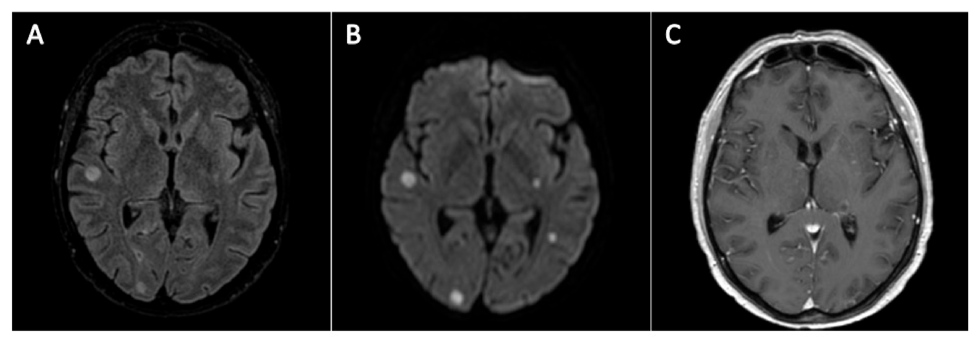
Figure 1. Magnetic resonance imaging of the brain for case 1. Findings were consistent with septic emboli. Pictures (A) and (B) show multifocal superficial and deep bilateral rounded lesions, some of which show nodular or ring-like enhancement after contrast injection (left thalamus in picture (C)). Lesions were localized at both sub- and sovra-tentorial level, with a miliary distribution, the largest ones involving the cerebellum and the brainstem with a maximum diameter of about 10 mm. In addition, phlogistic mucosal thickening of paranasal cavities was documented (not shown). (A): FLAIR sequence; (B): DWI sequence; (C): T1-post Gadolinium sequence.
Jof 07 00347 g001
Table 1. In vitro antifungal susceptibility tests.
Clinical Case 1Clinical Case 2
SpeciesLomentospora prolificansRhizopus microsporus
SamplePeripheral bloodSoft tissue biopsy
DrugsMICMIC
L-AMB>32 mg/L0.5 mg/L
Itraconazole>32 mg/L1.5 mg/L
Voriconazole>32 mg/L-
Posaconazole>32 mg/L0.19 mg/L
Isavuconazole>32 mg/L0.125 mg/L
Table 1 shows in vitro antifungal susceptibility tests of fungal strains isolated from patients (cases 1 and 2). Minimal inhibitory concentration was determined by E-test. Lacking validated MIC breakpoints for any of the drugs against Lomentospora and Mucor fungal genera, interpretation and determination of susceptibility categories was not possible. L-AMB: liposomal Amphotericin B.
Publisher’s Note: MDPI stays neutral with regard to jurisdictional claims in published maps and institutional affiliations.