Case Report

Fungal Invasive Co-Infection Due to Aspergillus fumigatus and Rhizopus arrhizus: A Rhino-Orbital Presentation

by
Juan Pablo Ramírez-Hinojosa
1,
Salvador Medrano-Ahumada
1,
Roberto Arenas
2,
Arturo Bravo-Escobar
3,
Sara Paraguirre-Martínez
4,
Juan Xicohtencatl-Cortes
5,
Erick Martínez-Herrera
6,7,* and
Rigoberto Hernández-Castro
8,*
1
Departamento de Infectología, Hospital General Dr. Manuel Gea González, Tlalpan 14080, Mexico
2
Servicio de Micología, Hospital General Dr. Manuel Gea González, Tlalpan 14080, Mexico
3
Servicio de Otorrinolaringología, Hospital General Dr. Manuel Gea González, Tlalpan 14080, Mexico
4
Departamento de Patología, Hospital General Dr. Manuel Gea González, Tlalpan 14080, Mexico
5
Laboratorio de Bacteriología Intestinal, Hospital Infantil de México Dr. Federico Gómez, Cuauhtémoc 06720, Mexico
6
Research Unit, Regional Hospital of High Specialty of Ixtapaluca, Ixtapaluca 56530, Mexico
7
Postgraduate Studies and Research Section, Higher School of Medicine, National Polytechnic Institute, Mexico City 11340, Mexico
8
Departamento de Ecología de Agentes Patógenos, Hospital General Dr. Manuel Gea González, Tlalpan 14080, Mexico
*
Authors to whom correspondence should be addressed.
J. Fungi 2021, 7(12), 1096; https://doi.org/10.3390/jof7121096
Submission received: 24 October 2021 / Revised: 10 December 2021 / Accepted: 11 December 2021 / Published: 20 December 2021
(This article belongs to the Special Issue Novel, Emerging and Neglected Fungal Pathogens for Humans and Animals)

Abstract

:
Aspergillosis and mucormycosis are filamentous fungal infections occurring predominantly in immunocompromised patients. Fulminant process with rapid infiltration of the contiguous tissue is distinctive for both type of fungi. The rhinocerebral co-infection by Aspergillus and Mucorales is very rare and is usually associated in immunocompromised patients with a high mortality rate. This rare co-infection leads to difficulties in diagnosis, and therapeutic delays can result in a poor prognosis. Overall, the treatment of choice is surgical debridement and liposomal amphotericin B. This paper describes a combined aspergillosis and mucormycosis case in a diabetes mellitus type 2 patient with chronic ulcerations of the palatal and cheek. To our knowledge, this is the first report of an uncommon co-infection of Aspergillus fumigatus and Rhizopus arrhizus in a rhino-orbital presentation.

1. Introduction

Mucormycosis and invasive aspergillosis infections are aggressive fungal infections with a high mortality rate. These infections have generally predilection among patients with risk factors as immunosuppression, untreated diabetes mellitus, kidney diseases, hematological malignancies or mayor trauma [1,2,3]. Aspergillosis is the clinical syndrome caused by diverse species of Aspergillus genus; A. fumigatus is the most important pathogenic fungus in humans; however, other species had been associated in different pathologies. Some clinical presentations are often similar among A. fumigatus related species, and are frequently misidentified [4,5].
Following aspergillosis, mucormycosis is the second most frequently mycosis caused by filamentous fungi. Agents of mucormycosis belong to the order Mucorales. Rhizopus, Mucor and Lichtheimia are the main genera implicated as cause of mucormycosis [6]. The most common clinical form is the rhinocerebral mucormycosis, and Rhizopus is the etiological agent of 70% of the reported cases [2,7].
Reports of combined mucormycosis and aspergillosis infections limited to the oral-rhinocerebral region are exceptional. Diagnosis is often based on histopatological examination and supported by fungal isolation. The peculiarity of this infection leads to difficulties in diagnosis and delays can result in a poor prognosis [8,9]. This paper describes the clinical features of combined aspergillosis and mucormycosis case in a diabetes mellitus type 2 patient with a chronic intra-oral ulceration of the palatal and cheek.

2. Case

A 52-year-old male patient with a history of diabetes mellitus type 2 with apparently regular control was presented to the emergency room in January of 2016 with a 2-month history of swelling of the left side of the face, left facial palsy, ptosis, and an impaired vision. At clinical examination, there was prominent tumefaction of the left hemi-face, involving the peri-orbital, zygomatic and maxillary areas, naso-sinusal congestion and diffuse ulcerative and necrotic lesion of the palatal and cheek mucosa (Figure 1A,B). The peripheral blood tests revealed mild neutropenia (WBC 14,800/mL, with 87% neutrophils, and glucose 180 mg/dL, glycosylated haemoglobin 6.7%), no other relevant abnormalities were observed. During examination, a KOH smear from the necrotic ulcer was performed, which revealed hyaline branched coenocytic hyphae. Amphotericin B deoxycolate was initiated (1 mg/kg per day), and rhino-orbital surgical debridement was performed, as well as an orbital exenteration with partial maxillectomy. The pathology of the palate, maxillary sinus and ethmoid bone samples were consistent with mucormycosis, but some histological specimens proved an invasive growth with septate hyphae (Figure 2A,B). Based on the clinical findings and the morphological features of the fungal organisms, a presumptive diagnosis of mucormycosis and concomitant aspergillosis was formulated. Samples for mycological culture (nasal and maxillar-sinus) were cut into very thin pieces and homogenized before being cultivated on Sabouraud agar (SDA) for 10 days at room temperature. Nevertheless, fungal isolation was repeatedly negative. We decided to add caspofungin as a salvage therapy; the combined (amphotericin B + caspofungin) therapy was given for five days. Finally, the patient died after six weeks of treatment.
This study was approved by the Institutional Review Board of Hospital General Dr. Manuel Gea Gonzalez, Mexico City, Mexico (24 August2018; IRB No. 06-77-2012-16).
Since the mycological culture was negative, it was decided to carry out the molecular identification in order to identify the causal agents associated with the patient. In this way, two paraffin-embedded tissue samples were analyzed, one sample from naso-sinus area and the second sample from maxillar sinus. The genomic DNA was isolated using a DNeasy blood and tissue kit (Qiagen, Ventura, CA, USA) according to the manufacturer’s instructions after preliminary removal of paraffin by extraction with xylene protocol. Molecular identification was performed by ITS rDNA region and beta-tubulin gene amplification and sequencing [10,11,12]. For ITS rDNA region amplification, a set of primers previously reported to identify fungi species (5′-TCCGTAGGTGAACCTTGCGG-3′) and ITS4 (5′-TCCTCCGCTTATTGATATGC-3′) were used [9,10]. The amplification program includes an initial denaturalization at 94 °C for 5 min; 33 cycles at 94 °C for 30 s, 55 °C for 35 s, 72 °C for 45 s, and a final extension for 5 min at 72 °C, with a no-template negative control. The reactions were performed in 50 µL reaction volume containing 25 µL of PCR master Mix (Qiagen, Ventura, CA, USA), 200 ng of DNA (2µL), 1 μL of each forward and reverse primer (25 pmol), and 21 μL nuclease-free water.
For beta-tubulin gene amplification primers (5′-AATTGGTGCCGCTTTCTGG-3′) and (5′-AGTTGTCGGGACGGAATAG-3′) were used [12]. PCR reaction was performed in a final volume of 50 μL, containing of 2 μL of DNA (200 ng), 25 μL of PCR master mix, 1 μL of 50 pmol of forward and reverse primers, and 21 μL of nuclease-free water. The amplification program includes an initial denaturation at 94 °C for 5 min, followed by 33 cycles of denaturation at 94 °C for 30 s, annealing at 59.5 °C for 40 s, and extension at 72 °C for 45 s with a final extension at 72 °C for 10 min. The PCR products were separated on 1% agarose gels stained with ethidium bromide.
For ITS rDNA region amplification, a 517 bp product was amplified from naso-sinusal sample, and 620 bp product from maxillar sample. The PCR products were purified and the nucleotide sequence was determined in both directions using the same primers with Taq FS dye terminator cycle sequencing fluorescence-based sequencing and analyzed on an applied biosystems 3730 xl DNA sequencing system. The consensus sequences homology search was performed with ex-type ITS rRNA fungi databases (https://blast.ncbi.nlm.nih.gov/Blast.cgi, accessed on 9 December 2021). using BLASTn analysis and displayed 98.65% of homology with A. fumigatus ATCC 1022, A. oerlinghausenensis CBS139183 (98.85%), A. fumisynnematus IFM42277 (99.80%), A. lentulus NRRL 35552 (99.81%), A. novofumigatus CBS 117250 (99.61%), A. fumigatiaffinis CBS 117186 (99.42%), A. fischeri NRLL 181 (99.23%), A. laciniosus KACC 41657 (99.23%), A. spinosus NRRL 5034 (99.23%), A. takakii IFM 53599 (99.23%), Aspergillus clavatus ATCC 1007 95.42% and, A. cervinus NRRL 5025 (93.15%), among others species from naso-sinusal sample and 100% of homology with Rhizopus arrhizus (syn. R. oryzae) HP25 and 8–3 M strains, among others from maxillar samples [13].
The ITS rDNA region does not allow species-level discrimination for the genus Aspergillus, so molecular identification was performed using the beta-tubulin gene (benA). A 485 bp fragment from naso-sinusal and maxillar sample was amplified and the nucleotide sequence was determined in both directions using the same primers. The sequences homology search was performed with ex-type strains sequences under section Aspergillus subgen. Fumigati using BLASTn analysis The sequence showed a 98.74% of homology with A. fumigatus ATCC 1022, A. oerlinghausenensis CBS139183 (94.20%), A. fischeri NRLL 181 (93.08%), A. spinosus NRRL 5034 (91.84%), A. laciniosus KACC 41657 (91.18%), A. takakii IFM 53599 (91.08%), A. lentulus NRRL 35552 (90.59%), A. fumigatiaffinis CBS 117186 (90.38%), A. novofumigatus CBS 117250 (89.98%), A. fumisynnematus IFM42277 (89.25%), Aspergillus clavatus ATCC 1007 (78.10%) and, A. cervinus NRRL 5025 (77.57%) [13]. The sequences obtained from ITS rDNA region were assigned Rhizopus arrhizus (syn. R. oryzae) GEA-44, Aspergillus fumigatus GEA-44, and Aspergillus fumigatus GEA-1 for benA gene, sequences were deposited in the GenBank under accession number MG029627, MT340910 and MT370439, respectively.

3. Discussion

Aspergillosis and mucormycosis are filamentous fungal infections occurring predominantly in immunocompromised patients, and both types of infection are characterized for being fulminant processes with rapid infiltration of contiguous tissues. The most common cause of invasive aspergillosis is A. fumigatus, followed by A. terreus, A. niger, A. flavus, and other species belonging to Fumigati section [12]. A. lentulus has been described recently as a new species of Aspergillus genus; genetic analysis indicated its close relation with A. fumigatus, and it has also been reported as a cause of invasive aspergillosis [5,12]. The correct identification of Aspergillus is a challenge for both clinical microbiology and mycology laboratories, which complicates epidemiological investigations leading often to its misidentification as an atypical A. fumigatus.
The classical fungi diagnosis involves a rapid microscopic identification using a KOH/chlorazol black preparation. The mycological culture is frequently reported as negative in half of the cases of mucormycosis. One of the main causes is the homogenization of the tissue since it can generate the loss of viability of the non-separated fragile hyphae of these fungi. In our clinical case, we failed at least three times in the mycological culture and one of the reasons was that we homogenized the tissue prior to culture. Therefore, it is recommended to slice and cut the tissue into very small and thin pieces before inoculation in the culture medium. Another problem associated with negative results is the use of a single culture medium (Sabouraud agar), so it is recommended to use other culture methods such as potato dextrose agar, Malt extract agar and Brain–heart–infusion (BHI) broth. The use of BHI broth provides optimal contact between the medium and the sample. In addition, growing at two temperatures (30 °C and 37 °C) can also increase yield of culture. Culture requires at least three days, and it needs mycology expertise for fungal description [14,15].
Histopathological examination displays characteristic fungal structures, such as large non-septate hyphae for Mucorales and septated branching hyphaes for Aspergillus; however, accurate and definitive identification is achieved by molecular methods [10,11,16]. Currently, one of the most used for fungi identification is the amplification and sequencing of ITS rRNA region, which allows the identification of a wide range of fungi. However, when this genetic marker cannot differentiate the species involved, it is necessary to use other genes such as beta-tubulin or Cadmoduline in the genus Aspergillus. For the molecular identification of Aspergillus fumigatus, experts recommend comparative sequence analysis of the ribosomal ITS region, specifically the ITS1 and ITS2 flanking regions of the 5.8S rDNA for the identification of the Aspergillus Fumigati section (intersection) and of the beta-tubulin or calmodulin genes for the identification of species level (intrasection). In this study, analysis of ITS rRNA region combined with amplification of benA gene, allowed the identification of the two etiologic fungal agents [5,12]. The correct identification of the causative agent is crucial to select the best systemic antifungal treatment which complements an aggressive surgical debridement of affected area. For both kinds of fungi, a fulminant process involving rapid infiltration of the neighboring tissues like the orbit and the anterior skull base is typical. A computed tomography (CT) scan is decisive to identify bone destruction, blood vessel compromise and spreading to adjacent structures (Figure 1C). MRI provides early detection of meningeal or intraparenchymatous spread as well as intracranial vascular occlusion.
Both mucormycosis and aspergillosis are usually severe infections, with mortality rates ranging from 20% to 40% in immune-compromised patients and approaching to 80–90% in those cases of disseminated infections or involvement of the brain structures [2,7,8]. Appropriate treatment must be instituted early, even before the results of culture and/or histopathological studies have been obtained. Currently, surgical debridement of the lesions as well as systemic administration of amphotericin B are considered the standard therapy; posaconazole and isavuconazole are promising alternatives.
Table 1 shows the reported cases of combined aspergillosis and mucormycosis. The rhinocerebral form was the most frequent clinical presentation, but oro-sinonasal, orofacial, rhino-orbital as well as sinus and brain abscess were also reported. The most common underlying disease were diabetes mellitus, Castleman disease, acute myeloid leukemia, cerebral trunk glioma and renal transplantation. Most cases show amphotericin B as treatment of choice; however, voriconazole, posaconazole, itraconazole, fluconazole and micafungin have also been used. The etiological agent combinations were very diverse and poorly defined.
Our case presented a rhino-orbital compromise, associated with diabetes mellitus, the most prevalent underlying disease, and was treated with amphotericin B, which is considered as a fundamental backbone of treatment for both agents; we also aimed to identify a mixed A. fumigatus and R. arrhizus infection. Herein, to our knowledge, this is the first case of rhino-obital infection due to this rare fungal combination.

Author Contributions

Conceived of or designed the study: J.P.R.-H., R.A., R.H.-C. Performed research: S.M.-A., A.B.-E., J.X.-C. Analyzed data: J.P.R.-H., S.P.-M., J.X.-C., R.H.-C. Wrote the paper: J.P.R.-H., R.H.-C., E.M.-H. E.M.-H., R.H.-C. had access to all of the raw data of the study, and reviewed the pertinent raw data on which the results and conclusions of this study are based. All authors have read and agreed to the published version of the manuscript.

Funding

The authors have not received any funding or benefits from industry or elsewhere to conduct this study.

Institutional Review Board Statement

The authors declare that the procedures followed were in accordance with the ethical standards of the responsible committee on human experimentation (institutional and national) Hospital General Dr. Manuel Gea Gonzalez, Mexico City, Mexico (24 August 2018; IRB No. 06-77-2012-16) and with the Helsinki Declaration of 1975, as revised in 2000.

Informed Consent Statement

The authors can certify that they have obtained all appropriate patient consent forms. In the form, the patient(s) has/have given his/her/their consent for his/her/their images and other clinical information to be reported in the journal. The patients understand that their names and initials will not be published, and due efforts will be made to conceal their identity, but anonymity cannot be guaranteed.

Data Availability Statement

The datasets used and/or analyzed during the present study are available from the corresponding author on reasonable request.

Conflicts of Interest

The authors declare that they have no conflict of interest.

References

  1. Bouza, E.; Muñoz, P.; Guinea, J. Mucormycosis: An emerging disease? Clin. Microbiol. Infect. 2006, 12, 7–23. [Google Scholar] [CrossRef] [Green Version]
  2. Farmakiotis, D.; Kontoyiannis, D.P. Mucormycoses. Infect. Dis. Clin. N. Am. 2016, 30, 143–163. [Google Scholar] [CrossRef]
  3. Chermetz, M.; Gobbo, M.; Rupel, K.; Ottaviani, G.; Tirelli, G.; Bussani, R.; Luzzati, R.; Di Lenarda, R.; Biasotto, M. Combined Orofacial Aspergillosis and Mucormycosis: Fatal Complication of a Recurrent Paediatric Glioma—Case Report and Review of Literature. Mycopathologia 2016, 181, 723–733. [Google Scholar] [CrossRef] [PubMed]
  4. Alcazar-Fuoli, L.; Mellado, E.; Alastruey-Izquierdo, A.; Cuenca-Estrella, M.; Rodriguez-Tudela, J.L. Aspergillus Section Fumigati: Antifungal Susceptibility Patterns and Sequence-Based Identification. Antimicrob. Agents Chemother. 2008, 52, 1244–1251. [Google Scholar] [CrossRef] [Green Version]
  5. Lamoth, F. Aspergillus fumigatus-Related Species in Clinical Practice. Front. Microbiol. 2016, 7, 683. [Google Scholar] [CrossRef] [Green Version]
  6. Walther, G.; Wagner, L.; Kurzai, O. Updates on the Taxonomy of Mucorales with an Emphasis on Clinically Important Taxa. J. Fungi 2019, 5, 106. [Google Scholar] [CrossRef] [Green Version]
  7. Roden, M.M.; Zaoutis, T.E.; Buchanan, W.L.; Knudsen, T.A.; Sarkisova, T.A.; Schaufele, R.L.; Sein, M.; Sein, T.; Chiou, C.C.; Chu, J.H.; et al. Epidemiology and Outcome of Zygomycosis: A Review of 929 Reported Cases. Clin. Infect. Dis. 2005, 41, 634–653. [Google Scholar] [CrossRef] [Green Version]
  8. Alfano, C.; Chiummariello, S.; Dessy, L.A.; Bistoni, G.; Scuderi, N. Combined mucormycosis and Aspergillosis of the rhinocerebral region. In Vivo 2006, 20, 311–315. [Google Scholar]
  9. Radowsky, J.S.; Strawn, A.A.; Sherwood, J.; Braden, A.; Liston, W. Invasive Mucormycosis and Aspergillosis in a Healthy 22-Year-Old Battle Casualty: Case Report. Surg. Infect. 2011, 12, 397–400. [Google Scholar] [CrossRef] [PubMed]
  10. Korabecná, M.; Liska, V.; Fajfrlík, K. Primers ITS1, ITS2 and ITS4 detect the intraspecies variability in the internal transcribed spacers and 5.8S rRNA gene region in clinical isolates of fungi. Folia Microbiol. 2003, 48, 233–238. [Google Scholar] [CrossRef]
  11. White, T.J.; Bruns, T.; Lee, S.; Taylor, J. Amplification and direct sequencing of fungal ribosomal RNA genes for phylogenetics. In PCR Protocols. A Guide to Methods and Applications; Innis, M.A., Gelfand, D.H., Sninsky, J.J., Eds.; Academic Press: San Diego, CA, USA, 1990; pp. 315–322. [Google Scholar]
  12. Balajee, S.A.; Gribskov, J.L.; Hanley, E.; Nickle, D.; Marr, K.A. Aspergillus lentulus sp. nov., a new sibling species of A. fumigatus. Eukaryot. Cell 2005, 4, 625–632. [Google Scholar] [CrossRef] [Green Version]
  13. Houbraken, J.; Kocsubé, S.; Visagie, C.M.; Yilmaz, N.; Wang, X.-C.; Meijer, M.; Kraak, B.; Hubka, V.; Bensch, K.; Samson, R.A.; et al. Classification of Aspergillus, Penicillium, Talaromyces and related genera (Eurotiales): An overview of families, genera, subgenera, sections, series and species. Stud. Mycol. 2020, 95, 5–169. [Google Scholar] [CrossRef] [PubMed]
  14. Cornely, O.A.; Alastruey-Izquierdo, A.; Arenz, D.; Chen, S.C.A.; Dannaoui, E.; Hochhegger, B.; Hoenigl, M.; Jensen, H.E.; Lagrou, K.; Lewis, R.E.; et al. Global guideline for the diagnosis and management of mucormycosis: An initiative of the European Confederation of Medical Mycology in cooperation with the Mycoses Study Group Education and Research Consortium. Lancet Infect. Dis. 2019, 19, e405–e421. [Google Scholar] [CrossRef]
  15. Lass-Flörl, C. Zygomycosis: Conventional laboratory diagnosis. Clin. Microbiol. Infect. 2009, 15, 60–65. [Google Scholar] [CrossRef] [Green Version]
  16. Arndt, S.; Aschendorff, A.; Echternach, M.; Daemmrich, T.D.; Maier, W. Rhino-orbital-cerebral mucormycosis and aspergillosis: Differential diagnosis and treatment. Eur. Arch. Oto-Rhino-Laryngol. 2008, 266, 71–76. [Google Scholar] [CrossRef]
  17. Severo, L.C.; Guindani, C.; Geyer, G.R. Chronic sinusitis caused by zygomycosis and aspergillosis. Eur. J. Clin. Microbiol. Infect. Dis. 1989, 8, 317–318. [Google Scholar] [CrossRef]
  18. Maiorano, E.; Favia, G.; Capodiferro, S.; Montagna, M.T.; Muzio, L.L. Combined mucormycosis and aspergillosis of the oro-sinonasal region in a patient affected by Castleman disease. Virchows Arch. 2004, 446, 28–33. [Google Scholar] [CrossRef]
  19. Pellacchia, V.; Terenzi, V.; Moricca, L.M.; Buonaccorsi, S.; Indrizzi, E.; Fini, G. Brain abscess by mycotic and bacterial infection in a diabetic patient: Clinical report and review of literature. J. Craniofac. Surg. 2006, 17, 578–584. [Google Scholar] [CrossRef] [PubMed]
  20. Virk, R.S.; Arora, P. Chronic Sinonasal Aspergillosis with Associated Mucormycosis. Ear Nose Throat J. 2007, 86, 22. [Google Scholar] [CrossRef] [PubMed] [Green Version]
  21. Kishel, J.J.; Sivik, J. Breakthrough invasive fungal infection in an immunocompromised host while on posaconazole prophylaxis: An omission in patient counseling and follow-up. J. Oncol. Pharm. Pract. 2008, 14, 189–193. [Google Scholar] [CrossRef] [PubMed]
  22. Rit, K.; Saha, R.; Dey, R.; Barik, G. Rhino-oculo-cerebral Aspergillus and mucor co-infections in an immunocompromised patient with type 2 diabetes mellitus. Med. J. Dr. DY Patil Univ. 2014, 7, 486. [Google Scholar] [CrossRef]
  23. Shashir, W.; Rupali, S.; Sujata, L.; Preeti, M. Concomitant Zygomycosis and Aspergillosis of the rhinocerebral region in a post renal transplant patient. Indian J. Transplant. 2013, 8, 25–27. [Google Scholar] [CrossRef]
  24. Goswami, S.; Vohra, R.; Raju, B.M.; Agarwal, A. Concomitant mucormycosis and aspergillosis of rhinocerebral region in a renal transplant patient air cooler being the culprit. Indian J. Med. Case Rep. 2016, 5, 30–34. [Google Scholar]
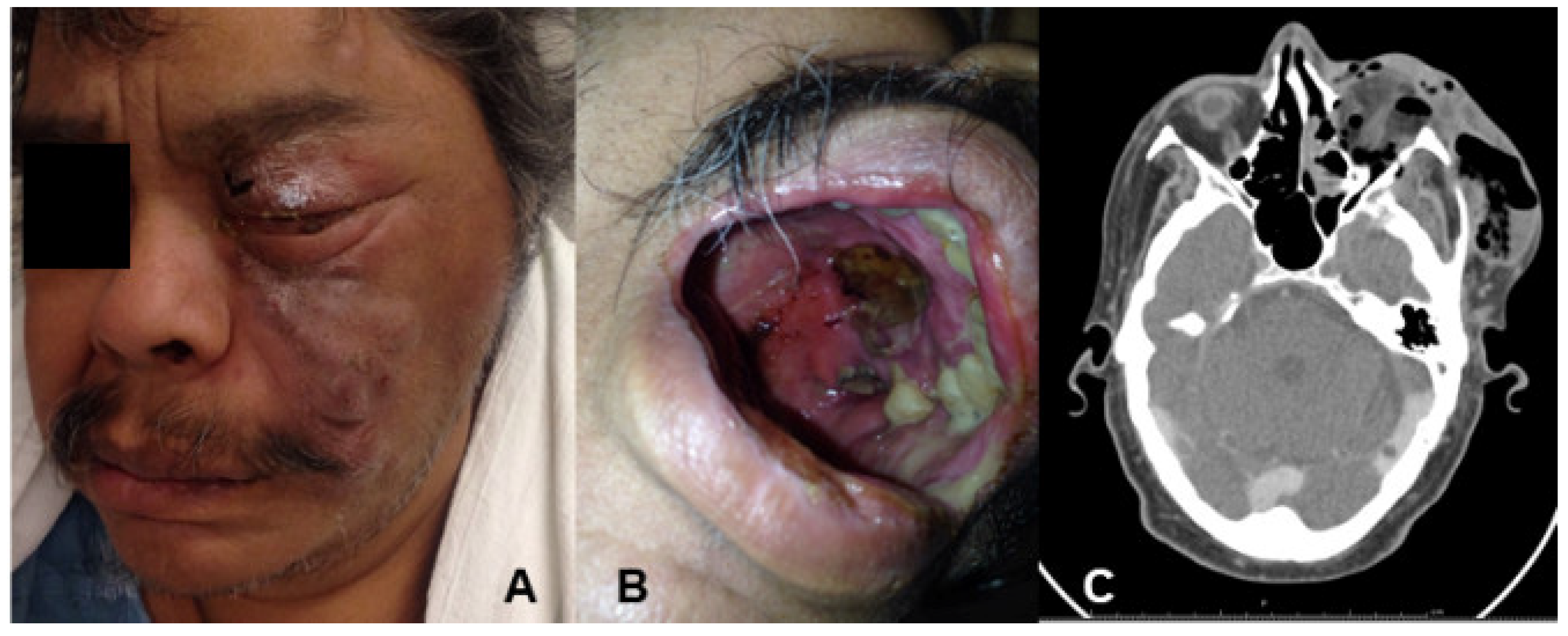
Figure 1. (A) Mucormycosis with facial infiltration and orbital involvement; (B) palatal necrotic lesions; (C) computed tomography with hypodense material involving multiple paranasal sinuses associated with bone erosion, and left orbit emphysema.
Jof 07 01096 g001
Figure 2. Nasal and maxillar-sinus tissue biopsies were paraffinembedded and formalin-fixed for histopathological examination. Three millimeter sections were stained with haematoxylin-eosin (H-e) and Gomori–Grocott methenamine-silver (G-G). (A) Nasal-sinus tissue sample showing the presnece of Asperguillus heads (H-E 40x); (B) Maxillar–sinus tissue sample showing presence of broad, non-septated hypahe (Gomori-Grocott 40x).
Jof 07 01096 g002
Table 1. Combined aspergillosis and mucormycosis cases.
CaseClinical PresentationUnderlying DiseaseTreatmentFungal AgentsReference
1SinusChronic sinusitisSurgeryAspergillus spp./Mucorales[17]
2Oro-sinonasalCastleman diseaseAmphotericin B, and itraconazoleAspergillus spp./Mucorales[18]
3RhinocerebralDiabetes mellitusVoriconazole, caspofungin, and amphotericin BA. fumigatus/Mucorales[8]
4Sinus and brain abscessDiabetes mellitusAmphotericin BA. fumigatus/Mucorales[19]
5SinonasalSinusitisItraconazole, and amphotericin BAspergillosis/Mucorales[20]
6SinusAcute myeloid leukemiaPosaconazole [21]
7Rhino-oculo-cerebralDiabetes mellitusVoriconazoleA. flavus/Mucorales[22]
8RhinocerebralDiabetes mellitus, and Renal transplantationAmphotericin BA. niger/R. arrhizus (syn. R. oryzae)[23]
9OrofacialCerebral trunk gliomaFluconazole, amphotericin B, and micafunginA. flavus/Mucorales[3]
10RhinocerebralRenal transplantationAmphotericin BA. fumigatus/
Mucorales
[24]
11Rhino-orbitalDiabetes mellitusAmphotericin BA. fumigatus/R. arrhizus (syn. R. oryzae)Present study
Publisher’s Note: MDPI stays neutral with regard to jurisdictional claims in published maps and institutional affiliations.