Article

Characteristics, Risk Factors, and Survival Analysis of Candida auris Cases: Results of One-Year National Surveillance Data from Oman

1
Central Public Health Laboratories, Directorate General for Disease Surveillance and Control (DGDSC), Ministry of Health, Muscat 393, Oman
2
Directorate General for Disease Surveillance and Control (DGDSC), Ministry of Health, Muscat 393, Oman
*
Authors to whom correspondence should be addressed.
J. Fungi 2021, 7(1), 31; https://doi.org/10.3390/jof7010031
Submission received: 15 November 2020 / Revised: 30 December 2020 / Accepted: 30 December 2020 / Published: 7 January 2021
(This article belongs to the Special Issue Candida auris 2.0)

Abstract

:
Background: Candida auris (C. auris) is an emerging healthcare-associated pathogen resulting in significant morbidity and mortality. The aim of this study is to report data from the national C. auris surveillance system for 2019 and conduct a survival analysis of the reported cohort. Methods: a retrospective analysis was conducted for all C. auris cases reported nationally to the Oman Antimicrobial Surveillance System (OMASS) in 2019, and isolates were sent to the Central Public Health Laboratories (CPHL). Clinical and demographic data were obtained through the E-Surveillance reporting system and the Electronic System (NEHR Al-Shifa) at CPHL. Statistical analysis was done using Kaplan–Meier analysis and Cox proportional hazard models. Results: One hundred and twenty-nine isolates of C. auris were grown from 108 inpatients; 87% were isolated from clinical samples, of which blood was the most common (38.9%). Forty (37%) were ≥65 years of age, 72 (66.7%) were males, and 85 (78.7%) were Omani nationals. Of the total isolates, 43.5% were considered as colonization; 56.5% were considered infection, of which 61.8% of them were candidemia. At least one risk factor was present in 98.1% of patients. The mean time from admission to infection was 1.7 months (SD = 2.8), and the mean length of hospital stay was 3.5 months (SD = 4). Totals of 94.8% and 96.1% of the isolates were non-susceptible to fluconazole and amphotericin, respectively. The variables found to be significantly associated with longer survival post C. auris diagnosis (p < 0.05) were age < 65 years, absence of comorbidities, length of stay < 3 months, colonization, and absence of candidemia. The infection fatality rate was 52.5%. Conclusion: Including C. auris in an ongoing antimicrobial surveillance program provides important data for the comprehensive management of this growing public health threat. The current study shows health care outbreaks of C. auris are ongoing, with 52.5% infection fatality, although our isolates remained sensitive to Echinocandins in vitro.

1. Introduction

Candida auris (C. auris) emerged as a resistant fungal pathogen for the first time in 2009. Since then, it has spread to all continents but Antarctica [1,2]. It is increasingly described as an agent of hospital-acquired infections and has gained global attention due to its multidrug-resistant properties and high crude mortality rate, up to 40% in cases of bloodstream infections [2,3,4,5]. In addition, C. auris strains are difficult to identify and can be misidentified as other phylogenetically related pathogens [6]. In Oman, the first report of C. auris isolates was in 2017, in which five cases were described with bloodstream infection [7]. After introducing diagnostic facilities, further hospital clusters were identified, and their control was a major challenge to the existing infection prevention and control measures [8]. In 2017, the Oman Antimicrobial Resistance Surveillance System (OMASS) included C. auris as an emerging pathogen at the national level, and a surveillance system was put in place to monitor the clinical, epidemiological, and drug susceptibility profiles to further guide the antimicrobial stewardship program and other AMR-related activities and interventions [9,10]. The Global Antimicrobial Resistance Surveillance System (GLASS) also includes invasive fungal bloodstream infections (BSIs) caused by the Candida species as part of the priority pathogens monitored for the AMR global action plan [11].
The threat of C. auris evolution to pan-resistance has become a reality and has been reported from multiple countries, resulting most of the time in fatalities [3,4,5,12,13]. This emphasizes the importance of monitoring this pathogen and its impact on morbidity and mortality. This study reports OMASS C. auris surveillance data from 2019 and conducts a survival analysis for infected/colonized patients reported during this year.

2. Materials and Methods

All cases of C. auris that had been entered into the national electronic surveillance system (E-Surveillance) and C. auris isolates received at the Central Public Health Laboratories (CPHL; the reference laboratory for the Antimicrobial Resistance Surveillance System) for confirmation and antimicrobial susceptibility testing (AST) between January and December 2019 were included in this study. These included all isolates from clinical and screening samples. Patient screening was conducted according to the national policy of infection prevention and control for C. auris. In general, screening is recommended for patients coming from affected hospitals locally, nationally, or internationally; patients admitted to units with newly identified and/or ongoing cases or colonization; and close contacts of newly diagnosed patients in intensive care or high-dependency units. All patient duplicates for the same episode of admission were removed, even if more than one positive sample was involved, considering only the most invasive sample. These duplicates were included when the interval between episodes of infection/colonization was ≥2 weeks for the same patient, which follows the national surveillance protocol for Multidrug-Resistant Organisms.

2.1. Clinical & Demographic Data

Clinical and demographic data were collected using the E-Surveillance reporting system and the Electronic System (NEHR Al-Shifa) at CPHL. The data included patient age, gender, nationality, diagnosis, ICU admission, risk factors, length of hospital stay, antifungal agents received and outcome, and specimen type. Risk factors included comorbidities such as hypertension (HTN), diabetes mellitus (DM), cardiovascular disease (CVD), chronic kidney disease (CKD), and immunodeficiency as well as presence of artificial ventilation, tracheostomy, indwelling catheter/device, ICU stay, and administration of multiple antibiotic courses during hospital stay. Indwelling catheters/devices included central venous line, external ventricular device (EVD), ventriculo-peritoneal (VP) shunt, percutaneous endoscopic gastrostomy (PEG) tube, and intracutaneous chest tube (ICD). The infection origin (hospital-acquired vs. community-acquired) was determined using the difference between the first positive specimen collection date (infection date) and admission date, which was further verified by the infection control team in the facility. Hospital-acquired infection was defined as infection occurring ≥ 48 h after admission or after transfer from another healthcare institution, otherwise it was labeled as community-acquired infection.
The infection fatality rate was calculated as the number of infected patients who died out of the total number of infected patients.

2.2. Identification & AST

Isolates were identified using MALDI-TOF (Maldi Biotyper MBT Compass 4.1.100, Bruker Daltoniks GmbH, Bremen, Germany) or Vitek 2 version 8.01 using a YST ID card. AST was done using Vitek 2 with the AST-YS07 card, which includes fluconazole, voriconazole, caspofungin, micafungin, amphotericin, and flucytosine. Susceptibility was interpreted according to CDC tentative breakpoints for Fluconazole, amphotericin, and echinocandins [14].

2.3. Data Analysis

The basic characteristics of the patients were analyzed using percentages (for categorical variables) and medians with interquartile ranges (for continuous variables). Age and length of stay were categorized into two factors each: young < 65 and old ≥ 65 for age; <3 months or ≥3 months for length of stay.
Survival times were estimated by Kaplan–Meier analysis, with the start point as the specimen collection date and the endpoint as the date of death. Univariate factors (age group, nationality, hospital length of stay, hospital, presence of comorbidities, presence of candidemia, and colonization versus infection) were compared using the log rank test. The simultaneous influences of these factors on survival time were examined using Cox proportional hazard models. All analyses were performed using R software version 4.0.
The study was approved by the Directorate General for Disease Surveillance and Control. Informed patient consent was not required because we utilized anonymous data collected for public health purposes, and no personal information was shared.

3. Results

A total of 129 C. auris isolates were identified in 108 patients during the study period. Eighteen patients had more than one episode of C. auris, and no duplicates needed to be removed according to the inclusion criteria. All patients were admitted to a hospital. Out of these isolates, 16 (12.4%) were screening samples obtained for infection control purposes, and 113 (87.6%) were clinical samples including 44 (38.9%) blood, 41 (36.3%) urine, 9 (8%) respiratory, 9 (8%) central line tip, 7 (6.2%) wound, and 3 (2.6%) other samples.
Among the 108 included patients, 40 (37%) were ≥65 years of age, 72 (66.7%) were males, and 85 (78.7%) were Omanis. The isolate was classified as colonization in 47 (43.5%) and infection in 61 (56.5%) patients, with 42 (68.8%) of them having candidemia. The mean time from admission to infection was 1.7 months (SD = 2.8) (Table 1). At least one risk factor was present in 106 of the infected/colonized patients: 74 had other comorbid diseases, 42 had an indwelling catheter/device, 41 had tracheostomy, 85 were on ventilators, 91 received multiple courses of antibiotics during their hospital stay, and 85 patients were admitted to the ICU at some point during hospital stay. The mean length of hospital stay for the included patients was 3.5 months (SD = 4).
In addition, of the 18 patients with multiple episodes of C. auris, 3 patients acquired infection followed by persistent colonization. On the other hand, 4 patients were found positive for C. auris in the screening samples and developed infection several days to several weeks after the initial screening.
Out of 53 infected patients with available treatment data, 47 received antifungal treatment (63.8% Echinocandins, 23.4% Azoles, 4.3% Amphotericin, and 8.5% multiple antifungal agents), and 15/49 died while on therapy. There were 6/53 patients who were not started on antifungal (4 died prior to results, 2 improved with source control). The overall fatality rate for the infected patients was 52.5%.
AST was conducted for 77/129 isolates (59.7%), and results are shown in Table 2. Echinofungins (caspofungin and micafungin) showed the lowest MIC among the antifungal classes tested, with 98.7% and 97.4% of tested isolates having MICs of <1 µg/mL and ≤1 µg/mL for caspofungin and micafungin, respectively. Fluconazole and amphotericin were found to have high MICs for the majority of tested isolates: 94.8% and 96.1%, respectively.
The infection origin was classified as healthcare for all patients in this report; all patients were infected/colonized ≥ 48 h after admission (100), or, in eight patients, C. auris was detected during routine screening, when transferred from another facility or readmitted after recent discharge from the same facility, taking into account the first C. auris isolate for each patient.
The isolates were received from seven hospitals in the country. The majority of these isolates (89.2%) were grown from patients admitted to three major hospitals—A, B, and C (a tertiary medical and surgical center, a tertiary trauma center, and a secondary medical and surgical center)—while the remaining 10.8% were from patients admitted to four other district hospitals. The epidemiological curve in Figure 1 shows the distribution of these isolates throughout the year. It is clear that these clusters were scattered across all months, without clear interruptions for the 3 major hospitals, A, B, and C which might indicate ongoing outbreaks.
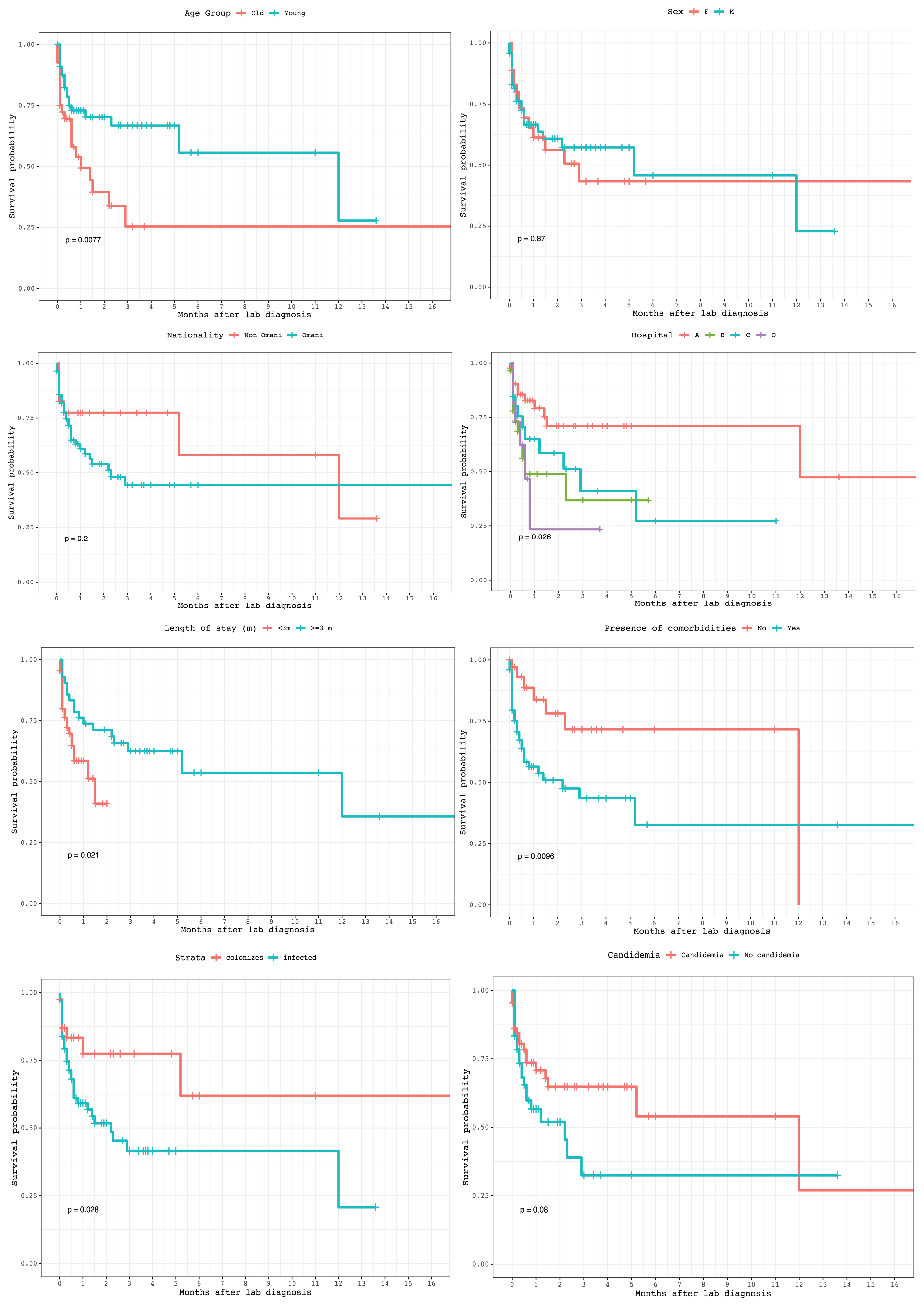
Using Kaplan–Meier analysis, we found age < 65 years, absence of comorbidities, hospital A, length of stay < 3 months, colonization, and absence of candidemia were significantly associated with longer survival post C. auris diagnosis (p < 0.05), whereas the other factors were not (Figure 2).
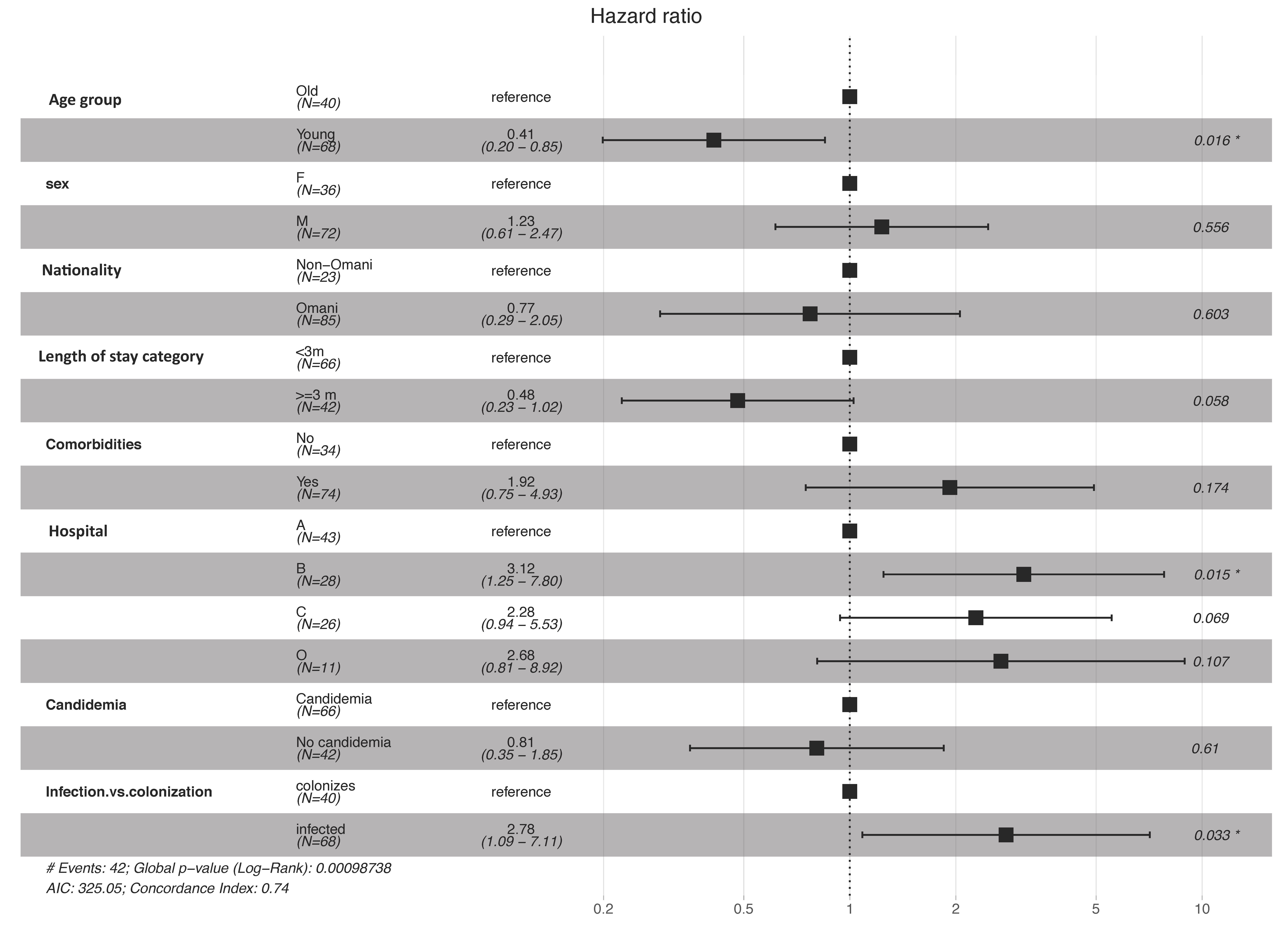
Adjusting for all studied factors, the multivariate Cox proportional hazard model shows that longer survival post diagnosis was significantly associated with younger age (<65 year) with an adjusted hazard ratio (AHR) of 0.41 (95% CI 0.20–0.85; p = 0.016). On the other hand, worse survival post diagnosis was found in infection compared to colonization groups (AHR 2.78, 95% CI 1.09–7.11, p = 0.033) and hospital B compared to A (AHR 3.12, 95% CI 1.25–7.80, p = 0.015; Figure 3).

4. Discussion

The current study describes the characteristics and outcomes of patients infected/colonized with C. auris at the national level from the OMASS data of 2019. Data clearly show the high burden and impact of this pathogen as a cause of healthcare-associated infection and ongoing outbreaks in the hospitals. The study underscores the importance of ongoing surveillance as a tool to assess the magnitude, burden, and impact of intervention on this growing healthcare challenge. Although our isolates for C. auris were susceptible to Echinocandins in vitro, and no pan-resistance had occurred, the fatality rate remained high at 52.5%.
Although the emergence of C. auris was first reported in 2009, a subsequent report identified C. auris in a sample collected back in 1996 [15]. Globally, it was under-reported, and it managed to surface as a significant AMR pathogen only in the last 5 years after several reports of health care outbreaks [16,17]. One theory that tried to explain this re-emergence was global warming, which might have led to narrowing the human ‘thermal restriction zone’ that protects humans from infection [18]. Many of the reported delays in the identification and mismanagement of patients are related to diagnostic challenges in identifying isolates and differentiating them from other Candida species [19,20]. At the national level, Candida isolates are diagnosed and subtyped at CPHL and some tertiary hospitals in the capital city of Muscat, but the delay in transporting samples from hospitals in other districts may have resulted in delaying appropriate intervention and increased the burden and fatalities.
All candida isolates from sterile sites should be identified at the species level to guide antifungal therapy [21]. In addition, the same should be done for candida isolated from non-sterile sites for infection control and surveillance purposes, as recommended by the Centers for Disease Control and Prevention (CDC) [22]. To differentiate C. auris from other candida species, culturing at 42 C or higher has been used [19]. Nevertheless, more accurate methods of identification are always required. Among these, MALDI-TOF and Vitek 2 YST ID card are accurate methods when C. auris is added to their databases [21,23]. In addition, PCR, real-time PCR [24,25,26], and sequencing methods have been evaluated and give excellent results to differentiate C. auris from other candida species. All C. auris isolates in our study were identified using either MALDI-TOF or Vitek 2 YST ID card, which are available in our reference laboratory. Early identification and reporting of C. auris, due to the availability of diagnostic tests, will improve AMR surveillance and better patient outcomes in many low- and middle-income countries. It will also provide better insight to the magnitude and burden of infection, which will impact global intervention plans [27].
A number of studies determined different possible risk factors for developing C. auris infection/colonization. These included the presence of tracheostomies, PEG tubes, and ventilators; prior antibiotic use; ICU stay; and comorbidities such as DM, CKD, and lung disease [28,29,30,31,32,33]. In our study, the majority of these patients had a tracheostomy (38.0%), indwelling catheters (38.9%), comorbidities (68.5%), were on a ventilator (78.7%), admitted to the ICU (78.7%), and/or received multiple courses of antibiotics during their hospital stay (84.3%). These factors are significant to consider when candida infection is suspected in order to guide management and infection control measures.
Although the hospitals in our study that reported C. auris are acute care institutions, they also provide long-term nursing care for cases involving stroke and multiple traumas because there is an absence of such long-term healthcare facilities in the country. This was reflected by the prolonged mean length of hospital stay of 3.5 months, which in turn affected the survival of these patients.
The reported mortality rates attributable to invasive C. auris infection range from 30% to 59% globally [31,34]. Crude in-hospital mortality rates for C. auris candidemia are estimated to range from 30% to 72% [20]. The overall infection fatality rate in our study was 52.5%, which was similar to the figure mentioned earlier from another study in Oman (53.1%) [8]. The majority of these patients required ICU admission.
The survival analysis showed that statistically significant factors negatively attributed to survival post diagnosis were old age, presence of comorbidities, C. auris infection rather than colonization, long hospital stay, and presence of candidemia. The effect of the latter on survival was analyzed and found to be statistically significant by Sayeed et al., while there was no difference in mortality between infected and colonized patients [33].
Our data clearly show there were ongoing outbreaks in three out of the seven hospitals reporting C. auris cases. The outbreak control of C. auris requires implementing multiple infection control measures, as mentioned in a previous report by Taori et al. [35] Despite the measures implemented in these three hospitals, the outbreaks were ongoing throughout the year, which clearly shows the difficulty in eradicating C. auris and the burden this organism can put on infection control programs at any healthcare facility.
Regarding antimicrobial susceptibility testing, there are no specific Breakpoints identified for C. auris; tentative breakpoints developed by CDC are currently in use [14]. Apart from the study by Kellie Arensman et al. showing a low rate of resistance to fluconazole (14%) and amphotericin B (3.6) [36], many reports detected a high rate of resistance to these antifungal classes. A recent review of C. auris cases from the Middle East showed in vitro antifungal susceptibility testing identified 100% of the strains to be resistant to fluconazole (MIC 32 ≥ 256 mg/L) while having variable resistance to other antifungal agents [37]. This is similar to reports by Ruiz-Gaitán et al. and Sayeed et al. [32,33]. A report from the USA showed that >90% of C. auris isolates were resistant to fluconazole, >60% were resistant to amphotericin, and 3.9% were resistant to echinocandins; three isolates were found to be pan-resistant (resistant to the three antifungal classes) [13]. Lockhart et al. found that 4% of the isolates studied were pan-resistant, involving patients from Pakistan, India, South Africa, and Venezuela [34].
According to our results, the isolates of C. auris were not susceptible to fluconazole, and only 5.2% had an MIC of ≤8 µg/mL. Similar findings were observed with amphotericin: only 3.9% of the isolates had an MIC of <1 µg/mL. On the other hand, the majority of isolates were susceptible to echinocandins (caspofungin and micafungin), with 98.7% and 97.4% having MICs of <1 µg/mL and ≤1 µg/mL, respectively. It is reassuring that our isolates remained sensitive to the Echinocandins group. This was similar to the above reports, but it differs from a report in India where 40% of the isolates were not susceptible to caspofungin [38].
The fact that 15 patients died while on antifungal treatment raises the possibility of treatment failure despite the MIC results in vitro. In analyzing 11 of these cases, for which AST data were available, 4 received antifungal agents with MICs in the non-susceptible range. A striking finding was that the remaining seven patients received echinocandins, and their C. auris AST results showed MICs to Echinocandins in the susceptible range. Acquiring resistance to echinocandins while on echinocandins has been reported previously [22]. However, this could not be proven here because no repeated samples taken with C. auris isolated and AST done for these patients while on treatment. It is still unclear how well in vitro AST results correlate with the in vivo performance of antifungal agents against C. auris infection. However, we must take into account other possible causes of death, such as underlying comorbidities and uncontrolled source of infection.
A limitation of this study is that it is retrospective and is based on voluntary reporting by the hospitals. In addition, no genotyping was performed to analyze the virulence and genetic background of its antifungal resistance. However, a previous report from Oman identified C. auris isolates as part of clade 1 (South Asian), and Muñoz et al. reported different resistance genes in this clade and other clades [8,39]. In addition, the small sample size might have affected the results and led to wide CIs, which might possibly explain the absence of significant p-values in some variables.

5. Conclusions

In conclusion, C. auris is an additional burden to the healthcare system and is the leading antimicrobial-resistant fungi. C. auris should be at the top of the AMR agenda because: it prefers the healthcare environment where many high-risk patients with complex health issues are managed; it can persist on medical devices and in the environment; it has developed resistance to many antifungals; pan-resistance is possible, and we are running out of treatment options; and it has high crude mortality rates. A comprehensive intervention program is urgently needed to decrease the burden of C. auris, and it should include an ongoing surveillance program that covers clinical, epidemiological, and drug susceptibility patterns on a regular basis, thereby strengthening the infection prevention and control programs in acute care hospitals. In addition, the fact that all cases in this cohort were hospital-acquired stresses the importance of antimicrobial stewardship programs in healthcare facilities.
Furthermore, this study emphasizes the importance of establishing a molecular genotyping service, such as Whole-Genome Sequencing, at CPHL for C. auris as part of the diagnostic toolkit during outbreak investigations and for surveillance. In addition, these data show the need to establish diagnostic services for C. auris at the district level.

Author Contributions

Conceptualization, A.A.-R., A.A.-M., A.A.-J., S.A.-A.; Methodology, A.A.-R., A.A.-W., A.A.; Validation, A.A.-M.; Formal Analysis, A.A.-W.; Investigation, A.A.-R., A.A.-M.; Data Curation, A.A.-R., A.A.-M., A.A.-W., A.A.; Writing—Original Draft Preparation, A.A.-R., A.A.-M., A.A.-W.; Writing—Review & Editing, A.A.-J., S.A.-A., A.A.; Supervision, A.A.-M., A.A.-J., S.A.-A.; Project Administration, A.A.-R., A.A.-M. All authors have read and agreed to the published version of the manuscript.

Funding

This research received no external funding.

Institutional Review Board Statement

Study has been approved by Directorate General For Disease Surveillance & Control, the owner of the data of national surveillance system.

Informed Consent Statement

Informed patient consent was not required because we utilized anonymous data collected for public health purposes, and no personal information was shared.

Data Availability Statement

Data in this study is available on request.

Acknowledgments

We would like to acknowledge Sandhyarani Ramamurthi for her support in providing patient data for the study. We would also like to acknowledge the Bacteriology Section at CPHL for their technical support in identifying the isolates and carrying out antimicrobial susceptibility testing.

Conflicts of Interest

The authors declare no conflict of interest.

References

  1. Satoh, K.; Makimura, K.; Hasumi, Y.; Nishiyama, Y.; Uchida, K.; Yamaguchi, H. Candida auris sp. nov., a novel ascomycetous yeast isolated from the external ear canal of an inpatient in a Japanese hospital. Microbiol. Immunol. 2009, 53, 41–44. [Google Scholar] [CrossRef]
  2. Du, H.; Bing, J.; Hu, T.; Ennis, C.L.; Nobile, C.J.; Huang, G. Candida auris: Epidemiology, biology, antifungal resistance, and virulence. PLoS Pathog. 2020, 16, e1008921. [Google Scholar] [CrossRef]
  3. Candida auris: Information for Laboratorians and Health Professionals. Atlanta, Georgia: US Department of Health and Human Services, Centers for Disease Control and Prevention. 2019. Available online: https://www.cdc.gov/fungal/candida-auris/health-professionals.html (accessed on 6 November 2020).
  4. Hori, Y.; Shibuya, K. Role of FKS Gene in the Susceptibility of Pathogenic Fungi to Echinocandins. Med. Mycol. J. 2018, 59, E31–E40. [Google Scholar] [CrossRef] [Green Version]
  5. Chowdhary, A.; Prakash, A.; Sharma, C.; Kordalewska, M.; Kumar, A.; Sarma, S.; Tarai, B.; Singh, A.; Upadhyaya, G.; Upadhyay, S.; et al. A multicentre study of antifungal susceptibility patterns among 350 Candida auris isolates (2009–17) in India: Role of the ERG11 and FKS1 genes in azole and echinocandin resistance. J. Antimicrob. Chemother. 2018, 73, 891–899. [Google Scholar] [CrossRef]
  6. Fasciana, T.; Cortegiani, A.; Ippolito, M.; Giarratano, A.; Di Quattro, O.; Lipari, D.; Graceffa, D.; Giammanco, A. Candida auris: An Overview of How to Screen, Detect, Test and Control This Emerging Pathogen. Antibiotics 2020, 9, 778. [Google Scholar] [CrossRef]
  7. Al-Siyabi, T.; Al Busaidi, I.; Balkhair, A.; Al-Muharrmi, Z.; Al-Salti, M.; Al’Adawi, B. First report of Candida auris in Oman: Clinical and microbiological description of five candidemia cases. J. Infect. 2017, 75, 373–376. [Google Scholar] [CrossRef] [PubMed]
  8. Al Maani, A.; Paul, H.; Al-Rashdi, A.; Al Wahaibi, A.; Al-Jardani, A.; Abri, A.; Albalushi, M.A.H.; Abri; Al Reesi, M.; Al Maqbali, A.; et al. Ongoing Challenges with Healthcare-Associated Candida auris Outbreaks in Oman. J. Fungi 2019, 5, 101. [Google Scholar] [CrossRef] [Green Version]
  9. Oman Antimicrobial Resistance Surveillance System (OMASS); The First National Annual Antimicrobial Resistance Report (2017); Directorate General for Disease Surveillance and Control, MoH: Muscat, Oman, 2018.
  10. Oman Antimicrobial Surveillance System (OMASS); The Annual Antimicrobial Resistance Report (2018); Directorate General for Disease Surveillance and Control, MoH: Muscat, Oman, 2019.
  11. Global Antimicrobial Resistance Surveillance System. April 2018. Available online: https://www.who.int/glass/events/AMR-in-invasive-candida-infections-meeting/en/ (accessed on 28 October 2020).
  12. Biagi, M.J.; Wiederhold, N.P.; Gibas, C.; Wickes, B.L.; Lozano, V.; Bleasdale, S.C.; Danziger, L. Development of High-Level Echinocandin Resistance in a Patient with Recurrent Candida auris Candidemia Secondary to Chronic Candiduria. Open Forum Infect. Dis. 2019, 6, ofz262. [Google Scholar] [CrossRef] [PubMed]
  13. Ostrowsky, B.; Greenko, J.; Adams, E.; Quinn, M.; O’Brien, B.; Chaturvedi, V.; Berkow, E.; Vallabhaneni, S.; Forsberg, K.; Chaturvedi, S.; et al. Candida auris Isolates Resistant to Three Classes of Antifungal Medications—New York, 2019. MMWR Morb. Mortal. Wkly. Rep. 2020, 69, 6–9. [Google Scholar] [CrossRef] [PubMed] [Green Version]
  14. Antifungal Susceptibility Testing and Interpretation, Candida auris, Centers for Disease Control & Prevention. Available online: https://www.cdc.gov/fungal/candida-auris/c-auris-antifungal.html (accessed on 28 October 2020).
  15. Oh, B.J.; Shin, J.H.; Kim, M.-N.; Sung, H.; Lee, K.; Joo, M.Y.; Shin, M.-G.; Suh, S.-P.; Ryang, D.W. Biofilm formation and genotyping of Candida haemulonii, Candida pseudohaemulonii, and a proposed new species (Candida auris) isolates from Korea. Med. Mycol. 2011, 49, 98–102. [Google Scholar] [CrossRef] [PubMed] [Green Version]
  16. Chowdhary, A.; Sharma, C.; Meis, J.F. Candida auris: A rapidly emerging cause of hospital-acquired multidrug-resistant fungal infections globally. PLoS Pathog. 2017, 13, e1006290. [Google Scholar] [CrossRef] [PubMed]
  17. Schelenz, S.; Hagen, F.; Rhodes, J.L.; Abdolrasouli, A.; Chowdhary, A.; Hall, A.; Ryan, L.; Shackleton, J.; Trimlett, R.; Meis, J.F.; et al. First hospital outbreak of the globally emerging Candida auris in a European hospital. Antimicrob. Resist. Infect. Control. 2016, 5, 1–7. [Google Scholar] [CrossRef] [PubMed] [Green Version]
  18. Misseri, G.; Ippolito, M.; Cortegiani, A. Global warming “heating up” the ICU through Candida auris infections: The climate changes theory. Crit. Care 2019, 23, 416. [Google Scholar] [CrossRef] [PubMed] [Green Version]
  19. Casadevall, A.; Kontoyiannis, D.P.; Robert, V. On the Emergence of Candida auris: Climate Change, Azoles, Swamps, and Birds. mBio 2019. [Google Scholar] [CrossRef] [PubMed] [Green Version]
  20. Cortegiani, A.; Misseri, G.; Fasciana, T.; Giammanco, A.; Giarratano, A.; Chowdhary, A. Epidemiology, clinical characteristics, resistance, and treatment of infections by Candida auris. J. Intensiv. Care 2018, 6, 69. [Google Scholar] [CrossRef] [PubMed]
  21. Spivak, E.S.; Hanson, K.E. Candida auris: An Emerging Fungal Pathogen. J. Clin. Microbiol. 2017, 56. [Google Scholar] [CrossRef] [PubMed] [Green Version]
  22. Division of Foodborne, Waterborne, and Environmental Diseases, National Center for Emerging and Zoonotic Infectious Diseases, Centers for Disease Control and Prevention. Candida auris Clinical Update: September 2017. Available online: https://www.cdc.gov/fungal/candida-auris/c-auris-alert-09-17.html (accessed on 15 November 2020).
  23. Identification of Candida auris, Centers for Disease Control & Prevention. Available online: https://www.cdc.gov/fungal/candida-auris/identification.html (accessed on 6 November 2020).
  24. Kordalewska, M.; Zhao, Y.; Lockhart, S.R.; Chowdhary, A.; Berrio, I.; Perlin, D.S. Rapid and Accurate Molecular Identification of the Emerging Multidrug-Resistant Pathogen Candida auris. J. Clin. Microbiol. 2017, 55, 2445–2452. [Google Scholar] [CrossRef] [Green Version]
  25. Ruiz-Gaitán, A.C.; Fernández-Pereira, J.; Valentin, E.; Tormo-Mas, M.Á.; Eraso, E.; Pemán, J.; De Groot, P.W.J. Molecular identification of Candida auris by PCR amplification of species-specific GPI protein-encoding genes. Int. J. Med. Microbiol. 2018, 308, 812–818. [Google Scholar] [CrossRef]
  26. Leach, L.; Zhu, Y.; Chaturvedi, S. Development and Validation of a Real-Time PCR Assay for Rapid Detection of Candida auris from Surveillance Samples. J. Clin. Microbiol. 2018. [Google Scholar] [CrossRef] [Green Version]
  27. Pierce, J.; Apisarnthanarak, A.; Schellack, N.; Cornistein, W.; Al Maani, A.; Adnan, S.; Stevens, M.P. Global Antimicrobial Stewardship with a Focus on Low- and Middle-Income Countries. Int. J. Infect. Dis. 2020, 96, 621–629. [Google Scholar] [CrossRef]
  28. Rozwadowski, F.; McAteer, J.; Chow, N.A.; Skrobarcek, K.; Forsberg, K.; Barrett, P.M.; Greeley, R.; Fulton, T.; Wells, J.; Welsh, R.M.; et al. 161. Prevalence and Risk Factors for Candida auris Colonization Among Patients in a Long-term Acute Care Hospital—New Jersey, 2017. Open Forum Infect. Dis. 2018, 5, S14. [Google Scholar] [CrossRef]
  29. Forsberg, K.; Woodworth, K.; Walters, M.; Berkow, E.L.; Jackson, B.; Chiller, T.; Vallabhaneni, S. Candida auris: The recent emergence of a multidrug-resistant fungal pathogen. Med. Mycol. 2019, 57, 1–12. [Google Scholar] [CrossRef] [PubMed] [Green Version]
  30. Adams, E.; Quinn, M.; Tsay, S.; Poirot, E.; Chaturvedi, S.; Southwick, K.; Greenko, J.; Fernandez, R.; Al, E.A.E.; Vallabhaneni, S.; et al. Candida auris in Healthcare Facilities, New York, USA, 2013–2017. Emerg. Infect. Dis. 2018, 24, 1816–1824. [Google Scholar] [CrossRef] [PubMed] [Green Version]
  31. Sekyere, J.O. Candida auris: A systematic review and meta-analysis of current updates on an emerging multidrug-resistant pathogen. Microbiologyopen 2018, 7, e00578. [Google Scholar] [CrossRef] [Green Version]
  32. Ruiz-Gaitán, A.; Martínez, H.; Moret, A.M.; Calabuig, E.; Tasias, M.; Alastruey-Izquierdo, A.; Zaragoza, Ó.; Mollar, J.; Frasquet, J.; Salavert-Lletí, M.; et al. Detection and treatment of Candida auris in an outbreak situation: Risk factors for developing colonization and candidemia by this new species in critically ill patients. Expert Rev. Antiinfect. Ther. 2019, 17, 295–305. [Google Scholar] [CrossRef]
  33. Sayeed, M.A.; Farooqi, J.; Jabeen, K.; Awan, S.; Mahmood, S.F. Clinical spectrum and factors impacting outcome of Candida auris: A single center study from Pakistan. BMC Infect. Dis. 2019, 19, 1–8. [Google Scholar] [CrossRef] [Green Version]
  34. Lockhart, S.R.; Etienne, K.A.; Vallabhaneni, S.; Farooqi, J.; Chowdhary, A.; Govender, N.P.; Colombo, A.L.; Calvo, B.; Cuomo, C.A.; Desjardins, C.A.; et al. Simultaneous Emergence of Multidrug-Resistant Candida auris on 3 Continents Confirmed by Whole-Genome Sequencing and Epidemiological Analyses. Clin. Infect. Dis. 2017, 64, 134–140. [Google Scholar] [CrossRef] [Green Version]
  35. Taori, S.K.; Khonyongwa, K.; Hayden, I.; Athukorala, G.D.A.; Letters, A.; Fife, A.; Desai, N.; Borman, A.M. Candida auris outbreak: Mortality, interventions and cost of sustaining control. J. Infect. 2019, 79, 601–611. [Google Scholar] [CrossRef]
  36. Arensman, K.; Miller, J.L.; Chiang, A.; Mai, N.; Levato, J.; Lachance, E.; Anderson, M.; Beganovic, M.; Pena, J.D. Clinical Outcomes of Patients Treated for Candida auris Infections in a Multisite Health System, Illinois, USA. Emerg. Infect. Dis. 2020, 26, 876–880. [Google Scholar] [CrossRef]
  37. Alfouzan, W.; Dhar, R.; Albarrag, A.; Al-Abdely, H. The emerging pathogen Candida auris: A focus on the Middle-Eastern countries. J. Infect. Public Health 2019, 12, 451–459. [Google Scholar] [CrossRef]
  38. Chowdhary, A.; Kumar, V.A.; Sharma, C.; Prakash, A.; Agarwal, K.; Babu, R.; Dinesh, K.R.; Karim, S.; Singh, S.K.; Hagen, F.; et al. Multidrug-resistant endemic clonal strain of Candida auris in India. Eur. J. Clin. Microbiol. Infect. Dis. 2014, 33, 919–926. [Google Scholar] [CrossRef] [PubMed]
  39. Muñoz, J.F.; Gade, L.; Chow, N.A.; Loparev, V.N.; Juieng, P.; Berkow, E.L.; Farrer, R.A.; Litvintseva, A.P.; Cuomo, C.A. Genomic insights into multidrug-resistance, mating and virulence in Candida auris and related emerging species. Nat. Commun. 2018, 9, 1–13. [Google Scholar] [CrossRef] [PubMed] [Green Version]
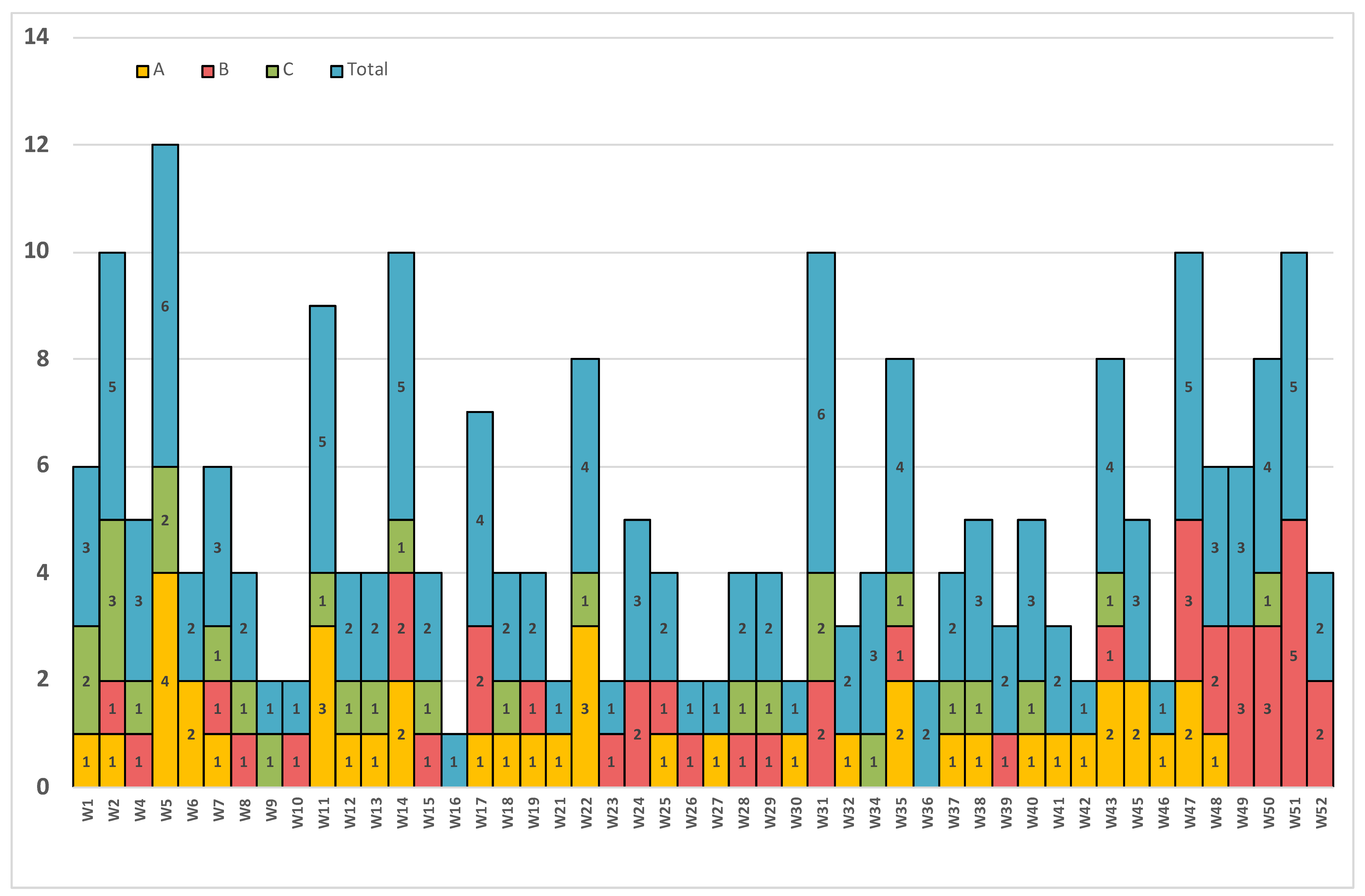
Figure 1. C. auris isolates by week over a 1 year period from (A) hospital A, (B) hospital B, (C) hospital C, and (Total) in all 7 hospitals. Numbers in the columns represent number of C.auris isolates per that week.
Jof 07 00031 g001
Figure 2. Kaplan–Meier survival analyses of age, sex, nationality, length of stay, presence of comorbidities, hospital where patients were admitted, candidemia, and colonization vs. infection.
Jof 07 00031 g002
Figure 3. Multivariate Cox proportional hazard model for survival post diagnosis by age, sex, nationality, length of stay, presence of comorbidities, hospital where patients were admitted, candidemia, and colonization vs. infection. Column 3 represent the hazard ratio (95% confidence interval), * statistically significant, p value < 0.05.
Jof 07 00031 g003
Table 1. Descriptive statistics of C. auris cases in 2019.
CategoriesOverall (N = 108)
Age group: ≥65 years40 (37%)
Males72 (66.7%)
Omani85 (78.7%)
Candidemia42 (38.9%)
Infection (not colonized)61 (56.5%)
Length of stay (months): <3 m66 (61.1%)
Comorbidities74 (68.5%)
Length of stay (months) after diagnosis: Mean (SD)1.7 (2.8)
ICU admission85 (78.7%)
Died (out of total patients)42 (38.9%)
Table 2. C. auris isolate (N = 77) distributions among different MIC values for the antifungal agents tested.
AntifungalFluconazoleVoriconazoleCaspofunginMicafunginAmphotericin BFlucytosine
MIC≤8 µg/mL
4 (5.2%)
≤1 µg/mL
68 (88.3%)
<1 µg/mL
76 (98.7%)
≤1 µg/mL
75 (97.4%)
<1 µg/mL
3 (3.9%)
≤8 µg/mL
70 (90.9%)
MIC16 µg/mL
34 (44.2%)
2 µg/mL
7 (9.1%)
1 µg/mL
0 (0.0%)
2 µg/mL
0 (0.0%)
1 µg/mL
9 (11.7%)
16 µg/mL
1 (1.3%)
MIC≥32 µg/mL
39 (50.6%)
≥4 µg/mL
2 (2.6%)
≥2 µg/mL
1 (1.3%)
≥4 µg/mL
2 (2.6%)
≥2 µg/mL
65 (84.4%)
≥32 µg/mL
6 (7.8%)
Publisher’s Note: MDPI stays neutral with regard to jurisdictional claims in published maps and institutional affiliations.