Sporotrichin Skin Test for the Diagnosis of Sporotrichosis

 ,
, 
 and
and
Abstract
:1. Introduction
2. Materials and Methods
2.1. Baseline Characteristics of the Study Population
2.2. Data Analysis
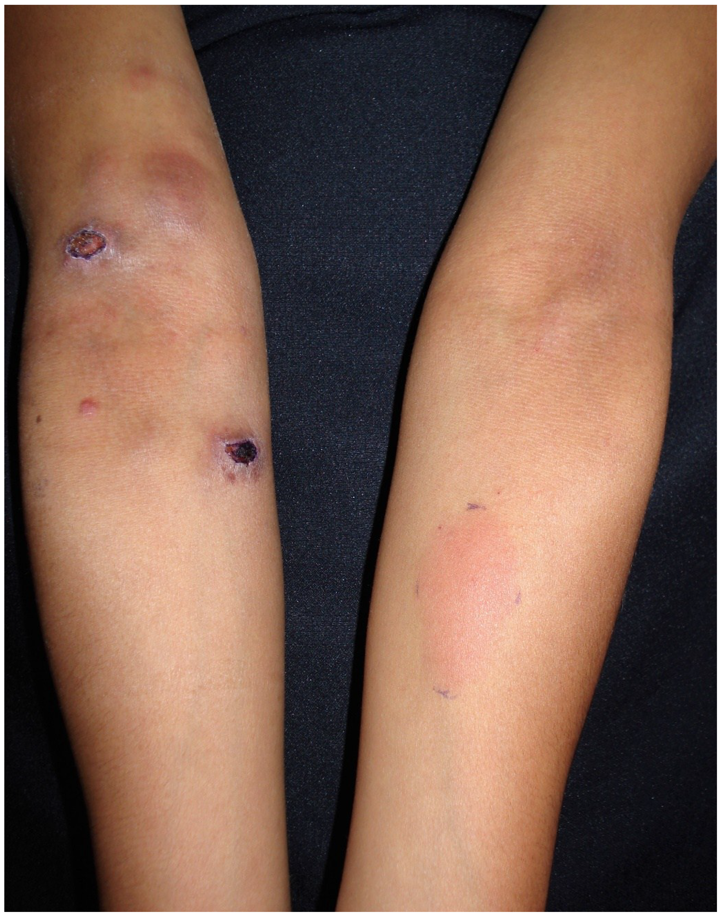
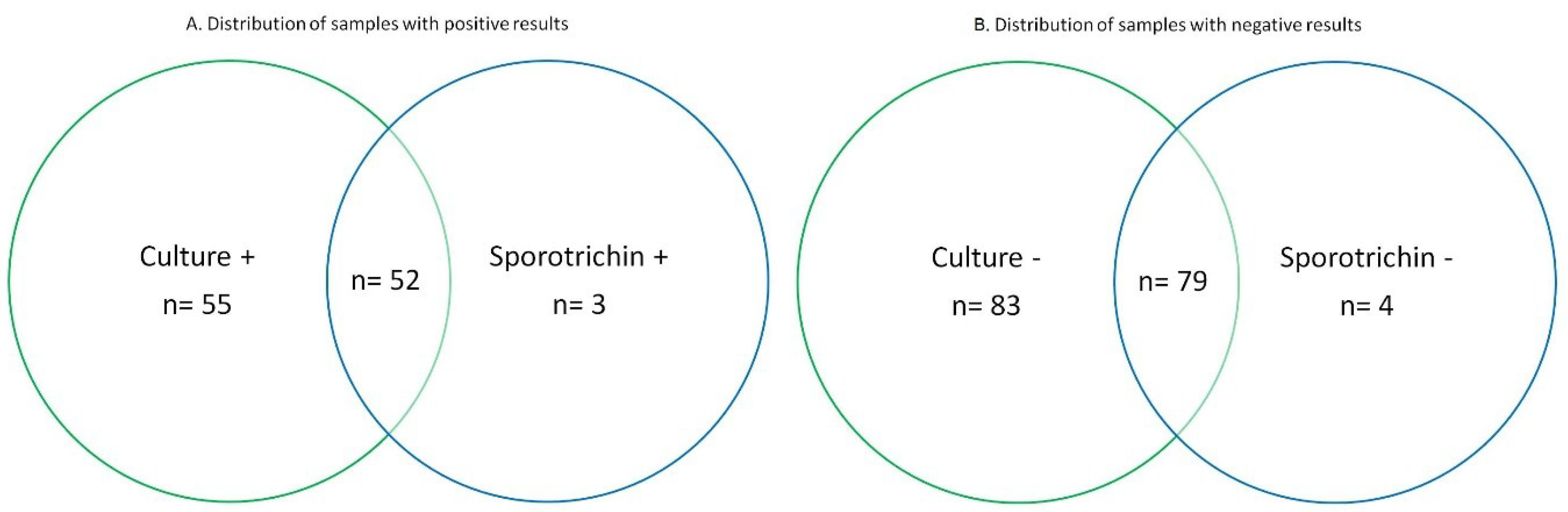
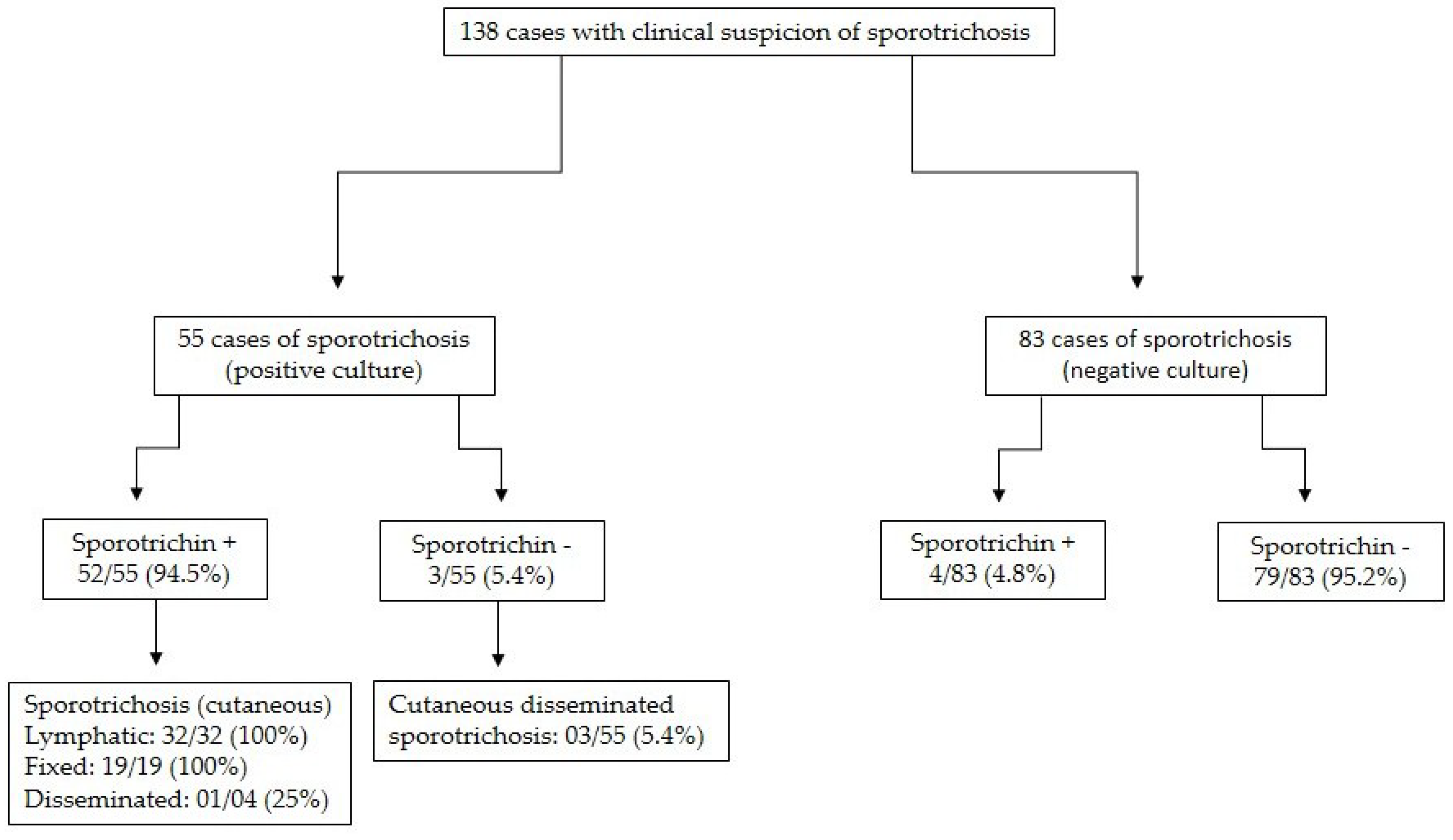
3. Results
4. Discussion
5. Conclusions
Author Contributions
Acknowledgments
Conflicts of Interest
References
- Barros, M.B.; de Almeida Paes, R.; Schubach, A.O. Sporothrix schenckii and Sporotrichosis. Clin. Microbiol. Rev. 2011, 24, 633–654. [Google Scholar] [CrossRef] [PubMed]
- Arenas, R. Sporotrichosis. In Topley & Wilsong’s Microbiology and Microbial Infections, 10th ed.; Merz, W.G., Hay, R., Eds.; Hodder-Arnold: London, UK, 2005; pp. 367–384. [Google Scholar]
- Bonifaz, A.; Vázquez-González, D. Sporotrichosis: An update. G. Ital. Dermatol. Venereol. 2010, 145, 659–673. [Google Scholar] [PubMed]
- Chakrabarti, A.; Bonifaz, A.; Gutierrez-Galhardo, M.C.; Mochizuki, T.; Li, S. Global epidemiology of sporotrichosis. Med. Mycol. 2015, 53, 3–14. [Google Scholar] [CrossRef] [PubMed]
- Ramírez-Soto, M.C. Sporotrichosis: The story of an endemic region in Peru over 28 Years (1985 to 2012). PLoS ONE 2015, 10, e0127924. [Google Scholar] [CrossRef] [PubMed]
- López-Romero, E.; Reyes-Montes, M.R.; Pérez-Torres, A.; Ruiz-Baca, E.; Villagómez-Castro, J.C.; Mora-Montes, H.M.; Flores-Carreón, A.; Toriello, C. Sporothrix schenckii complex and sporotrichosis, an emerging health problem. Future Microbiol. 2011, 6, 85–102. [Google Scholar] [CrossRef] [PubMed]
- Rodrigues, A.M.; de Hoog, S.; de Camargo, Z.P. Emergence of pathogenicity in the Sporothrix schenckii complex. Med. Mycol. 2013, 51, 405–412. [Google Scholar] [CrossRef] [PubMed]
- Quintella, L.P.; Passos, S.R.; do Vale, A.C.; Galhardo, M.C.; Barros, M.B.; Cuzzi, T.; Reis Rdos, S.; de Carvalho, M.H.; Zappa, M.B.; Schubach Ade, O. Histopathology of cutaneous sporotrichosis in Rio de Janeiro: A series of 119 consecutive cases. J. Cutan. Pathol. 2011, 38, 25–32. [Google Scholar] [CrossRef] [PubMed]
- González-Ochoa, A.; Soto-Figueroa, E. Polisacáridos de Sporotrichum schenckii. Intradermorreacción en el diagnóstico de la esporotricosis. Rev. Inst. Salub. Enferm. Trop. Mex. 1947, 8, 143–153. (In Spanish) [Google Scholar]
- González Ochoa, A. Valoración comparativa de los antígenos polisacáridos y celular de Sporothrix schenckii. Salud Pública Méx. 1970, 30, 303–315. (In Spanish) [Google Scholar]
- Bonifaz, A.; Araiza, J.; Pérez-Mejía, A.; Ochoa, L.A.; Toriello, C. Prueba intradérmica con esporotricina en una comunidad de la Sierra Norte de Puebla. Dermatol. Rev. Mex. 2013, 57, 428–432. (In Spanish) [Google Scholar]
- Arenas, G.; Toriello, C. Actividad inmunológica de antígenos miceliales y levaduriformes de diferentes fases de crecimiento de Sporothrix schenckii. Rev. Mex. Mic. 1986, 2, 131–144. (In Spanish) [Google Scholar]
- Madrid, H.; Cano, J.; Gené, J.; Bonifaz, A.; Toriello, C.; Guarro, J. Sporothrix globosa, a pathogenic fungus with widespread geographical distribution. Rev. Iberoam. Micol. 2009, 26, 218–222. [Google Scholar] [CrossRef] [PubMed]
- Dominguez-Soto, L.; Hojyo-Tomoka, M.T. The intradermal sporotrichin test and the diagnosis of sporotrichosis. Int. J. Dermatol. 1983, 22, 520. [Google Scholar] [CrossRef] [PubMed]
- Lopes-Bezerra, L.M. Sporothrix schenckii cell wall peptidorhamnomannans. Front. Microbiol. 2011, 2, 243. [Google Scholar] [CrossRef] [PubMed]
- Alba-Fierro, C.A.; Pérez-Torres, A.; Toriello, C.; Romo-Lozano, Y.; López-Romero, E.; Ruiz-Baca, E. Molecular components of the Sporothrix schenckii Complex that Induce immune response. Curr. Microbiol. 2016, 73, 292–300. [Google Scholar] [CrossRef] [PubMed]
- Lloyd, K.O.; Bitoon, M.A. Isolation and purification of a peptido-rhamnomannan from the yeast form of Sporothrix schenckii. Structural and immunochemical studies. J. Immunol. 1971, 107, 663–671. [Google Scholar] [PubMed]
- Kusuhara, M.; Hachisuka, H.; Sasai, Y. Statistical survey of 150 cases with sporotrichosis. Mycopathologia 1988, 102, 129–133. [Google Scholar] [CrossRef] [PubMed]
- Itoh, M.; Okamoto, S.; Kariya, H. Survey of 200 cases of sporotrichosis. Dermatologica 1986, 172, 209–213. [Google Scholar] [CrossRef] [PubMed]
- Rocha-Posada, H. Skin test with sporotrichin. Its sensitbity and specificity. Mycopathol. Mycol. Appl. 1968, 36, 42–54. [Google Scholar]
- Bonifaz, A.; Tirado-Sánchez, A.; Paredes-Solís, V.; Cepeda-Valdés, R.; González, G.M.; Treviño-Rangel, R.J.; Fierro-Arias, L. Cutaneous disseminated sporotrichosis: Clinical experience of 24 cases. J. Eur. Acad. Dermatol. Venereol. 2018, 32, 77–79. [Google Scholar] [CrossRef] [PubMed]
- Bonifaz, A.; Tirado-Sánchez, A. Cutaneous disseminated and extracutaneous sporotrichosis: Current status of a complex disease. J. Fungi 2017, 10, 6. [Google Scholar] [CrossRef] [PubMed]
- Sánchez-Alemán, M.A.; Araiza, J.; Bonifaz, A. Aislamiento y caracterización de cepas silvestres de Sporothrix schenckii e investigación de reactores a la esporotricina. Gac. Med. Mex. 2004, 140, 507–512. (In Spanish) [Google Scholar] [PubMed]
- Schneidau, J.D.; Lamar, L.M.; Harston, M.A. Cutaneous hypesensivity to sportrichin in Lousiana. JAMA 1964, 188, 371–373. [Google Scholar] [CrossRef] [PubMed]
- Rodrigues, M.T.; de Resende, M.A. Epidemiologic skin test survey of sensitivity to paracoccidioidin, histoplasmin and sporotrichin among gold mine workers of Morro Velho Mining, Brazil. Mycopathologia 1996, 135, 89–98. [Google Scholar] [CrossRef] [PubMed]
- Ghosh, A.; Chakrabarti, A.; Sharma, V.K.; Singh, K.; Singh, A. Sporotrichosis in Himachal Pradesh (north India). Trans. R. Soc. Trop. Med. Hyg. 1999, 93, 41–45. [Google Scholar] [CrossRef]


| Clinical Form | |||
|---|---|---|---|
| Cutaneous Lymphatic | Cutaneous Fixed | Cutaneous-Disseminated | |
| No. of patients | 32 (58.2%) | 19 (34.6%) | 4 (7.2%) |
| Positive response to sporotricin M | 32/32 (100%) | 19/19 (100%) | 1/4 (25%) |
| Normal-positive reaction (0.5–2 cm) | 22/32 (68.7%) | 2/19 (10.5%) | 1/4 (25.0%) |
| Hyperergic-positive reaction (>2 cm) | 10/32 (31.3%) | 17/19 (89.5%) | None |
© 2018 by the authors. Licensee MDPI, Basel, Switzerland. This article is an open access article distributed under the terms and conditions of the Creative Commons Attribution (CC BY) license (http://creativecommons.org/licenses/by/4.0/).
Share and Cite
Bonifaz, A.; Toriello, C.; Araiza, J.; Ramírez-Soto, M.C.; Tirado-Sánchez, A. Sporotrichin Skin Test for the Diagnosis of Sporotrichosis. J. Fungi 2018, 4, 55. https://doi.org/10.3390/jof4020055
Bonifaz A, Toriello C, Araiza J, Ramírez-Soto MC, Tirado-Sánchez A. Sporotrichin Skin Test for the Diagnosis of Sporotrichosis. Journal of Fungi. 2018; 4(2):55. https://doi.org/10.3390/jof4020055
Chicago/Turabian StyleBonifaz, Alexandro, Conchita Toriello, Javier Araiza, Max C. Ramírez-Soto, and Andrés Tirado-Sánchez. 2018. "Sporotrichin Skin Test for the Diagnosis of Sporotrichosis" Journal of Fungi 4, no. 2: 55. https://doi.org/10.3390/jof4020055
APA StyleBonifaz, A., Toriello, C., Araiza, J., Ramírez-Soto, M. C., & Tirado-Sánchez, A. (2018). Sporotrichin Skin Test for the Diagnosis of Sporotrichosis. Journal of Fungi, 4(2), 55. https://doi.org/10.3390/jof4020055