Article

Genomic Insights into the Molecular Basis of Broad Host Adaptability of the Entomopathogenic Fungus Conidiobolus coronatus (Entomophthoromycotina)

National Key Laboratory for Development and Utilization of Forest Food Resources, School of Forestry and Biotechnology, Zhejiang A&F University, Hangzhou 311300, China
*
Author to whom correspondence should be addressed.
Bai F and Yang T are joint first authors.
J. Fungi 2025, 11(8), 600; https://doi.org/10.3390/jof11080600
Submission received: 1 July 2025 / Revised: 13 August 2025 / Accepted: 16 August 2025 / Published: 19 August 2025
(This article belongs to the Special Issue New Perspectives on Insect-Associated Fungi)

Abstract

Conidiobolus coronatus (Entomophthorales), a fungal pathogen with a broad insect host range, is a promising candidate for biocontrol applications. We sequenced a C. coronatus strain isolated from a Rhopalomyia sp. cadaver using PacBio long-read sequencing to elucidate the molecular basis of its wide host adaptability. The newly assembled 44.21 Mb genome exhibits high completeness (BUSCO score: 93.45%) and encodes 11,128 protein-coding genes, with 23.1% predicted to mediate pathogen–host interactions. Comparative genomics with the aphid-obligate pathogen C. obscurus revealed significant expansions in gene families associated with host adaptation mechanisms, including host recognition, transcriptional regulation, degradation of host components, detoxification, and immune evasion. Functional annotation highlighted enrichment in cellular component organization and energy metabolism. Pfam annotation identified one hundred twenty-five seven-transmembrane receptors (putative GPCRs), sixty-seven fungus-specific transcription factors, three hundred sixty-one peptidases (one hundred ninety-eight serine proteases and one hundred three metalloproteases), one hundred twenty-seven cytochrome P450 monooxygenases (P450s), thirty-five cysteine-rich secretory proteins, and fifty-five tyrosinases. Additionally, four hundred thirty carbohydrate-active enzymes (CAZymes) across six major modules were characterized. Untargeted metabolomics detected 22 highly expressed terpenoids, consistent with terpenoid biosynthesis gene clusters in the genome. Collectively, these expansions underpin the broad host range of C. coronatus by enabling cross-host signal decoding and gene expression reprogramming, breaching diverse host physicochemical barriers, and expanding its chemical ecological niche. This study provides genomic insights into broad host adaptability in entomopathogenic fungi, facilitating further understanding of pathogen–host interactions.

1. Introduction

Entomopathogenic fungi, which invade insects by direct penetration of the host cuticle, serve as crucial natural regulators of insect populations [1]. Species from the genera Beauveria and Metarhizium, such as Beauveria bassiana and Metarhizium anisopliae, have been developed as commercial biopesticides for agricultural and forestry pest control [2]. Leveraging genomic advances, genetic modification can further enhance the efficacy of fungal insecticides [3]. For instance, engineering UV tolerance in Beauveria isolates expands their applicability and field persistence without compromising inherent virulence [4,5]. Beyond genetic improvements, their broad host ranges (generalist strategy) constitute a key factor in commercial success [6,7]. Unlike hypocrealean fungi (Ascomycota), entomopathogens within the subphylum Entomophthoromycotina (Zoopagomycota) exhibit higher virulence, manipulate host behavior to their advantage, and can trigger insect epizootics [8,9,10]. However, most are host-specialized (specialists) with narrow host ranges, limiting their practical application. Notable exceptions include entomophthoralean species, like Zoophthora radicans (five insect orders; Entomophthoraceae) and Conidiobolus coronatus (six insect orders as well as arthropods outside Insecta; Ancylistaceae), which demonstrate broad adaptability [8,11,12,13]. Elucidating the molecular mechanisms underlying the broad host range is critical for harnessing their biocontrol potential [14].
Omic approaches provide powerful tools to dissect host-range divergence between generalist and specialist entomopathogens [7,15]. In Hypocreales, generalists typically adapt via genome expansion, characterized by proliferation of secreted proteases (e.g., subtilisin and trypsin), G-protein-coupled receptors (GPCRs), detoxifying enzymes (e.g., cytochrome P450 monooxygenases, P450s), and acquisition of novel gene clusters encoding nonselective toxins [3,15]. Specialists, conversely, restrict the host range through genome reduction and contraction of key gene families [15]. Genome size alone is not a reliable predictor of the host range due to substantial influences from repetitive sequences and transposon activity [16]. For example, within Entomophthorales, the specialist fly-obligate pathogen Entomophthora muscae possesses a genome exceeding 1 Gb [16]. Such giant genomes are prevalent in the family Entomophthoraceae, contrasting sharply with average 40 Mb genomes in Ancylistaceae, reflecting divergent evolutionary trajectories [14,16,17,18].
The Conidiobolus species encompass saprophytes (e.g., C. heterosporus), obligate pathogens (e.g., aphid-obligate C. obscurus), and generalist pathogens (e.g., C. coronatus) [14,17,18]. As basal fungi, Conidiobolus spp. are typically multinucleate, with predominantly haploid somatic nuclei [14]. However, heterozygosity complicates genome assembly, often resulting in comparatively low BUSCO (Benchmarking Universal Single-Copy Ortholog) completeness scores [16]. Furthermore, conspecific strains exhibit significant intraspecific genome size variation (10–20%) [19,20]. A strain was recently isolated from a naturally infected Rhopalomyia sp. gall midge cadaver within the campus of Zhejiang A&F University (Hangzhou, China; 30.26° N, 119.73° E), followed by monosporic purification. Morpho-molecular characterization identified this strain as C. coronatus (Figure 1). Beyond displaying diverse sporulation patterns, this strain exhibited potent pathogenicity against Hyphantria cunea larvae (Supplementary Figure S1), confirming its broad-host-range trait. Pacbio Sequel II sequencing yielded a 44.2 Mb genome, larger than the 39.9 Mb reference genome of C. coronatus NRRL 28638. This study employs comparative genomics to elucidate the molecular basis underlying C. coronatus’s broad host adaptation, providing insights into Entomophthorales–host interactions.

2. Materials and Methods

2.1. Fungal Culture and DNA Extraction

The C. coronatus strain 20220622F was cultured on Sabouraud dextrose agar supplemented with yeast extract (40 g L−1 dextrose, 10 g L−1 peptone, 10 g L−1 yeast extract, and 15 g L−1 agar) in Petri dishes at 24 ± 1 °C with a 12:12 h light:dark photoperiod for 4 days. Culture pieces were mashed and transferred to 50 mL of liquid medium within a 150 mL flask, followed by incubation in a shaker at 120 rpm and 24 ± 1 °C for 3 days. Fresh mycelium from liquid culture was harvested, flash-frozen in liquid nitrogen, and pulverized into a fine powder for DNA extraction. The total genomic DNA was extracted using a TakaRa universal genomic DNA extraction kit (Tokyo, Japan) according to the manufacturer’s protocol. Purified DNA was subsequently sent to Biomarker Technologies (Beijing, China) for sequencing [18].

2.2. Genome Sequencing, Assembly, and Annotation

Whole-genome sequencing was performed on the Pacific Biosciences (PacBio) Sequel II platform (third-generation long-read sequencing) using circular consensus sequencing technology (CCS; https://github.com/PacificBiosciences/ccs, accessed on 30 July 2024), following the standard protocol [21]. Subreads were processed with the SMRT Link CCS program (parameters: min-passes, 5; min-rq, 0.9) to generate high-quality consensus reads [22]. The assembly was conducted using Hifiasm [23], followed by error-correction with Pilon using short-read sequencing data [24]. The genome assembly completeness was evaluated via alignment with BWA and BUSCO v2.0 against the Benchmarking Universal Single-Copy Ortholog database [25,26]. The assembled genome was deposited at GenBank under accession PRJNA1264516.
Due to the low conservation of repetitive sequences across species, constructing a species-specific repeat sequence database is essential for their accurate prediction. We built a repeat library using LTR_FINDER v1.05, MITE-Hunter, RepeatScout v1.0.5, and PILER-DF v2.4 [27,28,29,30]. This library was integrated with the Repbase database to form the final database [31]. Repeat sequences were then annotated using RepeatMasker v4.0.6 [32].
Gene prediction combined ab initio prediction- (Augustus v2.4, GlimmerHMM v3.0.4, GeneID v1.4, and SNAP) and homology-based methods (GeMoMa v1.3.1). Prediction results were integrated using EVidenceModeler (EVM) v1.1.1 [33]. Transfer RNA (tRNA) genes and ribosomal RNA (rRNA) genes were identified with tRNAscan-SE v1.3.1 and Infernal v1.1.1, respectively [34,35].
Functional annotation of protein-coding genes was performed by BLAST (v2.9.0) alignment (e-value cutoff value: 1 × 10⁻⁵) against KOG [36], KEGG [37], Swiss-Prot [38], Gene Ontology (GO) [39], Pfam [40], the Pathogen–Host Interaction database (PHI) [41], Carbohydrate-Active enZymes Database (CAZy) [42], and the Cytochrome P450 Engineering Database (CYPED) [43].
The protein sequences of all the predicted genes were analyzed using SignalP 4.0 to identify the proteins containing the signal peptides [44] and TMHMM to detect transmembrane domains [45]. Secreted proteins were identified as those containing predicted signal peptides but lacking transmembrane domains. For comparative analysis, we performed a re-annotation analysis of the C. obscurus strain ARSEF 7217 genome [18] using the methods described above.

2.3. Phylogenetics, Conserved Motifs, and Domain Analysis

Phylogenetic relationships for serine proteases, G-protein-coupled receptors (GPCRs), and fungus-specific transcription factors were inferred using MEGA v12 [46]. Maximum-likelihood phylogenetic trees were constructed under the Poisson correction model with 500 bootstrap replicates. Conserved motifs and domains were analyzed via MEME Suite (https://meme-suite.org, accessed on 30 August 2024) and CD-Search (https://www.ncbi.nlm.nih.gov/Structure/cdd/wrpsb.cgi, accessed on 30 August 2024), respectively.

2.4. Exploration of Terpene Metabolites in C. coronatus

Secondary-metabolite biosynthetic gene clusters (BGCs) were mined using antiSMASH v6.0 [47], identifying two terpenoid biosynthesis gene clusters. Untargeted metabolomic analysis was performed on 4-day liquid-cultured mycelia of C. coronatus 20220622F and C. obscurus ARSEF7217. Metabolites were extracted via standardized protocols and profiled using ultrahigh-performance liquid chromatography (UHPLC, Waters Acquity I-Class PLUS, Waters Corporation, Milford, MA, USA) coupled to a quadrupole time-of-flight high-resolution mass spectrometer (Q-TOF HRMS, Waters Xevo G2-XS, Waters Corporation, Milford, MA, USA) [48]. Raw data were processed using MassLynx (v4.2) for peak extraction and alignment. Metabolite identification was conducted using the METLIN and BMKGENE databases, supplemented by in silico fragment analysis. Following data preprocessing and quality control, differential expression analysis and functional annotation were conducted. High-dimensional data were analyzed using univariate (t-test) and multivariate (principal component analysis and orthogonal partial least squares discriminant analysis (OPLS-DA)) methods. Functional annotation utilized the KEGG database and LIPID Metabolites and Pathways Strategy (LIPID MAPS) for lipid classification. Annotated terpenoid compounds exhibiting significantly highly expression in C. coronatus were selected based on log2(Fold changes, FCs) ≥ 1, variable importance in projection ≥ 1 from OPLS-DA, and p < 0.05.

3. Results

3.1. Genomic Characteristics of C. coronatus

Whole-genome CCS was performed using the PacBio Sequel II platform, generating 3.84 Gb of CCS reads with an N50 length of 10,712 bp (Table 1; Supplementary Table S1). The C. coronatus genome assembly achieved a total length of 44.21 Mb at 86.85× coverage depth, comprising 77 scaffolds with an N50 length of 1,415,773 bp and a G + C content of 27.65% (Supplementary Table S2). BWA and BUSCO assessments demonstrated 99.91% genome coverage and 93.45% completeness (Supplementary Tables S3 and S4). Repeat sequence analysis using the custom repeat library identified 7,745,087 bp of repetitive sequences, representing 17.52% of the genome (Supplementary Table S5).
A total of 11,128 protein-coding genes were predicted through ab initio- and homology-based approaches, with an average gene length of 1348 bp (Supplementary Table S6). Gene structure annotation revealed 31,664 exons and 20,536 introns (Supplementary Table S7). Additionally, 1631 non-coding RNAs (ncRNAs) spanning 67 families were annotated, including 294 rRNAs, 1294 tRNAs, and 43 other ncRNAs (Table 1).
Comparative analysis of publicly available entomophthoralean genomes revealed that the genome assembly quality of C. coronatus strain 20220622F significantly exceeded that of the first-reported reference strain, C. coronatus NRRL 28638 (GenBank accession no. GCA_001566745.1; released February 24, 2016). Key assembly metrics, including sequencing coverage, scaffold N50 length, and BUSCO completeness, demonstrated substantial improvements (Table 2). Notably, genomic features, such as the protein-coding gene count and G + C content, were evolutionarily conserved with those of the aphid-specialist C. obscurus ARSEF7217. Pronounced divergence was observed relative to the fly-obligate pathogen E. muscae, which exhibits extreme genomic expansion (38,917 genes) and a gigabase-scale genome (1.03 Gb).

3.2. General Functional Annotation of C. coronatus Genes

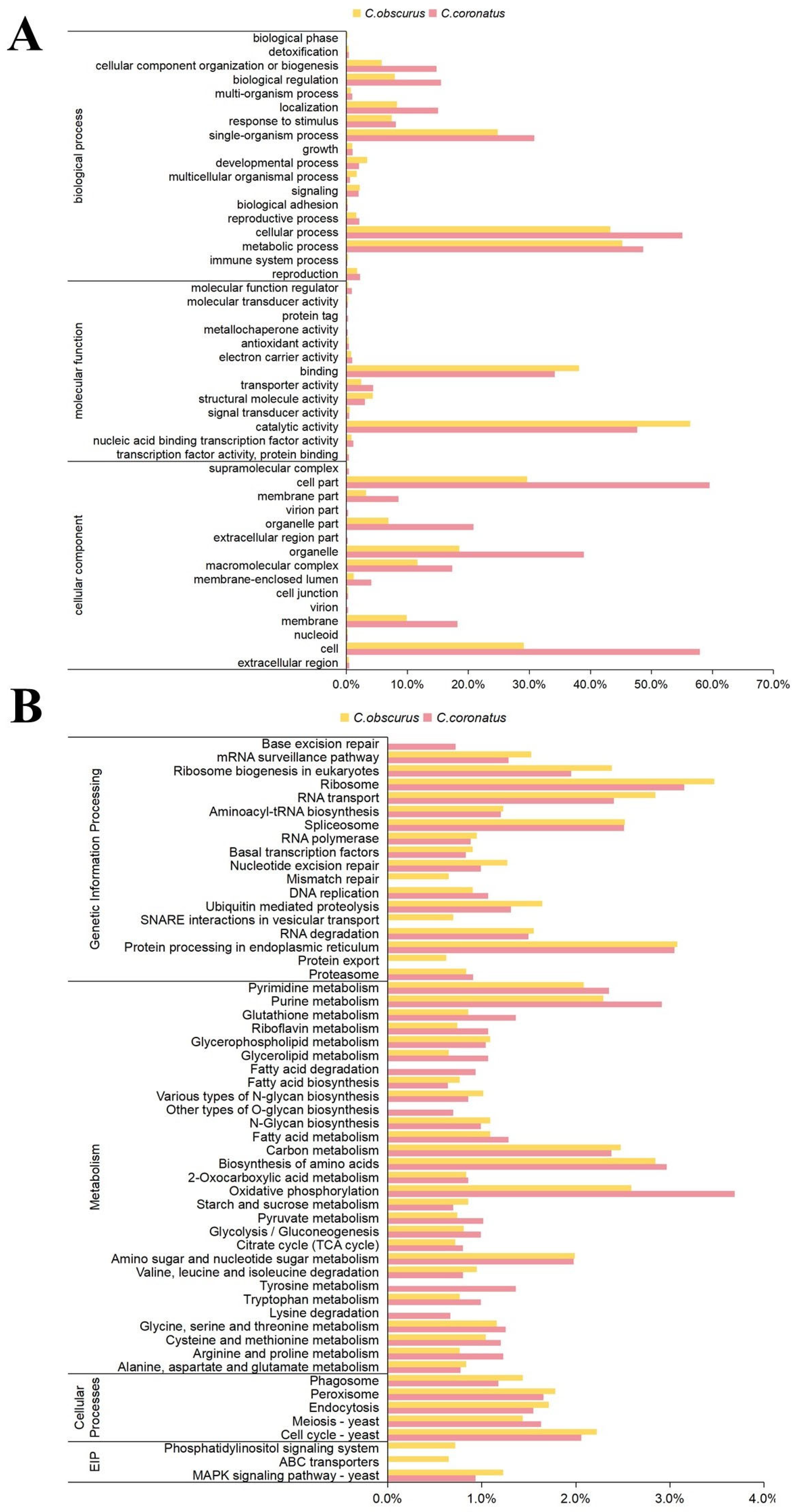
Among the 11,128 protein-coding genes identified in C. coronatus, 8025 genes (72.1%) received functional annotations through cross-database integration (Supplementary Table S8). Of these, 3702 genes were assigned GO terms (Figure 2A; Supplementary Table S9), distributed across molecular function (13 subcategories), cellular component (15 subcategories), and biological process (17 subcategories). The predominant GO terms were cell part (2205 genes), cell (2146), cellular process (2039), metabolic process (1801), and catalytic activity (1765). In contrast, C. obscurus exhibited significantly fewer GO annotations (2375 genes) [18], with its top terms being catalytic activity (1339), metabolic process (1074), and cellular process (1028). The expanded repertoire of cell-related genes in C. coronatus (e.g., cell: 2146 vs. 690 in C. obscurus) correlates with its enhanced capacity for conidial polymorphism. As shown in Figure 1E–H, C. coronatus produces diverse conidial forms, including microconidia and villose conidia, enabling adaptive fungal dispersal and survival.
Furthermore, three thousand seven hundred forty-two genes were annotated using the KEGG database (Figure 2B; Supplementary Table S10), covering metabolism (twenty-nine subcategories), genetic information processing (fifteen subcategories), cellular processes (five subcategories), and environmental information processing (one subcategory). The most enriched pathways in C. coronatus included oxidative phosphorylation (138 genes), ribosome (118), biosynthesis of amino acids (111), protein processing in endoplasmic reticula (114), and purine metabolism (109). For C. obscurus, the top enriched pathways were ribosome (150), protein processing in endoplasmic reticula (133), RNA transport (123), biosynthesis of amino acids (123), and oxidative phosphorylation (112). Additionally, 5571 genes of C. coronatus were annotated with KOG functional categories, fewer than the 6465 annotated in C. obscurus (Supplementary Table S11). The most prominent function contrasts between the two species were observed in energy production and conversion (391 vs. 339 genes), amino acid transport and metabolism (369 vs. 349 genes), and secondary-metabolite biosynthesis, transport, and catabolism (262 vs. 237 genes). In integrated KEGG and KOG analyses, C. coronatus exhibits significant advantages in energy metabolism (particularly oxidative phosphorylation) and secondary-metabolite biosynthesis, while also demonstrating strong amino acid metabolic capabilities.

3.3. Special Functional Annotation of C. coronatus Genes

3.3.1. CAZy Database Annotation

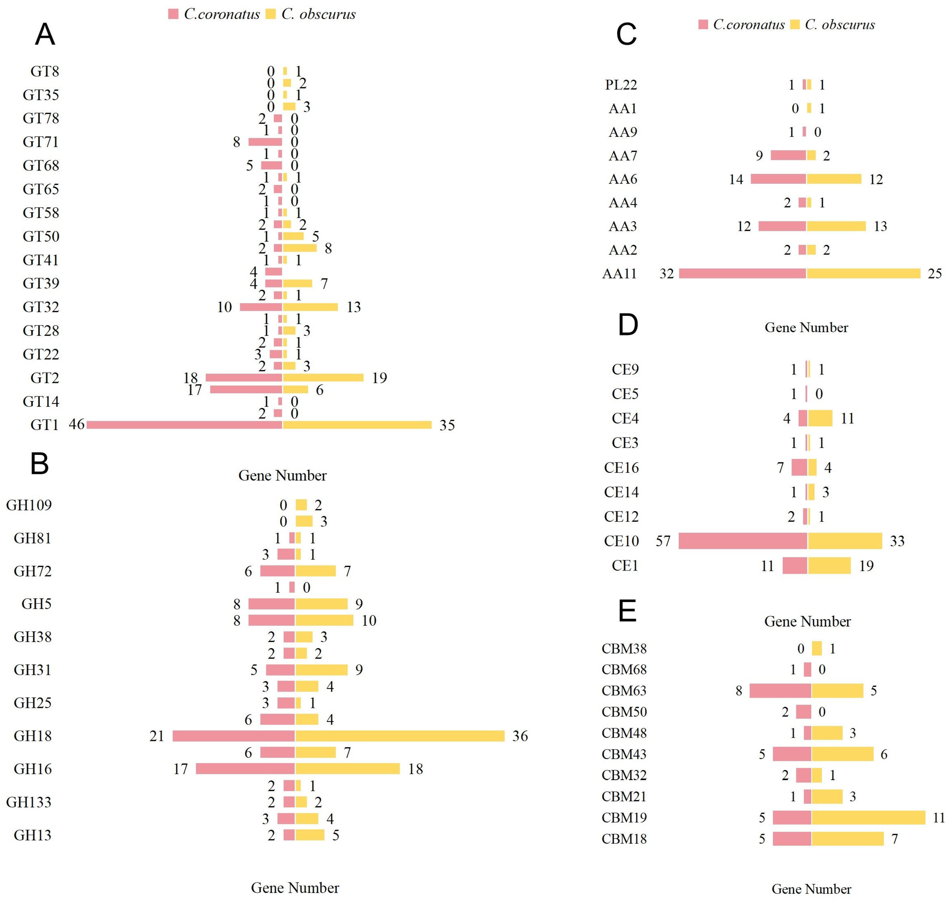
The CAZy database (http://www.cazy.org/, accessed on 30 August 2024) catalogs enzymes involved in glycosidic bond degradation, modification, and biosynthesis [42]. Analysis identified four hundred thirty CAZymes in C. coronatus, comprising five major enzyme classes alongside carbohydrate-binding modules (Figure 3): glycoside hydrolases (GHs), one hundred one enzymes (nineteen families, 23.48%); glycosyl transferases (GTs), one hundred forty-one enzymes (twenty-seven families, 32.79%); polysaccharide lyases (PLs), one enzyme (one family, 0.23%); carbohydrate esterases (CEs), eighty-five enzymes (nine families, 19.76%); auxiliary activities (AAs), seventy-two enzymes (seven families, 16.74%); and thirty carbohydrate-binding modules (CBMs, nine families, 6.97%). C. coronatus exhibits significant expansion in the GT1, GT71, CE10, AA7, and AA11 families compared to C. obscurus, which displayed high diversity in the GH18 (36 genes) and CBM19 (11 genes) families (Supplementary Table S12).

3.3.2. PHI-Base Annotations

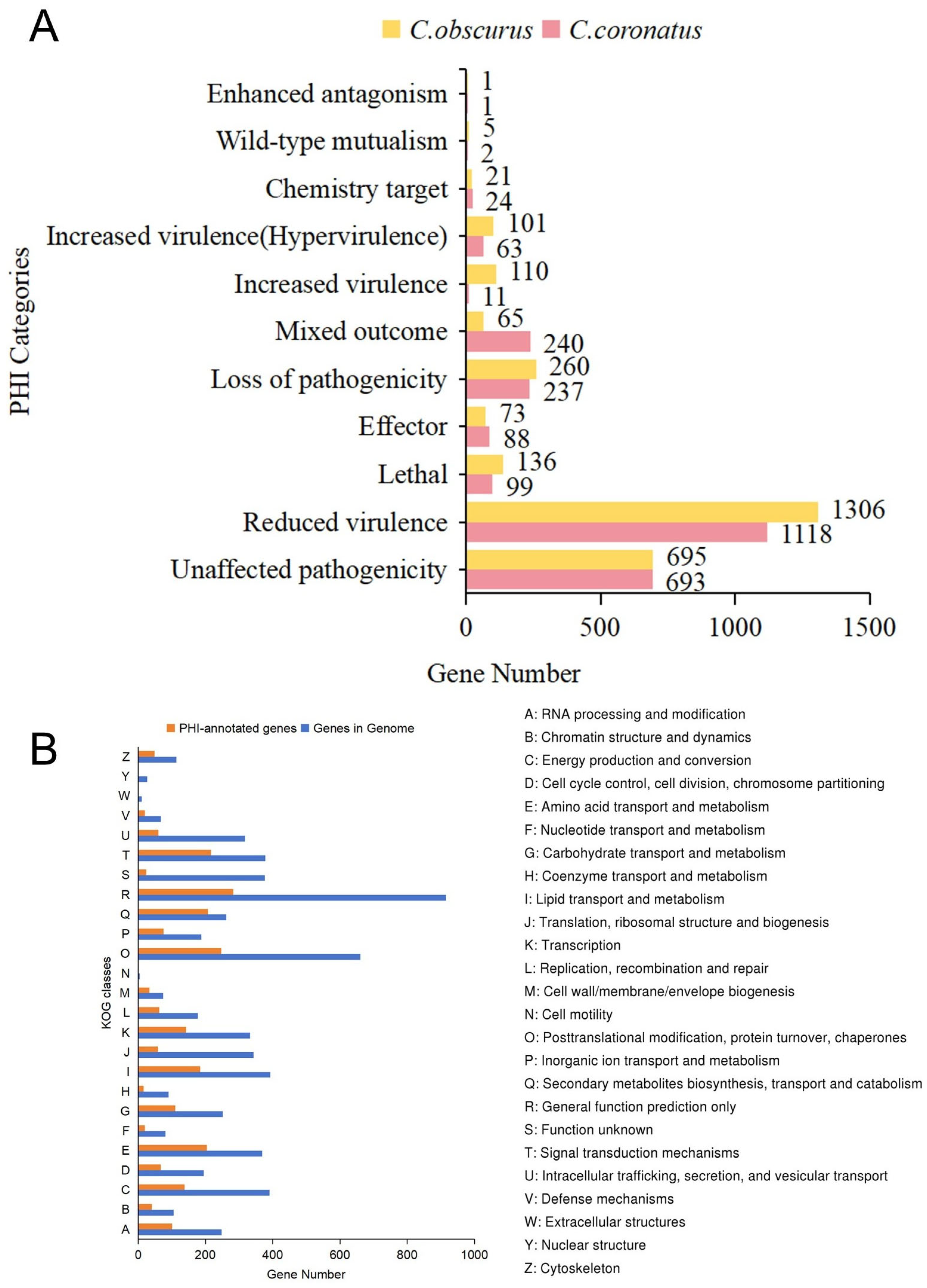
PHI-base curates experimentally validated or literature-reported pathogenic genes, virulence factors, and effector proteins from bacterial, fungal, and other pathogens infect plants, fungi, or insects [41]. Based on the C. coronatus genome, 2575 genes were annotated in PHI-base, with 73.2% predicted to associate with fungal pathogenicity (Figure 4A). These included 1118 genes (43.4%) linked to reduced virulence (weakening pathogenicity), 74 genes (2.9%) linked to increased virulence, and 99 genes (3.8%) linked to host lethality. Compared to C. obscurus, C. coronatus possessed more genes related to mixed outcomes (240 vs. 65 genes) and effectors (88 vs. 73 genes), whereas C. obscurus had higher numbers of genes associated with increased virulence (110 vs. 11 genes) and lethality (136 vs. 99 genes).
Further analysis of PHI-annotated genes in C. coronatus via KOG classification revealed significant enrichment within the O functional category (Posttranslational modification, protein turnover, and chaperones), accounting for 32.7% of all the PHI-associated genes (Figure 4B). This enrichment probably reflects adaptations in the secretory virulence machinery. We identified 1316 secreted proteins harboring signal peptides but lacking transmembrane domains. Functional classification highlighted their roles in the host’s barrier degradation and immune suppression, especially exhibiting expansions in metallopeptidases M28 and M36 (twenty-three vs. zero genes in C. obscurus), tyrosinase (fifty-five vs. twenty-four), and cysteine-rich secretory proteins (twenty-three vs. fifteen) (Supplementary Table S13).

3.3.3. Protein Family (Pfam) Domain Annotation

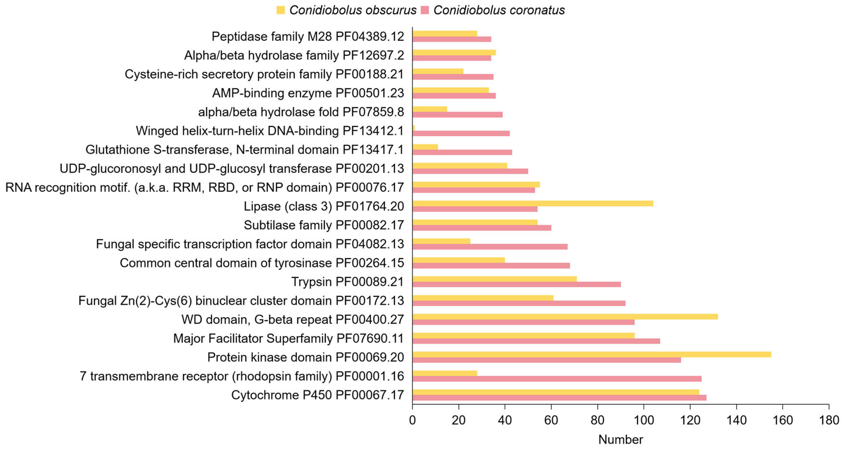
Comparative Pfam domain analysis reveals that C. coronatus exhibits distinct protein family features associated with host adaptation (Figure 5). Notably, C. coronatus possesses more genes encoding seven-transmembrane receptors (PF00001.16; one hundred twenty-five vs. twenty-eight genes in C. obscurus), fungus-specific transcription factors (PF04082.13; sixty-seven vs. twenty-five), glutathione S-transferase N-terminal domains (PF13417.1; forty-three vs. eleven), winged-helix–turn-helix DNA-binding domains (PF13412.1; forty-two vs. one), and alpha/beta-hydrolase folds (PF07859.8; thirty-nine vs. fifteen). Meanwhile, C. coronatus shows comparable abundance in P450s (PF00067.17; 127 vs. 124). Integrated CYPED analysis further identifies CYP51 as the most abundant subfamily (Supplementary Figure S2). These findings suggest that C. coronatus has evolved enhanced capabilities for environmental sensing, xenobiotic detoxification, and transcriptional regulation, probably supporting its adaptive flexibility in diverse ecological niches.

3.4. Abundant Serine Proteases and Metallopeptidases in C. coronatus

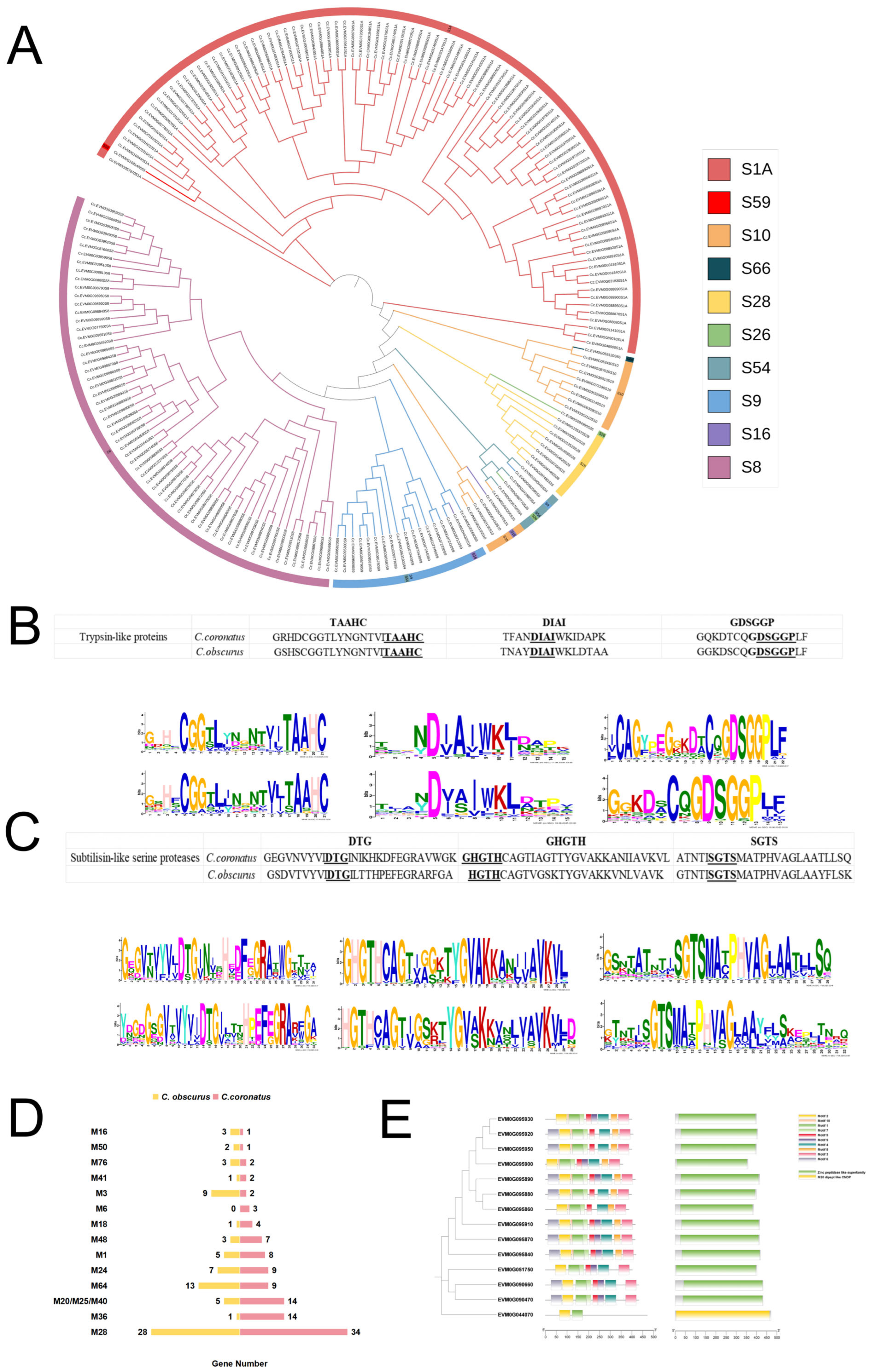
Proteolytic enzymes play a pivotal role in fungal insect infections, where degradation of insect epidermal components requires the synergistic action of diverse peptidases, directly influencing the host range specificity. Generalist entomopathogenic fungi are reported to encode more peptidases than specialists [3]. Pfam annotation identified one hundred ninety-eight serine protease genes in C. coronatus, distributed across ten families: trypsin (S1A, ninety genes), subtilisin (S8, sixty), prolyl oligopeptidase (S9, eighteen), carboxypeptidase Y (S10, twelve), lon protease (S16, two), signal peptidase I (S26, two), acid prolyl endopeptidase (S28, eight), rhomboid (S54, four), nucleoporin (S59, one), and LD-carboxypeptidase (S66, one) (Figure 6A). Compared to C. obscurus, C. coronatus exhibited a higher number of genes within trypsin (S1A) and subtilisin (S8) families (Supplementary Table S14).
MEME-based motif analysis confirmed conserved sequences flanking the catalytic triad residues (DTG, GHGTH, and SGTS) in subtilisin of both species (Figure 6C). Likewise, trypsin exhibited conserved active-site residues (His, Asp, and Ser; HDS motif) with highly analogous motif architectures (Figure 6B).
The C. coronatus genome also displays significant expansion in metallopeptidase genes (107 vs. 78 in C. obscurus, Figure 6D). Notably, it exhibits an increased number of M20/M25/M40 domains (fourteen vs. five, Figure 6E; PF01546.23), which cleave zinc-dependent peptide bonds at N-termini, suggesting their expansion facilitates adaptive proteolysis across diverse host substrates. Additionally, C. coronatus possesses a markedly expanded repertoire of M36 metallopeptidase genes (fourteen vs. one).

3.5. Expansion of G-Protein-Coupled Receptors in C. coronatus

Fungal GPCRs mediate host recognition and activate downstream signaling pathways critical for infection initiation [49]. Pfam database annotation identified 125 GPCR-encoding genes in the C. coronatus genome, significantly exceeding the 28 observed in C. obscurus. This expansion suggests that broad-host-range entomopathogenic fungi employ increased GPCR diversity to detect heterogeneous host environmental signals.
Further analysis of the 125 GPCRs in C. coronatus, using MEME suite, revealed 10 conserved motifs (Figure 7A). Among these, motif 10 exhibited high conservation, with an aspartic acid residue present in 123 GPCRs (Figure 7B). Domain annotation classified one hundred twelve functionally annotated GPCRs into three categories: seven tm class-A rhodopsin-like, seven tm GPCR superfamily, and seven tm 1 superfamily.

3.6. Expansion of Transcription Factors in C. coronatus

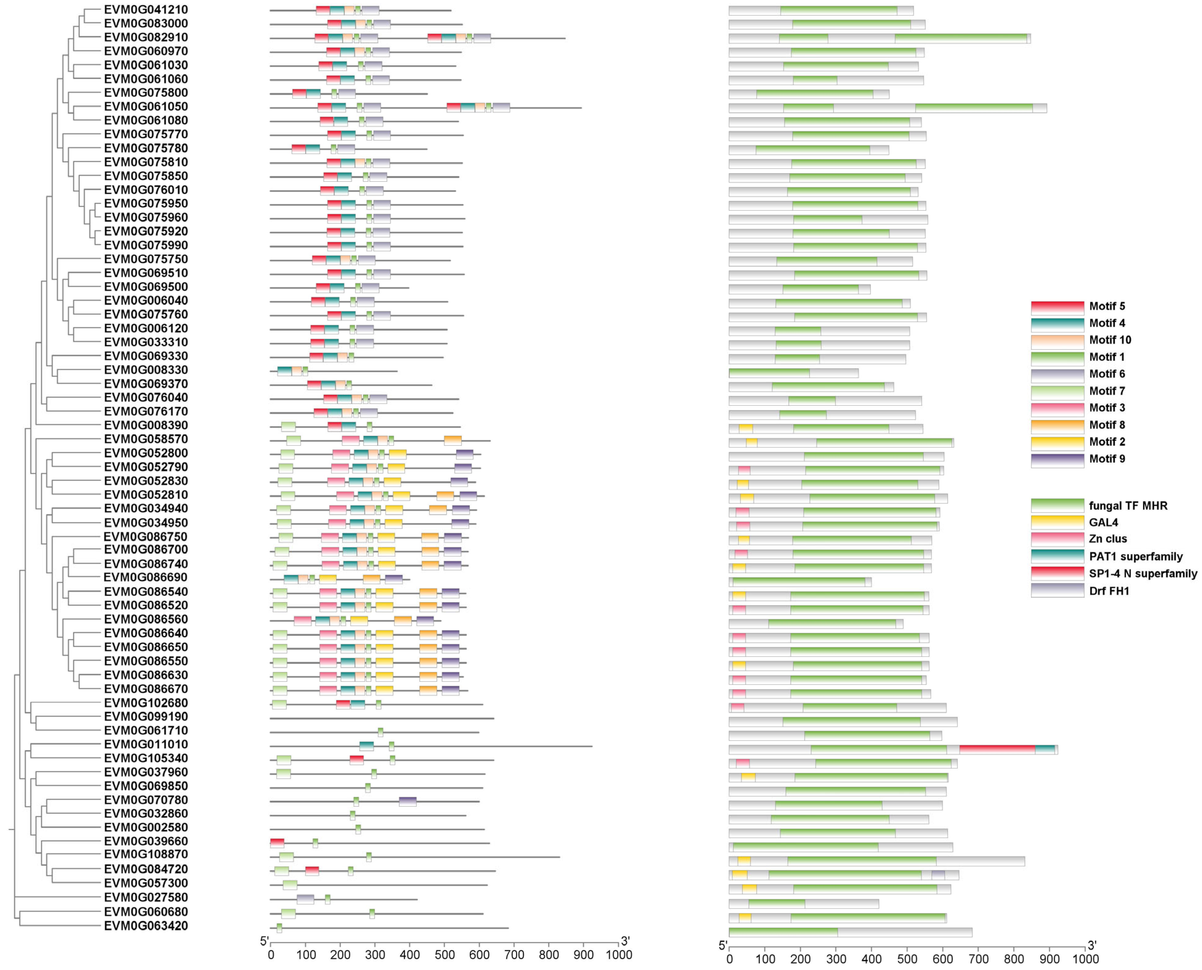
Pfam annotation identified diverse transcription factors in C. coronatus (Supplementary Table S16), including fungus-specific transcription factor domain, bZIP transcription factor, histone-like transcription factor, fungal Zn2-Cys6 binuclear cluster domain, and zinc-finger double domain. This repertoire highlights the complexity of its transcriptional network. Compared to C. obscurus, C. coronatus contains 67 genes encoding the fungus-specific transcription factor domain (Figure 8). Among these, 11 transcription factors were annotated using the Swiss-Prot database, with most involved in regulating nitrogen assimilation processes (Supplementary Table S17). This abundance suggests their role as core regulatory elements in C. coronatus responses to environmental signals, potentially facilitating its adaptation to multiple hosts.

3.7. Metabolomic Profiling Reveals Enhanced Terpenoid Biosynthesis in C. coronatus

Genome mining using antiSMASH v6.0 identified eleven biosynthetic gene clusters in C. coronatus, including two dedicated to terpenoid biosynthesis (Supplementary Table S18). Untargeted metabolomic profiling via LC-quadrupole time-of-flight detected 1073 metabolites in negative ion mode and 2062 metabolites in positive ion mode (Supplementary Figure S3). Subsequent annotation against the KEGG and LIPID MAPS databases classified 51 metabolites as terpenoid-related compounds. The OPLS-DA model revealed 28 terpenoid-like metabolites exhibiting differential abundance between C. coronatus and C. obscurus, based on the variable importance in projection values. Of these, 22 metabolites were significantly upregulated in C. coronatus (Table 3), including monoterpenes, sesquiterpenes, diterpenes, tetraterpenes, and terpendoles.

4. Discussion

Fungi of the genus Conidiobolus exhibit polyphyletic development, encompassing diverse ecological niches, including soil saprotrophy, insect parasitism, plant symbiosis, and even human infection [17,18,50]. Recent studies integrating molecular phylogenetic reconstruction, morphological and ecological traits, and evolutionary trajectories have prompted the reclassification of Conidiobolus into multiple families, such as Neoconidiobolaceae [11]. From the perspective of insect pathogenesis, successful host infection involves three critical stages: host recognition, host invasion, and host nutrient adaptation/utilization [51]. These processes rely on activation of fungal infection machinery triggered by host-specific signals. Conversely, exposure to non-host substrates typically triggers infective conidia to produce secondary conidia for dispersal—a hallmark of entomophthoralean fungi that employ diverse spore types to facilitate transmission and survival [1,52]. Upon signal perception, fungal gene expression patterns are dramatically reconfigured via signal transduction mechanisms to adapt to heterogeneous host environments [3,53]. Genomic analysis herein revealed that C. coronatus possesses expanded repertoires of host recognition molecules, transcription regulators, and host-invading enzymes, thus comprehensively enabling its infection continuum (Supplementary Table S19).
Cell surface GPCRs mediate host recognitions in insect pathogens by detecting chemical/physical signals (e.g., isoflavones and hydrophobicity) and regulating infection processes [49,51,54]. Comparative genomic analysis shows that the generalist M. anisopliae encodes significantly more GPCR genes (61) than the specialist M. acridum (37). Furthermore, ablation of individual GPCR genes impairs infection structure formation, suggesting GPCRs in broad-spectrum host recognition [51,55,56]. We identified 125 GPCRs with diverse motif architectures in C. coronatus, suggesting a molecular basis for its broad host range.
GPCRs activate infection-related enzymes via MAPK, cAMP-PKA, or calcium signal pathways [51]. However, functional genomics in entomophthoralean fungi remain constrained by limited genetic tools. Current knowledge primarily extrapolates from model fungi. Although C. coronatus encodes diverse transcription factor families, functional validation is scarce. Nevertheless, many are putatively associated with pathogenicity, such as prz1 (Swiss-Prot ID), involved in regulating calcium ion homeostasis for germination [57]; TUP1, repressing pathogenesis-related genes [58]; TRY3, involved in fungal cell adherence to substrates [59]; and GIS1, involved in the Ras/cAMP pathway for nutrient adaptation [60]. Additionally, long non-coding RNAs have been reported to regulate virulence attenuation in C. obscurus [61]. Given these gaps, transcriptomic profiling of core gene clusters and non-coding regulators during C. coronatus infection is imperative.
Prior to successful colonization of the insect hemocoel, entomopathogenic fungi must overcome the host’s physicochemical barriers and evade immune defenses. The insect cuticle comprises a protein–chitin matrix embedded with lipids [62]. To breach this barrier, entomopathogenic fungi secrete cuticle-degrading enzymes (CDEs), including serine proteases (subtilisin), chitinases (GH18), and lipases [18,53]. Genomic analysis in these CDE families supports C. coronatus’ capacity to invade diverse hosts. Functional annotations indicate synergistic roles; e.g., CE10 esterases remove ester-based modifications from chitin substrates to facilitate chitinase activity; protease–chitinase complexes hydrolyze cuticular matrices; and lipases degrade epicuticular waxes [18,63]. Notably, individual subtilisin contributes 19–29% to virulence in the generalist B. bassiana [64]. The pronounced CDE expansion in C. coronatus suggests exceptional cuticle penetration adaptability, underpinning its broad host range.
Meanwhile, our study has revealed numerous effector molecules that counteract diverse host immune defenses, including P450s, tyrosinases, and cysteine-rich secretory proteins (CRISPs). In C. coronatus, P450s, such as CYP51, facilitate fungal colonization by synthesizing ergosterol for membrane integrity and detoxify host-derived compounds [65]. Tyrosinases, copper-containing oxidases, catalyze melanin biosynthesis, which protects against the host’s hydrolytic enzymes, UV radiation, and oxidative stress by scavenging free radicals [66]. CRISPs, belonging to the CAP superfamily, disrupt host immunity through ion channel modulation and sterol binding, thereby enhancing pathogen virulence [67,68]. Additionally, C. coronatus produces non-ribosomal peptides, coronatin-1/2, which directly incapacitate host hemocytes by inducing cytoskeletal disassembly and apoptosis [69,70].
Host-genotype-specific variations modulate interaction outcomes. In our study, 240 PHI-annotated genes correlate with “mixed outcome” phenotypes, probably attributable to C. coronatus’ broad host range. Generalist pathogens target conserved host features while dynamically adapting virulence strategies to specific host genotypes [15]. The acquisition of genes encoding nonselective toxins via horizontal gene transfer (HGT) enhances pathogenic virulence [3]. For example, CytCo (a cytolytic-like gene in the aphid-obligate pathogen C. obscurus), probably originating from bacterial pathogens, induces toxicity in Galleria mellonella hemocytes and nematodes by disrupting calcium homeostasis through interacting with calcium-transporting ATPase [71,72,73,74]. Destruxins, broad-spectrum toxins targeting nerve–muscle ion channels and disrupting hemocyte-mediated immunity in diverse insects, were acquired via HGT of the dtx gene cluster from a plant-associated ancestor resembling Alternaria species [56]. C. coronatus’s metabolites also exhibit insecticidal activity across hosts, e.g., harman and norharman alkaloids exhibit efficacy against Lepidoptera hosts [75]. In this study, sesquiterpene metabolites produced via terpenoid biosynthetic gene clusters in C. coronatus suggest insecticidal and plant-growth-promoting effects, warranting experimental validation.
Conclusively, our integrated genomic and metabolomic analyses reveal divergent evolutionary adaptations underpinning host-range plasticity in Conidiobolus pathogens: C. coronatus (a generalist) exhibits genomic plasticity through expansions in host recognition (GPCRs), invasion (protease–CAZyme synergies), and niche exploitation (terpenoid–P450 networks), enabling broad host ranges across insect taxa, whereas C. obscurus (an aphid-obligate specialist) relies on focused virulence toolkits. Current limitations in genetic tools for Entomophthorales constrain functional validation of these candidates; integrating multiomic approaches will elucidate how C. coronatus dynamically reprograms its infectivity to adapt to diverse insect hosts. The broad-spectrum efficacy of C. coronatus positions it for deployment against multi-pest complexes (e.g., lepidopteran–dipteran infestations), leveraging its polyphagous adaptions for targeted biocontrol applications.

Supplementary Materials

The following supporting information can be downloaded at: https://www.mdpi.com/article/10.3390/jof11080600/s1, Figure S1: Images of Hyphantria cunea larvae infected by Conidiobolus coronatus after conidial inoculation; Figure S2: P450 family classification statistics of C. coronatus; Figure S3: Volcano plots of differentially abundant metabolites between C. coronatus and C. obscurus; Table S1: CCS-read data volume statistics of Conidiobolus coronatus; Table S2: Statistics of genome assembly results; Table S3: Comparison result statistics; Table S4: BUSCO evaluation statistics; Table S5: Statistics of repeat sequence prediction results; Table S6: Statistics of genetic prediction results; Table S7: Basic genetic information statistics; Table S8: Gene function annotation statistics; Table S9: Comparison of GO functional annotations between C. coronatus and C. obscurus; Table S10: KEGG functional annotation statistics of C. coronatus and C. obscurus; Table S11: The KOG classes in C coronatus vs. C. obscurus; Table S12: The different numbers of carbohydrate enzyme family genes in C. coronatus and C. obscurus; Table S13: T20-secreted protein of Pfam annotation; Table S14: Serine protease gene numbers in C. coronatus and C. obscurus; Table S15: Metallopeptidase categories of C. coronatus; Table S16: Transcription factor categories of C. coronatus; Table S17: Swiss-Prot-annotated functions of genes with fungus-specific transcription factor domain; Table S18: Gene clusters for secondary-metabolite biosynthesis in C. coronatus genome; Table S19: Molecular mechanisms underlying Conidiobolus coronatus’s broad host adaptability across infection stages.

Author Contributions

X.Z. conceived the idea and aims and designed the experiments. L.Z., T.Y., and X.C. conducted the fungal culture and DNA extraction. X.Z. conducted the formal analysis. F.B. and J.Y. conducted the data curation. F.B. performed writing—original draft preparation. X.Z. performed writing—review and editing. All authors have read and agreed to the published version of the manuscript.

Funding

This research was funded by a grant from the National Natural Science Foundation of China (Grant No. 32271883).

Institutional Review Board Statement

Not applicable.

Informed Consent Statement

Not applicable.

Data Availability Statement

The PacBio-II-assembled genome was deposited at GenBank, with accession PRJNA1264516. All the data generated or analyzed during this study are included in the published article, accessions, and Supplementary Material file.

Conflicts of Interest

The authors declare no conflicts of interest.

References

  1. de Bekker, C.; Beckerson, W.C.; Elya, C. Mechanisms behind the madness: How do zombie-making fungal entomopathogens affect host behavior to increase transmission? mBio 2021, 12, e0187221. [Google Scholar] [CrossRef]
  2. Wang, C.S.; Feng, M.G. Advances in fundamental and applied studies in China of fungal biocontrol agents for use against arthropod pests. Biol. Control 2014, 68, 129–135. [Google Scholar] [CrossRef]
  3. Wang, C.; Wang, S. Insect pathogenic fungi: Genomics, molecular interactions, and genetic improvements. Annu. Rev. Entomol. 2017, 62, 73–90. [Google Scholar] [CrossRef]
  4. Lovett, B.; St Leger, R.J. Genetically engineering better fungal biopesticides. Pest Manag. Sci. 2018, 74, 781–789. [Google Scholar] [CrossRef] [PubMed]
  5. Tong, S.M.; Feng, M.G. Molecular basis and regulatory mechanisms underlying fungal insecticides’ resistance to solar ultraviolet irradiation. Pest Manag. Sci. 2022, 78, 30–42. [Google Scholar] [CrossRef]
  6. Faria, M.R.; Wraight, S.P. Mycoinsecticides and mycoacaricides: A comprehensive list with worldwide coverage and international classification of formulation types. Biol. Control 2007, 43, 237–256. [Google Scholar] [CrossRef]
  7. Brodeur, J. Host specificity in biological control: Insights from opportunistic pathogens. Evol. Appl. 2012, 5, 470–480. [Google Scholar] [CrossRef]
  8. Sacco, N.E.; Hajek, A.E. Diversity and breadth of host specificity among arthropod pathogens in the Entomophthoromycotina. Microorganisms 2023, 11, 1658. [Google Scholar] [CrossRef] [PubMed]
  9. Zhou, X. Winter prevalence of obligate aphid pathogen Pandora neoaphidis mycosis in the host Myzus persicae populations in southern China: Modeling description and biocontrol implication. Braz. J. Microbiol. 2012, 43, 325–331. [Google Scholar] [CrossRef] [PubMed]
  10. Elya, C.; Lok, T.C.; Spencer, Q.E.; McCausland, H.; Martinez, C.C.; Eisen, M. Robust manipulation of the behavior of Drosophila melanogaster by a fungal pathogen in the laboratory. eLife 2018, 7, e34414. [Google Scholar] [CrossRef]
  11. Gryganskyi, A.P.; Nie, Y.; Hajek, A.E.; Hodge, K.T.; Liu, X.Y.; Aadland, K.; Voigt, K.; Anishchenko, I.M.; Kutovenko, V.B.; Kava, L.; et al. The early terrestrial fungal lineage of Conidiobolus-transition from saprotroph to parasitic lifestyle. J. Fungi 2022, 8, 789. [Google Scholar] [CrossRef] [PubMed]
  12. Boguś, M.I.; Kazek, M.; Drozdowski, M.; Kaczmarek, A.; Wrońska, A.K. The entomopathogenic fungus Conidiobolus coronatus has similar effects on the cuticular free fatty acid profile of sensitive and resistant insects. Insects 2023, 14, 895. [Google Scholar] [CrossRef] [PubMed]
  13. Torres Acosta, R.I.; Humber, R.A.; Sánchez-Peña, S.R. Zoophthora radicans (Entomophthorales), a fungal pathogen of Bagrada hilaris and Bactericera cockerelli (Hemiptera: Pentatomidae and Triozidae): Prevalence, pathogenicity, and interplay of environmental influence, morphology, and sequence data on fungal identification. J. Invertebr. Pathol. 2016, 139, 82–91. [Google Scholar] [CrossRef]
  14. De Fine Licht, H.H.; Hajek, A.E.; Eilenberg, J.; Jensen, A.B. Utilizing genomics to study entomopathogenicity in the fungal phylum Entomophthoromycota: A review of current genetic resources. Adv. Genet. 2016, 94, 41–65. [Google Scholar]
  15. St Leger, R.J. The evolution of complex Metarhizium-insect-plant interactions. Fungal Biol. 2024, 128, 2513–2528. [Google Scholar] [CrossRef]
  16. Stajich, J.E.; Lovett, B.; Lee, E.; Macias, A.M.; Hajek, A.E.; de Bivort, B.L.; Kasson, M.T.; De Fine Licht, H.H.; Elya, C. Signatures of transposon-mediated genome inflation, host specialization, and photoentrainment in Entomophthora muscae and allied entomophthoralean fungi. eLife 2024, 12, RP92863. [Google Scholar] [CrossRef]
  17. Wang, Y.; Nie, Y.; Yu, D.; Xie, X.; Qin, L.; Yang, Y.; Huang, B. Genome-wide study of saprotrophy-related genes in the basal fungus Conidiobolus heterosporus. Appl. Microbiol. Biotechnol. 2020, 104, 6261–6272. [Google Scholar] [CrossRef]
  18. Zhang, L.; Yang, T.; Yu, W.; Wang, X.; Zhou, X.; Zhou, X. Genome-wide study of conidiation-related genes in the aphid-obligate fungal pathogen Conidiobolus obscurus (Entomophthoromycotina). J. Fungi 2022, 8, 389. [Google Scholar] [CrossRef]
  19. Chen, E.C.H.; Morin, E.; Beaudet, D.; Noel, J.; Yildirir, G.; Ndikumana, S.; Charron, P.; St-Onge, C.; Giorgi, J.; Krüger, M.; et al. High intraspecific genome diversity in the model arbuscular mycorrhizal symbiont Rhizophagus irregularis. New Phytol. 2018, 220, 1161–1171. [Google Scholar] [CrossRef]
  20. Hane, J.K.; Anderson, J.P.; Williams, A.H.; Sperschneider, J.; Singh, K.B. Genome sequencing and comparative genomics of the broad host-range pathogen Rhizoctonia solani AG8. PLoS Genet. 2014, 10, e1004281. [Google Scholar] [CrossRef] [PubMed]
  21. Lang, D.; Zhang, S.; Ren, P.; Liang, F.; Sun, Z.; Meng, G.; Tan, Y.; Li, X.; Lai, Q.; Han, L.; et al. Comparison of the two up-to-date sequencing technologies for genome assembly: HiFi reads of Pacific Biosciences Sequel II system and ultralong reads of Oxford Nanopore. Gigascience 2020, 9, giaa123. [Google Scholar] [CrossRef] [PubMed]
  22. Congrains, C.; Bremer, F.; Dupuis, J.R.; Barr, N.B.; Garzón-Orduña, I.J.; Rubinoff, D.; Doorenweerd, C.; Jose, M.S.; Morris, K.; Kauwe, A.; et al. CCS-Consensuser: A haplotype-aware consensus generator for PacBio amplicon sequences. Mol. Ecol. Resour. 2025, 4, e14113. [Google Scholar] [CrossRef]
  23. Cheng, H.; Concepcion, G.T.; Feng, X.; Zhang, H.; Li, H. Haplotype-resolved de novo assembly using phased assembly graphs with hifiasm. Nat. Methods 2021, 18, 170–175. [Google Scholar] [CrossRef]
  24. Walker, B.J.; Abeel, T.; Shea, T.; Priest, M.; Abouelliel, A.; Sakthikumar, S.; Cuomo, C.A.; Zeng, Q.; Wortman, J.; Young, S.K.; et al. Pilon: An integrated tool for comprehensive microbial variant detection and genome assembly improvement. PLoS ONE 2014, 9, e112963. [Google Scholar] [CrossRef]
  25. Li, H.; Durbin, R. Fast and accurate short read alignment with Burrows-Wheeler transform. Bioinformatics 2009, 25, 1754–1760. [Google Scholar] [CrossRef]
  26. Simão, F.A.; Waterhouse, R.M.; Ioannidis, P.; Kriventseva, E.V.; Zdobnov, E.M. BUSCO: Assessing genome assembly and annotation completeness with single-copy orthologs. Bioinformatics 2015, 31, 3210–3212. [Google Scholar] [CrossRef]
  27. Xu, Z.; Wang, H. LTR_FINDER: An efficient tool for the prediction of full-length LTR retrotransposons. Nucleic Acids Res. 2007, 35, W265–W268. [Google Scholar] [CrossRef] [PubMed]
  28. Han, Y.; Wessler, S.R. MITE-Hunter: A program for discovering miniature inverted-repeat transposable elements from genomic sequences. Nucleic Acids Res. 2010, 38, e199. [Google Scholar] [CrossRef]
  29. Price, A.L.; Jones, N.C.; Pevzner, P.A. De novo identification of repeat families in large genomes. Bioinformatics 2005, 21, i351–i358. [Google Scholar] [CrossRef]
  30. Edgar, R.C.; Myers, E.W. PILER: Identification and classification of genomic repeats. Bioinformatics 2005, 21, i152–i158. [Google Scholar] [CrossRef] [PubMed]
  31. Bao, W.; Kojima, K.K.; Kohany, O. Repbase Update, a database of repetitive elements in eukaryotic genomes. Mob. DNA 2015, 6, 11. [Google Scholar] [CrossRef]
  32. Tarailo-Graovac, M.; Chen, N. Using RepeatMasker to identify repetitive elements in genomic sequences. Curr. Protoc. Bioinform. 2009, 25, 4.10.1–4.10.14. [Google Scholar] [CrossRef]
  33. Haas, B.J.; Salzberg, S.L.; Zhu, W.; Pertea, M.; Allen, J.E.; Orvis, J.; White, O.; Buell, C.R.; Wortman, J.R. Automated eukaryotic gene structure annotation using EVidenceModeler and the program to assemble spliced alignments. Genome Biol. 2008, 9, R7. [Google Scholar] [CrossRef]
  34. Nawrocki, E.P.; Eddy, S.R. Infernal 1.1: 100-fold faster RNA homology searches. Bioinformatics 2013, 29, 2933–2935. [Google Scholar] [CrossRef] [PubMed]
  35. Lowe, T.M.; Eddy, S.R. tRNAscan-SE: A program for improved detection of transfer RNA genes in genomic sequence. Nucleic Acids Res. 1997, 25, 955–964. [Google Scholar] [CrossRef] [PubMed]
  36. Tatusov, R.L.; Galperin, M.Y.; Natale, D.A.; Koonin, E.V. The COG database: A tool for genome-scale analysis of protein functions and evolution. Nucleic Acids Res. 2000, 28, 33–36. [Google Scholar] [CrossRef]
  37. Kanehisa, M.; Goto, S.; Kawashima, S.; Okuno, Y.; Hattori, M. The KEGG resource for deciphering the genome. Nucleic Acids Res. 2004, 32, D277–D280. [Google Scholar] [CrossRef] [PubMed]
  38. Boeckmann, B.; Bairoch, A.; Apweiler, R.; Blatter, M.C.; Estreicher, A.; Gasteiger, E.; Martin, M.J.; Michoud, K.; O’Donovan, C.; Phan, I.; et al. The SWISS-PROT protein knowledgebase and its supplement TrEMBL in 2003. Nucleic Acids Res. 2003, 31, 365–370. [Google Scholar] [CrossRef] [PubMed]
  39. Ashburner, M.; Ball, C.A.; Blake, J.A.; Botstein, D.; Butler, H.; Cherry, J.M.; Davis, A.P.; Dolinski, K.; Dwight, S.S.; Eppig, J.T.; et al. Gene ontology: Tool for the unification of biology. The Gene Ontology Consortium. Nat. Genet. 2000, 25, 25–29. [Google Scholar] [CrossRef]
  40. Finn, R.D.; Coggill, P.; Eberhardt, R.Y.; Eddy, S.R.; Mistry, J.; Mitchell, A.L.; Potter, S.C.; Punta, M.; Qureshi, M.; Sangrador-Vegas, A.; et al. The Pfam protein families database: Towards a more sustainable future. Nucleic Acids Res. 2016, 44, D279–D285. [Google Scholar] [CrossRef]
  41. Urban, M.; Cuzick, A.; Rutherford, K.; Irvine, A.; Pedro, H.; Pant, R.; Sadanadan, V.; Khamari, L.; Billal, S.; Mohanty, S.; et al. PHI-base: A new interface and further additions for the multi-species pathogen-host interactions database. Nucleic Acids Res. 2017, 45, D604–D610. [Google Scholar] [CrossRef]
  42. Lombard, V.; Golaconda Ramulu, H.; Drula, E.; Coutinho, P.M.; Henrissat, B. The carbohydrate-active enzymes database (CAZy) in 2013. Nucleic Acids Res. 2014, 42, D490–D495. [Google Scholar] [CrossRef] [PubMed]
  43. Sirim, D.; Wagner, F.; Lisitsa, A.; Pleiss, J. The cytochrome P450 engineering database: Integration of biochemical properties. BMC Biochem. 2009, 10, 27. [Google Scholar] [CrossRef] [PubMed]
  44. Petersen, T.N.; Brunak, S.; von Heijne, G.; Nielsen, H. SignalP 4.0: Discriminating signal peptides from transmembrane regions. Nat. Methods 2011, 8, 785–786. [Google Scholar] [CrossRef] [PubMed]
  45. Krogh, A.; Larsson, B.; von Heijne, G.; Sonnhammer, E.L. Predicting transmembrane protein topology with a hidden Markov model: Application to complete genomes. J. Mol. Biol. 2001, 305, 567–580. [Google Scholar] [CrossRef]
  46. Kumar, S.; Stecher, G.; Suleski, M.; Sanderford, M.; Sharma, S.; Tamura, K. MEGA12: Molecular evolutionary genetic analysis version 12 for adaptive and green computing. Mol. Biol. Evol. 2024, 41, msae263. [Google Scholar] [CrossRef]
  47. Blin, K.; Shaw, S.; Kloosterman, A.M.; Charlop-Powers, Z.; van Wezel, G.P.; Medema, M.H.; Weber, T. antiSMASH 6.0: Improving cluster detection and comparison capabilities. Nucleic Acids Res. 2021, 49, W29–W35. [Google Scholar] [CrossRef]
  48. Want, E.J.; Wilson, I.D.; Gika, H.; Theodoridis, G.; Plumb, R.S.; Shockcor, J.; Holmes, E.; Nicholson, J.K. Global metabolic profiling procedures for urine using UPLC-MS. Nat. Protoc. 2010, 5, 1005–1018. [Google Scholar] [CrossRef]
  49. Lara-Martínez, D.; Tristán-Flores, F.E.; Cervantes-Montelongo, J.A.; Silva-Martínez, G.A. Fungal stress responses and the importance of GPCRs. J. Fungi 2025, 11, 213. [Google Scholar] [CrossRef]
  50. Vilela, R.; Mendoza, L. Human pathogenic entomophthorales. Clin. Microbiol. Rev. 2018, 31, e00014–e00018. [Google Scholar] [CrossRef]
  51. Hong, S.; Shang, J.; Sun, Y.; Tang, G.; Wang, C. Fungal infection of insects: Molecular insights and prospects. Trends Microbiol. 2024, 32, 302–316. [Google Scholar] [CrossRef]
  52. Zhou, X.; Montalva, C.; Arismendi, N.; Hong, F. Neozygites linanensis sp. nov. a fungal pathogen infecting bamboo aphids in southeast China. Mycotaxon 2017, 132, 305–315. [Google Scholar] [CrossRef]
  53. Wang, J.; Zhou, X.; Guo, K.; Zhang, X.; Lin, H.; Montalva, C. Transcriptomic insight into pathogenicity-associated factors of Conidiobolus obscurus, an obligate aphid-pathogenic fungus belonging to Entomopthoromycota. Pest. Manag. Sci. 2018, 74, 1677–1686. [Google Scholar] [CrossRef]
  54. Xue, C.; Hsueh, Y.P.; Heitman, J. Magnificent seven: Roles of G protein-coupled receptors in extracellular sensing in fungi. FEMS Microbiol. Rev. 2008, 32, 1010–1032. [Google Scholar] [CrossRef]
  55. Ying, S.H.; Feng, M.G.; Keyhani, N.O. A carbon responsive G-protein coupled receptor modulates broad developmental and genetic networks in the entomopathogenic fungus, Beauveria bassiana. Environ. Microbiol. 2013, 15, 2902–2921. [Google Scholar] [CrossRef]
  56. Hu, X.; Xiao, G.; Zheng, P.; Shang, Y.; Su, Y.; Zhang, X.; Liu, X.; Zhan, S.; St Leger, R.J.; Wang, C. Trajectory and genomic determinants of fungal-pathogen speciation and host adaptation. Proc. Natl. Acad. Sci. USA 2014, 111, 16796–16801. [Google Scholar] [CrossRef] [PubMed]
  57. Hirayama, S.; Sugiura, R.; Lu, Y.; Maeda, T.; Kawagishi, K.; Yokoyama, M.; Tohda, H.; Giga-Hama, Y.; Shuntoh, H.; Kuno, T. Zinc finger protein Prz1 regulates Ca2+ but not Cl homeostasis in fission yeast. Identification of distinct branches of calcineurin signaling pathway in fission yeast. J. Biol. Chem. 2003, 278, 18078–18084. [Google Scholar] [CrossRef]
  58. Braun, B.R.; Head, W.S.; Wang, M.X.; Johnson, A.D. Identification and characterization of TUP1-regulated genes in Candida albicans. Genetics 2000, 156, 31–44. [Google Scholar] [CrossRef] [PubMed]
  59. Finkel, J.S.; Xu, W.; Huang, D.; Hill, E.M.; Desai, J.V.; Woolford, C.A.; Nett, J.E.; Taff, H.; Norice, C.T.; Andes, D.R.; et al. Portrait of Candida albicans adherence regulators. PLoS Pathog. 2012, 8, e1002525. [Google Scholar] [CrossRef]
  60. Pedruzzi, I.; Bürckert, N.; Egger, P.; De Virgilio, C. Saccharomyces cerevisiae Ras/cAMP pathway controls post-diauxic shift element-dependent transcription through the zinc finger protein Gis1. EMBO J. 2000, 19, 2569–2579. [Google Scholar] [CrossRef] [PubMed]
  61. Ye, G.F.; Zhang, L.H.; Zhou, X. Long noncoding RNAs are potentially involved in the degeneration of virulence in an aphid-obligate pathogen, Conidiobolus obscurus (Entomophthoromycotina). Virulence 2021, 21, 1705–1716. [Google Scholar] [CrossRef] [PubMed]
  62. Włóka, E.; Boguś, M.I.; Wrońska, A.K.; Drozdowski, M.; Kaczmarek, A.; Sobich, J.; Gołębiowski, M. Insect cuticular compounds affect Conidiobolus coronatus (Entomopthorales) sporulation and the activity of enzymes involved in fungal infection. Sci. Rep. 2022, 12, 13641. [Google Scholar] [CrossRef]
  63. Nakamura, A.M.; Nascimento, A.S.; Polikarpov, I. Structural diversity of carbohydrate esterase. Biotechnol. Res. Innov. 2017, 1, 35–51. [Google Scholar] [CrossRef]
  64. Gao, B.J.; Mou, Y.N.; Tong, S.M.; Ying, S.H.; Feng, M.G. Subtilisin-like Pr1 proteases marking the evolution of pathogenicity in a wide-spectrum insect-pathogenic fungus. Virulence 2020, 11, 365–380. [Google Scholar] [CrossRef]
  65. Ngwenya, M.L.; Chen, W.; Basson, A.K.; Shandu, J.S.; Yu, J.H.; Nelson, D.R.; Syed, K. Blooming of unusual cytochrome P450s by tandem duplication in the pathogenic fungus Conidiobolus coronatus. Int. J. Mol. Sci. 2018, 19, 1711. [Google Scholar] [CrossRef]
  66. Seo, S.Y.; Sharma, V.K.; Sharma, N. Mushroom tyrosinase: Recent prospects. Agric. Food Chem. 2003, 51, 2837–2853. [Google Scholar] [CrossRef] [PubMed]
  67. Gibbs, G.M.; Roelants, K.; O’Bryan, M.K. The CAP superfamily: Cysteine-rich secretory proteins, antigen 5, and pathogenesis-related 1 proteins--roles in reproduction, cancer, and immune defense. Endocr. Rev. 2008, 29, 865–897. [Google Scholar] [CrossRef] [PubMed]
  68. Zhao, M.; Zhang, Y.; Guo, H.; Gan, P.; Cai, M.; Kang, Z.; Cheng, Y. Identification and functional analysis of CAP genes from the wheat stripe rust fungus Puccinia striiformis f. sp. tritici. J. Fungi 2023, 9, 734. [Google Scholar] [CrossRef]
  69. Boguś, M.I.; Wieloch, W.; Ligęza-Żuber, M. Coronatin-2 from the entomopathogenic fungus Conidiobolus coronatus kills Galleria mellonella larvae and incapacitates hemocytes. B. Entomol. Res. 2017, 107, 66–76. [Google Scholar] [CrossRef]
  70. Wieloch, W.; Boguś, M.I.; Ligęza, M.; Koszela-Piotrowska, I.; Szewczyk, A. Coronatin-1 isolated from entomopathogenic fungus Conidiobolus coronatus kills Galleria mellonella hemocytes in vitro and forms potassium channels in planar lipid membrane. Toxicon 2011, 58, 369–379. [Google Scholar] [CrossRef]
  71. Wang, Y.; Chen, S.; Wang, J.; Zhou, X. Characterization of a cytolytic-like gene from the aphid-obligate fungal pathogen Conidiobolus obscurus. J. Invertebr. Pathol. 2020, 173, 107366. [Google Scholar] [CrossRef] [PubMed]
  72. Zhang, L.H.; Yang, T.; Siu, X.; Zhang, X.Q.; Zhou, X. Debilitation of Galleria mellonella hemocytes using CytCo a cytolytic-like protein derived from the entomopathogen Conidiobolus obscurus. Pestic. Biochem. Phys. 2023, 193, 105418. [Google Scholar] [CrossRef]
  73. Liu, X.; Liu, X.; Chen, S.; Chen, Y.; Su, X.; Zhang, X.; Guo, K.; Zhou, X. Calcium leakage involved in nematotoxic effects of the Conidiobolus obscurus CytCo protein on the pine wood nematode, Bursaphelenchus xylophilus. Pest Manag. Sci. 2024, 80, 6366–6374. [Google Scholar] [CrossRef] [PubMed]
  74. Zhu, J.; Fu, Y.; Zhang, L.; Yang, T.; Zhou, X. Transcriptomic profiling of bean aphid Megoura crassicauda upon exposure to the aphid-obligate entomopathogen Conidiobolus obscurus (Entomophthoromycotina) and screening of CytCo-binding aphid proteins through a pull-down assay. Insects 2024, 15, 388. [Google Scholar] [CrossRef]
  75. Wrońska, A.K.; Boguś, M.I. Harman and norharman, metabolites of the entomopathogenic fungus Conidiobolus coronatus (Entomophthorales), affect the serotonin levels and phagocytic activity of hemocytes, insect immunocompetent cells, in Galleria mellonella (Lepidoptera). Cell Biosci. 2019, 9, 29. [Google Scholar] [CrossRef] [PubMed]
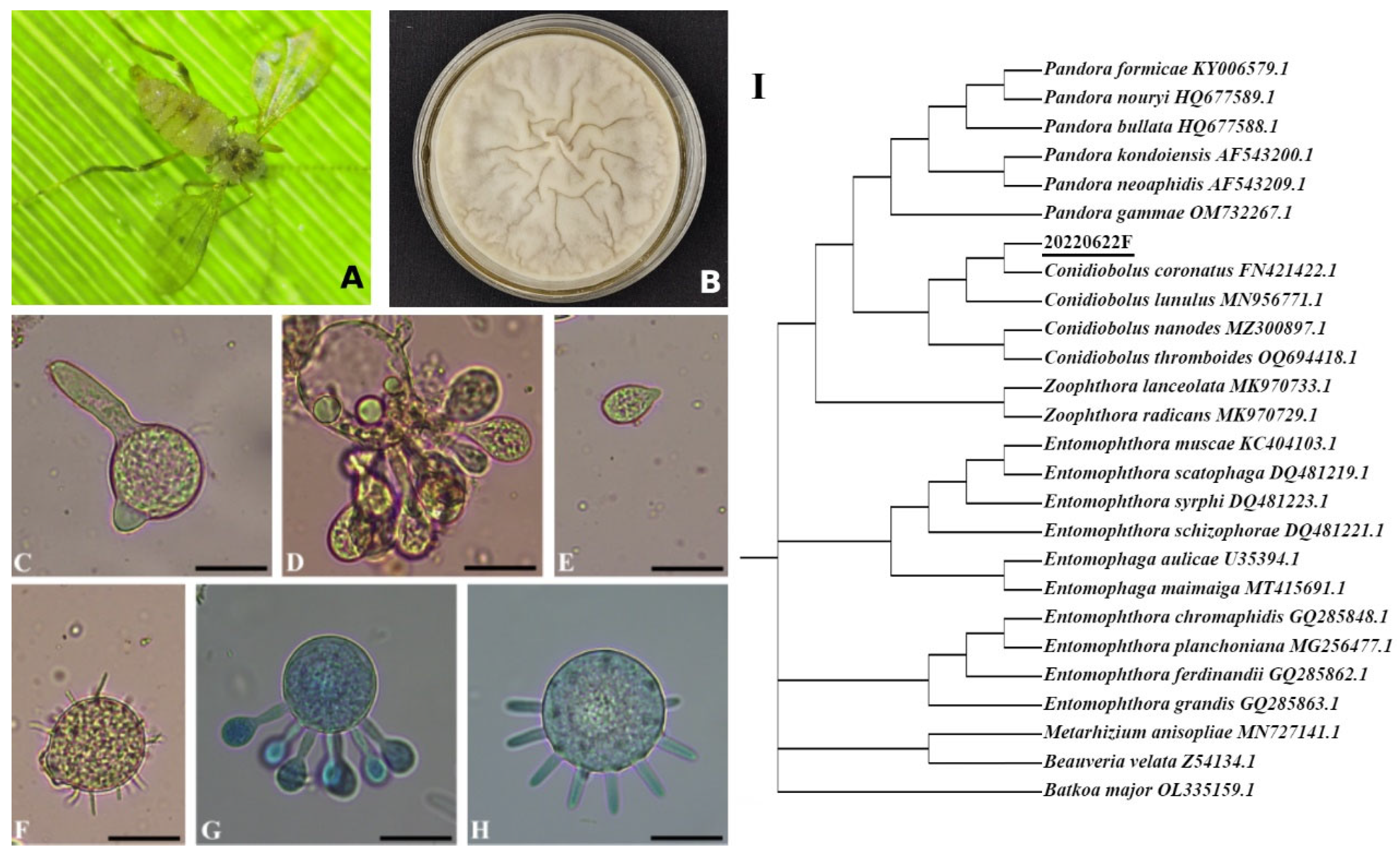
Figure 1. Morphological and molecular identification of Conidiobolus sp. strain 20220622F. (A) Cadaver of a naturally infected Rhopalomyia sp. gall midge attached to a bamboo leaf. (B) Purified culture on Sabouraud dextrose agar plus yeast extract in a 60 mm diameter Petri dish. (CH) Micrographs of conidia stained with lactophenol-aceto-orcein (CF) and cotton blue (G,H): (C) germinating primary conidium; (D,G,H) conidia at different sporulation stages; (E) secondary conidium; (F) villose conidium; (G,H) forming microconidia. (I) Phylogenetic analysis based on ITS sequences comparing Conidiobolus sp. strain 20220622F with other Conidiobolus species from GenBank. Beauveria and Metarhizium spp. served as the outgroup. Scale bar: 20 μm for (CH).
Jof 11 00600 g001
Figure 2. Functional divergence between the generalist C. coronatus and the specialist C. obscurus. (A) GO enrichment across biological process, molecular function, and cellular component categories. (B) KEGG pathway enrichment in genetic information processing, metabolism, and cellular processes. Bar lengths indicate the percentages of enriched genes relative to the total annotated genes. EIP: environmental information processing.
Jof 11 00600 g002
Figure 3. The different numbers of carbohydrate enzyme family genes in C. coronatus and C. obscurus. (A) Glycosyl transferases, (B) glycoside hydrolases, (C) polysaccharide lyases and auxiliary activities, (D) carbohydrate esterases, and (E) carbohydrate-binding modules.
Jof 11 00600 g003
Figure 4. Comparative functional annotation of pathogenicity-associated genes in C. coronatus and C. obscurus. (A) Distribution of PHI (pathogen–host interaction) categories related to virulence phenotypes. Gene counts are shown for 11 functional categories associated with pathogenic outcomes. (B) Functional classification of PHI-based genes in C. coronatus via KOG annotation.
Jof 11 00600 g004
Figure 5. Top 20 Pfam annotation results for C. coronatus and C. obscurus.
Jof 11 00600 g005
Figure 6. Diversity and expansion of peptidase repertoires in the C. coronatus genome based on Pfam annotation. (AC) Serine protease repertoire: (A) Phylogenetic tree for the serine proteases in C. coronatus, constructed using the maximum likelihood method in MEGA v12, illustrating evolutionary relationships among these families; (B) conserved motifs identified in trypsins, highlighting key functional residues; (C) conserved motifs in subtilisins, emphasizing catalytic domains. (D) Comparative analysis of metalloproteinase gene families between C. coronatus and C. obscurus, showing differential expansion patterns. (E) Phylogenetic tree and conserved motif visualization for proteases containing the M20/M25/M40 domain (Pfam ID: PF01546.23), based on sequence alignment and domain architecture.
Jof 11 00600 g006
Figure 7. Phylogenetic classification and structural characterization of GPCRs in Conidiobolus coronatus. (A) Integrated analysis of G-protein-coupled receptors (GPCRs): (Left) Maximum likelihood phylogenetic tree of GPCR sequences, revealing evolutionary clustering patterns. (Right) Distribution of conserved structural motifs and domains across GPCR sequences. (B) Sequence conservation analysis of a representative motif 10 in GPCR domain.
Jof 11 00600 g007
Figure 8. Phylogenetic classification and structural characterization of fungus-specific transcription factor in Conidiobolus coronatus. (Left) Maximum likelihood phylogenetic tree of sequences of fungus-specific transcription factor, revealing evolutionary clustering patterns. (Right) Distribution of conserved structural motifs and domains across these sequences.
Jof 11 00600 g008
Table 1. Genome characteristics of Conidiobolus coronatus 20220622F.
SpeciesC. coronatus
Genome assembly (Mb)44.21
Coverage (×)82.44
Scaffold number77
Scaffold N50 length (bp)1,415,773
Scaffold N90 length (bp)755,109
G + C content (%)27.65
Protein-encoding genes11,128
Average number of exons per gene2.85
Repeat sequences (bp)7,745,087
rRNA, family number294, 4
tRNA, family number1294, 46
Other ncRNA, family number43, 17
Table 2. Genomic characteristics of different entomophthoralean species.
Species [Ref.]C. coronatus 20220622FC. coronatus NRRL28638 [16]C. heterosporus RCEF6331 [17]C. obscurus ARSEF
7217 [18]
E. muscae ARSEF
13514 [16]
Genome size (Mb)44.2139.933.437.61030
Coverage (×)82.4416.3146631883.12
Complete fungal BUSCOs93.45%~80%/80.0%81.3%
Scaffolds771050961677810
Scaffold N50 (bp)1,415,773102,4101,660,4671,104,530329,600
Scaffold N90 (bp)755,10919,42286,18175,75179,400
G + C content (%)27.6527.536.8826.4641.03
Repeat sequence (%)17.52/12.917.4490.9%
Protein-coding genes11,12810,56810,85710,26238,917
Table 3. Highly expressed terpene compounds detected in C. coronatus vs. C. obscurus.
Compound NameAnnotation
(LIPID MAPS/KEGG)
Relative AbundanceLog2(FC)
C. coronatusC. obscurus
Limonene-1,2-diolC10 isoprenoids (monoterpenes) (PR0102)658.1286.91.20
(1S,4R)-1-Hydroxy-2-oxolimonene390.1271.50.52
6,10-Dimethyl-9-methylene-undec-5E-en-2-oneC15 isoprenoids (sesquiterpenes) (PR0103)881.71.19.71
5-(1-Oxopropan-2-yl)isolongifol-5-ene17,817.5320.65.80
5-(1-Hydroxypropan-2-yl)isolongifol-5-ene281.68.35.09
Dihydrophaseic acid1613.9215.52.90
(2-trans,6-trans)-Farnesol1753.0427.32.04
Gibberellin A8-cataboliteC20 isoprenoids (diterpenes) (PR0104)3087.461.95.64
(-)-Reiswigin A1965.6144.13.77
Gibberellin A34-catabolite2408.01187.81.02
(-)-2,7-Dolabelladiene-6beta,10alpha,18-triol12,478.56731.40.89
Gibberellin A3632,494.926,459.10.30
AstaxanthinC40 isoprenoids (tetraterpenes) (PR0107)2557.29.98.02
Adonixanthin2844.9110.64.69
1′-Hydroxytorulene30,533.43964.02.95
Octaprenyl diphosphate3803.31120.31.76
Neurosporaxanthin1082.4438.61.30
Echinenone260.3132.90.97
Thiothece-47422,722.812,189.40.90
Canthaxanthin841.7499.20.75
Terpendole KIndole diterpene alkaloid biosynthesis (ko00403)177.350.11.82
Terpendole G2762.21725.80.68
Disclaimer/Publisher’s Note: The statements, opinions and data contained in all publications are solely those of the individual author(s) and contributor(s) and not of MDPI and/or the editor(s). MDPI and/or the editor(s) disclaim responsibility for any injury to people or property resulting from any ideas, methods, instructions or products referred to in the content.