Article

Detection of Pestalotiopsis abbreviata sp. nov., the Causal Agent of Pestalotiopsis Leaf Blight on Camellia japonica Based on Metagenomic Analysis

1
Institute of Agriculture and Life Science, Gyeongsang National University, Jinju 52828, Republic of Korea
2
Forest Entomology and Pathology Division, National Institute of Forest Science, Seoul 02455, Republic of Korea
3
Department of Forest Environmental Resources, Gyeongsang National University, Jinju 52828, Republic of Korea
4
Department of Environment and Forest Resources, Chungnam National University, Daejeon 34134, Republic of Korea
*
Authors to whom correspondence should be addressed.
These authors contributed equally to this study as first authors.
J. Fungi 2025, 11(8), 553; https://doi.org/10.3390/jof11080553
Submission received: 14 June 2025 / Revised: 19 July 2025 / Accepted: 23 July 2025 / Published: 25 July 2025

Abstract

Tree diseases affecting Camellia japonica have emerged as a significant threat to the health and longevity of this ornamental tree, particularly in countries where this tree species is widely distributed and cultivated. Among these, Pestalotiopsis spp. have been frequently reported and are considered one of the most impactful fungal pathogens, causing leaf blight or leaf spot, in multiple countries. Understanding the etiology and distribution of these diseases is essential for effective management and conservation of C. japonica populations. The traditional methods based on pathogen isolation and pure culture cultivation for diagnosis of tree diseases are labor intensive and time-consuming. In addition, the frequent coexistence of the major pathogens with other endophytes within a single C. japonica tree, coupled with inconsistent symptom expression and the occurrence of pathogens in asymptomatic hosts, further complicates disease diagnosis. These challenges highlight the urgent need to develop more rapid, accurate, and efficient diagnostic or monitoring tools to improve disease monitoring and management on trees, including C. japonica. To address these challenges, we applied a metagenomic approach to screen fungal communities within C. japonica trees. This method enabled comprehensive detection and characterization of fungal taxa present in symptomatic and asymptomatic tissues. By analyzing the correlation between fungal dominance and symptom expression, we identified key pathogenic taxa associated with disease manifestation. To validate the metagenomic approach, we employed a combined strategy integrating metagenomic screening and traditional fungal isolation to monitor foliar diseases in C. japonica. The correlation between dominant taxa and symptom expression was confirmed. Simultaneously, traditional isolation enabled the identification of a novel species, Pestalotiopsis, as the causal agent of leaf spot disease on C. japonica. In addition to confirming previously known pathogens, our study led to the discovery and preliminary characterization of a novel fungal taxon with pathogenic potential. Our findings provide critical insights into the fungal community of C. japonica and lay the groundwork for developing improved, rapid diagnostic tools for effective disease monitoring and management of tree diseases.

1. Introduction

Pestalotiopsis (Pestalotiopsidaceae and Amphisphaeriales) is an ascomycetous genus introduced by Steyaert [1] to accommodate asexual fungi that produce conidia bearing both apical and basal appendages. The genus was typified by Pestalotiopsis guepinii, which was first described from isolates obtained from the stems and leaves of Camellia japonica in France. Pestalotiopsis, a species-rich asexual genus mostly lacking sexual morphs, is widely distributed throughout tropical and temperate regions [2,3,4]. Pestalotiopsis exhibits a diverse range of ecological characteristics, functioning as saprophytes and either parasitic or pathogenic fungi. In some cases where the species within this genus are regarded as phytopathogens, they cause a variety of diseases on foliage, stems, and roots, leading to considerable economic losses [4,5,6,7,8], while this group is also commonly isolated as endophytes [4,9,10].
Species of Pestalotiopsis can be differentiated by conidial morphology, including conidial length, width, length and color of median cells, and length of the apical appendages from its closely related groups, including Neopestalotiopsis and Pseudopestalotiopsis [11,12]. However, given the fact that identification based solely on morphological characters proved to be unreliable, phylogenetic species delimitation based on the introduction of multi-locus DNA sequence analyses is currently accepted to assure the identity at the species level [3,4,6].
Recent breakthroughs in bioinformatics, especially metagenomic-based approaches to detect plant pathogens, have revolutionized microbial diagnosis by offering expedited and remarkably sensitive protocols, compared with traditional microbiological approaches including culture-based pathogen isolation protocols [13]. The majority of these culture-independent and high-throughput techniques prioritize assessing entire microbial communities rather than isolating individual or closely related taxa to promptly and effectively detect phytopathogens based on the profile of phytobiome [13,14,15,16].
The genus Camellia (Theaceae) includes more than 250 plants (“The Plant List”, retrieved from 10th June 2025), and is widely distributed in tropical and subtropical regions including East Asian countries [17]. Of these, C. japonica is one of the most popular tree species for essential oil production, ornamental, and medicinal purposes [18]. Camellia japonica is a tree species widely distributed and commonly found in the southern regions of South Korea, especially for a variety of ecological and ornamental purposes, including as a hedge plant alongside roads in urban areas in the country.
Due to the popularity of C. japonica for various purposes in the country, disease surveillance and monitoring on C. japonica were performed during the summer of 2021 and 2022 to implement containment programs for pathogens on the tree. Severely infected leaves of C. japonica, showing symptoms including leaf spots and tips and marginal blight upon investigation, were revealed. As part of this study, microbial profiles on the leaves of C. japonica were determined using a metagenomic approach to achieve high-throughput identification of a causal agent of leaf spot disease observed on the tree. The occurrence and abundance of the causal pathogen would be higher in the diseased tissues of C. japonica. In order to validate the results obtained through metagenomic analysis and to achieve species-level identification of the pathogen, culture-based isolation techniques were additionally applied.

2. Materials and Methods

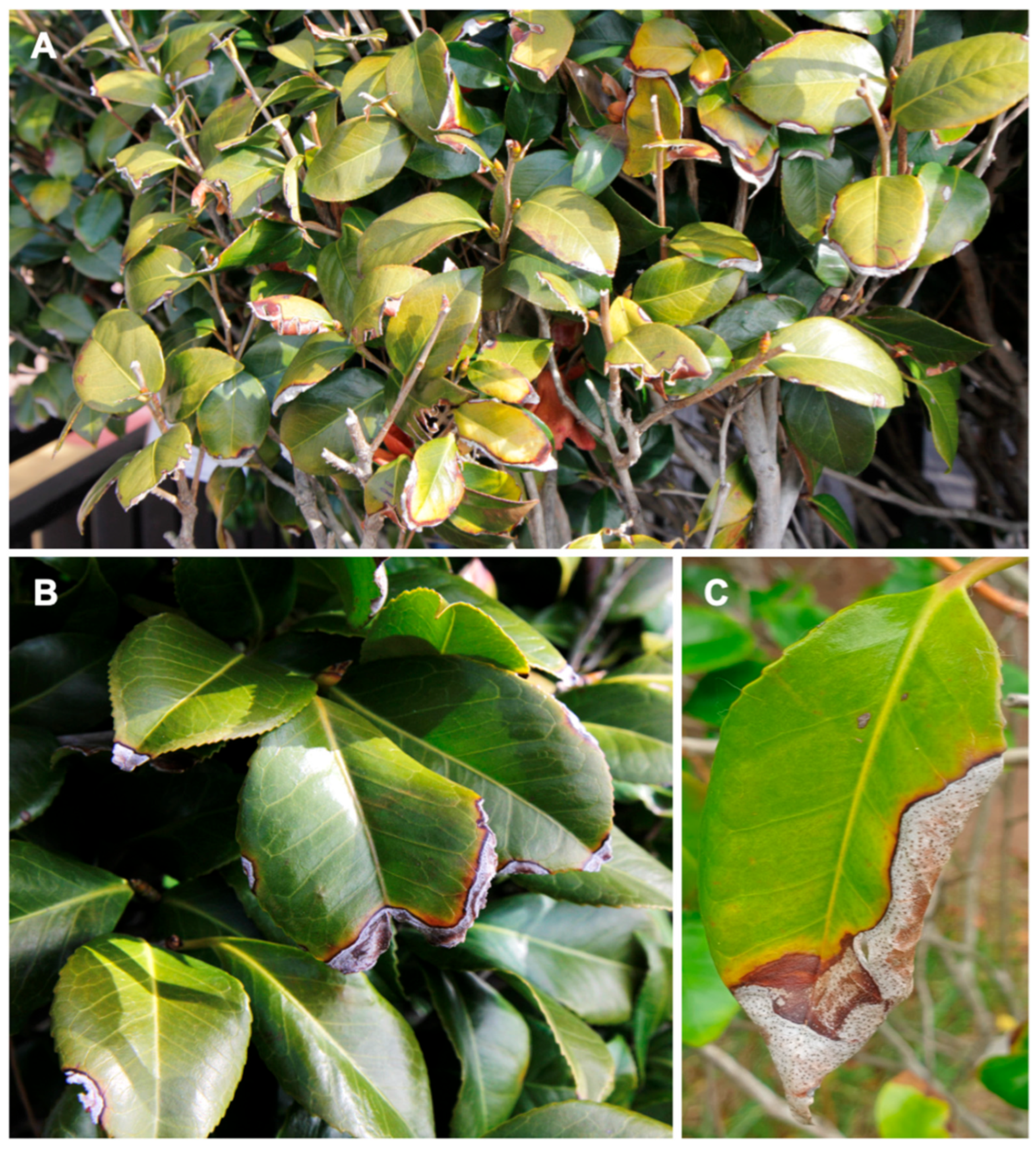
2.1. Study Site Description and Sample Collection

The two sampling sites were located in Busan, a city in the southern part of South Korea (35°09′13.4″ N 129°09′03.5″ E, 35°09′08.2″ N 129°09′09.5″ E), which were approximately 100 m apart from each other. The study areas were dominated by C. japonica, which were naturally propagated near the sea, with scattered Pinus thunbergii trees.
Approximately 10 to 15 leaves per tree of C. japonica showing leaf spot symptoms (Figure 1A–C) were randomly sampled, and samples were collected from each of the ten trees at each study site. Samples were placed in individual paper bags and transported to the laboratory for DNA extraction and fungal isolation.

2.2. Genomic DNA Extraction and PCR Amplifications, Sequencing

For metagenomic analysis, a total of 24 samples were subjected to DNA extraction. Genomic DNA was extracted from approximately 0.25 g (fresh weight) of each homogenized leaf sample using a DNeasy Plant Mini Kit (Qiagen, Valencia, CA, USA) following the manufacturer’s instructions. The genomic DNAs extracted were quantified using a Nanodrop ND-1000 spectrophotometer (Nanodrop, Wilmington, DE, USA) and adjusted to a final concentration of 20 ng µL−1. To obtain a metabarcoding profile on the leaves of C. japonica, PCR amplification was achieved using the ITS3 and ITS4 primers [19], targeting the internal transcribed spacer region 2 (ITS2), which is known to have no insertions commonly found in ITS1, producing a 320 bp amplicon in length on average in fungi [20,21]. These were then transported on dry ice to Macrogen Inc. (Seoul, Republic of Korea) for paired-end sequencing using the Illumina MiSeq platform.
For culture-based isolation of the fungal pathogen on C. japonica, fungal isolations were directly made to 2% potato-dextrose agar (PDA, Difco Laboratories, Inc., Detroit, MI, USA) supplemented with 100 mg/L−1 streptomycin sulphate (Sigma–Aldrich, St. Louis, MO, USA) by transferring spores from the infected leaves of the tree, where, in most cases, conidiomata were abundantly sporulated. Cultures were incubated at 25 °C for three weeks in the dark to allow for sufficient mycelial growth. The mycelium was then scraped from the surface of the agar with sterilized surgical scalpel blades and transferred to 1.5 mL Eppendorf tubes. Genomic DNA was then extracted using a ZR Fungal/Bacterial DNA MiniPrep kit (Zymo Research, Irvine, CA, USA) following the manufacturer’s instructions. The quantity and quality of DNA extracted were evaluated with a spectrophotometer (ND-1000; NanoDrop Technologies, Wilmington, DE, USA) to calibrate the concentration and purity of DNA as PCR templates.

2.3. Bioinformatic Processing

Following sequencing, a total of 2,439,406 raw reads were downloaded and processed using QIIME2 version 2022.2 [22]. Denoising was performed with the DADA2 plugin [23] in single-read mode, due to the size of the target amplicon. Sequences were trimmed and quality filtered, with full-length duplicates removed and reads sorted by abundance. For ITS sequences, denoised reads were used to infer amplicon sequence variants (ASV), and chimeric sequences were removed using the UNITE database as a reference [24], which served as the basic units of observation. Taxonomic classification was performed using a classifier trained with the scikit-learn library, based on the UNITE database (release 8.0). For classification, only ASV with a minimum of 10 sequence reads were included. The final processed ASV table comprised 1,166,426 reads across 1704 ASV.

2.4. Data Preparation and Metagenomic Statistical Analysis
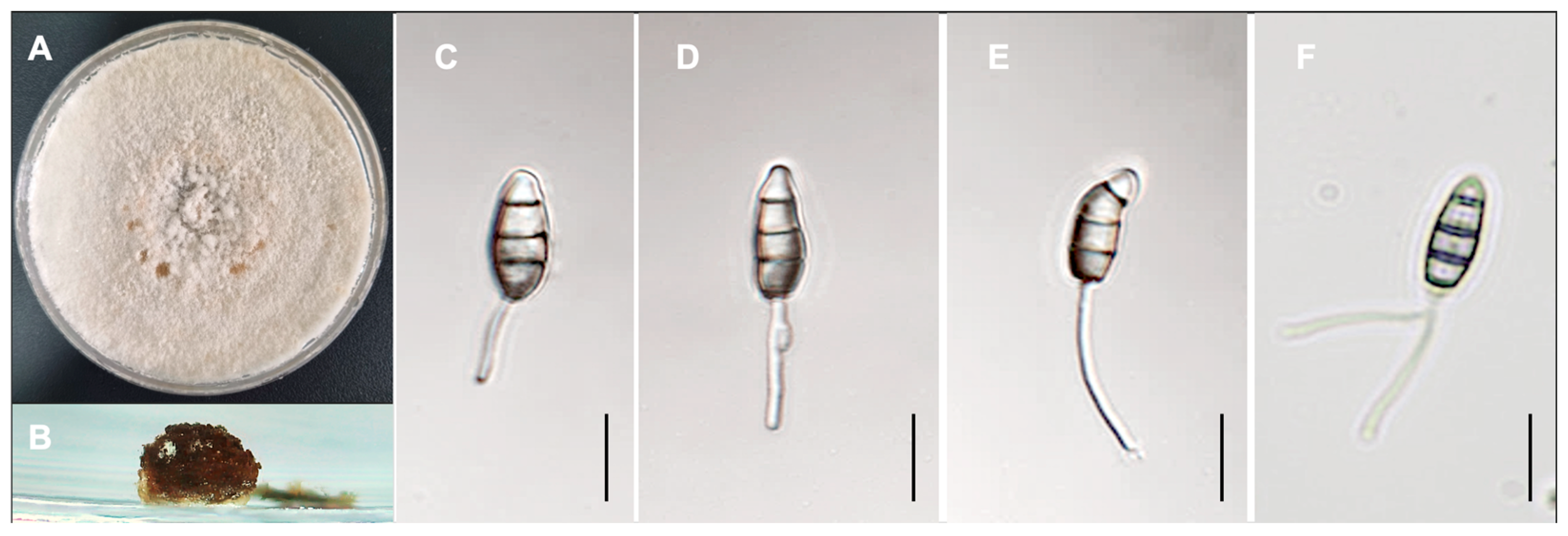
Fungal diversity profiling was conducted to evaluate the effects of one factor—diseased vs. healthy. Subsequently, the dataset processed from QIIME2 was imported into R and converted into a phyloseq object for further analyses. Taxonomic composition, diversity metrics, and ordination analyses were subsequently performed to facilitate interactive visualization and statistical interpretation.
Permutational multivariate analysis of variance (PERMANOVA) was performed to assess if significant differences (p < 0.05) existed between healthy and diseased trees of C. japonica in the relative abundance of fungal microbiota, based on the number of ASV, in relation to the factors under study.
PERMANOVA analyses using the ‘adonis2() function’ from the vegan package were performed based on a Bray–Curtis similarity matrix [25]. When the number of unique permutations was fewer than 15, Monte Carlo permutation p-values were reported. In cases where significant interaction effects were observed, a posteriori pairwise comparisons were conducted using 9999 permutations under a reduced model.
Subsequently, differential abundance testing was performed on the top 10 most abundant taxa to identify statistically significant differences in their relative abundances across samples through PERMANOVA analysis based on the ASV dataset.
These were all achieved using the microViz package (version 0.12.7) [26], implemented in R version 3.6.1. ASV occurring fewer than two times were removed from the dataset, and features present in only one sample were excluded. Unless otherwise specified, all procedures were performed using default settings.

2.5. Microscopy

Fungal structures were mounted on microscope slides in water and later replaced with 85% lactic acid for further observation. The structures were examined using a Zeiss AX10 Imager A2 (Carl Zeiss Microscopy GmbH, Göttingen, Germany) equipped with an Axiocam 506 color digital camera. At least 30 measurements were taken for taxonomically relevant structures when possible.

2.6. Multi-Gene Phylogenetic Analyses

To confirm the result obtained from the metagenomic analysis, the culture-based isolation approaches were employed to ensure the identity of the fungal pathogen isolated from C. japonica at the species level.
Two isolates, GNUFP284 and GNUFP285, representing each sampling location, obtained in this study, were subjected to sequencing and phylogenetic analyses based on three gene regions. These included the internal transcribed spacer region (ITS), partial β-tubulin (tub2), and partial translation elongation factor 1-alpha (tef1), which were amplified using the primers ITS5/ITS4 [19], T1/Bt-2b [27,28], and EF1-728F/EF-2 [29,30], respectively. Amplification conditions for ITS and TEF followed Crous et al. [5] and for TUB, Lee et al. [31]. The resulting PCR products were submitted to Macrogen (Seoul, Korea) for forward and reverse sequencing reactions.
The sequences of Pestalotiopsis spp. were retrieved from GenBank (Table 1). Sequences for each of the three gene regions, ITS, TUB, and TEF, were then aligned using the online interface of MAFFT version 7 (http://mafft.cbrc.jp/alignment/server, accessed on 29 May 2025) [32], with the iterative refinement method (FFT-NS-i settings) selected. Sequence alignments were manually edited in MEGA7 [33], producing a concatenated dataset of the three gene regions.
Two different phylogenetic analyses were employed, including maximum parsimony (MP) analyses using MEGA7 and maximum likelihood (ML) tests using RAxML HPC BlackBox version 8.1.11 [34,35], using the default option with the GTR substitution model implemented in the CIPRES cluster server (https://www.phylo.org/, accessed on 31 May 2025) at the San Diego Supercomputing Center. For both MP and ML analyses, Pestalotiopsis jesteri was used as the outgroup taxon.

3. Results

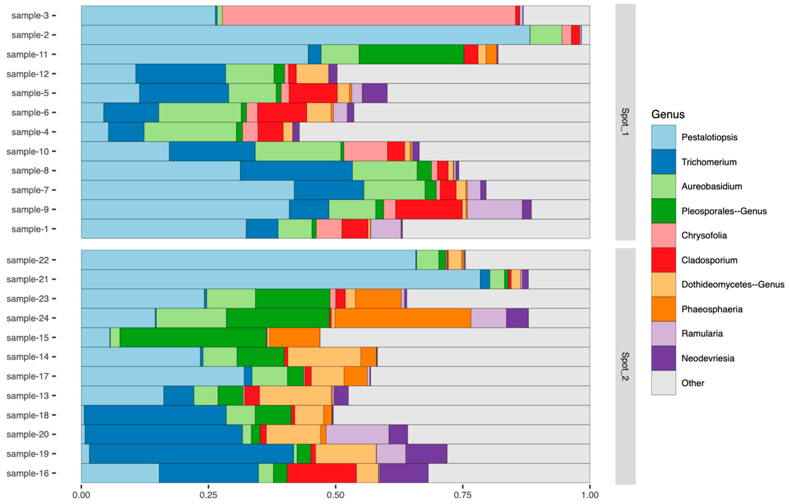
3.1. Metagenomic Assessment of Fungal Diversity on Camellia japonica

Deep sequencing of fungal-targeted ITS1 amplicons yielded 1,166,426 high-quality reads, from which 1704 unique amplicon sequence variants (ASV) were inferred. For downstream analysis, a subset of 961 ASV assigned to the phylum Ascomycota was extracted from the processed dataset and used for further analyses, and an overview of the fungal community composition is presented in Table 2.
The dataset was subsequently filtered using the tax_filter() function with default parameters from the microViz package (v0.12.7) [26] in R, in order to retain the most abundant taxa associated with C. japonica. This consequently resulted in a total of 90 taxa with a relative abundance exceeding 0.10%.

3.2. Detection of a Putative Fungal Pathogen Associated with Leaf Blight on Camellia japonica Based on Metagenomic Analysis

Within the phylum Ascomycota on C. japonica, the most abundant genus was Pestalotiopsis with a total abundance of 31.8%. This tendency was most often observed regardless of the sampling location (Figure 2).
In addition to Pestalotiopsis sp., several other putative fungal pathogens, including Aureobasidium sp. and Cladosporium sp., etc., that might cause leaf spot disease on C. japonica, were observed. However, these taxa can likely be excluded based on the relatively lower abundance than that observed from Pestalotiopsis sp., as well as the characteristic symptoms and signs observed on the leaf surfaces of the affected trees (Figure 2).
Among the 10 most prevalent fungal taxa observed from C. japonica, differential abundance testing was conducted based on PERMANOVA analysis, and it was clearly shown that Pestalotiopsis sp. was identified as the predominant taxon, showing statistically significant differences in its relative abundance between healthy and diseased C. japonica trees (Figure 3) and within the diseased C. japonica trees (Figure 4).
To assess the pathogenicity of Pestalotiopsis sp. that was identified through metagenomic analysis, inoculation trials were conducted using a single isolate (GNUFP284). A conidial suspension (1 × 10⁵ conidia/mL) was sprayed onto the leaves of C. japonica seedlings, while control plants were treated with sterile distilled water. Two weeks after inoculation, characteristic leaf spot symptoms developed on the inoculated seedlings, and Pestalotiopsis sp. was successfully re-isolated from the lesions, thereby fulfilling Koch’s postulates.

3.3. Identification of a Putative Fungal Pathogen Associated with Leaf Blight on Camellia japonica

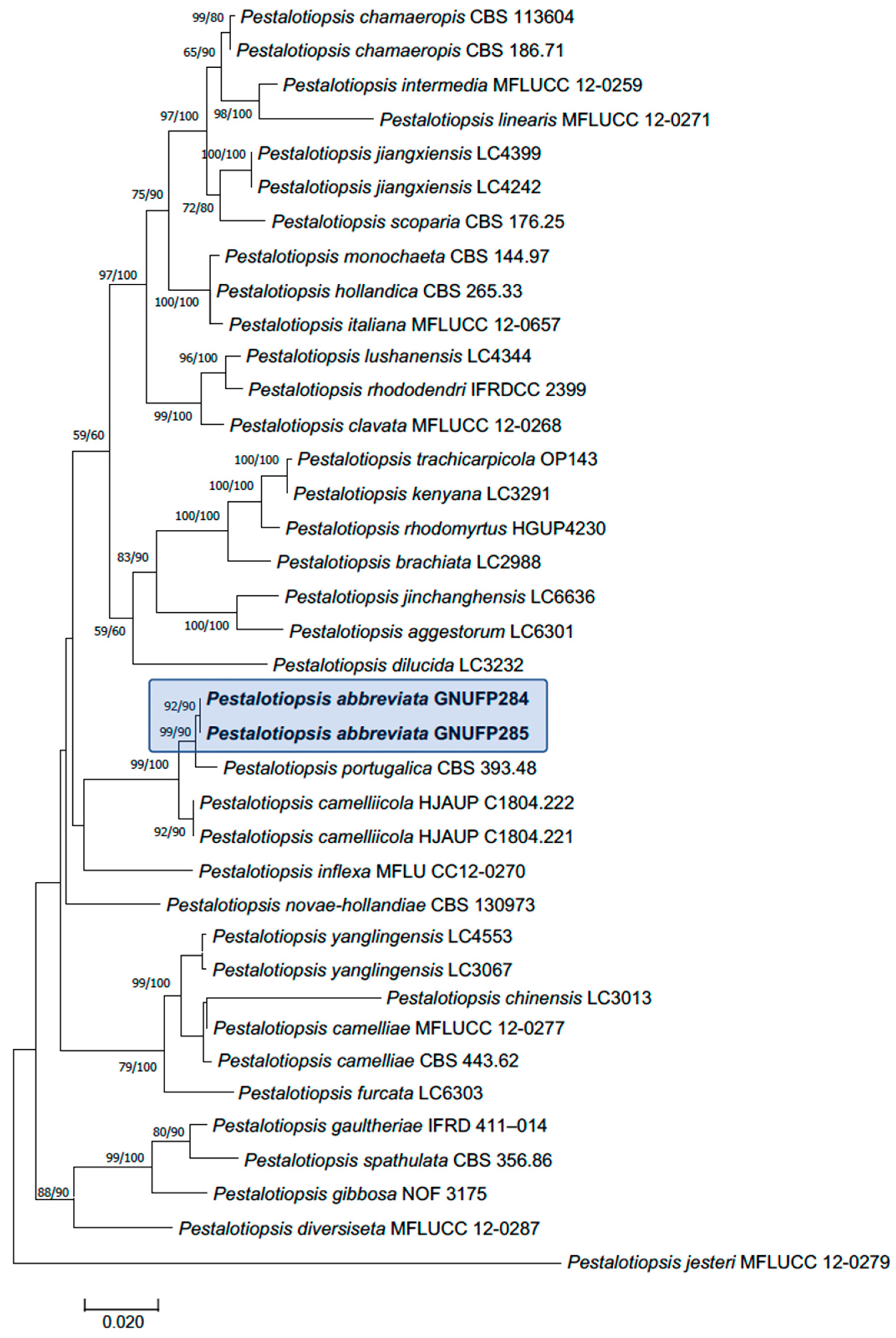
3.3.1. Multi-Gene Phylogenetic Analyses

Three gene regions, including ITS, tub2, and tef1, were successfully sequenced and deposited in GenBank under accession numbers OR478187–188 (ITS), OR480768–769 (tub2), and OR480770–771 (tef1). The sequences obtained from each gene region were aligned with those of closely related Pestalotiopsis species, as identified through BLAST searches (version BLAST+ 2.16.0) in the NCBI nucleotide database. Phylogenetic analyses based on the concatenated sequences of ITS, tub2, and tef1 yielded a tree (Figure 5). Although minor differences were observed between the topologies generated by the maximum likelihood (ML) and maximum parsimony (MP) methods, both analyses consistently supported that the isolates from C. japonica represent a previously undescribed species. This novel species is closely related to P. portugalica but is phylogenetically distinct from it and all other described Pestalotiopsis species.

3.3.2. Taxonomy

Morphological comparisons, including conidiomata and conidia, and phylogenetic inference based on three gene regions provided sufficient evidence that the Pestalotiopsis abbreviata sp. nov. isolated from Camellia japonica represents an undescribed species. This species is described as follows:
Pestalotiopsis abbreviata D.Hyeon Lee & S.E. Cho, sp. nov. MB 859637. Figure 6.
Etymology: The epithet refers to the reduced or absent basal appendages observed in this species.
Typus: Republic of Korea, Busan, Haeundae-gu, U-dong, 708-7, 35°09′13.4″ N, 129°09′03.5″ E, on leaves of C. japonica, 24 June 2021. D.H. Lee (holotype KACC 410709, ex-holotype culture GNUFP284).
Description: Conidiomata developed on PDA, pycnidial, globose to clavate, appearing aggregated or scattered, partially embedded to erumpent, dark brown to black, and measuring up to 550 μm in diameter; they released dark brown to black, globose conidial masses. Conidiophores often absent or reduced to conidiogenous cells; when present, septate near the base, unbranched or basally branched, subcylindrical, and hyaline. Conidiogenous cells discrete or integrated, ampulliform to clavate or subcylindrical, hyaline, smooth-walled, broad-based, and measure 7–35 × 3–5 μm. Conidia fusoid to ellipsoid, straight to slightly curved, 4-septate, and slightly constricted at the septa, measuring 13–18 × 4.5–7.1 μm. Basal cell hyaline, conic to obconic with a truncate base, thin-walled and verruculose, 3–6 μm long. Median cells doliiform, pale brown to brown with darker septa, totaling 11–14.5 μm. Apical cell hyaline, obconic, thin-walled, and verruculose, 3.0–5.0 μm long. Apical appendages one to two unbranched (mostly one), filiform and arise from the apex, 9–23 μm long. Centric basal appendage absent. Sexual states not observed.
Culture characteristics: Colonies, on 2% PDA, white, with flocculent aerial mycelium, which grow relatively fast, reaching the edge of the Petri dish 7 to 10 days after incubation at 25 °C in darkness. Scattered black conidiomata formed on 2% PDA.
Additional specimen examined: Republic of Korea, Busan, Haeundae-gu, U-dong, 710-1, 35°09′08.2″ N 129°09′09.5″ E, on leaves of C. japonica, 10 Sep. 2022. D.H. Lee culture GNUFP285.
Notes: Pestalotiopsis abbreviata is distinguished from P. portugalica by several distinct morphological characters. This includes non-proliferating, ampulliform to clavate or subcylindrical conidiogenous cells lacking a collarette, while P. portugalica shows percurrently proliferating conidiogenous cells (2–6 times) with a distinct collarette and periclinal thickening. The apical appendages in P. abbreviata are unbranched (mostly one) and arise directly from the apex, whereas those in P. portugalica are 1–3, often branched, and arise from an apical crest. The basal appendage is consistently absent in P. abbreviata but occasionally present in P. portugalica. In addition, the conidiomata of P. abbreviata are larger (up to 550 μm), aggregated, and erumpent, compared with the smaller (200–400 μm), solitary, and semi-immersed conidiomata of P. portugalica [4].

4. Discussion

The application of advanced molecular approaches—particularly microbial profiling targeting rRNA DNA regions based on metagenomics in plant pathology and applied microbiology—has become essential for the accurate and rapid detection of plant pathogens. This is particularly important given the increasing emergence of virulent strains, which drive the prevalence and severity of plant diseases [16]. This study evaluated the potential application of metagenomic analysis for effective monitoring of tree diseases, particularly for leaf blight disease caused by Petalotiopsis spp. on Camellia japonica, which is the tree disease that has been reported on C. japonica from several countries [36,37]. To validate the reliability of this approach, metagenomic profiling was performed in parallel with conventional culture-based isolation and identification of pathogens. In comparing the two methods, this study aimed to assess the feasibility of employing metagenomics as a complementary tool for disease surveillance and diagnosis.
In recent years, metagenomic approaches have emerged as powerful tools for investigating plant-associated microbial communities, offering high-resolution insights into pathogen diversity and community dynamics without the limitations of culture-based methods. This is partly due to the fact that it is associated with increasing accessibility, allowed either through the lower cost of these methods or the simplification of the processes involved. Despite its limitations in resolving certain genera with low interspecific variability—such as Aspergillus, Cladosporium, Fusarium, Penicillium, and Trichoderma, which exhibit narrow or absent barcode gaps within the internal transcribed spacer (ITS) regions—ITS remain the primary and most widely accepted molecular markers for fungal identification [38].
In this study, the application of metagenomic analysis targeting the ITS region enabled the detection and taxonomic assignment of Pestalotiopsis sp. within the fungal community of C. japonica. Although additional loci such as tub2 or tef1 may offer higher resolution for species-level identification within the genus Pestalotiopsis, the ITS regions remain the most widely used DNA barcode for fungal taxonomy, including for the members of this genus [38]. In the present study, ITS-based metagenomic analysis proved sufficient to reveal the clear predominance of Pestalotiopsis species in symptomatic C. japonica tissues, thereby supporting its role as a primary pathogen associated with leaf blight symptoms in this host. These findings demonstrate the effectiveness of ITS-based metagenomic profiling for identifying key pathogenic taxa within complex plant-associated microbiomes.
As expected, a diverse group of fungi associated with C. japonica was detected in this study. Among the top ten most dominant fungal taxa identified in the leaves of C. japonica, several genera were consistently abundant, including both commonly reported endophytic or pathogenic taxa—such as Pestalotiopsis, Cladosporium, and Alternaria—as well as less frequently observed genera such as Epicoccum, Didymella, Nigrospora, Colletotrichum, Fusarium, Phoma, and Aspergillus. Notably, the prominence of Pestalotiopsis in these metagenomic data was consistent with results from culture-based isolation, as well as with the characteristic morphological features of this genus observed on symptomatic leaf tissues. Together, these findings provide strong evidence supporting its role as a causal pathogen of leaf blight in C. japonica.
Pestalotiopsis species, including P. portugalica and P. camelliicola, have been reported as causal agents of leaf spot disease on Camellia spp. in China [36] and the Republic of Korea [39]. Additionally, our study confirmed the presence of a novel fungal species within the genus Pestalotiopsis, identified as a causal agent of leaf spot disease on Camellia spp. This newly described pathogen, designated as P. abbreviata, expands the current understanding of the diversity and pathogenicity of Pestalotiopsis species associated with Camellia hosts. Although phylogenetic analysis revealed P. portugalica as the closest relative of the novel species, clear morphological differences were observed between them. Moreover, both maximum parsimony (MP) and maximum likelihood (ML) analyses distinctly supported the classification of this novel species as a separate taxon. In addition, comparative evaluation with P. cameliicola, previously reported from C. japonica in China [36], demonstrated notable differences in conidial size and other morphological characteristics, further substantiating its distinction as a new species. These findings highlight the need for further investigation into the epidemiology and management of leaf spot diseases caused by this emerging pathogen in the country.

5. Conclusions

This study evaluated the applicability of metagenomic analysis as a tool for effective disease diagnosis and monitoring in Camellia japonica, a widely cultivated ornamental tree increasingly affected by foliar diseases such as leaf blight and leaf spot. By combining high-throughput sequencing of the ITS region with conventional culture-based isolation, we comprehensively characterized the fungal communities associated with symptomatic and asymptomatic tissues. The clear dominance of Pestalotiopsis species in symptomatic samples strongly supports their role as primary pathogens responsible for disease manifestation in C. japonica. Furthermore, traditional isolation methods enabled the identification and pathogenicity validation of a previously undetermined species of Pestalotiopsis, reinforcing the reliability of the metagenomic approach. These findings demonstrate that ITS-based metagenomic profiling can effectively complement classical diagnostic methods by enhancing detection speed, accuracy, and resolution in complex plant microbiomes. The integration of both approaches provides a robust framework for early pathogen detection, disease surveillance, and the development of targeted management strategies. Overall, this study underscores the practical and scientific value of metagenomic tools in forest pathology and highlights their potential for improving disease management systems in ornamental and ecologically significant tree species.

Author Contributions

Conceptualization, D.-H.L. and S.-E.C.; methodology, D.-H.L. and S.-E.C.; validation, D.-H.L., S.-E.C., K.H.P. and K.S.; formal analysis, D.-H.L., S.-E.C. and K.S.; investigation, D.-H.L., S.-E.C. and K.S.; resources, D.-H.L., S.-E.C. and K.S.; data curation, D.-H.L., S.-E.C. and K.H.P.; writing—original draft preparation, D.-H.L., S.-E.C., K.H.P. and K.S.; writing—D.-H.L., S.-E.C., K.H.P. and K.S.; visualization, D.-H.L. and K.S.; supervision, D.-H.L. and K.S.; project administration, D.-H.L.; funding acquisition, D.-H.L. and K.S. All authors have read and agreed to the published version of the manuscript.

Funding

This work was supported by a project (PNo. FE0703-2023-02-2025) funded by the National Institute of Forest Science, Republic of Korea. In addition, this work was supported by the National Research Foundation of Korea (NRF) (No. RS-2023-00250589) and the research fund for the next generation of academics of Gyeongsang National University in 2024.

Data Availability Statement

The original contributions presented in this study are included in the article. The raw sequence data generated through metagenomic sequencing have been deposited in the NCBI Sequence Read Archive (SRA) under the BioProject accession number PRJNA1293874. Further inquiries can be directed to the corresponding authors.

Conflicts of Interest

The authors declare no conflicts of interest.

References

  1. Steyaert, R.L. Contributions à l’étude monographique de Pestalotia de Not. et Monochaetia Sacc. (Truncatella gen. nov. et Pestalotiopsis gen. nov.). Bull. Jard. Bot. Brux. 1949, 19, 285–354. [Google Scholar] [CrossRef]
  2. Bate-Smith, E.C.; Metcalfe, C.R. Leucanthocyanins.3. The nature and systematic distribution of tannin in dicotyledonous plants. J. Linn. Soc. 1957, 55, 669–705. [Google Scholar] [CrossRef]
  3. Maharachchikumbura, S.S.; Guo, L.D.; Chukeatirote, E.; Bahkali, A.H.; Hyde, K.D. Pestalotiopsis—Morphology, phylogeny, biochemistry and diversity. Fungal Divers. 2011, 50, 167–187. [Google Scholar] [CrossRef]
  4. Maharachchikumbura, S.S.; Hyde, K.D.; Groenewald, J.Z.; Xu, J.; Crous, P.W. Pestalotiopsis revisited. Stud. Mycol. 2014, 79, 121–186. [Google Scholar] [CrossRef]
  5. Crous, P.W.; Summerell, B.A.; Swart, L.; Denman, S.; Taylor, J.E.; Bezuidenhout, C.M.; Palm, M.E.; Marincowitz, S.; Groenewald, J.Z. Fungal pathogens of Proteaceae. Pers. Mol. Phylogeny Evol. Fungi 2011, 27, 20–45. [Google Scholar] [CrossRef]
  6. Maharachchikumbura, S.S.; Guo, L.D.; Cai, L.; Chukeatirote, E.; Wu, W.P.; Sun, X.; Crous, P.W.; Bhat, D.J.; McKenzie, E.H.C.; Bahkali, A.H.; et al. A multi-locus backbone tree for Pestalotiopsis, with a polyphasic characterization of 14 new species. Fungal Divers. 2012, 56, 95–129. [Google Scholar] [CrossRef]
  7. Maharachchikumbura, S.S.; Chukeatirote, E.; Guo, L.D.; Crous, P.W.; Mckenzie, E.H.; Hyde, K.D. Pestalotiopsis species associated with Camellia sinensis (tea). Mycotaxon 2013, 123, 47–61. [Google Scholar] [CrossRef]
  8. Zhang, Y.M.; Maharachchikumbura, S.S.; Wei, J.G.; McKenzie, E.; Hyde, K.D. Pestalotiopsis camelliae, a new species associated with grey blight of Camellia japonica in China. Sydowia 2012, 64, 335–344. [Google Scholar]
  9. Watanabe, K.; Motohashi, K.; Ono, Y. Description of Pestalotiopsis pallidotheae: A new species from Japan. Mycoscience 2010, 51, 182–188. [Google Scholar] [CrossRef]
  10. Debbab, A.; Aly, A.H.; Proksch, P. Mangrove derived fungal endophytes–a chemical and biological perception. Fungal Divers. 2013, 61, 1–27. [Google Scholar] [CrossRef]
  11. Jeewon, R.; Liew, E.C.; Simpson, J.A.; Hodgkiss, I.J.; Hyde, K.D. Phylogenetic significance of morphological characters in the taxonomy of Pestalotiopsis species. Mol. Phylogenet. Evol. 2003, 27, 372–383. [Google Scholar] [CrossRef]
  12. Hu, H.; Jeewon, R.; Zhou, D.; Zhou, T.; Hyde, K.D. Phylogenetic diversity of endophytic Pestalotiopsis species in Pinus armandii and Ribes spp.: Evidence from rDNA and β-tubulin gene phylogenies. Fungal Divers. 2007, 24, 1–22. [Google Scholar]
  13. Gökdemir, F.Ş.; İşeri, Ö.D.; Sharma, A.; Achar, P.N.; Eyidoğan, F. Metagenomics next generation sequencing (mNGS): An exciting tool for early and accurate diagnostic of fungal pathogens in plants. J. Fungi 2022, 8, 1195. [Google Scholar] [CrossRef]
  14. Jongman, M.; Carmichael, P.C.; Bill, M. Technological advances in phytopathogen detection and metagenome profiling techniques. Curr. Microbiol. 2020, 77, 675–681. [Google Scholar] [CrossRef] [PubMed]
  15. Singh, D.P.; Prabha, R.; Rai, A.; Arora, D.K. Bioinformatics-assisted microbiological research: Tasks, developments and upcoming challenges. Am. J. Bioinform. 2012, 1, 10–19. [Google Scholar] [CrossRef]
  16. Jongman, M.; Chidamba, L.; Korsten, L. Bacterial biomes and potential human pathogens in irrigation water and leafy greens from different production systems described using pyrosequencing. J. Appl. Microbiol. 2017, 123, 1043–1053. [Google Scholar] [CrossRef] [PubMed]
  17. Zhao, D.W.; Hodkinson, T.R.; Parnell, J.A. Phylogenetics of global Camellia (Theaceae) based on three nuclear regions and its implications for systematics and evolutionary history. J. Syst. Evol. 2023, 61, 356–368. [Google Scholar] [CrossRef]
  18. Pereira, A.G.; Garcia-Perez, P.; Cassani, L.; Chamorro, F.; Cao, H.; Barba, F.J.; Simal-Gandara, J.; Prieto, M.A. Camellia japonica: A phytochemical perspective and current applications facing its industrial exploitation. Food Chem. X 2022, 13, 100258. [Google Scholar] [CrossRef] [PubMed]
  19. White, T.J.; Bruns, T.; Lee, S.; Taylor, J.W. Amplification and direct sequencing of fungal ribosomal rna genes for phylogenetics. In PCR Protocols: A Guide to Methods and Applications; Innis, M.A., Gelfand, D.H., Sninsky, J.J., White, T.J., Eds.; Academic Press Inc.: New York, NY, USA, 1990; pp. 315–322. [Google Scholar]
  20. Martin, K.J.; Rygiewicz, P.T. Fungal-specific PCR primers developed for analysis of the ITS region of environmental DNA extracts. BMC Microbiol. 2005, 5, 28. [Google Scholar] [CrossRef]
  21. Bazzicalupo, A.L.; Balint, M.; Schmitt, I. Comparison of ITS1 and ITS2 rDNA in 454 sequencing of hyperdiverse fungal communities. Fungal Ecol. 2013, 6, 102–109. [Google Scholar] [CrossRef]
  22. Bolyen, E.; Rideout, J.R.; Dillon, M.R.; Bokulich, N.A.; Abnet, C.C.; Al-Ghalith, G.A.; Alexander, H.; Alm, E.J.; Arumugam, M.; Asnicar, F. Reproducible, interactive, scalable and extensible microbiome data science using QIIME 2. Nat. Biotechnol. 2019, 37, 852–857. [Google Scholar] [CrossRef]
  23. Callahan, B.J.; McMurdie, P.J.; Rosen, M.J.; Han, A.W.; Johnson, A.J.A.; Holmes, S.P. DADA2: High-resolution sample inference from Illumina amplicon data. Nat. Methods 2016, 13, 581–583. [Google Scholar] [CrossRef] [PubMed]
  24. Nilsson, R.H.; Larsson, K.H.; Taylor, A.F.S.; Bengtsson-Palme, J.; Jeppesen, T.S.; Schigel, D.; Kennedy, P.; Picard, K.; Glöckner, F.O.; Tedersoo, L.; et al. The UNITE database for molecular identification of fungi: Handling dark taxa and parallel taxonomic classifications. Nucleic Acids Res. 2019, 47, D259–D264. [Google Scholar] [CrossRef]
  25. Oksanen, J.; Kindt, R.; Legendre, P.; O’Hara, B.; Stevens, M.H.H.; Oksanen, M.J.; Suggests, M.A.S.S. The vegan package. Commun. Ecol. Pack. 2007, 10, 631–637. [Google Scholar]
  26. Barnett, D.J.; Arts, I.C.; Penders, J. microViz: An R package for microbiome data visualization and statistics. J. Open Source Softw. 2021, 6, 3201. [Google Scholar] [CrossRef]
  27. Glass, N.L.; Donaldson, G.C. Development of primer sets designed for use with the PCR to amplify conserved genes from filamentous ascomycetes. Appl. Environ. Microbiol. 1995, 61, 1323–1330. [Google Scholar] [CrossRef] [PubMed]
  28. O’Donnell, K.; Cigelnik, E. Two divergent intragenomic rDNA ITS2 types within a monophyletic lineage of the fungus Fusarium are nonorthologous. Mol. Phylogenet. Evol. 1997, 7, 103–116. [Google Scholar] [CrossRef]
  29. O’Donnell, K.; Kistler, H.C.; Cigelnik, E.; Ploetz, R.C. Multiple evolutionary origins of the fungus causing Panama disease of banana: Concordant evidence from nuclear and mitochondrial gene genealogies. Proc. Natl. Acad. Sci. USA 1998, 95, 2044–2049. [Google Scholar] [CrossRef]
  30. Carbone, I.; Kohn, L.M. A method for designing primer sets for speciation studies in filamentous ascomycetes. Mycologia 1999, 91, 553–556. [Google Scholar] [CrossRef]
  31. Lee, S.; Groenewald, J.Z.; Crous, P.W. Phylogenetic reassessment of the coelomycete genus Harknessia and its teleomorph Wuestneia (Diaporthales), and the introduction of Apoharknessia gen. nov. Stud. Mycol. 2004, 50, 235–252. [Google Scholar]
  32. Katoh, K.; Misawa, K.; Kuma, K. MAFFT: A novel method for rapid multiple sequence alignment based on fast Fourier transform. Nucleic Acids Res. 2002, 30, 3059–3066. [Google Scholar] [CrossRef]
  33. Kumar, S.; Stecher, G.; Tamura, K. MEGA7: Molecular evolutionary genetics analysis version 7.0 for bigger datasets. Mol. Biol. Evol. 2016, 33, 1870–1874. [Google Scholar] [CrossRef]
  34. Stamatakis, A. RAxML-VI-HPC: Maximum likelihood-based phylogenetic analyses with thousands of taxa and mixed models. Bioinformatics 2006, 22, 2688–2690. [Google Scholar] [CrossRef] [PubMed]
  35. Stamatakis, A.; Hoover, P.; Rougemont, J. A rapid bootstrap algorithm for the RAxML Web servers. Syst. Biol. 2008, 57, 758–771. [Google Scholar] [CrossRef] [PubMed]
  36. Luo, X.X.; Liao, M.G.; Zhang, K.; Castañeda-Ruíz, R.F.; Ma, J.; Xu, Z.H. Morphological and phylogenetic analyses reveal eight novel species of Pestalotiopsis (Sporocadaceae, Amphisphaeriales) from southern China. MycoKeys 2024, 109, 207. [Google Scholar] [CrossRef]
  37. Florea, A.M.; Gafencu, A.M.; Lipșa, F.D.; Gabur, I.; Ulea, E. Occurrence of Pestalotiopsis sp. on ornamental plants Camellia japonica L. in Romanian public gardens. Horticulturae 2025, 11, 93. [Google Scholar] [CrossRef]
  38. Schoch, C.L.; Seifert, K.A.; Huhndorf, S.; Robert, V.; Spouge, J.L.; Levesque, C.A.; Chen, W.; Bolchacova, E.; Voigt, K.; Crous, P.W.; et al. Nuclear ribosomal internal transcribed spacer (ITS) region as a universal DNA barcode marker for Fungi. Proc. Natl. Acad. Sci. USA 2012, 109, 6241–6246. [Google Scholar] [CrossRef] [PubMed]
  39. Korean Society of Plant Pathology. Korea’s List of Plant Diseases, 6th ed.; Korean Society of Plant Pathology: Suwon, Republic of Korea, 2023; pp. 123–124. [Google Scholar]
Figure 1. Symptoms of leaf spot and leaf blight on Camellia japonica. (A) Landscape view of diseased leaves of C. japonica. (B,C) Close-up view of diseased leaves of C. japonica.
Jof 11 00553 g001
Figure 2. The 10 most abundant taxa within the phylum Ascomycota observed on C. japonica.
Jof 11 00553 g002
Figure 3. Significant differences in the dominance of Pestalotiopsis sp. in its relative abundance between healthy and diseased C. japonica trees.
Jof 11 00553 g003
Figure 4. Significant differences in the dominance of Pestalotiopsis sp. in its relative abundance within the diseased C. japonica trees. Each circle (node) represents a taxonomic unit from phylum to genus level. Red branches indicate taxa significantly abundant in diseased samples, while blue branches indicate taxa that are less abundant. The intensity of the color corresponds to the LDA score (estimate), and the size of each dot reflects the relative prevalence of the taxon across samples. Nodes with a black border denote statistical significance (p < 0.05).
Jof 11 00553 g004
Figure 5. Phylogenetic trees based on maximum likelihood (ML) analysis of a combined dataset of ITS, tub2, and tef1 gene sequences for Pestalotiopsis species. Isolates in bold and highlighted are the new species of Pestalotiopsis described in this study. Bootstrap values > 50% for MP and maximum likelihood (ML) are presented above branches as ML/MP. The scale bar indicates 0.02 changes.
Jof 11 00553 g005
Figure 6. Pestalotiopsis abbreviate sp. nov. (A) Colony on PDA after 10 days at 25 °C. (B) Conidiomata, (CF) Conidia. Scale bars: 10 µm (CF).
Jof 11 00553 g006
Table 1. Isolates and GenBank accession numbers used for phylogenetic analyses in this study.
SpeciesIsolatesITSTUBTEF
P. abbreviataGNUFP284OR478187OR480768OR480770
P. abbreviataGNUFP285OR478188OR480769OR480771
P. aggestorumLC6301KX895015KX895348KX895234
P. brachiataLC2988KX894933KX895265KX895150
P. camelliaeCBS 433.62KM199336KM199424KM199512
P. camelliaeMFLUCC 12-0277JX399010JX399041JX399074
P. camelliicolaHJAUP C1804.221PP962357PP952229PP952236
P. camelliicolaHJAUP C1804.222PP962358PP952230PP952235
P. chamaeropisCBS 113604KM199323KM199389KM199471
P. chamaeropisCBS 186.71KM199326KM199391KM199473
P. chinensisLC3013KX894939KX895271KX895156
P. clavataMFLUCC 12-0268JX398990JX399025JX399056
P. dilucidaLC3232KX894961KX895293KX895178
P. diversisetaMFLUCC 12-0287NR 120187JX399040JX399073
P. furcataLC6303KX895016KX895349KX895235
P. gaultheriaeIFRD 411–014KC537805KC537819KC537812
P. gibbosaNOF 3175LC311589LC311590LC311591
P. hollandicaCBS 265.33KM199328KM199388KM199481
P. inflexaMFLUCC 12-0270JX399008JX399039JX399072
P. intermediaMFLUCC 12-0259JX398993JX399028JX399059
P. italianaMFLUCC 12-0657KP781878KP781882KP781881
P. jesteriMFLUCC 12-0279JX399012JX399043JX399076
P. jiangxiensisLC4242KX895035KX895327KX895213
P. jiangxiensisLC4399KX895009KX895341KX895227
P. jinchanghensisLC6636KX895028KX895361KX895247
P. kenyanaLC3291KX894962KX895294KX895179
P. linearisMFLUCC 12-0271JX398992JX399027JX399058
P. lushanensisLC4344KX895005KX895337KX895223
P. monochaetaCBS 144.97KM199327KM199386KM199479
P. novae-hollandiaeCBS 130973NR_147557KM199425KM199511
P. portugalicaCBS 393.48KM199335KM199422KM199510
P. rhododendriIFRDCC 2399KC537804KC537818KC537811
P. rhodomyrtusHGUP4230KF412648 KF412642KF412645
P. scopariaCBS 176.25KM199330KM199393KM199478
P. spathulataCBS 356.86NR 147558KM199423KM199513
P. trachicarpicolaOP143KC537809 KC537823KC537816
P. yanglingensisLC3067KX894949KX895281KX895166
P. yanglingensisLC4553KX895012KX895345KX895231
Table 2. Overview of fungal microbiota composition on Camellia japonica.
ClassOrderFamilyGenus
Classified ASV876798692626
Identified taxa1147119180
Disclaimer/Publisher’s Note: The statements, opinions and data contained in all publications are solely those of the individual author(s) and contributor(s) and not of MDPI and/or the editor(s). MDPI and/or the editor(s) disclaim responsibility for any injury to people or property resulting from any ideas, methods, instructions or products referred to in the content.

