Article

Foliar Epichloë gansuensis Endophyte and Root-Originated Bacillus subtilis LZU7 Increases Biomass Accumulation and Synergistically Improve Nitrogen Fixation in Achnatherum inebrians

State Key Laboratory of Herbage Improvement and Grassland Agro-Ecosystems, Key Laboratory of Grassland Livestock Industry Innovation, Ministry of Agriculture and Rural Affairs, Engineering Research Center of Grassland Industry, Ministry of Education, Gansu Tech Innovation Centre of Western China Grassland Industry, Centre for Grassland Microbiome, College of Pastoral Agriculture Science and Technology, Lanzhou University, Lanzhou 730000, China
*
Authors to whom correspondence should be addressed.
J. Fungi 2025, 11(7), 466; https://doi.org/10.3390/jof11070466
Submission received: 22 April 2025 / Revised: 8 June 2025 / Accepted: 18 June 2025 / Published: 20 June 2025
(This article belongs to the Section Environmental and Ecological Interactions of Fungi)

Abstract

:
Although drunken horse grass (Achnatherum inebrians) can be simultaneously infected by the foliar endophyte Epichloë gansuensis and colonized by Bacillus subtilis, it remains unclear whether Epichloë endophyte symbiosis influences B. subtilis colonization, as well as how their interaction affects nitrogen fixation and assimilation. The purpose of the present study was to investigate whether E. gansuensis endophyte infection facilitates the colonization of B. subtilis in the roots of host plants, with a focus on understanding the interaction effects of the E. gansuensis endophyte and B. subtilis on plant growth and nutrient absorption. In this study, we measured the colony growth rate of B. subtilis LZU7 when co-cultured with E. gansuensis strains. In addition to an in vitro test, we investigated the root colonization of Epichloë endophyte-infected plants (E+) and Epichloë endophyte-free plants (E−) with the GFP-tagged B. subtilis LZU7 in an inoculation test. Furthermore, we evaluated the interactions between E. gansuensis endophyte symbiosis and B. subtilis LZU7 colonization on the dry weight, nitrogen fixation, nitrogen converting-enzyme activity, and nutrients for E+ and E− plants by labeling with 15N2. The results showed that the growth rates of B. subtilis LZU7 were altered and increased in a co-culture with the E. gansuensis endophyte. A significantly greater colonization of GFP-tagged B. subtilis LZU7 was detected in the roots of E+ plants compared with the roots of E− plants, suggesting that E. gansuensis endophyte symbiosis enhances the colonization of beneficial microorganisms. The combination of E. gansuensis endophyte symbiosis and B. subtilis LZU7 inoculation significantly altered the expression of the nitrogenase (nifH) gene, thereby promoting increased biological nitrogen fixation (BNF). The E. gansuensis endophyte infection and inoculation with B. subtilis LZU7 significantly increased δ15NAir in plants. Co-inoculation with the E. gansuensis endophyte and B. subtilis LZU7 significantly elevated NH4+ accumulation in the roots, depleted the NH4+ availability in the surrounding soil, and showed no measurable impact on the foliar NH4+ content. The observed alterations in the NH4+ content were linked to nitrogen-fixing microorganisms that promoted nitrogen fixation, thereby enhancing nitrogen uptake and contributing to greater biomass production in A. inebrians. Our findings highlighted the fact that a foliar symbiosis with the E. gansuensis endophyte enhances the recruitment of beneficial bacteria, and that the resulting interaction significantly impacts nitrogen fixation, assimilation, and allocation in host plants.

1. Introduction

Mutualistic interactions with symbionts are being increasingly recognized for the important roles they play in ameliorating stressors and facilitating the ecosystem services that their host organisms provide [1,2]. Such symbioses influence various processes in host plants, ranging from nutrient acquisition to interactions with pollinators, seed dispersers, and the biotic and abiotic environment [3,4,5]. Microorganisms are among the most important mutualists of plants. One group of important microbial symbionts that often interact mutualistically with their hosts comprises Epichloë, a fungal endophyte of grasses [6]. These fungi are asexual, are vertically transmitted, and live asymptomatically within the plant tissues of 20–30% of grass species worldwide, including many economically important forage and turf grasses [7]. Endophyte symbioses with grasses can increase host resistance to insect pests through the production of alkaloids [8], increase N absorption [9,10], enhance drought and salt tolerance [11,12,13], and confer protection from super-infections caused by fungal pathogens [14]. Consequently, these symbiotic relationships have been widely exploited in the breeding of resistant varieties, ecological restoration, and grassland improvement [15,16]. Much is known about the enhanced stress tolerance conferred by endophyte symbiosis; despite this, little is known about whether there are foliar endophyte infections that interact with other root beneficial microbes in these processes [17].
Root-associated microbiomes have been recognized as the “second genome” of plants, especially plant growth-promoting bacteria, which communicate and participate in interplay to increase nitrogen fixation, provide mineralizing nutrients, modulate the plant hormonal balance, and defend against pathogens [18,19,20,21]. For instance, Bacillus species are major industrial workhorse microorganisms and have been widely used in agriculture. They provide important services to the plant, such as preventing pathogen infections [22] and enhancing the colonization and N2 fixation of rhizobia in roots [23,24], thus promoting the yield of the associated plants [25]. B. subtilis is the best-characterized member of the genus Bacillus, which comprises Gram-positive bacteria that can modulate microbial interactions to regulate N transport and assimilation, thus improving N efficiency and sustainable agricultural development [26].
Nitrogen is a major growth-limiting nutrient for plant defense and growth as well as net primary productivity in terrestrial ecosystems, and the vast majority of the nitrogen (78%) in the atmosphere exists in the form of a single molecule, which is not directly available to plants [27]. Specific beneficial microbe taxas (e.g., nitrogen-fixing bacteria) are able to convert N2 into plant-available forms via nitrogen nitrogenase [28]. An endophyte infection in plants can not only modify these microbial community structures by mediating root exudates and plant litter but also reduce the N loss from the soil and improve the nitrogen use efficiency in host plants through these microbes [29]. Microbial community structure and composition are usually influenced by changes in N availability [10,30,31]. Endophyte infection induces host plants to use N for the synthesis of their characteristic bioprotective alkaloids, establishing N as an important currency of mutualism in vertically transmitted foliar Epichloë endophyte symbioses [32,33]. This positive feedback between the Epichloë endophyte and N is expected to stimulate N fixation by cycling microorganisms, thereby increasing N bioavailability and plant growth. The magnitude of these effects depends on the feedback strength toward microbes, which varies with different endophytic fungal symbionts [10,34]. To date, the ecological mechanisms explaining how Epichloë endophyte infections in grasses contribute to nutrient cycling and plant growth through interactions with beneficial microbes (Bacillus sp.) have been seldom explored [35]. Given this background, we hypothesized that in A. inebrians, an E. gansuensis endophyte infection would have a strong impact on nitrogen fixation and plant productivity by affecting the colonization of B. subtilis in the roots.
To test this hypothesis, we isolated and characterized the specificity and function of root endophytic bacteria from low-NH4+ -treated A. inebrians and assessed the interactions between E. gansuensis endophyte strains and endosymbiotic bacteria strains. We also monitored the colonization of plant roots by the GFP-tagged B. strains LZU7 strain in the Epichloë endophyte-infected (E+) plants relative to the Epichloë endophyte-free (E−) plants. To test for an effect of the interactions between the E. gansuensis endophyte infection and B. subtilis LZU7 inoculation on plant growth and nutrient absorption, we measured a number of indicators involved in plant growth as well as nitrogen fixation, transformation, and accumulation. Furthermore, we used models to quantify the contribution of the endophyte infection and B. subtilis LZU7 inoculation to plant nutrient accumulation and productivity.

2. Methods and Materials

2.1. Plant Materials

Different ecotypes of drunken horse grass (A. inebrians) were selected for this study based on their contrasting rhizosphere nitrogen transformation capacity, as previously described [34]. Briefly, a screening experiment was performed on 5 ecotypes by assessing their rhizosphere nitrogen transformation capacity, using the copy number of the nitrogen-cycle-related genes (i.e., amoA-AOB, amoA-AOA, nirS, nirK, and nosZ) [34]. The A. inebrians from Tianzhu in Gansu Province, China, was chosen to study its endosymbiotic bacteria and the interactions with the Epichloë endophyte on host nitrogen uptake and growth.

2.2. Detection and Isolation of E. gansuensis Endophyte

Sequencing and a comparison with the National Center for Biotechnology Information (NCBI) identified that the A. inebrians from Tianzhu was infected with the E. gansuensis endophyte [36]. Epichloë endophyte-infected (E+) and Epichloë endophyte-free (E−) A. inebrians were space-planted in field plots at 2 locations in Gansu, China, in 2017. E+ and E− seeds were harvested in each plot over of a 6-year study (2017–2023) and examined for endophyte hyphae through seed staining. Microscopic detection procedures and PCR analyses using the following Epichloë-specific PCR primer pair: tub2-exon 1d-1: GAGAAAATGCGTGAGATTGT, tub2-exon 4u-2: GTTTCGTCCGAGTTCTCGAC. The E+ and E− seeds used for fungal endophyte isolation in this study were collected during July 2023, and stored at 4 °C to maintain endophyte viability.
After peeling, the E+ seeds were surface-sterilized with 75% ethanol for 30 s and 1.0% NaOCl for 1 min, followed by a sterile water rinse 4–5 times. They were then dried on sterile filter paper and stored at 4 °C until use. To evaluate the sterilization effect, the final rinse water was dripped into Luria–Bertani medium (5 g of yeast extract, 10 g of tryptone, 10 g of NaCl, 18 g of agar, 1000 mL, and a pH of 7.0) and trypticase soy agar medium (15 g of pancreatic peptone, 5 g of soy peptone, 5 g of NaCl, 18 g of agar, 1000 mL, and a pH of 7.3 ± 0.2). After one week, no bacterial colonies were formed on the LB or TSA plates, indicating that the surface of the seeds was sterilized thoroughly. The sterilized seeds were placed on potato dextrose agar media (200 g of potato, 20 g of glucose, and 18 g of agar, with volumization to 1000 mL) using a sterilized tweezer. After a period of growth, the seedlings were directly cut using a sterilized scalpel, and then the incision was allowed to come into contact with the culture medium. The mycelium grew at the incision site after continued incubation. The purified fungus was obtained by inoculating the mycelium on new PDA media from 10 to 30 days after incubation, with 3 repetitions, and there were no other stray bacteria.
In order to determine whether the isolated fungal strain was the E. gansuensis endophyte, we carried out linear amplification and sequencing. In brief, fungal DNA was extracted using the D3195-01 HP Fungal DNA Mini Kit (Omega Biotek Inc., Norcross, GA, USA) according to the manufacturer’s directions. The fungal ITS2 region of the ribosomal DNA was amplified using the universal primers ITS1 and ITS4 (ITS1: TCCGTAGGTGAACCTGCGC, and ITS4: TCCTCCGCTTATTGATATGC) [37]. The PCR products were sequenced to obtain the internal transcribed spacer (ITS) sequence from the ribosomal DNA of the fungal strain. The amplified DNA sequence was analyzed using the Basic Local Alignment Search Tool (BLAST v2.16.0) to identify homologous sequences and assess the sequence similarity to existing sequences in the NCBI database (http://www.ncbi.nlm.nih.gov/).

2.3. Isolation and Identification of Endophytic Bacteria

The roots of the E+ plants treated with 0.01 mol/L ammonium nitrogen in the preliminary experiment were rinsed several times with sterile water to remove impurities attached to the surface of the roots [29], and they were then cut into shreds. Sterilization and an evaluation of the sterilizing effect were conducted in the same manner as in the above seed treatments. An amount of 500 mg of the root sample was accurately weighed and placed into a sterilized mortar. Sterilized quartz sand was added to disrupt and homogenize the roots sufficiently by grinding. After grinding, 5 mL of a 0.9% NaCl solution was added and the mixture was transferred to a 50 mL sterile conical centrifuge tube, shaken well, and sonicated for 1 min with occasional shaking. An amount of 1 mL of the supernatant was taken and extracting solutions were made with concentrations of 10−6, 10−7, and 10−8. A total of 100 μL of each concentration was spread onto LB agar plates, with three parallel replicates for each concentration. The Petri dishes were incubated in a dark incubator at 25 °C for 3–5 d. The endosymbiotic bacteria were isolated and purified by the plant tissue culture and scribing method. Briefly, a small amount of a bacterial colony was transferred aseptically using a pick-up loop to an LB solid medium for district zoning. To obtain more independently distributed individual cells, the colony was diluted using the point-to-line method. To clarify the morphological characteristics of the isolated strains, isolated pure-culture endophytic bacteria were stained using a Gram-stain kit (Beso Biotech, Zhuhai, China). The bacterial strains were coated onto a slide and fixed by heating over an alcohol lamp. The four steps of pre-staining, mordanting, decoloration, and re-staining were carried out. After each step, the slide was washed with water, blotted up, and covered with a glass sheet. The images were taken using an Olympus CX22 LED microscope (Shinjuku, Japan). Gram-positive bacteria were stained purple, while Gram-negative bacteria were stained red. The shape, size, surface, and color of the bacterial strains were observed with the naked eye.
The total DNAs was extracted from samples of the pure bacterial cultures with an Ezup Column Bacteria Genomic DNA Purification Kit (Sangon Biotech, Shanghai, China) by following the manufacturer’s instructions. The DNA concentration and purity were measured using a NanoDropND-1000 spectrophotometer (Thermo Fisher Scientific, Waltham, MA, USA). The 16S rRNA gene of the bacterial strains was amplified using the universal primers 27F (5′-GAGTTTGATCATGGCTCAG-3′) and 1492R (5′-GGTTACCTTGTTACGATC-3′). Each DNA template was amplified in triplicate in a 25 μL reaction mixture containing primers. The PCR conditions were as follows: 12.5 µL of 2 × EasyTaq PCR SuperMix (Sangon Biotech, Shanghai, China), 1.0 µL of forward primers, 1.0 µL of barcoded reverse primers, 1.0 µL of template DNA, and 9.5 µL of ddH2O. The PCR amplification program was as follows: 95 °C for 5 min; 30 cycles of 95 °C for 60 s, 52.5 °C for 60 s, and 72 °C for 2 min; and 72 °C for 10 min. The PCR product was stored at 4 °C until use. The PCR products were purified and sent to Sangon Biotech for double-end sequencing and splicing. A homologous comparison was performed between nucleotide sequences of the 16S rRNA gene in the sequenced strains and published sequences in the NCBI database. The phylogenetic tree was constructed with the MEGA7 software (v7.0.14) using the neighbor joining method with 1000 bootstrap values and was visualized using FigTree. v.1.4.2 [37].

2.4. Functional Characterization of Endophytic Bacteria

2.4.1. IAA Determination

Single colonies from the plates were picked and added to LB broth (10 g of tryptone, 5 g of yeast, 10 g of NaCl, 1 L of distilled water, and pH 7.2) containing 5 mmol·L−1 of L-tryptophan, and the control group was without the addition of bacterial colonies. The bacteria were incubated in a shaker at 120 r.min−1 and 28 °C for 5 d. After the incubation period, 1 mL of the bacterial solution was put into a 1.5 mL centrifuge tube and centrifuged (12,000 rpm, 5 min) to obtain the supernatant. A total of 200 μL of supernatant was aspirated and mixed with 200 μL of Salkowski’s color development solution (50 mL of 35% HClO4 ± 1 mL of 0.5 mol·L−1 FeCl3), and the color change of the solution was observed after avoiding light for 30 min. A standard solution of IAA was used as the control. Then, 200 μL of the color reaction mixture was transferred into a 96-well plate, and three technical replicates were used to obtain an OD reading on a microplate reader at OD530. The IAA content of the strains was computed using the standard curve method.

2.4.2. The Measurement of Siderophores

Single colonies from the plates were inoculated onto sterilized MKB medium (15 mL of glycerin, 3.28 g of FeCl3·6H2O, 5 g of acid hydrolyzed casein, 2.5 g ofMgSO4·7H2O, and 18 g of agar powder) and incubated for 2 d in an incubator at 28 °C in an inverted position. After 2 d, 10 mL of a CAS test solution (0.06 g/L chrome azurol sulphonate, 0.073 g/L hexadecyl trimethyl-ammonium bromide, and 0.0027 g/L FeCl3.6H2O) (60 °C) was added to each inoculated MKB plate; the color change of each plate was observed after 1 h.

2.4.3. Determination of Nitrogen Fixation Potential

The isolated strains were inoculated into Ashby’s nitrogen-free medium (10 g of sugar, 0.2 g of KH2PO4, 0.2 g of MgSO4·7H2O, 0.2 g of NaCl, 0.1 g of CaSO4·2H2O, 5 g of CaCO3, 15 g of agar, 1000 mL, and a pH of 7.0–7.2) for inverted incubation at 28 °C for 4–7 d. If the bacterial strain had a nitrogen-fixation ability, the colonies were grown on a nitrogen-free medium.

2.4.4. Real-Time Amplification Assay of nifH

To investigate whether the isolated bacterial strains were nitrogen-fixing, the nifH gene was detected. The nifH gene of the bacterial strains was amplified using the primers nifHF (5′-AAAGGYGGWATCGGYAARTCCACCAC-3′) and nifHR (5’-TTGTTSGCSGCRTACATSGCCATCAT-3’) [38]. Each DNA template was amplified in triplicate in a 25 μL reaction system (2.5 μL of 2 × Taq Master Mix1, 8.5 μL of ddH2O, 2.0 µL of DNA,1.0 µL of forward primers, and 1.0 µL of barcoded reverse primers). The PCR amplification program was as follows: 98 °C for 2 min; 30 cycles of 98 °C for 15 s, 55 °C for 30 s, and 72 °C for 30 s; and 72 °C for 5 min. The amplified PCR products were detected using 2% agarose gel electrophoresis.

2.5. Plate Confrontation Experiments Between Epichloë Endophyte and Endophytic Bacterial Strains

The plate confrontation experiments were conducted as follows: To assess the interactions between the Epichloë endophyte and endosymbiotic bacterial strains, 6 mm E. gansuensis endophyte cakes were inoculated in solid YEDP medium, and then endophytic bacteria were inoculated around the E. gansuensis using the crisscross method. After incubating for another 7 d, the inhibitory effects on colony growth were observed. The double plate method was as follows: E. gansuensis endophyte cakes (6 mm) were cultured in a 500 mL flask containing 200 mL of liquid YEDP medium at 150 rpm for 10 d at 23 °C. The culture solution was filtered through a 0.22 μm membrane and then coated onto a nitrogen-free medium to solidify. YEDP liquid medium filtered through a 0.22 μm membrane was used as a blank control. A liquid with 200 μL 10−5 and 10−6 cells mL−1 (OD = 0.5, 600 nm) was coated onto a nitrogen-free medium containing the E. gansuensis endophyte to count the number of single colonies.

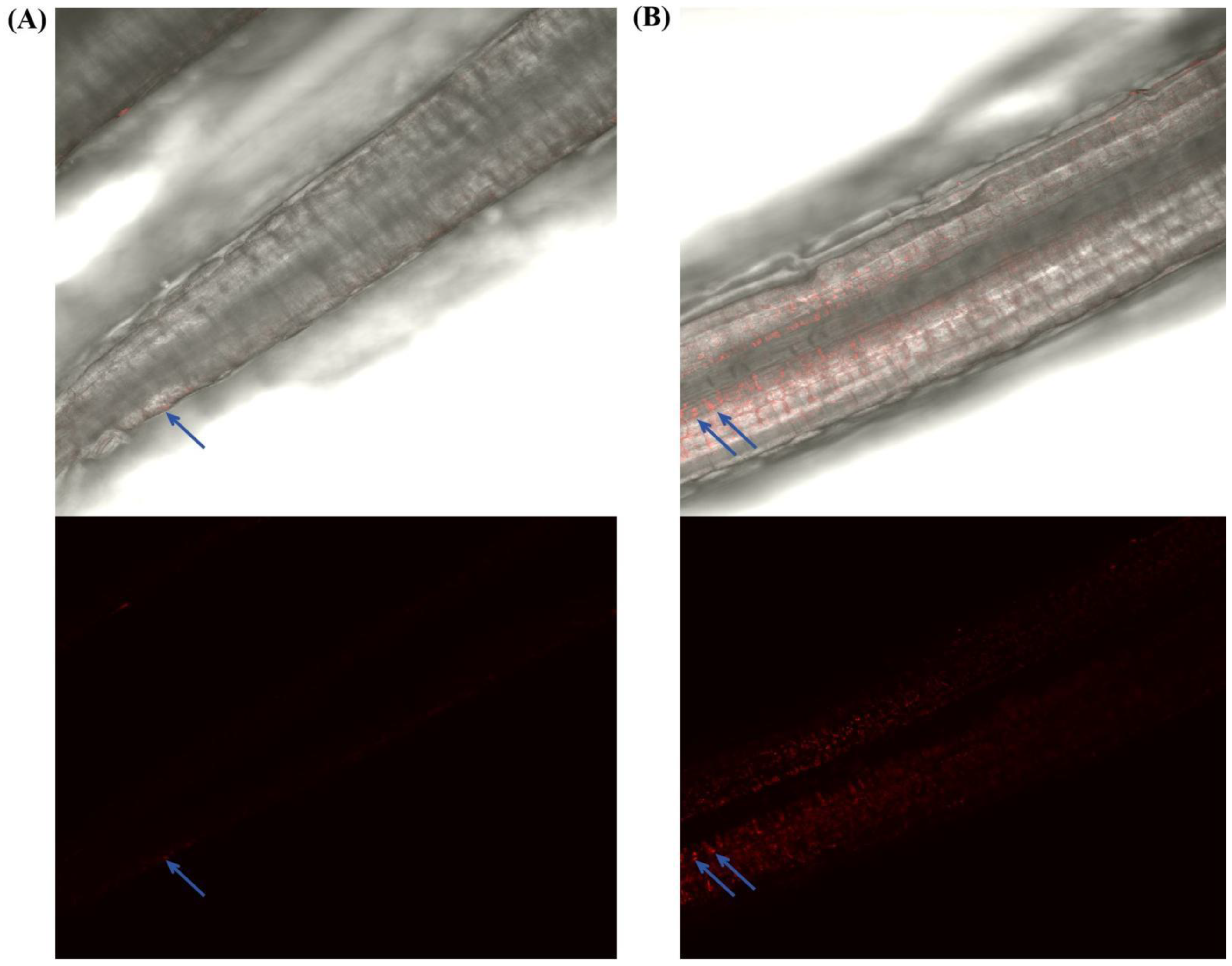
2.6. Colonization of A. inebrians Roots by GFP-Tagged Endophytic Bacteria

Soil sterilization was performed as follows: A 100 g sample of unplanted soil from Jinniu Mountain in Yuzhong, Gansu, China (pH of 8.2, 53 mg·g−1 of total nitrogen, 0.96 mg·g−1 of total phosphorus, 0.03 mg·g−1 of NO3, and 0.03 mg·g−1 of NH4+) was transferred into a 250 mL serum vial, which was then consecutively sterilized twice by autoclaving at 121 °C for 20 min, and sterilized again after 24 h. After sterilization, the soil was cooled in an ultra-clean test bench, and its moisture content was kept at 60% of the maximum water holding capacity.
The glasshouse experiments were conducted as follows: E+ and E− seeds were surface-sterilized and then placed on sterilized nitrogen-free MS medium to germinate. After 14 d, E+ and E− seedlings that exhibited uniform growth were transplanted into an experimental set up with sterilized soil, with four plants in each plot and four replicates. The roots were completely encapsulated in the soil. The E+ and E− seedlings were cultivated in a greenhouse under long-day conditions (16 h photoperiod, 25 ± 2 °C, light/dark cycle, with a light intensity of approximately 600 μmol·m−2·s−1).
To verify the effects of the E. gansuensis endophyte on endophytic bacteria colonization, the endophytic bacteria were tagged with red fluorescent protein (GFP) (vector pBE2R-mRFP1). For inoculation, the GFP-tagged endophytic bacteria strain was activated in LB medium, and colonies with 10−5 cells·mL−1 (OD600 = 0.5) were incubated for 24 h. A total of 100 μL of the bacterial suspension was applied to the potted plants by adding it to the bottom of the container 3 days after transplanting, and 100 μL of liquid medium was used as a blank control. After 15 days, root samples from the E+ and E− plants were surface-sterilized with 70% ethanol, collected, and then sectioned. Thin sections were observed under a confocal laser scanning microscope (CLSM, Olympus FV3000 confocal microscope, Shinjuku, Japan) using a condition excitation wavelength of 552 nm.

2.7. 15N Isotope Dilution Method

To verify the nitrogen conversion efficiency (NCE) of the Epichloë endophyte and the endophytic bacteria in A. inebrians, N2 was labeled with 15N. The 15N2 (98%) was purchased from Shanghai Chemical Reagent Research Institute Co., Ltd., Shanghai, China. The experimental set up tube was plugged with rubber sealing and the mouth was sealed with a three-way valve to prevent the entry of microorganisms and air (Figure S1). The set-up was evacuated with a vacuum pump and cleaned using argon to remove N2, and then 78% 15N2, 21% O2, and 1% argon were passed through. The incubation was carried out at 25 °C and 70% humidity for 15 days. The plant and soil samples were harvested separately from each treatment. The N content and 15N enrichment of the plants and soil were determined using a flow injection analyzer (FIAstar 5000 Analyzer, Foss, Hilleroed, Denmark) and a stable isotope ratio mass spectrometer (Mat 253, Thermo Fisher Scientific, Waltham, MA, USA). Biological nitrogen fixation (BNF) was calculated using the following equations: BNF (ug·g−1 DW) = TN (ug·g−1 DW) × atom%15Nexcess (atom%15Nexcess = atom%15Nsample-atom%15Ncontrol).

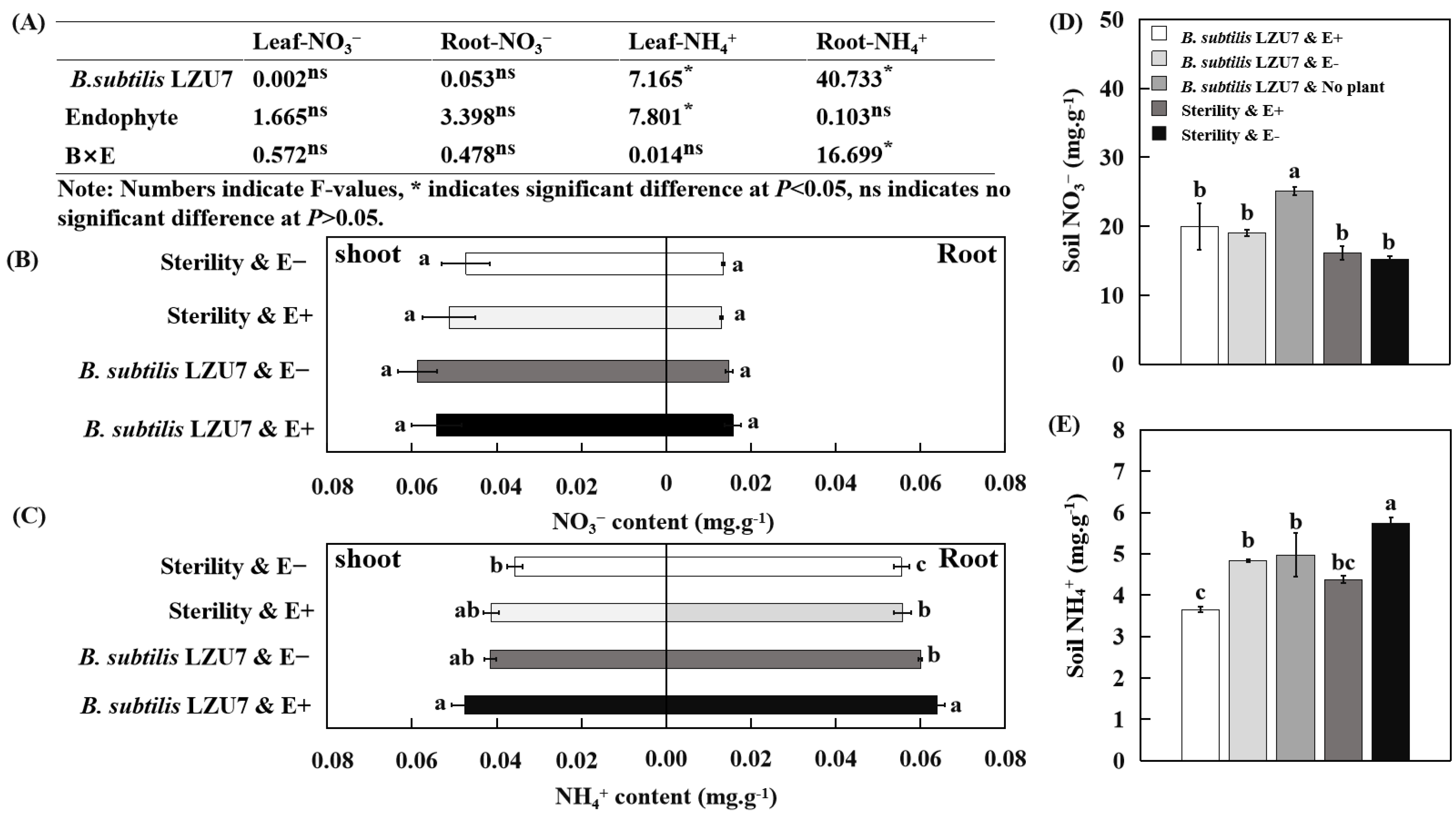
2.8. Plant and Soil Nutrient Determination

The gases were collected from each treatment (four replicates) on day 15 and detected using gas chromatography (GC-2014C, Shimadzu Limited, Kyoto, Japan) within 36 h. Nitrogen-transformation-related enzyme activities were determined using an ELISA detection kit and an ELISA reader (Thermo Fisher). The organic matter was measured using the K2CrO7–H2SO4 oxidation–reduction titration method. A FlashEA 1112 series CHNS/O analyzer (Thermo Fisher, Waltham, MA, USA) was used to determine the total C (TC) and total P (TP) contents in the shoots, roots, and soils. To determine the plant dry weight, all the plant samples were oven-dried at 80 °C until they reached a constant weight (9). Ergonovine was detected using an Agilent ChemStation for GC−LC Rev.A.10.01 systems (Agilent Technologies, Santa Clara, CA, USA) [39]. The mycelium concentration of the E. gansuensis endophyte in A. inebrians was measured using quantitative polymerase chain reaction (QPCR) [40].

2.9. Statistical Analysis

All the statistical analyses were primarily performed with the R v.4.0.3 (http://www.r-project.org/) and IBM SPSS Statistics v.21 software. A normal distribution and the homogeneity of variances of the data were tested with the Shapiro–Wilk’s test and Levene’s test. The effect of the Epichloë endophyte and endophytic bacteria on the plant weight, tiller, plant nutrient uptake, and nifH gene abundance was assessed using a two-factor ANOVA followed by Tukey’s HSD test. If no significant interactions were detected between the Epichloë endophyte and the endophytic bacteria, significant differences in their individual effects on parameter variances were evaluated using an independent samples t-test. Indoleacetic acid was assessed for variation among the endophytic bacteria using a one-way analysis of variance (ANOVA) followed by Tukey’s HSD test. Spearman’s correlation coefficients were calculated among the plant weight, tiller, plant and soil nutrients, nifH gene abundance, ergonovine, and mycelium concentration to explore the relationship between the Epichloë endophyte and the endophytic bacteria and to determine whether their interactions could promote nitrogen transformation in the A. inebrians host.

3. Results

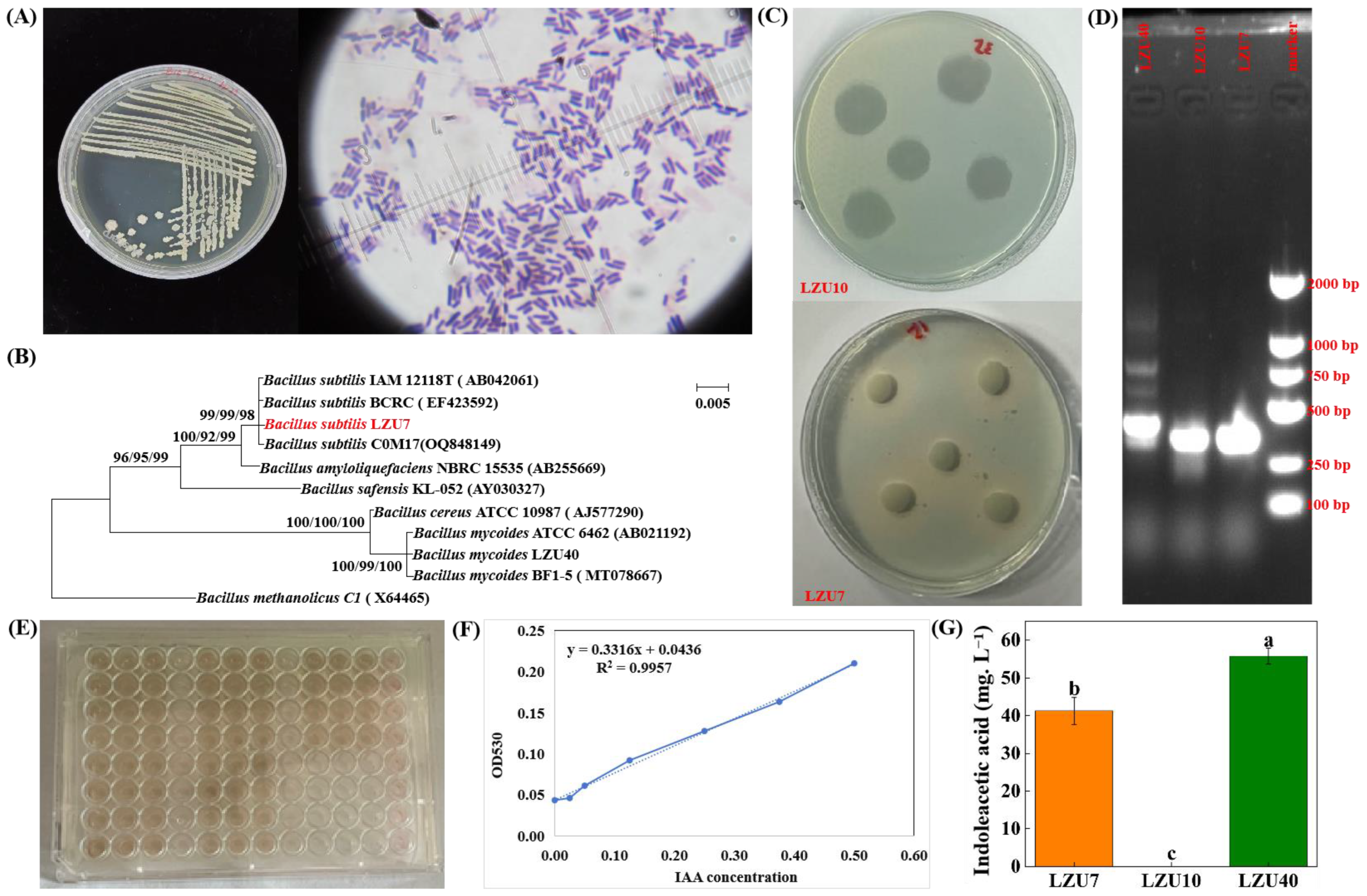
3.1. Community Structure and Function of Endophytic Bacteria in Roots of A. inebrians

We isolated and identified a total of 46 bacterial isolates from A. inebrians roots, and mainly focused on bacterial isolates belonging to the phylum Actinomycetota (1 isolate), Firmicutes (21 isolates), and Proteobacteria (24 isolates) (Table S1 and Figure S3). Five Proteobacteria and two Firmicutes isolates had the ability to secrete IAA, and seven Firmicutes and two Proteobacteria had the ability to produce siderophores (Table S1). In particular, the two tested Firmicutes isolates had the capacity to secrete growth hormones and siderophores, were able to grow in a nitrogen-free medium, and contained the nifH gene across all the tested isolates (Figure 1). However, the E. gansuensis endophyte strains had no growth-promoting effects (Figure S2).

3.2. E. gansuensis Endophyte Promotes the Growth and Colonization of Bacillus subtilis LZU7

The total plate count of B. subtilis LZU7 and B. mycoides LZU40 from the phylum Firmicutes with a growth-promoting effect was measured using the plate confrontation method. Compared with the sterile water and PDA culture medium treatments, the fungal endophyte fluid significantly increased the number of B. subtilis LZU7 colonies, but there was no significant effect on the number of B. mycoides LZU40 colonies (Table 1). We observed no inhibition of the colony growth for B. subtilis LZU7 or B. mycoides LZU40 by the E. gansuensis endophyte (Figure S4). There was less GFP-tagged B. subtilis LZU7 in the roots when the plants were not infected with the E. gansuensis endophyte (Figure 2A). When plants were infected with the E. gansuensis endophyte, more GFP-tagged B. subtilis LZU7 was detected in the roots of A. inebrians (Figure 2B).

3.3. E. gansuensis Endophyte and B. subtilis LZU7 Promote A. inebrians Growth and Increase Yield

The biomass of the plants inoculated with B. subtilis LZU7 reached 1.41 ± 0.20 mg, which was significantly higher than the biomass of the non-inoculated plants (1.16 ± 0.08) (p < 0.001) (Figure 3A). The biomass of the plants infected with the E. gansuensis endophyte was 1.37 ± 0.11 mg, which was significantly greater than that of non-infected plants (1.20 ± 0.01 mg) (p < 0.05) (Figure 3B). However, no significant effects on plant biomass were observed due to the interaction between an endophyte infection and B. subtilis LZU7 inoculation (Figure 3C). The results indicated that neither an endophyte infection nor B. subtilis LZU7 inoculation significantly promoted tillering and total carbon (Figure 3D,E). (Figure 3C). Total nitrogen was significantly higher in E+ plants (29.69 ± 0.87 mg. g−1) under the B. subtilis LZU7 inoculation treatment than in E+ (27.01 ± 0.67 mg. g−1) and E− (27.57 ± 0.54 mg. g−1) plants under the non-inoculation treatment, and in E− (27.18 ± 0.32 mg. g−1) plants under the inoculation treatment (Figure 3D).

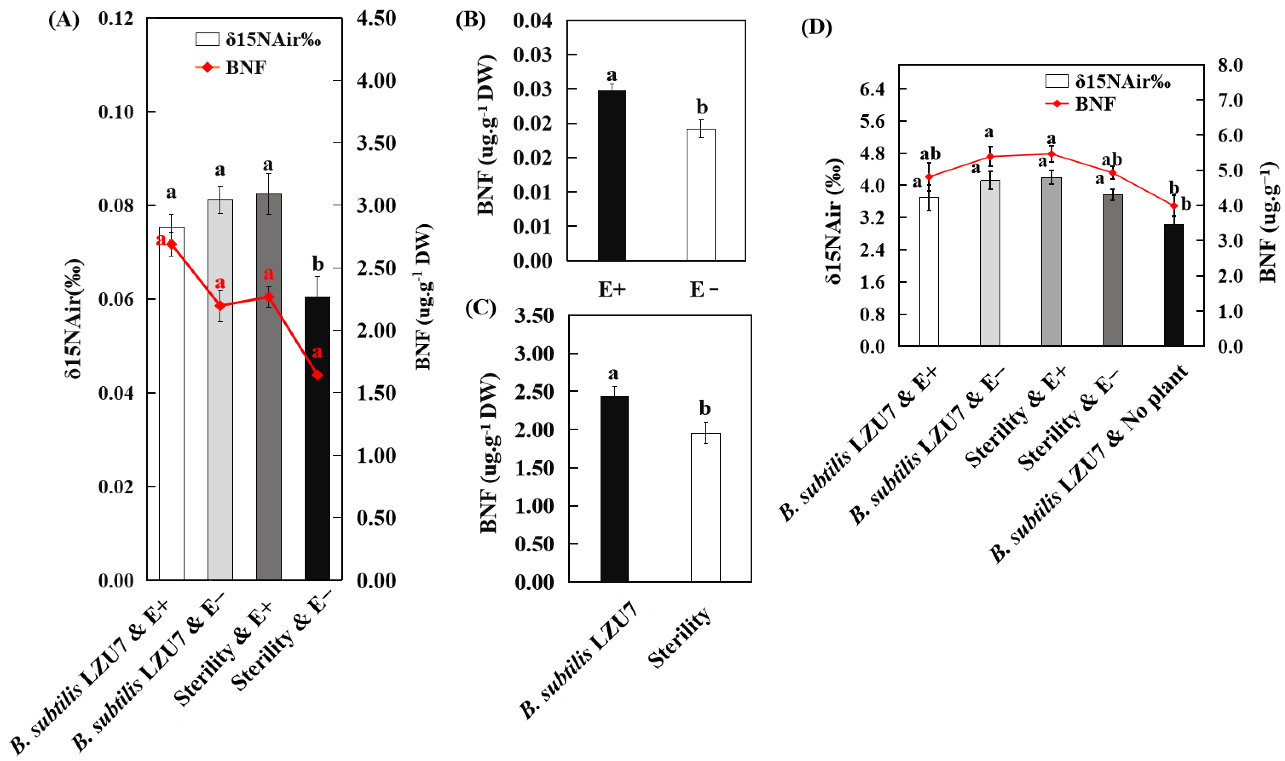
3.4. Interactions Between E. gansuensis and B. subtilis LZU7 Enhance Nitrogen Fixation in A. inebrians

The leaf NH4+ content of E+ plants under the B. subtilis LZU7 inoculation condition were found to be much higher than in E− plants under the B. subtilis LZU7 non-inoculation condition, while the plants infected with E. gansuensis endophyte under the non-inoculation condition and non-infected plants under the inoculation condition showed no significant differences (Figure 4A,C). For root NH4+, the E. gansuensis endophyte-infected plants under the B. subtilis LZU7 inoculation condition showed a significantly higher content than the E− plants under inoculation or non-inoculation conditions, and the E+ plants under the non-inoculation condition (Figure 4A,C). The inoculation of B. subtilis LZU7 and its combination with an endophyte infection did not change the NO3 content in the leaves, roots, or soil of A. inebrians (Figure 4 A,B,D). However, these treatments had a strong effect on the soil NH4+ content (Figure 4E).
For the inoculation treatment with B. subtilis LZU7, the root nitrogenase activity was significantly greater in E+ plants than in E− plants, while it was not significantly affected by an endophyte infection under the non-inoculation treatment (Figure S5A). The E. gansuensis endophyte infection significantly increased the nitrate reductase activity in the roots of A. inebrians (Figure S5B). The nitrate reductase and nitrite reductase activities in the roots of A. inebrians plants grown in soil inoculated with B. subtilis LZU7 were significantly greater than that in the roots of A. inebrians plants grown in soil without inoculated B. subtilis LZU7 soil (Figure S5C,D). Likewise, the glutamine synthetase activity in the roots was significantly (p < 0.05) greater in E+ plants compared with E− plants (Figure S5E), despite the fact that this activity in E+ and E− plants was not significantly affected by inoculation with B. subtilis LZU7. However, four enzymatic activities in the roots were not significantly affected by the interactions between the E. gansuensis infection and the B. subtilis LZU7 inoculation.
When the soil was not inoculated with B. subtilis LZU7, the CO2 and CH4 fluxes did not change, regardless of E. gansuensis infection. When the soil was inoculated with B. subtilis LZU7, the CO2 and CH4 fluxes were significantly (p < 0.05) increased (Figure S6A,B). The CO2 and CH4 fluxes were significantly increased when planting in soil inoculated with B. subtilis LZU7 compared with planting in soil that was not inoculated with B. subtilis LZU7; however, they were not significantly affected by the presence of the E. gansuensis endophyte (Figure S6A,B). The N2O flux did not change significantly between soils planted with E+ and E− plants under the conditions of the inoculation or non-inoculation with B. subtilis LZU7. However, there was a significant difference in the N2O flux between the soils inoculated and non-inoculated with B. subtilis LZU7 (Figure S6C).
In the 0–5 cm soil layer, the nifH gene copy number clearly increased, with a significantly higher nifH gene copy number in soil inoculated with B. subtilis LZU7 than in soil non-inoculated with B. subtilis LZU7. However, the soil nifH gene copy number was not increased by endophyte-infected plants compared with endophyte-free plants, under both the inoculated and non-inoculated treatments (Figure 5A). In the 5–10 cm soil layer, the treatment inoculated with B. subtilis LZU7 also had an increased soil nifH gene copy number compared with the treatment not inoculated with B. subtilis LZU7. Furthermore, the nifH gene copy number was significantly higher in the soil with E+ plants than in the soil with E− plants under the treatment inoculated with B. subtilis LZU7 (Figure 5A). In the roots, the interactions between an E. gansuensis infection and B. subtilis LZU7 inoculation significantly increased the nifH gene copy number (Figure 5B).
The δ 15N of the plants was impacted by an E. gansuensis infection and B. subtilis LZU7 inoculation, with a significant increase in the environments where the E+ and E− plants grew under the treatment of B. subtilis LZU7 inoculation, and in the environments where the E+ plants grew under non-inoculation conditions (Figure 6A). The presence of the E. gansuensis endophyte resulted in significantly increased plant BNF, which was 0.03 ± 0.005 μg·g−1 DW, compared with the endophyte-free plants, with a content of 0.019 ± 0.008 μg·g−1 DW (Figure 6B). The plant BNF significantly increased by 24.5% in response to B. subtilis LZU7 inoculation (Figure 6C). However, the combined cultivation of E+ plants and inoculation with B. subtilis LZU7 did not lead to a significant effect on the concentration of δ 15N or BNF in the soil (Figure 6D).

3.5. E. gansuensis Endophyte Increased the Nitrogen and Biomass Accumulation of A. inebrians by Interacting with B. subtilis LZU7

We observed that the copy number of the nifH gene in the roots and the plant BNF were significantly and positively correlated with the biomass; the concentration of TN in the plant; the CO2 flux; the NH4+ content in the leaves and roots; and the nitrate reductase, alkaloid, and hyphal concentrations, whereas a negative association was observed between the plant BNF and the soil NH4+ content (p < 0.05) (Figure 7). Moreover, the plant BNF showed significant positive correlations with nitrogenase (Figure 7). The alkaloid and hyphal concentrations showed a better correlation with plant BNF (Figure 7), even though there was no statistically significant difference in the E+ plants under the B. subtilis LZU7 inoculation treatment (Figure S7). No significant correlations were detected between the content of plant 15N and other variables, except for a negative correlation with the soil NH4+ content (Figure 7). The interaction of the E. gansuensis endophyte with B. subtilis LZU7 increased the NH4+ accumulation in the leaves and roots compared with the control, which showed positive correlations with the biomass, plant TN, CO2 flux, nifH gene copy number in the leaves and roots, and plant BNF (p < 0.05) (Figure 7). Furthermore, we observed a positive relationship between biomass accumulation and the plant TN, the NH4+ in the leaves and roots, the CO2 flux, the leaf NO3, nitrogenase, and nitrate reductase, but a strong negative correlation between biomass accumulation and the soil NH4+ content (Figure 7).
We used SEM to decipher how an endophyte infection affects plant performance by interacting with B. subtilis LZU7 (Figure 8). The SEM results indicated that the E. gansuensis endophyte had positive effects on plant performance by recruiting B. subtilis LZU7 colonization to increase the root N accumulation (Figure 8). The E. gansuensis endophyte and B. subtilis LZU7 had direct effects on plant biomass. Furthermore, changes in the soil nifH gene mediated by the E. gansuensis endophyte and B. subtilis LZU7 further affected the soil NH4+ content and led to a change in the root NH4+ content, and, thus, increased biomass accumulation (Figure 8). The root NH4+ content and the soil nifH gene were the main factors affecting plant biomass (Figure 8).

4. Discussion

The symbiotic fungal endophyte of A. inebrians, the E. gansuensis endophyte, has been found to affect the composition of root-associated bacteria by mediating root metabolism, thereby resulting in a positive response to low-nitrogen stress [29]. In this study, we found that the foliar E. gansuensis endophyte promoted the colonization of A. inebrians roots by root-originated B. subtilis LZU7, resulting in an increase in the N accumulation and biomass of the host plants.
Initially, we studied whether inhibition was present between the E. gansuensis endophyte strains and the B. subtilis LZU7 and B. mycoides LZU40 strains under in vitro culture conditions. The strains of the E. gansuensis endophyte lacked the ability to secrete IAA and siderophores and possessed no nitrogen fixation gene (nifH gene) (Figure S2), but the B. subtilis LZU7 and B. mycoides LZU40 strains had these functions (Figure 1). The effect of the E. gansuensis endophyte on the growth of B. subtilis LZU7 and B. mycoides LZU40 was explored from many angles. First, we used a solid medium to test the inhibition between both strains. The results showed that there was no inhibitory effect between the E. gansuensis endophyte and B. subtilis LZU7 or B. mycoides LZU40, but a growth-promoting effect was not obvious. Second, the fermentation broth of E. gansuensis endophyte strains increased the number of B. subtilis LZU7 colonies, but had no effect on B. mycoides LZU40, suggesting that the E. gansuensis endophyte has the potential to improve the growth of LZU7 strains. However, the strains and species used are taxonomically diverse and possibly metabolically different. The probiotic substances secreted by the Epichloë endophyte should be considered for further study.
Second, we demonstrated that an E. gansuensis endophyte infection could promote the colonization of A. inebrians roots by B. subtilis LZU7. We found that a large amount of GFP-tagged B. subtilis LZU7 aggregated in the roots of the symbionts of E. gansuensisA. inebrians (Figure 2). These results suggest that the Epichloë endophyte plays an important role in the recruitment of beneficial microbes. Epichloë endophyte-induced changes and the enrichment of beneficial microbes were mainly attributed to two mechanisms related to altering the host’s metabolism. First, the endophytic-fungal-mediated metabolites of host plants could change the bacterial community composition directly [41,42], as symbiotic microorganisms have been shown to exhibit metabolic differences between different symbionts. For example, the E. gansuensis endophyte has been shown to mediate the production of secondary metabolite products, while alkaloids produced by A. inebrians could increase the colonization of beneficial root microbiomes and increase the complexity of interactions [29]. However, a change in beneficial microbes was inconsistent with the results of Rojas et al. [43], which showed that the infection of Schedonorus arundinaceus with E. coenophiala leads to a reduction in arbuscular mycorrhizal colonization. This suggests that the effects of an Epichloë endophyte infection on beneficial microbes may vary depending on the endophyte and/or host species.
Positive effects of the Epichloë endophyte on plant growth, fitness, defense against pathogens, tolerance to drought stress, and herbivore resistance have been documented [14,44,45,46], but the effect of its interactions with beneficial root microbes on the productivity and nutrient absorption of host plants is unknown. Zhao et al. [35] reported that the interaction between the E. gansuensis endophyte and Bacillus strains increased seed germination and the growth of A. inebrians. This increase might result from secondary metabolites from probiotic metabolism [47]. Our study demonstrates that productivity and nutrient absorption are also affected by the E. gansuensis endophyte and B. subtilis LZU7. Contrary to Zhao et al. [35], we observed that endophyte- or rhizobacteria-enhanced plant growth manifested as increased host biomass; E. gansuensis endophyte-infected plants or plants grown in soil inoculated with B. subtilis LZU7 gained more plant biomass compared with E. gansuensis-free plants or plants grown in soil not inoculated with B. subtilis LZU7, but tillers were not affected by an E. gansuensis endophyte infection or B. subtilis LZU7 inoculation (Figure 3). Therefore, the foliar E. gansuensis endophyte and root B. subtilis can improve plant productivity in addition to promoting seed germination.
The effects of the E. gansuensis endophyte on the nitrogen uptake efficiency in host plants and on soil nitrogen and phosphorus cycling may depend on plant enzyme activities and beneficial rhizobacteria interactions [30,48,49]. Importantly, we demonstrated that the root NH4+ concentrations were significantly increased for E. gansuensis endophyte-infected plants grown in soil inoculated with B. subtilis LZU7 compared with E. gansuensis endophyte-infected plants grown in non-inoculated soils and E. gansuensis endophyte-free plants grown in inoculated and non-inoculated soils (Figure 4C). The root NH4+ concentration had a significantly positive association with plant nitrogenase and nitrate reductase activities and with the nifH gene copy number in the roots and soil (Figure 7), suggesting that the interactions between the E. gansuensis endophyte and B. subtilis LZU7 promote nitrogen fixation and transformation; this is also supported by the significantly high enzyme activity and BNF (Figure 5 and Figure 6 and S5).
The synthesis of large amounts of alkaloids was associated with the depletion of plant N, in accordance with previous studies (31, 32) in which no significant differences were observed in the leaf NH4+ concentration between E+ and E− plants grown in inoculated or non-inoculated soils, but higher leaf NH4+ concentrations were observed for E+ plants grown in soils inoculated with B. subtilis LZU7 relative to E− plants grown in soils not inoculated with B. subtilis LZU7. Ren et al. [50] found that an endophyte infection tended to reduce the shoot nitrogen (N) concentration but caused a significant increase in the fractions of N allocated by the host plants. Furthermore, we found 62.31 mg.kg−1 and 59.06 mg.kg−1 of ergot alkaloids in E+ plants grown in soils inoculated and not inoculated with B. subtilis LZU7, respectively (Figure S7). Both of these ergot alkaloid concentrations increased significantly in response to an endophyte infection in interactive systems (Figure 8). These results indicate that an endophyte infection can promote nitrogen uptake and utilization efficiency in host plants.
The interactions of the E. gansuensis endophyte with B. subtilis LZU7 will change the soil NH4+ concentration, which will alter the root NH4+ concentration and lead to a biomass shift (Figure 8). Under low-nutrient conditions, an endophyte infection could be used to increase the root micro-nutrient content (e.g., K and Ca) to promote biomass accumulation and alleviate nutrient starvation stress (9). This tripartite interaction involving foliar fungal endophytes, host plants, and root bacteria can stimulate nitrogen-fixing microorganism activity, thereby improving the N bioavailability by decreasing molecular N and increasing BNF (Figure 4, Figure 5, Figure 6 and Figure 7). This process not only conducts N fixation, but also N transformation, leading to positive feedback between both. The relationship among endophyte infections, the nutrient availability, N acquisition, and biomass accumulation could be negative or positive. We also found that the interaction between the E. gansuensis endophyte and B. subtilis LZU7 increased the soil nifH gene copy number, which in turn affected the NH4+ content in the roots and soil and ultimately led to an increase in biomass (Figure 8). As previously reported, a low soil nutrient condition is suggested to benefit N acquisition and biomass accumulation by stimulating the effects of endophyte infection in enhancing cooperation and interactions among root bacteria [10,29]. However, there is also a contrasting perspective that high nutrient levels provide a greater opportunity for host plants to thrive, leading to positive correlations among nutrient resource availability, endophyte infections, and biomass accumulation [51,52]. This is because the Epichloë endophyte is a heterotrophic microorganism that competes with the host for nutrients or photosynthate at a metabolic cost at a low nutrient level [6,53]. Taken together, changes in the nutrient contents in the plants and soil were confirmed to contribute significantly to plant N acquisition and growth mediated by endophyte infections. This implies that E. gansuensis endophyte interactions with B. subtilis LZU7 are intricate at the interface between above-ground and below-ground plant parts. We, therefore, conclude that nutrient fraction connections driven by the E. gansuensis endophyte and B. subtilis LZU7 among different tissues is a pathway as important as the positive feedback between fungal endophyte interactions with B. subtilis LZU7 in enhancing plant performance.

5. Conclusions

This study demonstrates that the E. gansuensis endophyte can promote the growth of the plant growth-promoting rhizobacterium B. subtilis LZU7 in an in vitro culture and enhance its colonization in the roots of A. inebrians plants. We also confirmed that the E. gansuensis endophyte interactions with B. subtilis LZU7 had positive effects on nitrogenase (nifH) gene expression, which could lead to an increase in biological nitrogen fixation (BNF). These results also suggest that E. gansuensis infection and B. subtilis LZU7 inoculation regulate the interaction with nutrient fractions leading to biomass accumulation. The root NH4+ concentration was positively correlated with both E. gansuensis endophyte infection and B. subtilis LZU7 inoculation, and eventually improved the production of A. inebrians. This, in turn, led to negative feedback with soil N accumulation through affecting the copy number of the soil nifH gene, leading to a further increase in the root NH4+ content, and enhanced biomass accumulation. Our results provide a novel demonstration that interactions between the foliar Epichloë endophyte and the beneficial root-originated bacterium B. subtilis can have major effects on the uptake and utilization of an essential nutrient, and they suggest that the source of N resources can be from biological nitrogen fixation, which is mediated by nitrogen-fixing microorganisms.

Supplementary Materials

The following supporting information can be downloaded at: https://www.mdpi.com/article/10.3390/jof11070466/s1, Figure S1 Schematic representation (A) and physical pictures (B) of experimental set-up; Figure S2 Functional identification of Epichloë gansuensis endophyte strains. (A) The surface and reverse views of E. gansuensis endophyte strain. (B) Gel electrophoresis map of the nifH gene in E. gansuensis endophyte strains. (C) Siderophores measurement of E. gansuensis endophyte strains. (D) Promoting ability of E. gansuensis endophyte strains; Figure S3 The genetic relationships analysis among 46 strains endophytic bacterium by building the phylogenetic tree; Figure S4 Plate confrontation test of the E. gansuensis endophyte strains and endophytic bacteria. (A) B. Subtilis LZU7 strains. (B) B. mycoides LZU40; Figure S5 Effects of E. gansuensis endophyte infection and B. Subtilis LZU7 inoculation on nitrogenase, nitrate reductase, nitrite reductase and glutamine synthetase of A. inebrians. (A) The two-factor analysis of enzymatic activity. (B,E) The one-factor analysis of nitrate reductase and nitrite reductase between E+ and E- plants. (C,D) The one-factor analysis of nitrate reductase and glutamine synthetase between inoculated and non-inoculated B. subtilis LZU7; Figure S6 The effects of the E. gansuensis endophyte infection and B. Subtilis LZU7 inoculation influences soil fluxes of the greenhouse gases CO2, CH4 and N2O. (A) CO2 flux; (B) CH4 flux; (C) N2O flux; Figure S7 The effects of the E. gansuensis endophyte infection and B. Subtilis LZU7 inoculation on mycelial gene copy number and alkaloid content. (A) Mycelial gene copy number; (B) Alkaloid content; Table S1 Isolation, identification and functional characterization of endophytic bacteria in A. inebrians.

Author Contributions

Visualization, Methodology, Investigation, Data curation, Investigation, Formal analysis, Conceptualization, Writing—original draft, and Writing—review and editing, Y.J.; Conceptualization, Software, Methodology, Resources, Data curation, Writing—review and editing, and Funding acquisition, Z.C.; Editing, K.M.; Conceptualization, Resources, Supervision, Writing—review and editing, and Funding acquisition, C.L. All authors have read and agreed to the published version of the manuscript.

Funding

The work supported by the China Postdoctoral Science Foundation (2024M761243), Gansu Province Outstanding Doctoral Students Project (22JR5RA434), Intellectual Property Plan (Targeted Organization) Project of Gansu Administration for Market Regulation (22ZSCQD01), The Fundamental Research Funds for the Central Universities (lzujbky-2022-kb02, lzujbky-2023-49 and lzujbky-2025-jdzx09) and Gansu Province Grassland Monitoring and Evaluation Technology Support Project of Gansu Province Forestry and Grassland Administration ([2021]794).

Institutional Review Board Statement

Not applicable.

Informed Consent Statement

Not applicable.

Data Availability Statement

The original contributions presented in this study are included in the article/Supplementary Materials. Further inquiries can be directed to the corresponding authors.

Conflicts of Interest

The authors declare no conflict of interest.

References

  1. Chepick, G.P.; Cho, R. Interactive effects of fungal endophyte infection and host genotype on growth and storage in Lolium perenne. New Phytol. 2003, 158, 183–191. [Google Scholar] [CrossRef]
  2. Rey, T.; Chatterjee, A.; Buttay, M.; Toulotte, J.; Schornack, S. Medicago truncatula symbiosis mutants affected in the interaction with a biotrophic root pathogen. New Phytol. 2015, 206, 497–500. [Google Scholar] [CrossRef] [PubMed]
  3. Ivanov, S.; Harrison, M.J. Receptor-associated kinases control the lipid provisioning program in plant-fungal symbiosis. Science 2024, 383, 443–448. [Google Scholar] [CrossRef]
  4. Parniske, M. Arbuscular mycorrhiza: The mother of plant root endosymbioses. Nat. Rev. Microbiol. 2008, 6, 763–765. [Google Scholar] [CrossRef] [PubMed]
  5. Schardl, C.L.; Leuchtmann, A.; Spiering, M.J. Symbioses of grasses with seedborne fungal endophytes. Annu. Rev. Plant Biol. 2004, 55, 315–340. [Google Scholar] [CrossRef]
  6. Clay, K. Fungal endophyte symbiosis and plant diversity in successional fields. Science 1999, 285, 1742–1744. [Google Scholar] [CrossRef]
  7. Leuchtmann, A. Systematics, distribution, and host specificity of grass endophytes. Nat. Toxins 1992, 1, 150–162. [Google Scholar] [CrossRef]
  8. Popay, A.J.; Thom, E.R. Endophyte effects on major insect pests in Waikato dairy pasture. N. Z. Grassl. Assoc. 2009, 71, 121–126. [Google Scholar] [CrossRef]
  9. Vázquez-de-Aldana, B.R.; García-Ciudad, A.; García-Criado, B.; Vicente-Tavera, S.; Zabalgogeazcoa, I. Fungal endophyte (Epichloë festucae) alters the nutrient content of Festuca rubra regardless of water availability. PLoS ONE 2013, 18, e84539. [Google Scholar] [CrossRef]
  10. Guo, J.Q.; Mcculley, R.L.; Phillips, T.D.; Mcnear, D.H. Fungal endophyte and tall fescue cultivar interact to differentially affect bulk and rhizosphere soil processes governing c and n cycling. Soil Biol. Biochem. 2016, 101, 165–174. [Google Scholar] [CrossRef]
  11. Cavazos, B.R.; Bohner, T.F.; Donald, M.L.; Sneck, M.E.; Shadow, A.; Omacini, M.; Rudgers, J.A.; Miller, T.E.X. Testing the roles of vertical transmission and drought stress in the prevalence of heritable fungal endophytes in annual grass populations. New Phytol. 2018, 219, 1075–1084. [Google Scholar] [CrossRef]
  12. Oberhofer, M.; Güsewell, S.; Leuchtmann, A. Effects of natural hybrid and non-hybrid Epichloë endophytes on the response of Hordelymus europaeus to drought stress. New Phytol. 2024, 201, 242–253. [Google Scholar] [CrossRef] [PubMed]
  13. Pereira, E.C.; Aldana, B.R.V.D.; Arellano, J.B.; Zabalgogeazcoa, I. The Role of fungal microbiome components on the adaptation to salinity of Festuca rubra subsp. pruinosa. Front. Plant Sci. 2021, 12, 695717–695732. [Google Scholar] [CrossRef] [PubMed]
  14. Dupont, P.Y.; Eaton, C.J.; Wargent, J.J.; Fechtner, S.; Solomon, P.; Schmid, J.; Day, R.C.; Scott, B.; Cox, M. Fungal endophyte infection of ryegrass reprograms host metabolism and alters development. New Phytol. 2015, 208, 1227–1240. [Google Scholar] [CrossRef]
  15. Purahong, W.; Hyde, K.D. Effects of fungal endophytes on grass and non-grass litter decomposition rates. Fungal Divers. 2011, 47, 1–7. [Google Scholar] [CrossRef]
  16. Young, C.A.; Hume, D.E.; McCulley, R.L. Forages and pastures symposium: Fungal endophytes of tall fescue and perennial ryegrass: Pasture friend or foe? J. Anim. Sci. 2013, 91, 2379–2394. [Google Scholar] [CrossRef]
  17. Li, F.; Deng, J.; Nzabanita, C.; Li, Y.Z.; Duan, T.Y. Growth and physiological responses of perennial ryegrass to an AMF and an Epichloë endophyte under different soil water contents. Symbiosis 2019, 79, 151–161. [Google Scholar] [CrossRef]
  18. Guo, S.; Geisen, S.; Mo, Y.N.; Yan, X.Y.; Huang, R.L.; Liu, H.J.; Gao, Z.L.; Tao, C.Y.; Deng, X.H.; Xiong, W.; et al. Predatory protists impact plant performance by promoting plant growth-promoting rhizobacterial consortia. ISME J. 2024, 18, wrae180. [Google Scholar] [CrossRef]
  19. Kennedy, I.R.; Choudhury, A.T.M.A.; Kecskés, M.L. Non-symbiotic bacterial diazotrophs in crop-farming systems: Can their potential for plant growth promotion be better exploited? Soil Biol. Biochem. 2004, 36, 1229–1244. [Google Scholar] [CrossRef]
  20. Lugtenberg, B.; Kamilova, F. Plant-growth-promoting rhizobacteria. Annu. Rev. Microbiol. 2009, 63, 541–556. [Google Scholar] [CrossRef]
  21. Wang, W.B.; Portal-Gonzalez, N.; Wang, X.; Li, J.L.; Li, H.; Portieles, R.; Borras-Hidalgo, O.; He, W.X.; Santos-Berrmudez, R. Metabolome-driven microbiome assembly determining the health of ginger crop (Zingiber officinalel. roscoe) against rhizome rot. Microbiome 2024, 12, 167–187. [Google Scholar] [CrossRef] [PubMed]
  22. Radhakrishnan, R.; Hashem, A.; Abd_Allah, E.F. Bacillus: A biological tool for crop improvement through bio-molecular changes in adverse environments. Front. Physiol. 2017, 8, 667. [Google Scholar] [CrossRef] [PubMed]
  23. Figueredo, M.S.; Tonelli, M.L.; Taurian, T.; Angelini, J.; Ibañez, F.; Valetti, L.; Muñoz, V.; Anzuay, S.; Ludueña, L.; Fabra, A. Interrelationships between Bacillus sp. CHEP5 and Bradyrhizobium sp. SEMIA6144 in the induced systemic resistance against Sclerotium rolfsii and symbiosis on peanut plants. J. Biosci. 2014, 39, 877–885. [Google Scholar] [CrossRef]
  24. Kaschuk, G.; Auler, A.C.; Vieira, C.E.; Dakora, F.D.; Jaiswal, S.K.; Da-Cruz, S.P. Coinoculation impact on plant growth promotion: A review and meta-analysis on coinoculation of rhizobia and plant growth-promoting bacilli in grain legumes. Braz. J. Microbiol. 2022, 53, 2027–2037. [Google Scholar] [CrossRef]
  25. Kalantari, S.; Marefat, A.; Naseri, B.; Hemmati, R. Improvement of bean yield and Fusarium root rot biocontrol using mixtures of Bacillus, Pseudomonas and Rhizobium. Trop. Plant Pathol. 2018, 43, 499–505. [Google Scholar] [CrossRef]
  26. Wang, T.Q.; Chen, Q.Q.; Liang, Q.; Zhao, Q.; Lu, X.; Tian, J.H.; Guan, Z.D.; Liu, C.; Li, J.F.; Zho, M.; et al. Bacillus suppresses nitrogen efficiency of soybean–rhizobium symbiosis through regulation of nitrogen-related transcriptional and microbial patterns. Plant Cell Environ. 2024, 47, 4305–4322. [Google Scholar] [CrossRef]
  27. Soundararajan, S.; Jedd, G.; Li, X.L.; Ramos-Pamploña, M.; Chua, N.H.; Naqvia, N.I. Woronin body function in magnaporthe grisea is essential for efficient pathogenesis and for survival during nitrogen starvation stress. Plant Cell 2004, 16, 1564–1574. [Google Scholar] [CrossRef]
  28. Li, Y.; Zhang, L.; Cheng, Z.Q.; Jin, L.Y.; Zhang, T.; Liang, L.M.; Cheng, L.J.; Zhang, Q.Y.; Xu, X.H.; Lan, C.H.; et al. Microbiota and functional analyses of nitrogen-fixing bacteria in root-knot nematode parasitism of plants. Microbiome 2023, 11, 48. [Google Scholar] [CrossRef]
  29. Jin, Y.Y.; Chen, Z.J.; White, J.F.; Malik, K.; Li, C.J. Interactions between Epichloë endophyte and the plant microbiome impact nitrogen responses in host Achnatherum inebrians plants. Microbiol. Spectr. 2024, 12, e0257423. [Google Scholar] [CrossRef]
  30. Liu, L.; Gundersen, P.; Zhang, T.; Mo, J.M. Effects of phosphorus addition on soil microbial biomass and community composition in three forest types in tropical China. Soil Biol. Biochem. 2012, 44, 31–38. [Google Scholar] [CrossRef]
  31. Yang, H.J.; Li, Y.; Wu, M.Y.; Zhang, Z.; Li, L.H.; Wan, S.Q. Plant community responses to nitrogen addition and increased precipitation: The importance of water availability and species traits. Glob. Change Biol. 2011, 17, 2936–2944. [Google Scholar] [CrossRef]
  32. Faeth, S.H.; Fagan, W.F. Fungal endophytes: Common host plant symbionts but uncommon mutualists. Integr. Comp. Biol. 2002, 42, 360–368. [Google Scholar] [CrossRef]
  33. Schardl, C.L.; Young, C.A.; Pan, J.; Florea, S.; Takach, J.E.; Panaccione, D.G.; Farman, M.L.; Webb, J.S.; Jaromczyk, J.; Charlton, N.D.; et al. Currencies of mutualisms: Sources of alkaloid genes in vertically transmitted Epichloë. Toxins 2013, 5, 1064–1088. [Google Scholar] [CrossRef] [PubMed]
  34. Jin, Y.Y.; Chen, Z.J.; He, Y.L.; White, J.F.; Malik, K.; Chen, T.X.; Li, C.J. Effects of Achnatherum inebrians ecotypes and endophyte status on plant growth, plant nutrient, soil fertility and soil microbial community. Soil Sci. Soc. Am. J. 2022, 86, 1028–1042. [Google Scholar] [CrossRef]
  35. Zhao, H.T.; Nie, X.M.; Zhang, W.; Zhang, X.X.; Ju, Y.W.; Li, Y.Z.; Christesen, M.J. Effects of interaction of Epichloë gansuensis and Bacillus strains on the seed germination and seedling growth in Achnatherum inebrians plants. Res. Sq. 2023. [Google Scholar] [CrossRef]
  36. Jin, Y.Y. Microbial Mechanisms of Nitrogen Effective Use in Achnatherum inebrians-Fungal Endphyte Symbiont. Ph.D. Dissertation, Lanzhou University, Lanzhou, China, 2024. [Google Scholar]
  37. Tamura, K.; Peterson, D.; Peterson, N.; Stecher, G.; Nei, M.; Kumar, S. MEGA5: Molecular evolutionary genetics analysis using maximum likelihood, evolutionary distance, and maximum parsimony methods. Mol. Biol. Evol. 2011, 28, 2731–2739. [Google Scholar] [CrossRef] [PubMed]
  38. Zhou, G.P.; Fan, K.K.; Li, G.L.; Gao, S.J.; Chang, D.N.; Liang, T.; Li, S.; Liang, H.; Zhang, J.D.; Che, Z.X.; et al. Synergistic effects of diazotrophs and arbuscular mycorrhizal fungi on soil biological nitrogen fixation after three decades of fertilization. iMeta 2023, 2, e81. [Google Scholar] [CrossRef] [PubMed]
  39. Cheplick, G.P.; Clay, K.; Marks, S. Interactions between fungal endophyte infection and nutrient limitation in the grasses Lolium perenne and Festuca arundinacea. New Phytol. 1989, 111, 89–98. [Google Scholar] [CrossRef]
  40. Chen, T.X.; Simpson, W.R.; Nan, Z.B.; Li, C.J. NaCl stress modifies the concentrations of endophytic fungal hyphal and peramine in Hordeum brevisubulatum seedlings. Crop Pasture Sci. 2021, 73, 214–221. [Google Scholar] [CrossRef]
  41. Patchett, A.; Newman, J.A. Comparison of plant metabolites in root exudates of Lolium perenne infected with different strains of the fungal endophyte Epichloë festucae var. lolii. J. Fungi 2021, 7, 148–177. [Google Scholar] [CrossRef]
  42. Liang, J.J.; Gao, G.Y.; Zhong, R.; Liu, B.W.; Christensen, M.J.; Ju, Y.W.; Zhang, W.; Wu, Z.; Li, C.; Li, C.J.; et al. Effect of Epichloë gansuensis endophyte on seed-borne microbes and seed metabolites in Achnatherum inebrians. Microbiol. Spectr. 2023, 11, e01350-22. [Google Scholar] [CrossRef] [PubMed]
  43. Rojas, X.; Guo, J.Q.; Leff, J.W.; McNear, D.; Fierer, N.; McCulley, R.L. Infection with a shoot-specific fungal endophyte (Epichloë) alters tall fescue soil microbial communities. Microb. Ecol. 2016, 72, 197–206. [Google Scholar] [CrossRef] [PubMed]
  44. Brem, D.; Leuchtmann, A. Epichloë grass endophytes increase herbivore resistance in the woodland grass Brachypodium sylvaticum. Oecologia 2001, 126, 522–530. [Google Scholar] [CrossRef]
  45. Bultman, T.L.; Bell, G.; Martin, W.D. A fungal endophyte mediates reversal of wound induced resistance and constrains tolerance in a grass. Ecology 2004, 85, 679–685. [Google Scholar] [CrossRef]
  46. Powell, R.G.; Petroski, R.J. Alkaloid toxins in endophyte-infected grasses. Nat. Toxins 2010, 1, 163–170. [Google Scholar] [CrossRef]
  47. Njoroge Kimotho, R.; Zheng, X.; Li, F.R.; Chen, Y.J.; Li, X.F. A potent endophytic fungus Purpureocillium lilacinum YZ1 protects against Fusarium infection in field-grown wheat. New Phytol. 2024, 243, 1899–1916. [Google Scholar] [CrossRef]
  48. Hou, W.P.; Xia, C.; Christensen, M.J.; Wang, J.F.; Li, X.Z.; Chen, T.; Nan, Z.B. Effect of Epichloë gansuensis endophyte on rhizosphere bacterial communities and nutrient concentrations and ratios in the perennial grass species Achnatherum inebrians during three growth seasons. Crop Pasture Sci. 2020, 71, 1050–1066. [Google Scholar] [CrossRef]
  49. Wang, J.F.; Nan, Z.B.; Christensen, M.J.; Zhang, X.X.; Tian, P.; Zhang, Z.X.; Niu, X.L.; Gao, P.; Chen, T.; Ma, L.X. Effect of Epichloë gansuensis endophyte on the nitrogen metabolism, nitrogen use efficiency, and stoichiometry of Achnatherum inebrians under nitrogen limitation. J. Agric. Food Chem. 2018, 66, 4022–4031. [Google Scholar] [CrossRef]
  50. Ren, A.Z.; Wei, M.Y.; Yin, L.J.; Wu, L.J.; Zhou, Y.; Li, X.; Cao, Y.B. Benefits of a fungal endophyte in Leymus chinensis depend more on water than on nutrient availability. Environ. Exp. Bot. 2014, 108, 71–78. [Google Scholar] [CrossRef]
  51. Li, X.; Zhou, Y.; Mace, W.; Qin, J.H.; Liu, H.; Chen, W.; Ren, A.Z.; Gao, Y.B. Endophyte species influence the biomass production of the native grass Achnatherum sibiricum (L.) keng under high nitrogen availability. Ecol. Evol. 2016, 6, 8595–8606. [Google Scholar] [CrossRef]
  52. Murphy, B.R.; Doohan, F.M.; Hodkinson, T.R. Yield increase induced by the fungal root endophyte Piriformospora indica in barley grown at low temperature is nutrient limited. Symbiosis 2014, 62, 29–39. [Google Scholar] [CrossRef]
  53. Hamilton, C.E.; Dowling, T.E.; Faeth, S.H. Hybridization in endophyte symbionts alters host response to moisture and nutrient treatments. Microb. Ecol. 2010, 59, 768–775. [Google Scholar] [CrossRef] [PubMed]
Figure 1. Morphological characterization and functional analysis of Bacillus subtilis LZU7. (A) Colony morphology, colony color, shape and size observations, and Gram staining. Purple indicates Gram-positive bacteria, while red indicates Gram-negative bacteria. (B) Determining genetic relationships among endophytic bacteria by building a phylogenetic tree based on the 16S rDNA sequences of the bacteria and its GenBank allies. (C) Ferritin analysis. Change in color indicates that the strain has the ability to secrete ferritin. (D) Gel electrophoresis map of the nifH gene in endophytic bacteria. The DNA fragment size of the nifH gene is 480 bps. The presence of a band at 480 bp indicates that the strain contains the nifH gene. (E) The determination of plant hormone indole-3-acetic acid. (F,G) The standard curve and concentration of indole-3-acetic acid. Values are means ± standard error. Soild and dotted lines: fitted and measured standard curves. Different lowercase letters indicate significant differences (p < 0.05) in the concentration among different strains.
Jof 11 00466 g001
Figure 2. Colonization of GFP-tagged Bacillus subtilis LZU7 in Achnatherum inebrians roots. (A) Root tissues of endophyte-free (E−) plants. (B) Root tissues of endophyte-infected (E+) plants. Arrows indicate the presence of B. subtilis LZU7.
Jof 11 00466 g002
Figure 3. The effects of endophytic fungal (Epichloë gansuensis) and bacterial (Bacillus subtilis LZU7) interactions on plant performance metrics. (A) Biomass differences in plants grown in B. subtilis LZU7-inoculated versus non-inoculated soils. Different lowercase letters indicate significant differences (p < 0.05) between the inoculation and non-inoculation treatments according to Student’s t-test. (B) Biomass between endophyte-infected (E+) plants and endophyte-free (E−) plants. Different lowercase letters indicate significant differences (p < 0.05) between E+ and E− plants according to Student’s t-test. (CF) Biomass, tiller number, total nitrogen, and total carbon between E+ and E− plants grown in inoculated and non-inoculated B. subtilis LZU7 soils. Different lowercase letters indicate significant difference (p < 0.05) by two-factor analysis of endophyte infection and B. subtilis inoculation.
Jof 11 00466 g003
Figure 4. Changes in the NH4+ content in the leaves, roots, and soil due to an E. gansuensis infection and B. subtilis LZU7 inoculation. (A) A two-factor analysis of the effects of E. gansuensis and B. subtilis LZU7 on the NH4+ and NO3 contents. (B,C) The concentration of NH4+ and NO3 in the leaves and roots between E+ and E− plants grown in inoculated and non-inoculated B. subtilis LZU7 soils. The left side represents the leaf content, while the right side represents the root content. (D,E) The soil NH4+ and NO3 concentrations after planting E+ and E− plants in inoculated and non-inoculated B. subtilis LZU7 soils. Different lowercase letters indicate significant differences between treatments (p <  0.05).
Jof 11 00466 g004
Figure 5. Potential nitrogen-fixing ability of roots and soil by E. gansuensis infection and B. subtilis LZU7 inoculation. (A) The copy number of the nifH gene in soil. (B) The copy number of nifH gene in roots of E+ and E− plants. Different lowercase letters indicate significant differences between treatments (p  <  0.05).
Jof 11 00466 g005
Figure 6. The effects of E. gansuensis endophyte infection and B. subtilis LZU7 inoculation on δ15N signatures and biological nitrogen fixation (BNF) in plant–soil systems. (A) δ 15N and biological nitrogen fixation (BNF) between E+ and E− plants grown in inoculated and non-inoculated B. subtilis LZU7 soils. Bar graph represents δ 15N. Red polyline represents BNF. (B) BNF between E+ and E− plants. Different lowercase letters indicate significant difference (p < 0.05) between E+ and E− plants by Student’s t-test. (C) Plant BNF between inoculated and non-inoculated B. subtilis LZU7 treatments. (D) Soil δ 15N and biological nitrogen fixation.
Jof 11 00466 g006
Figure 7. Correlation analysis among plant biomass, nutrients, greenhouse gases, nitrogen-conversion-related enzymes, hyphal concentration, alkaloid, nifH gene, δ 15N, and BNF. * Indicates significant correlations. The size of positive and negative numbers indicates the positive and negative correlation coefficients. Red circles indicate a positive correlation, while blue circles indicate a negative correlation. The larger the circle, the stronger the correlation between both. TN: plant total N content; CO2: CO2 flux; L-AN: leaf NH4+ content; R-AN: root NH4+ content; S-NN: soil NO3 content; S-AN: soil NH4+ content; L-NN: leaf NO3 content; P-BNF and S-BNF: biological nitrogen fixation in plant and soil; R-nifH and S-nifH: the copy number of the nifH gene in root and soil samples; P-15N and S-15N: the concentration of δ 15N in plant and soil.
Jof 11 00466 g007
Figure 8. Structural equation modeling (SEM) shows the potential pathways that the interactions of E. gansuensis infection and B. subtilis LZU7 inoculation enhance plant performance. Overall, 4 treatments (B. subtilis LZU7 and E+, B. subtilis and E−, sterility and E+, and sterility and E−) with 4 replicates per treatment in the glasshouse experiment (16 datapoints) were included in this model. In this model, we quantified endophyte infection and B. subtilis LZU7 as their detected number. NH4+ content in leaves, roots, and soil, total BNF, soil nifH gene, and alkaloid were quantified as their response variables. A. inebrians performance was quantified as the variable of plant dry biomass. Red arrows indicate a positive correlation; blue arrows indicate a negative correlation. In the model, numbers on arrows are standardized path coefficients (SPC), indicating the strength of the relationship (* p < 0.05, and *** p < 0.001). The width of arrows is proportional to the strength of path coefficients. The numbers (R2) on the top of the response variables represent the proportion of explained variance. Results of model fitting: Chi-square (χ2) = 7.150, degrees of freedom (df) = 13, and probability level (p) = 0.894.
Jof 11 00466 g008
Table 1. The changes in total plate count of B. Subtilis LZU7 and B. mycoides LZU40 treated with fungal endophyte solution.
Bacteria CoatingTreatment10−510−6
B. subtilis LZU7Fungal endophyte fluid59.25 ± 1.31 a19.25 ± 3.98 a
PDA culture medium44.50 ± 3.88 b12.00 ± 3.13 b
Sterile water44.50 ± 3.88 b7.00 ± 0.91 bc
B. mycoides LZU40Fungal endophyte fluid36.00 ± 5.30 b8.00 ± 0.91 bc
PDA culture medium40.25 ± 4.03 b7.75 ± 0.63 bc
Sterile water19.75 ± 0.85 c4.00 ± 250.85 c
Values are means ± standard error. Different lowercase letters indicate significant difference (p < 0.05) among different treatments.
Disclaimer/Publisher’s Note: The statements, opinions and data contained in all publications are solely those of the individual author(s) and contributor(s) and not of MDPI and/or the editor(s). MDPI and/or the editor(s) disclaim responsibility for any injury to people or property resulting from any ideas, methods, instructions or products referred to in the content.

