Review

The Role of Oral Yeasts in the Development and Progression of Oral Squamous Cell Carcinoma: A Scoping Review

Melbourne Dental School, University of Melbourne, Carlton, VIC 3053, Australia
*
Author to whom correspondence should be addressed.
J. Fungi 2025, 11(4), 260; https://doi.org/10.3390/jof11040260
Submission received: 15 February 2025 / Revised: 25 March 2025 / Accepted: 26 March 2025 / Published: 27 March 2025

Abstract

:
The role of oral yeasts in oral squamous cell carcinoma (OSCC) has gained attention due to evidence linking fungal dysbiosis to carcinogenesis. While Candida albicans has been the primary focus, emerging studies highlight the importance of non-Candida species yeast genera. This scoping review synthesises the evidence on the role of oral yeasts, including Candida spp. and non-Candida species, in the development and progression of OSCC. A PRISMA-ScR-guided search was conducted in Medline, Embase, EBM Reviews, and CINAHL. Observational and experimental studies involving humans with OSCC, oral potentially malignant disorders (OPMDs), or oral epithelial dysplasia (OED) were included. This review analysed 75 studies. Research on oral yeast in OSCC has progressed since the 1970s, with advancements in identification techniques—from conventional culture methods to metagenomic sequencing and multi-omics approaches—alongside improved animal and cellular models of OSCC. These methodological advancements have identified notable distinctions in the oral mycobiome between carcinomatous and healthy states. Clinical findings reinforce the hypothesis that oral yeasts, particularly Candida spp., actively contribute to the dysplasia–carcinoma sequence. Emerging evidence suggests that oral yeasts may significantly modulate events contributing to OSCC progression. However, further mechanistic studies and robust clinical evidence are essential to establish causality and clarify their role in OSCC.

1. Introduction

The vital role of the microbiome in cancer development has become increasingly evident. Dysbiotic microbiomes, characterised by shifts in composition and function toward an unhealthy state, have been observed in numerous instances of carcinoma. Evidence suggests that microbial dysbiosis may not only result from disordered tissue architecture but could also precede it [1]. In the context of oral squamous cell carcinoma (OSCC), the role of prominent fungi, such as Candida spp., in oral epithelial oncogenesis has been debated for decades, with discussions originating from as early as the 1970s and likely even earlier. These early investigations provided a foundation for understanding the interplay between fungal colonisation and oral epithelial abnormal transformation [2,3,4,5,6,7].
Emerging evidence has highlighted that Candida spp., particularly Candida albicans, is highly prevalent in patients with oral potentially malignant disorders (OPMDs) and OSCC [8,9]. Their colonisation has been associated with more severe dysplastic changes [10,11,12]. Furthermore, hyphal invasion rates were observed to be higher in tissues exhibiting pronounced oncogenic changes [10,13]. These findings spurred research into the mechanisms by which Candida spp. may contribute to oncogenic transformation. Experimental studies have revealed that metabolic by-products, proteolytic enzymes, and other virulence factors of Candida spp. could promote events relevant to cancer development and progression [14,15,16]. Despite this, the role of fungi in oral carcinogenesis remains controversial, with ongoing debates regarding the strength of evidence, the precise mechanisms involved, and the lack of robust clinical evidence on the significance of fungal involvement in oral carcinogenesis [17,18].
Advancements in metagenomic and metaproteomic technologies have propelled further exploration of this topic, allowing for a more comprehensive investigation of microbial communities and their functional roles in oncogenesis [19]. Tools like high-throughput sequencing and protein identification software have enabled researchers to uncover not only Candida spp. but also other genera, such as Aspergillus spp. and Malassezia spp., which are increasingly being implicated in OSCC progression. The diversity of fungal genera was observed to be different in OSCC compared to normal tissue or benign lesions, suggesting that various other genera may contribute to the progression of OSCC. Such findings underscore the need to explore beyond Candida spp., as these diverse genera might act either synergistically or independently in promoting carcinogenesis [20,21,22,23].
The diverse study approaches used to investigate the role of oral yeasts in oral epithelial carcinogenesis underscore the need for a comprehensive review to synthesise existing evidence on both Candida spp. and non-Candida yeasts in this context. This scoping review was conducted to assess the breadth of available evidence, examine variations in study methodologies and findings, and identify research gaps to guide future investigations. Specifically, the review aimed to describe the following:
  • The techniques and methodologies used to investigate oral yeast colonisation and/or infection in the context of oral epithelial carcinogenesis.
  • The role of Candida spp. colonisation and/or infection in oral epithelial carcinogenesis.
  • The role of non-Candida yeast species colonisation and/or infection in oral epithelial carcinogenesis.

2. Materials and Methods

We followed the established PRISMA-ScR guidelines in developing the predefined protocol and the final review report [24].

2.1. Protocol and Registration

The protocol for this scoping review has been registered with the Open Science Framework (OSF) and is accessible at https://osf.io/m893q (accessed on 25 March 2025).

2.2. Eligibility Criteria

The research question was developed using the Population–Concept–Context (PCC) framework. The population included individuals diagnosed with OSCC, oral epithelial dysplasia (OED), or OPMDs, as well as experimental models of these conditions. The concept focused on the role of oral yeasts, and the context explored their impact on OSCC development and progression. The resulting research question was as follows: “What is the available evidence on the role of oral yeasts (Candida spp. and non-Candida spp.) in the development and progression of oral squamous cell carcinoma?”
Eligible studies included observational, experimental, or clinical research in English involving humans or models of OSCC, OED, or OPMDs, examining the presence, role, or involvement of oral yeasts, particularly their prevalence, pathogenic mechanisms, or interactions with the host or microbiota.

2.3. Selection of Sources of Evidence

The search strategy for this scoping review, conducted on 15 August 2024, targeted relevant studies across databases including Medline (via Ovid), Embase (via Ovid), EBM Reviews, and CINAHL. Keyword strings, tailored for each database, combined controlled vocabulary and free-text terms related to oral yeasts (e.g., Candida spp.), OSCC, OED, and OPMD, using Boolean operators, truncation, and filters. The strategy, developed with a professional librarian, ensured specificity and sensitivity (keyword strings detailed in Supplementary Materials). Records were de-duplicated using Zotero (v7.0.15.) and uploaded to Covidence®.
Screening and selection were conducted by two reviewers. Titles and abstracts were screened against eligibility criteria, with relevant articles undergoing a full-text review. Disagreements were resolved through discussion or consultation with a third reviewer. These processes were conducted in Covidence®.

2.4. Data Items and Data Charting Process

The data charting process was conducted in Covidence® to systematically capture relevant information aligned with the study’s objectives. Charting forms were calibrated for clarity and consistency, and two reviewers independently performed data extraction, resolving discrepancies through discussion or consultation with a third reviewer. The extracted data included study design, objectives, methods, and statistical analyses related to oral yeasts’ role in oral epithelial carcinogenesis.

2.5. Synthesis of Results

The synthesis of results involved systematically organising and summarising the charted data to align with the scoping review’s objectives. The extracted data were collated in a standardised spreadsheet and categorised by study design, objective, method of yeast investigation, and outcome. Bibliometric data were separately organised for analysis.
Descriptive summaries, including the frequencies and distributions of the study characteristics (e.g., study types, geographic locations, and sample collection methods), were tabulated and visually presented. The key findings on yeast prevalence, pathogenic mechanisms, and host–microbiota interactions were summarised narratively. Inferential statistics from the included studies were synthesised to highlight significant associations.

3. Results

From the initial pool of 4042 records, 75 articles [2,3,4,5,6,7,8,9,10,11,12,13,14,16,20,21,22,23,25,26,27,28,29,30,31,32,33,34,35,36,37,38,39,40,41,42,43,44,45,46,47,48,49,50,51,52,53,54,55,56,57,58,59,60,61,62,63,64,65,66,67,68,69,70,71,72,73,74,75,76,77,78,79,80,81] met the inclusion criteria and were included in the qualitative synthesis (Figure 1). The included studies comprised a mix of observational and experimental studies, detailed in Tables S1–S3. Observational studies dominated, accounting for 61 out of the 75 included articles.

3.1. Study Characteristics

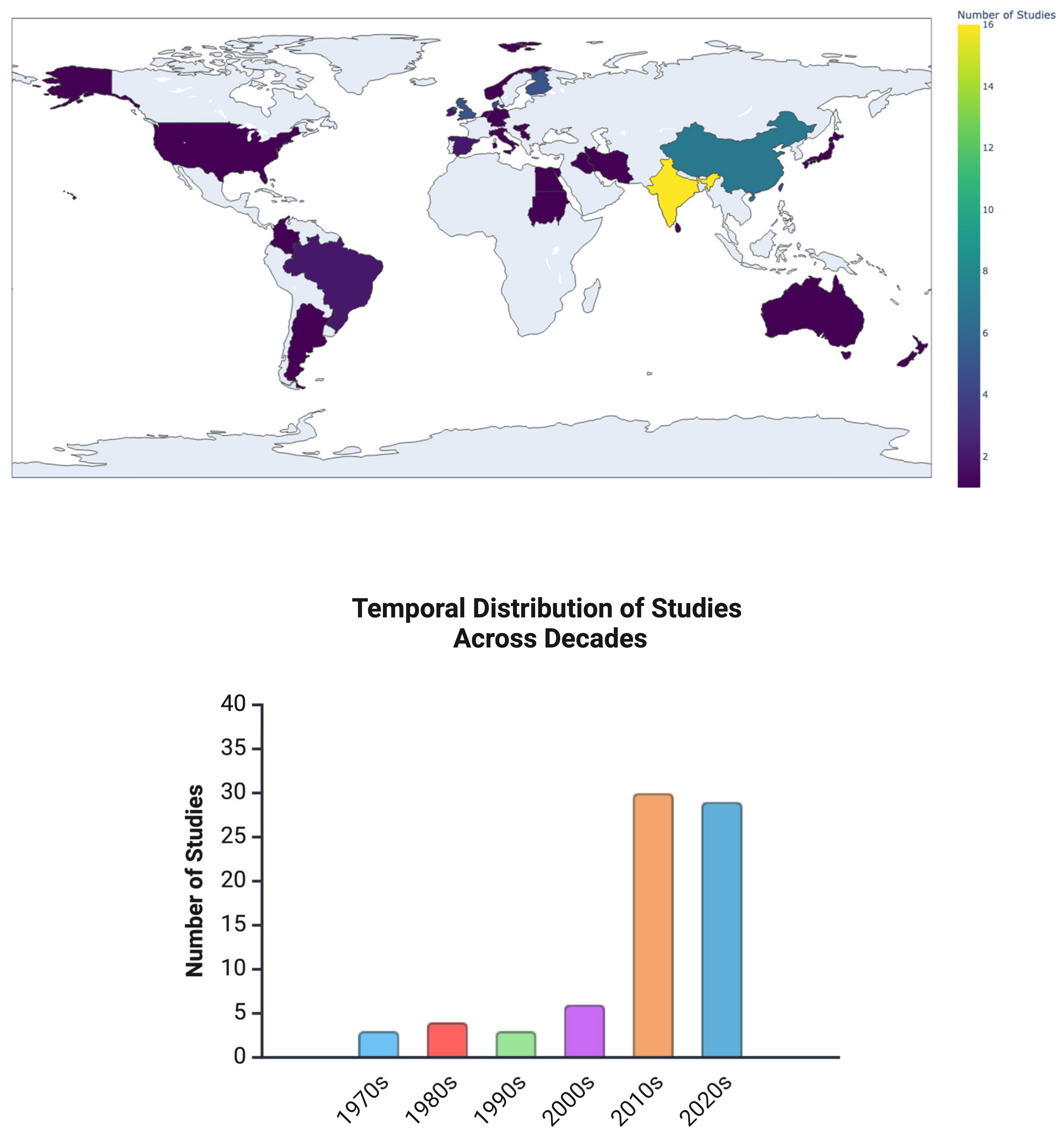
Figure 2 illustrates the geographical and temporal trends in the included studies. Most observational studies were cross-sectional (n = 41), followed by case–control (n = 12) and cohort (n = 9). Additionally, experimental studies included both in vitro (n = 9) and in vivo (n = 8) models.
The publication dates ranged from 1970 to 2024, with studies originating from diverse regions. While older studies provided historical insights into the role of Candida spp. in oral disease progression, recent research has increasingly focused on molecular mechanisms, host interactions, and mycobiome diversity using advanced sequencing and proteomic approaches.

3.2. Objective 1: The Techniques and Methodologies Used to Investigate Oral Yeast Colonisation and/or Infection in the Context of Oral Epithelial Carcinogenesis

The study of oral yeasts in oral epithelial carcinogenesis has evolved significantly over the past few decades, transitioning from traditional culture-based methods to advanced molecular and functional analyses (Figure 3). These methodologies aim to characterise fungal diversity, quantify fungal load, examine their pathogenic potentials, and assess functional interactions between fungi and host tissues.

3.2.1. Culture-Based and Phenotype-Based Identification Techniques

Historically, the identification of oral yeasts relied on culture-based techniques, which remain widely used for fungal isolation and characterisation. Standard culture methods involve plating oral samples on agar-based media, particularly on SDA. Microscopic techniques such as PAS staining and GMS staining have been employed in histological samples to visualise fungal structures in OSCC tissues [22].
For phenotype-based species identification, CHROMagar is commonly used, followed by colony morphology examination and biochemical tests such as germ tube formation, sugar assimilation, and fermentation assays [8,9,32,62]. While these approaches allow for the differentiation of Candida spp., they have limitations in detecting mixed fungal communities and low-abundance species.
In clinical settings, automated systems such as Vitek® 2 and Phoenix™ are widely used for yeast identification. These platforms integrate biochemical profiling with advanced algorithms to rapidly and accurately identify fungal species, offering standardisation and efficiency in hospital laboratories. However, such automated methods are generally limited to clinical diagnostics and are rarely used in research applications, particularly in the context of this scoping review. Among the included studies, only Saxena et al. (2021) [48] reported the use of the Vitek® 2 system for yeast identification.

3.2.2. Molecular and Genotypic Characterisation

Advances in molecular biology techniques have enabled more precise identification of fungal species and strains. PCR-based methods have been adopted for detecting and differentiating Candida spp. For example, two studies employed PCR-Restriction Fragment Length Polymorphism (PCR-RFLP) and species-specific PCR to identify and distinguish NAC species, such as C. tropicalis, C. glabrata, and C. parapsilosis [8,55]. Moreover, Alnuaimi et al. (2015) [44] described a novel technique called PCR high-resolution melting curve analysis to discriminate species of Candida spp. These techniques offer higher sensitivity and specificity compared to phenotype-based approaches. However, the use of PCR-based characterisation of oral yeasts, especially Candida spp. is mainly limited to research purposes and is not commonly used in routine clinical diagnostics.

3.2.3. High-Throughput Sequencing and Metagenomic Analyses

With the advent of next-generation sequencing (NGS) technologies, researchers have been able to characterise the oral mycobiome with an unprecedented level of detail. Internal Transcribed Spacer (ITS) sequencing, particularly ITS2-based metagenomics, has become the gold standard for profiling fungal communities in OSCC tissues and saliva samples [21].
High-throughput sequencing studies have revealed significant dysbiosis in the oral mycobiome of OSCC patients, with a reduction in fungal diversity and an enrichment of specific species such as C. albicans, C. etchellsii, Malassezia restricta, Aspergillus tamarii, and Cyberlindnera spp. [22,23]. These findings suggest that certain fungal taxa may play a role in cancer progression or immune modulation.
Additionally, shotgun metagenomic sequencing has been used to achieve the strain-level resolution of fungal populations, allowing researchers to study genetic variations that may contribute to virulence and acetaldehyde production. Studies using metatranscriptomics (RNA-seq) have provided further insights into fungal metabolic activity and gene expression profiles, helping to elucidate functional interactions between fungi and host cells [79].

3.2.4. Fungal–Bacterial Interaction Studies

Emerging evidence suggests that fungi interact with bacterial communities in the oral cavity, potentially influencing cancer progression. Several studies have utilised both metagenomic and metatranscriptomic approaches to elucidate these interactions. Metagenomic studies, as described by [78,80,81], have employed parallel sequencing of 16S rRNA (bacterial) and ITS (fungal) regions to profile the oral microbiome and assess inter-kingdom microbial relationships. Meanwhile, an integrative metatranscriptomic approach, as employed by Jain et al. (2023) [79], simultaneously characterised both microbial and host transcriptomes. This approach aimed to uncover the mechanisms by which the microbiome (both fungi and bacteria) contributes to oral epithelial carcinogenesis.

3.2.5. Proteomic and Metabolomic Approaches

In addition to genetic and microbiome analyses, proteomic and metabolomic techniques have been employed to investigate fungal contributions to carcinogenesis. Mass spectrometry-based proteomics has been used to compare fungal protein expression between tumour and adjacent non-tumour tissues, identifying fungal proteins associated with immune modulation, oxidative stress, and tissue invasion [20].
Metabolomic studies have also focused on acetaldehyde production, a key carcinogenic metabolite generated by Candida spp. Using gas chromatography–mass spectrometry (GC-MS), researchers have quantified acetaldehyde levels in saliva and tissue samples, demonstrating that C. albicans can produce mutagenic concentrations of acetaldehyde in the presence of ethanol [16,57,58].

3.2.6. In Vitro and In Vivo Experimental Models

Functional studies have been conducted using both in vitro and in vivo models to assess the tumour-promoting effects of fungi, focusing solely on Candida spp.
  • In Vitro Studies: Oral squamous cell carcinoma cell lines (e.g., SCC25, SCC15, CAL27) and a dysplastic cell model (i.e., DOK) have been co-cultured with Candida spp. to examine their effects on cell proliferation, migration, cytokine secretion, and oncogenic signalling [14,36,56,69,73,74,76,77]. Additionally, co-culture models have been developed to study the effects of Candidal biofilm or combined fungal–bacterial biofilm on OSCC cell lines and oral epithelial cells [56,76].
  • In Vivo Studies: OSCC models (e.g., 4NQO-induced and xenografted) have been used to assess the impact of fungal colonisation on tumour development. Studies have shown that C. albicans-colonised animals develop more severe dysplasia and tumours, accompanied by increased immune suppression and PD-L1 expression. These findings indicate that Candida spp. may contribute to immune evasion and tumour progression.

3.3. Objective 2: The Role of Candida spp. Colonisation and/or Infection in Oral Epithelial Carcinogenesis

3.3.1. Prevalence of Candida spp. in OSCC and OPMDs and Its Clinical Implications

The presence of Candida spp. in OSCC and OPMDs has been consistently documented across multiple observational studies (Table S1), with a significantly higher prevalence in malignant tissues than in non-malignant mucosa. This suggests a strong association between fungal colonisation and disease progression. McCullough et al. (2002) [10] detected Candida spp. in 74.7% of patients with OED or OSCC, compared to 34.2% of those without dysplasia, with higher fungal loads observed in moderate-to-severe dysplasia cases. Similarly, Alnuaimi et al. (2015) [44] identified Candida spp. in 69.2% of OSCC cases, compared to 42.3% of controls, with C. albicans being the dominant species.

3.3.2. Clinical Impact of Candida spp. Colonisation

The clinical impact of Candida spp. colonisation on pre-cancerous and cancerous conditions was described in several studies. A higher fungal burden has been associated with an increased risk of recurrence in premalignant lesions. Chiu et al. (2011) [13] reported that MOLs with Candida spp. infection had a significantly higher recurrence rate (67.7%) compared to non-infected MOLs (32.3%) (OR = 4.2, p < 0.05). In another study, Mohamed et al. (2021) [21] observed that a higher Candida spp. burden was correlated with poorer overall survival in OSCC patients (p = 0.043), suggesting its potential as an independent prognostic factor. Additionally, research by Bansal et al. (2018) [46] highlighted that Candida spp. was present in 88.6% of OSCC cases and 45.7% of pre-cancerous lesions, and was completely absent in healthy individuals. Further supporting this, Alnuaimi et al. (2015) [44] found that the presence of Candida spp. was identified as a significant risk factor for OSCC (OR = 3.242), with high colonisation further increasing the risk (OR = 3.587).

3.3.3. Prevalence of Non-albicans Candida spp. (NAC) Species in OSCC and OPMDs

While C. albicans is the most frequently detected species, several studies have highlighted the presence of NAC species in OSCC and OPMDs (Table S1), suggesting a broader role for fungal diversity in disease progression. Sankari and Mahalakshmi (2019) [9] found that C. albicans and NAC species were equally prevalent (41.26%) in OSCC patients, whereas C. albicans was more common in healthy controls (55.9%), indicating a greater role of NAC species in malignancy.
Among NAC species, Bansal et al. (2018) [46] reported that C. krusei (19.6%) and C. tropicalis (9.7%) were the most frequently detected NAC species in OSCC. Similarly, Makinen et al. (2018) [47] found that although C. albicans predominated (84%) in OSCC, other NAC species, including C. dubliniensis, C. tropicalis, and C. glabrata, were also present.
Notably, Sankari et al. (2020) [8] observed that NAC species dominated across OSCC and OPMD samples, with C. krusei (21%), C. tropicalis (21%), and P. anomala (21%) being the most prevalent in OSCC. The greater biofilm-forming capabilities of NAC species, compared to C. albicans, may contribute to persistent colonisation, enhanced resistance to antifungal treatments, and tumour-promoting effects. These findings suggest that NAC species play a significant role in oral carcinogenesis and warrant further investigation into their pathogenic potential [8].

3.3.4. Mechanisms of Candida spp. in Oral Epithelial Carcinogenesis

Several mechanisms have been proposed to explain the oncogenic potential of Candida spp., including acetaldehyde production, virulence factor expression, biofilm formation, and interactions with bacterial communities.

3.3.5. Acetaldehyde Production and Carcinogenicity

One of the most well-documented mechanisms is the ability of C. albicans to metabolise ethanol into acetaldehyde, a recognised carcinogen (Table S2). Gainza-Cirauqui et al. (2013) demonstrated that C. albicans isolates from potentially malignant oral lesions produced acetaldehyde above the carcinogenic threshold (>100 µM) in the presence of ethanol, particularly in smokers [57]. This finding aligns with research by Bakri et al. (2014), who identified elevated ADH1 gene expression in C. albicans isolates from chronic hyperplastic candidosis (CHC) lesions, reinforcing its role in localised acetaldehyde production [30].
Further supporting these findings, Marttila et al. (2013) reported that C. albicans isolates from OSCC patients produced significantly higher acetaldehyde levels under low-oxygen conditions compared to isolates from controls and APECED patients. Notably, acetaldehyde production was correlated with increased expression of ALD6 and ACS1, genes involved in the pyruvate bypass pathway, while ADH1 and ADH2 levels were also upregulated in OSCC-derived strains [58]. These findings suggest that metabolic adaptations in C. albicans within the tumour microenvironment may enhance its carcinogenic potential. Additionally, a case–control study assessing Candida spp. isolates from 52 OSCC patients and 104 matched healthy controls found that a higher percentage of isolates from OSCC patients produced higher mutagenic acetaldehyde levels (>40 µM) compared to the controls [16].

3.3.6. Virulence Factors and Tissue Invasion

In addition to acetaldehyde metabolism, C. albicans employs several virulence factors that may contribute to tissue invasion, immune modulation, and tumour progression. Proteolytic enzyme secretion, particularly aspartyl proteinases and phospholipases, has been implicated in epithelial barrier disruption and inflammatory modulation (Table S2). Rehani et al. (2011) reported that aspartyl proteinase activity was significantly elevated in OSCC tissues compared to non-cancerous controls, supporting its role in tissue degradation [59].
Furthermore, biofilm formation has been shown to enhance fungal persistence and resistance to host immune responses. Alnuaimi et al. (2016) observed that Candida spp. isolates from OSCC exhibited increased biofilm formation, phospholipase production, and metabolic activity, which may facilitate the persistent colonisation and local inflammatory responses that drive carcinogenesis [16].
Further evidence of C. albicans virulence in precancerous and dysplastic tissues was provided by Krögh et al. (1987), who found that lesion-derived C. albicans strains exhibited higher nitrosation potential, converting N-benzylmethylamine into carcinogenic NBMA. Additionally, these strains were more efficient in nitrate-to-nitrite reduction, further facilitating nitrosamine formation, a process implicated in malignant transformation [51].

3.3.7. Immunomodulatory Properties

Beyond its ability to invade tissues and alter metabolic pathways, multiple studies confirm the involvement of Candida spp. in shaping the tumour immune microenvironment and in promoting immune evasion and chronic inflammation, which may promote OSCC progression (Table S2). By interacting with immune receptors, cytokines, and tumour-associated immune cells, Candida spp. suppresses anti-tumour immunity while inducing immunosuppressive conditions conducive to cancer development.
Candida spp. colonisation has been linked to immune dysregulation in OSCC tissues, particularly through the Toll-like receptor (TLR) and NF-κB pathways. Rusanen et al. (2024) found that OSCC tissues exhibited reduced expression of TLR1–5, TLR7, and TLR8 in the basement membrane and endothelium, while infiltrative tumour zones showed increased TLR1, TLR2, TLR4, TLR8, and TLR9 expression, suggesting localised immune activation. Notably, the presence of Candida spp. was correlated with higher TLR4 expression in the basement membrane (p = 0.012), linking fungal colonisation to inflammatory signalling. Additionally, NF-κB activation was elevated in OSCC infiltrative zones, correlating with increased TLR9 and TLR10 expression, reinforcing its role in chronic inflammation and tumour progression [65].
Another study described the capacity of Candida spp. in recruiting immunosuppressive myeloid cells, facilitating tumour immune escape. Wang et al. (2023) demonstrated that in a 4NQO-induced OSCC model, Candida spp. infection increased tumour incidence and progression, along with higher tumour-associated macrophages (TAMs), particularly M2-like macrophages, which suppress T cell-mediated immunity. This effect was driven by IL-17A signalling, which upregulated CCL2, a chemokine that recruits macrophages. Neutralising IL-17A reduced macrophage infiltration and tumour progression, confirming the role of Candida spp. in immune suppression [71].
Further supporting this, Wang et al. (2024) found that Candida spp. infection led to an increase in IL-17A+ CD4+ T cells and γδ T cells, both involved in chronic inflammation and tumour progression. Additionally, Candida spp. colonisation expanded polymorphonuclear myeloid-derived suppressor cells (PMN-MDSCs), which actively suppress T cell activation. Blocking CCL2 signalling significantly reduced MDSC infiltration and restored anti-tumour immunity [72].
Beyond shaping the tumour microenvironment, Candida spp. has been implicated in upregulating programmed death-ligand 1 (PD-L1), a key immune checkpoint molecule that inhibits T cell-mediated tumour clearance. Wang et al. (2022) showed that Candida spp. infection significantly increased PD-L1 expression in OSCC cells through the TLR2/MyD88 and NF-κB pathways, linking fungal infection to chronic-inflammation-driven immune suppression. Notably, in vivo studies confirmed that PD-L1 expression was highest in OSCC tissues colonised by Candida spp., indicating its role in tumour immune escape [73].
In addition to local immune modulation, Candida spp. infection has been linked to systemic immune suppression, particularly in aged and immunocompromised individuals. Bhaskaran et al. (2021) found that aged mice infected with Candida spp. developed OSCC more rapidly, with higher Treg:CD8 ratios, suppressing anti-tumour immunity; increased MDSC infiltration, further dampening T cell activity; and elevated IL-1β levels, promoting chronic inflammation [70]. Similarly, Lee et al. (2020) demonstrated that Candida spp., when co-cultured with Fusobacterium nucleatum, enhanced IL-1β secretion, activating the PI3K/Akt pathway and worsening tumour-promoting inflammation [36].
Moreover, emerging evidence suggests that Candida spp. may also impair the efficacy of immune checkpoint inhibitors (ICIs). Wang et al. (2024) showed that Candida spp. infection weakened PD-1 blockade therapy, leading to T cell exhaustion and reduced tumour control, highlighting fungal colonisation as a potential factor in immunotherapy resistance [72].

3.3.8. Fungal–Bacterial Interactions in OSCC Progression

Microbiome profiling of OSCC tissues by Mukherjee et al. (2017) revealed significant microbial dysbiosis, with reduced fungal diversity and strong inter-kingdom correlations. Candida albicans and Lichtheimia spp. were positively associated with Fusobacterium spp. and Porphyromonas spp., suggesting cooperative interactions that may drive inflammation and tumour progression [78]. Similarly, Jain et al. (2023) identified transcriptionally active fungi, particularly Malassezia restricta, which interacted with pro-tumorigenic bacteria and modulated host pathways linked to proliferation, immune evasion, and carcinogenesis. Their findings further demonstrated that fungal–bacterial consortia were associated with the upregulation of proliferation-related pathways in host cells [79].
Experimental models have further demonstrated how fungal–bacterial interactions enhance tumour progression. Arzmi et al. (2019) showed that polymicrobial biofilms containing C. albicans, Streptococcus mutans, and Actinomyces naeslundii increased oral cancer cell proliferation and migration, accompanied by the upregulation of IL-6 and MMP1, key mediators of tumour invasion [56]. Lee et al. (2020) further found that co-culturing C. albicans with Fusobacterium nucleatum led to increased IL-1β secretion via the PI3K/Akt/GSK-3β pathway, a mechanism implicated in chronic inflammation and immune suppression [36].
Beyond individual interactions, Heng et al. (2022) reported that C. tropicalis was enriched in both OPMDs and OSCC, frequently coexisting with dysbiotic bacterial species linked to biofilm formation and inflammation. This suggests that fungal–bacterial consortia may sustain tumour-supportive conditions, promote persistent infections, and alter immune responses, reinforcing OSCC progression [81].

3.4. Objective 3: The Role of Non-Candida Yeast Species Colonisation and/or Infection in Oral Epithelial Carcinogenesis

While Candida spp. has been the primary focus of research on fungal involvement in OSCC, increasing evidence suggests that other yeast species may also play a role in carcinogenesis (a list of studies with this scope are presented in Table S3). Advances in metagenomic sequencing and fungal proteomics have uncovered a more complex fungal community within OSCC tissues, shifting the perspective from a single-species model to one involving broader fungal dysbiosis. This paradigm shift suggests that interactions between different fungal taxa and the host may contribute to tumour progression through diverse mechanisms.
Recent studies have highlighted key non-Candida spp. yeast species that appear to be enriched in OSCC tissues compared to non-malignant counterparts. Perera et al. (2017) demonstrated that while fungal diversity in OSCC tissues was generally lower, carcinogenic fungi such as Candida etchellsii- and Hannaella luteola-like species were overrepresented. Conversely, fibro-epithelial polyps—considered non-malignant—exhibited an abundance of Malassezia restricta, Aspergillus tamarii, and Alternaria alternata. These findings indicate that while some fungal species might promote tumour progression, others could serve as commensals or even exert protective effects [22]. Further supporting this notion, Mohamed et al. (2021) found that higher Malassezia carriage was associated with improved survival outcomes in OSCC patients, suggesting a potentially complex, context-dependent role of certain fungal species [21].
Beyond simply being present in OSCC tissues, several yeast species appear to exhibit oncogenic potential through metabolic and immune-modulatory pathways. Sankari et al. (2020) reported that C. krusei, C. tropicalis, and P. anomala were significantly more prevalent in OSCC than in healthy controls, with C. famata exclusively detected in OSCC and OPMDs. The absence of C. famata in healthy individuals suggests that it may contribute to malignant transformation rather than merely existing as an opportunistic coloniser [8]. Jain et al. (2023) further demonstrated that transcriptionally active Malassezia restricta influenced host cell signalling pathways involved in inflammation and immune evasion, reinforcing the idea that non-Candida spp. fungi can modulate the tumour microenvironment [79].
One of the most intriguing aspects of non-Candida spp. yeast involvement in OSCC is their interaction with bacterial communities. Mukherjee et al. (2017) observed that Lichtheimia spp. and Malassezia spp. Were correlated with pathogenic bacterial genera such as Fusobacterium spp. and Porphyromonas spp., both of which are implicated in OSCC progression. These findings suggest that fungal species may not act in isolation, but rather, contribute to a broader microbial dysbiosis that influences tumorigenesis [78]. Sami et al. (2023) also reported that Aspergillus spp., a known aflatoxin producer, was enriched in OSCC samples from Toombak users—a population known for its high oral cancer risk—while Candida spp. were more prevalent in non-users. This raises questions about how different fungal taxa interact with external carcinogenic exposures such as smokeless tobacco [80].
Proteomic analyses provide additional insights into the potential functional roles of non-Candida yeast species in OSCC. He et al. (2023) identified fungal proteins enriched in OSCC tissues that were associated with tissue invasion, oxidative stress, and immune modulation. Lichtheimia corymbifera, Aspergillus fumigatus, and Verruconis gallopava were particularly abundant in OSCC samples, and their metabolic profiles suggested potential contributions to carcinogenic processes [20]. Similarly, Jain et al. (2023) found that non-Candida spp. yeasts interacted with host immune pathways, potentially driving inflammation and tumour progression [79].
These findings collectively underscore the need to reassess the role of fungal communities in OSCC. While Candida spp. (especially, C. albicans) remains the most well-characterised fungal species in oral cancer, the presence and activity of other yeast species suggest a more nuanced landscape in which multiple fungal taxa, in concert with bacteria and host factors, contribute to disease progression.

4. Discussion

This scoping review synthesises the current evidence on oral yeasts in OSCC, revealing a transition from prevalence-focused studies to mechanistic investigations exploring fungal virulence and interactions within the tumour microenvironment. The data consistently indicate that C. albicans is the most frequently detected species in OPMDs, OEDs, and OSCC, with its prevalence increasing in tandem with disease severity [10,11,12,43]. However, recent studies have drawn attention to the role of NAC species and other fungal taxa, challenging the conventional view that C. albicans is the primary fungal agent implicated in OSCC development [8,20,21,22,23,60,79,80,81].
The elevated detection of Candida spp. in dysplastic and OSCC lesions compared to healthy controls has been widely documented, with some studies reporting a three- to four-fold higher prevalence in OSCC patients. While C. albicans remains dominant, NAC species, particularly C. tropicalis, C. glabrata, and C. parapsilosis, have been frequently identified in precancerous and malignant lesions [8,46,47,48,49,50]. Beyond Candida spp., other fungal genera have also been detected in different compositions in OSCC tissues, including Rhodotorula spp., Saccharomyces spp., Kloeckera spp., Malassezia spp., and Lichtheimia spp. These findings indicate that shifts in fungal species composition, rather than the presence of C. albicans alone, may contribute to OSCC pathogenesis. Differences in fungal diversity between OSCC and healthy oral mucosa have also been observed, with some studies reporting reduced fungal diversity in OSCC, while others indicate a distinct fungal profile with functionally relevant species alterations. This observation implies that certain fungal communities may be more adapted to tumour microenvironments, potentially facilitating carcinogenesis. However, the involvement of non-Candida spp. genera in OSCC has only been described in observational studies, and their carcinogenic potential remains unexplored in experimental settings [20,22,23,78,81].
A central debate in this field concerns whether oral yeasts actively contribute to carcinogenesis or merely colonise dysregulated epithelial environments opportunistically. Several studies support an active pathogenic role for oral yeasts, particularly Candida spp., highlighting their production of carcinogenic metabolites like acetaldehyde [44,57,58] and N-nitrosobenzylmethylamine [51,82], as well as fungal invasion observed histopathologically in dysplastic and OSCC tissues. Virulence factors including fungal proteases, phospholipases, and immune evasion strategies further suggest their involvement in immune modulation, epithelial degradation, and tissue invasion [11,35,36,59,72,73]. Other virulence factors, including biofilm formation, aspartyl proteases, and phospholipases, enhance immune evasion, epithelial degradation, and tissue invasion, further supporting the notion of a pathogenic role for Candida spp. in OSCC [16,55,56,59,60]. Recent studies have expanded this discussion beyond Candida spp., identifying other yeasts such as Malassezia spp., Rhodotorula spp., Lichtheimia spp., Aspergillus spp., and Saccharomyces spp., which potentially contribute through chronic inflammation, cell cycle disruption, oxidative stress, and epithelial damage [20,79,80,81]. Additionally, fungal–bacterial interactions have emerged as significant, with Candida spp. frequently co-occurring alongside pro-inflammatory bacteria like Fusobacterium nucleatum and Streptococcus mutans, suggesting a possible synergistic effect in OSCC pathogenesis. These findings highlight the complexity of fungal involvement in OSCC, underscoring the need for longitudinal and experimental studies to clarify the exact nature of fungal contributions to carcinogenesis [36,56,78,79,81].
Conversely, an alternative perspective suggests that fungal colonisation in OSCC may be a secondary consequence of tumour-induced immune suppression and epithelial barrier breakdown rather than a primary driver of carcinogenesis. The frequent detection of C. albicans and other yeasts in non-dysplastic conditions, such as OLK and OLP, indicates that fungal presence alone might be insufficient to initiate malignant transformation. However, the current evidence remains divided, underscoring the necessity for longitudinal studies and experimental models to clarify whether the role of fungi in OSCC is causal or merely opportunistic [15,83,84].
While most studies agree that Candida spp. prevalence increases with disease severity, the extent to which fungi actively contribute to carcinogenesis remains contested. C. albicans, in particular, has been frequently associated with OSCC, producing carcinogenic metabolites such as acetaldehyde and nitrosamines, and exhibiting increased hyphal invasion in dysplastic and malignant tissues compared to non-dysplastic oral mucosa [10,16,35,44,54]. However, emerging evidence underscores the need to move beyond a Candida species-centric perspective and consider the broader oral mycobiome in carcinogenesis. There is ongoing debate about fungal diversity’s role in OSCC, with some studies noting reduced diversity, potentially favouring high-virulence Candida spp. strains, while others suggest that diversity changes may be methodological artefacts. Recent evidence highlights that specific fungal communities, including NAC species, could modulate metabolic pathways, host immune responses, and microbial interactions within the tumour microenvironment. Furthermore, the observed increased prevalence of NAC species in OSCC could reflect a shift in microbial ecology rather than direct oncogenic activity [84,85]. Clarifying the exact contribution of fungi, including the broader oral mycobiome, remains essential and underscores the importance of moving beyond a Candida species-centric view. The potential impact of antifungal treatments on OSCC risk is another critical area requiring further investigation through longitudinal and experimental studies [17].

Limitations of This Review

Although this scoping review comprehensively maps the available evidence, several limitations should be acknowledged. A key limitation is the absence of a formal risk-of-bias assessment, which is not typically conducted in scoping reviews. However, potential biases such as publication bias, selective reporting, and methodological inconsistencies across studies may influence the findings. For instance, studies with positive associations between Candida spp. and OSCC may be more likely to be published, while studies with null findings might be underreported. Additionally, variations in study design, sample size, and diagnostic methods could introduce selection bias, measurement bias, or confounding factors that affect the interpretation of results.
Furthermore, methodological inconsistencies across the included studies limit the direct comparability of findings. Variability in fungal detection methods, including differences in sampling techniques, culture conditions, and molecular identification approaches, has resulted in significant discrepancies in prevalence rates. The reliance on cross-sectional and in vitro studies restricts the ability to establish causality, while heterogeneity in patient populations, including differences in age, lifestyle factors such as smoking and alcohol consumption, and geographic variability, complicates direct comparisons. Additionally, inconsistencies in histopathological classification criteria for dysplasia and OSCC further affect study comparability, making it difficult to draw definitive conclusions. The inclusion of both observational and experimental studies introduces further heterogeneity, as differences in study design may influence the strength and reliability of the evidence base.

5. Conclusions and Implications

This review highlights the complexity of oral yeast involvement in OSCC and the ongoing debate over whether these fungi are active carcinogenic agents or opportunistic colonisers. The growing recognition that fungal diversity in OSCC differs from that in healthy individuals suggests a potential functional role in tumour development, particularly through alterations in microbial interactions, metabolic activity, and immune modulation. However, whether these fungal changes are a driver or consequence of carcinogenesis remains uncertain. Addressing these knowledge gaps through well-designed longitudinal studies, standardised methodologies, and advanced multi-omics analyses will be essential in refining our understanding of fungal contributions to OSCC and identifying potential diagnostic and therapeutic strategies.

Supplementary Materials

The following supporting information can be downloaded at: https://www.mdpi.com/article/10.3390/jof11040260/s1, Table S1: Prevalence and characterisation of Candida spp. in oral mucosal lesions and OSCC; Table S2: Mechanistic findings of Candida spp. and oral carcinogenesis; Table S3: Findings from mycobiome studies on oral squamous cell carcinoma; Table S4: Keywords strings (Medline by Ovid); Table S5: Keywords strings (Embase by Ovid); Table S6: Keywords strings (EBM Reviews); Table S7: Keywords strings (Web of Science).

Author Contributions

Conceptualisation, S.W., M.M., and T.Y.; Methodology, S.W., M.M., and T.Y.; Investigation, S.W. and Z.B.N.; Formal Analysis, S.W. and Z.B.N.; Writing—Original Draft Preparation, S.W.; Writing—Review and Editing, S.W., M.M., and T.Y.; Visualisation, S.W.; Supervision, M.M. and T.Y.; Funding Acquisition, T.Y. All authors have read and agreed to the published version of the manuscript.

Funding

This study received funding from Lembaga Pengelola Dana Pendidikan (grant number 202403220801271) and Melbourne Dental School, The University of Melbourne.

Data Availability Statement

No new data were created or analyzed in this study.

Acknowledgments

We thank Lindy Cochrane, a University of Melbourne Librarian, for her valuable assistance in formulating the search strategy. We also acknowledge the Melbourne Dental School’s Oral Medicine and Oral Cancer (OMOC) research group for their input in developing the protocol of this study.

Conflicts of Interest

The authors declare no conflicts of interest.

Abbreviations

The following abbreviations are used in this manuscript:
CFUColony-Forming Unit
DNADeoxyribonucleic Acid
EBMEvidence-Based Medicine
EGFREpidermal Growth Factor Receptor
EMTEpithelial–Mesenchymal Transition
FFPEFormalin-Fixed Paraffin-Embedded
GMSGomori Methenamine Silver
HNSCCHead and Neck Squamous Cell Carcinoma
HRPHorseradish Peroxidase
IHCImmunohistochemistry
IL-6Interleukin-6
JAK-STATJanus Kinase-Signal Transducer and Activator of Transcription
KEGGKyoto Encyclopedia of Genes and Genomes
LFCLogarithmic Fold Change
MMPMatrix Metalloproteinase
mRNAMessenger Ribonucleic Acid
NACNon-albicans Candida
NF-κBNuclear Factor Kappa B
OEDOral epithelial dysplasia
OPMDOral potentially malignant disorder
OSCCOral squamous cell carcinoma
OTSCCOral tongue squamous cell carcinoma
PASPeriodic Acid–Schiff
PCRPolymerase Chain Reaction
PRISMA-ScRPreferred Reporting Items for Systematic Reviews and Meta-Analyses for Scoping Reviews
qPCRQuantitative Polymerase Chain Reaction
RPKMReads Per Kilobase of Exon Model per Million Mapped Reads
RT-PCRReal-Time Polymerase Chain Reaction
SDASabouraud’s Dextrose Agar
scSeqSingle-Cell Sequencing
TLRToll-like receptor
TNF-αTumour Necrosis Factor Alpha
TGF-βTransforming Growth Factor Beta
URMUpstream Regulatory Mechanism
WntWingless-Related Integration Site (Wnt Signalling Pathway)

References

  1. Lythgoe, M.P.; Mullish, B.H.; Frampton, A.E.; Krell, J. Polymorphic Microbes: A New Emerging Hallmark of Cancer. Trends Microbiol. 2022, 30, 1131–1134. [Google Scholar] [CrossRef] [PubMed]
  2. Renstrup, G. Occurrence of Candida in Oral Leukoplakias. Acta Pathol. Microbiol. Scand. B Microbiol. Immunol. 1970, 78, 421–424. [Google Scholar] [CrossRef] [PubMed]
  3. Roed-Petersen, B.; Renstrup, G.; Pindborg, J.J. Candida in Oral Leukoplakias A Histologic and Exfoliative Cytologic Study. Eur. J. Oral Sci. 1970, 78, 323–328. [Google Scholar] [CrossRef]
  4. Daftary, D.K.; Mehta, F.S.; Gupta, P.C.; Pindborg, J.J. The Presence of Candida in 723 Oral Leukoplakias among Indian Villagers. Eur. J. Oral Sci. 1972, 80, 75–79. [Google Scholar] [CrossRef]
  5. Hornstein, O.P.; Gräßel, R.; Schirner, E. Prevalence Rates of Candidosis in Leukoplakias and Carcinomas of the Oral Cavity. Arch. Dermatol. Res. 1979, 266, 99–102. [Google Scholar] [CrossRef]
  6. Silverman, S., Jr.; Gorsky, M.; Ms, F.L. Dds, Oral Leukoplakia and Malignant Transformation. A Follow-up Study of 257 Patients. Cancer 1984, 53, 563–568. [Google Scholar] [CrossRef]
  7. Krogh, P.; Holmstrup, P.; Thorn, J.J.; Vedtofte, P.; Pindborg, J.J. Yeast Species and Biotypes Associated with Oral Leukoplakia and Lichen Planus. Oral Surg. Oral Med. Oral Pathol. 1987, 63, 48–54. [Google Scholar] [CrossRef]
  8. Sankari, S.L.; Mahalakshmi, K.; Kumar, V.N. A Comparative Study of Candida Species Diversity among Patients with Oral Squamous Cell Carcinoma and Oral Potentially Malignant Disorders. BMC Res. Notes 2020, 13, 488. [Google Scholar] [CrossRef]
  9. Sankari, S.L.; Mahalakshmi, K. Oral Candidal Carriage among Patients with Oral Squamous Cell Carcinoma: A Case-Control Study. J. Orofac. Sci. 2019, 11, 55–58. [Google Scholar] [CrossRef]
  10. McCullough, M.; Jaber, M.; Barrett, A.W.; Bain, L.; Speight, P.M.; Porter, S.R. Oral Yeast Carriage Correlates with Presence of Oral Epithelial Dysplasia. Oral Oncol. 2002, 38, 391–393. [Google Scholar] [CrossRef]
  11. Tamgadge, S.; Tamgadge, A.; Pillai, A.; Chande, M.; Acharya, S.; Kamat, N. Association of Candida Sp. with the Degrees of Dysplasia and Oral Cancer: A Study by Calcofluor White under Fluorescent Microscopy. Iran. J. Pathol. 2017, 12, 348–355. [Google Scholar] [PubMed]
  12. Hebbar, P.; Pai, A.; Sujatha, D. Mycological and Histological Associations of Candida in Oral Mucosal Lesions. J. Oral Sci. 2013, 55, 157–160. [Google Scholar] [CrossRef] [PubMed]
  13. Chiu, C.-T.; Li, C.-F.; Li, J.-R.; Wang, J.; Chuang, C.-Y.; Chiang, W.-F.; Huang, S.-C.; Chang, S.-W. Candida Invasion and Influences in Smoking Patients with Multiple Oral Leucoplakias—A Retrospective Study. Mycoses 2011, 54, e377–e383. [Google Scholar] [CrossRef] [PubMed]
  14. Vadovics, M.; Ho, J.; Igaz, N.; Alföldi, R.; Rakk, D.; Veres, É.; Szücs, B.; Horváth, M.; Tóth, R.; Szücs, A.; et al. Candida albicans Enhances the Progression of Oral Squamous Cell Carcinoma In Vitro and In Vivo. mBio 2022, 13, e03144-21. [Google Scholar] [CrossRef]
  15. Wang, X.; Zhang, W.; Wu, W.; Wu, S.; Young, A.; Yan, Z. Is Candida albicans a Contributor to Cancer? A Critical Review Based on the Current Evidence. Microbiol. Res. 2023, 272, 127370. [Google Scholar] [CrossRef]
  16. Alnuaimi, A.D.; Ramdzan, A.N.; Wiesenfeld, D.; O’Brien-Simpson, N.M.; Kolev, S.D.; Reynolds, E.C.; McCullough, M.J. Candida Virulence and Ethanol-Derived Acetaldehyde Production in Oral Cancer and Non-Cancer Subjects. Oral Dis. 2016, 22, 805–814. [Google Scholar] [CrossRef]
  17. Shukla, K.; Vun, I.; Lov, I.; Laparidis, G.; McCamley, C.; Ariyawardana, A. Role of Candida Infection in the Malignant Transformation of Oral Leukoplakia: A Systematic Review of Observational Studies. Transl. Res. Oral Oncol. 2019, 4, 2057178X1982822. [Google Scholar] [CrossRef]
  18. Ayuningtyas, N.F.; Mahdani, F.Y.; Pasaribu, T.A.S.; Chalim, M.; Ayna, V.K.P.; Santosh, A.B.R.; Santacroce, L.; Surboyo, M.D.C. Role of Candida albicans in Oral Carcinogenesis. Pathophysiology 2022, 29, 650–662. [Google Scholar] [CrossRef]
  19. Navgire, G.S.; Goel, N.; Sawhney, G.; Sharma, M.; Kaushik, P.; Mohanta, Y.K.; Mohanta, T.K.; Al-Harrasi, A. Analysis and Interpretation of Metagenomics Data: An Approach. Biol. Proced. Online 2022, 24, 18. [Google Scholar] [CrossRef]
  20. He, S.; Chakraborty, R.; Ranganathan, S. Metaproteomic Analysis of an Oral Squamous Cell Carcinoma Dataset Suggests Diagnostic Potential of the Mycobiome. Int. J. Mol. Sci. 2023, 24, 1050. [Google Scholar] [CrossRef]
  21. Mohamed, N.; Litlekalsøy, J.; Ahmed, I.A.; Martinsen, E.M.H.; Furriol, J.; Javier-Lopez, R.; Elsheikh, M.; Gaafar, N.M.; Morgado, L.; Mundra, S.; et al. Analysis of Salivary Mycobiome in a Cohort of Oral Squamous Cell Carcinoma Patients from Sudan Identifies Higher Salivary Carriage of Malassezia as an Independent and Favorable Predictor of Overall Survival. Front. Cell. Infect. Microbiol. 2021, 11, 673465. [Google Scholar] [CrossRef]
  22. Perera, M.; Al-Hebshi, N.N.; Perera, I.; Ipe, D.; Ulett, G.C.; Speicher, D.J.; Chen, T.; Johnson, N.W. A Dysbiotic Mycobiome Dominated by Candida albicans Is Identified within Oral Squamous-Cell Carcinomas. J. Oral Microbiol. 2017, 9, 1385369. [Google Scholar] [CrossRef]
  23. Banerjee, S.; Tian, T.; Wei, Z.; Peck, K.N.; Shih, N.; Chalian, A.A.; O’Malley, B.W.; Weinstein, G.S.; Feldman, M.D.; Alwine, J.; et al. Microbial Signatures Associated with Oropharyngeal and Oral Squamous Cell Carcinomas. Sci. Rep. 2017, 7, 4036. [Google Scholar] [CrossRef]
  24. Tricco, A.C.; Lillie, E.; Zarin, W.; O’Brien, K.K.; Colquhoun, H.; Levac, D.; Moher, D.; Peters, M.D.J.; Horsley, T.; Weeks, L.; et al. PRISMA Extension for Scoping Reviews (PRISMA-ScR): Checklist and Explanation. Ann. Intern. Med. 2018, 169, 467–473. [Google Scholar] [CrossRef]
  25. Barrett, A.W.; Kingsmill, V.J.; Speight, P.M. The Frequency of Fungal Infection in Biopsies of Oral Mucosal Lesions. Oral Dis. 1998, 4, 26–31. [Google Scholar] [CrossRef]
  26. Vuckovic, N.; Bokor-Bratic, M.; Vuckovic, D.; Picuric, I. Presence of Candida albicans in Potentially Malignant Oral Mucosal Lesions. Arch. Oncol. 2004, 12, 51–54. [Google Scholar] [CrossRef]
  27. Dany, A.; Kurian, K.; Shanmugam, S. Association of Candida in Different Stages of Oral Leukoplakia. J. Indian Acad. Oral Med. Radiol. 2011, 23, 14–16. [Google Scholar] [CrossRef]
  28. Odedra, S.; Chawda, J.; Rupapara, R.; Rupakar, P. Presence of Candida in Oral Dysplastic Lesions—A Casual Involvement or a Causal Role? Indian J. Public Health Res. Dev. 2013, 4, 267–271. [Google Scholar] [CrossRef]
  29. Wu, L.; Feng, J.; Shi, L.; Shen, X.; Liu, W.; Zhou, Z. Candidal Infection in Oral Leukoplakia: A Clinicopathologic Study of 396 Patients from Eastern China. Ann. Diagn. Pathol. 2013, 17, 37–40. [Google Scholar] [CrossRef]
  30. Bakri, M.M.; Cannon, R.D.; Holmes, A.R.; Rich, A.M. Detection of Candida albicans ADH1 and ADH2 mRNAs in Human Archival Oral Biopsy Samples. J. Oral Pathol. Med. 2014, 43, 704–710. [Google Scholar] [CrossRef]
  31. Singh, S.K.; Gupta, A.; Rajan, S.Y.; Padmavathi, B.N.; Mamatha, G.P.; Mathur, H.; Bhuvaneshwari, S.; Soundarya, S. Correlation of Presence of Candida and Epithelial Dysplasia in Oral Mucosal Lesions. J. Clin. Diagn. Res. JCDR 2014, 8, ZC31–ZC35. [Google Scholar] [CrossRef] [PubMed]
  32. Sarkar, R.; Rathod, G.P. Clinicopathologic Assessment of Candida Colonization of Oral Leukoplakia. Indian J. Dermatol. Venereol. Leprol. 2014, 80, 413–418. [Google Scholar] [CrossRef] [PubMed]
  33. Brouns, E.; Baart, J.A.; Karagozoglu, K.H.; Aartman, I.H.A.; Bloemena, E.; Van der Waal, I. Malignant Transformation of Oral Leukoplakia in a Well-Defined Cohort of 144 Patients. Oral Dis. 2014, 20, e19–e24. [Google Scholar] [CrossRef]
  34. Hongal, B.; Kulkarni, V.; Deshmukh, R.; Joshi, P.; Karande, P.; Shroff, A. Prevalence of Fungal Hyphae in Potentially Malignant Lesions and Conditions-Does Its Occurrence Play a Role in Epithelial Dysplasia? J. Oral Maxillofac. Pathol. 2015, 19, 10. [Google Scholar] [CrossRef]
  35. Hafed, L.; Farag, H.; El-Rouby, D.; Shaker, O.; Shabaan, H.-A. Candida albicans Alcohol Dehydrogenase 1 Gene in Oral Dysplasia and Oral Squamous Cell Carcinoma. Pol. J. Pathol. 2019, 70, 210–216. [Google Scholar] [CrossRef]
  36. Lee, C.-H.; Hung, P.-F.; Liu, K.-J.; Chung, H.-L.; Yang, W.-C.; Hsu, K.-C.; Fong, T.-H.; Lo, H.-J.; Chen, Y.-P.; Yang, J.-R.; et al. LDOC1 Suppresses Microbe-Induced Production of IL-1β in Human Normal and Cancerous Oral Cells through the PI3K/Akt/GSK-3β Axis. Cancers 2020, 12, 3148. [Google Scholar] [CrossRef]
  37. Erira, A.T.; Navarro, A.F.R.; Robayo, D.A.G. Human Papillomavirus, Epstein-Barr Virus, and Candida albicans Co-Infection in Oral Leukoplakia with Different Degrees of Dysplasia. Clin. Exp. Dent. Res. 2021, 7, 914–923. [Google Scholar] [CrossRef]
  38. Zhang, W.; Wu, S.; Wang, X.; Gao, Y.; Yan, Z. Malignant Transformation and Treatment Recommendations of Chronic Hyperplastic Candidiasis-A Six-Year Retrospective Cohort Study. Mycoses 2021, 64, 1422–1428. [Google Scholar] [CrossRef]
  39. Yang, S.-W.; Lee, Y.-C.; Lee, Y.-S.; Chang, L.-C.; Lai, Y.-R. Risk Assessment of Malignant Transformation of Oral Leukoplakia in Patients with Previous Oral Squamous Cell Carcinoma. Int. J. Oral Maxillofac. Surg. 2022, 51, 1394–1400. [Google Scholar] [CrossRef]
  40. Rezazadeh, F.; Beirami, M.; Zareshahrabadi, Z.; Sedarat, H.; Zomorodian, K. Evaluation of the Distribution of Candida Species in Patients with Dysplastic and Nondysplastic Oral Lichen Planus Lesions. BioMed Res. Int. 2022, 2022, 8100352. [Google Scholar] [CrossRef]
  41. Saraneva, O.; Furuholm, J.; Hagström, J.; Sorsa, T.; Rita, V.; Tervahartiala, T.; Välimaa, H.; Ruokonen, H. Oral Potentially Malignant Disorders and Candida in Oral Tongue Squamous Cell Carcinoma Patients. Dent. J. 2023, 11, 170. [Google Scholar] [CrossRef] [PubMed]
  42. Saigal, S.; Bhargava, A.; Mehra, S.K.; Dakwala, F. Identification of Candida albicans by Using Different Culture Medias and Its Association in Potentially Malignant and Malignant Lesions. Contemp. Clin. Dent. 2011, 2, 188–193. [Google Scholar] [CrossRef] [PubMed]
  43. Gall, F.; Colella, G.; Di Onofrio, V.; Rossiello, R.; Angelillo, I.F.; Liguori, G. Candida Spp. in Oral Cancer and Oral Precancerous Lesions. New Microbiol. 2013, 36, 283–288. [Google Scholar] [PubMed]
  44. Alnuaimi, A.D.; Wiesenfeld, D.; O’Brien-Simpson, N.M.; Reynolds, E.C.; McCullough, M.J. Oral Candida Colonization in Oral Cancer Patients and Its Relationship with Traditional Risk Factors of Oral Cancer: A Matched Case-Control Study. Oral Oncol. 2015, 51, 139–145. [Google Scholar] [CrossRef]
  45. Hulimane, S.; Maluvadi-Krishnappa, R.; Mulki, S.; Rai, H.; Dayakar, A.; Kabbinahalli, M. Speciation of Candida Using CHROMagar in Cases with Oral Epithelial Dysplasia and Squamous Cell Carcinoma. J. Clin. Exp. Dent. 2018, 10, e657–e660. [Google Scholar] [CrossRef]
  46. Bansal, R.; Pallagatti, S.; Sheikh, S.; Aggarwal, A.; Gupta, D.; Singh, R. Candidal Species Identification in Malignant and Potentially Malignant Oral Lesions with Antifungal Resistance Patterns. Contemp. Clin. Dent. 2018, 9, S309–S313. [Google Scholar] [CrossRef]
  47. Makinen, A.; Nawaz, A.; Makitie, A.; Meurman, J.H. Role of Non-Albicans Candida and Candida albicans in Oral Squamous Cell Cancer Patients. J. Oral Maxillofac. Surg. 2018, 76, 2564–2571. [Google Scholar] [CrossRef]
  48. Saxena, A.; Nagi, R.; Sandeep, T.; Patil, D.J.; Choudhary, R.; Kaur, A. Identification of Candida albicans and Non-albicans Candida Resistant Species in Tobacco Users and Oral Squamous Cell Carcinoma Patients: Comparison of HiCrome Agar and Automated VITEK 2 System. J. Oral Maxillofac. Pathol. JOMFP 2021, 25, 551–552. [Google Scholar] [CrossRef]
  49. Abidullah, M.; Bhosle, S.; Komire, B.; Sharma, P.; Swathi, K.; Karthik, L. Investigation of Candidal Species among People Who Suffer from Oral Potentially Malignant Disorders and Oral Squamous Cell Carcinoma. J. Pharm. Bioallied Sci. 2021, 13, S1050–S1054. [Google Scholar] [CrossRef]
  50. Ilhan, B.; Vural, C.; Gurhan, C.; Vural, C.; Veral, A.; Wilder-Smith, P.; Ozdemir, G.; Guneri, P. Real-Time PCR Detection of Candida Species in Biopsy Samples from Non-Smokers with Oral Dysplasia and Oral Squamous Cell Cancer: A Retrospective Archive Study. Cancers 2023, 15, 5251. [Google Scholar] [CrossRef]
  51. Krogh, P.; Hald, B.; Holmstrup, P. Possible Mycological Etiology of Oral Mucosal Cancer: Catalytic Potential of Infecting Candida albicans and Other Yeasts in Production of N-Nitrosobenzylmethylamine. Carcinogenesis 1987, 8, 1543–1548. [Google Scholar] [CrossRef] [PubMed]
  52. Rindum, J.L.; Stenderup, A.; Holmstrup, P. Identification of Candida albicans Types Related to Healthy and Pathological Oral Mucosa. J. Oral Pathol. Med. 1994, 23, 406–412. [Google Scholar] [CrossRef] [PubMed]
  53. Williams, D.W.; Bartie, K.L.; Potts, A.J.; Wilson, M.J.; Fardy, M.J.; Lewis, M.A. Strain Persistence of Invasive Candida albicans in Chronic Hyperplastic Candidosis That Underwent Malignant Change. Gerodontology 2001, 18, 73–78. [Google Scholar] [CrossRef] [PubMed]
  54. Abdulrahim, M.H.; McManus, B.A.; Flint, S.R.; Coleman, D.C. Genotyping Candida albicans from Candida Leukoplakia and Non-Candida Leukoplakia Shows No Enrichment of Multilocus Sequence Typing Clades but Enrichment of ABC Genotype C in Candida Leukoplakia. PLoS ONE 2013, 8, e73738. [Google Scholar] [CrossRef]
  55. Weerasekera, M.-M.; Wijesinghe, G.-K.; Sampath, A.; Dilhari, A.; Madhumal, T.; Dilrukshi, R.; Willaddara, R.; Karunathilaka, S.; Gunasekara, C.; Fernando, N.; et al. The Genotypes and Virulence Attributes of C. albicans Isolates from Oral Leukoplakia. Med. Oral Patol. Oral Cirugia Bucal 2021, 26, e786–e794. [Google Scholar] [CrossRef]
  56. Arzmi, M.H.; Cirillo, N.; Lenzo, J.C.; Catmull, D.V.; O’Brien-Simpson, N.; Reynolds, E.C.; Dashper, S.; McCullough, M. Monospecies and Polymicrobial Biofilms Differentially Regulate the Phenotype of Genotype-Specific Oral Cancer Cells. Carcinogenesis 2019, 40, 184–193. [Google Scholar] [CrossRef]
  57. Gainza-Cirauqui, M.L.; Nieminen, M.T.; Novak Frazer, L.; Aguirre-Urizar, J.M.; Moragues, M.D.; Rautemaa, R. Production of Carcinogenic Acetaldehyde by Candida albicans from Patients with Potentially Malignant Oral Mucosal Disorders. J. Oral Pathol. Med. 2013, 42, 243–249. [Google Scholar] [CrossRef]
  58. Marttila, E.; Bowyer, P.; Sanglard, D.; Uittamo, J.; Kaihovaara, P.; Salaspuro, M.; Richardson, M.; Rautemaa, R. Fermentative 2-Carbon Metabolism Produces Carcinogenic Levels of Acetaldehyde in Candida albicans. Mol. ORAL Microbiol. 2013, 28, 281–291. [Google Scholar] [CrossRef]
  59. Rehani, S.; Rao, N.N.; Rao, A.; Carnelio, S.; Ramakrishnaiah, S.H.; Prakash, P.Y. Spectrophotometric Analysis of the Expression of Secreted Aspartyl Proteinases from Candida in Leukoplakia and Oral Squamous Cell Carcinoma. J. Oral Sci. 2011, 53, 421–425. [Google Scholar] [CrossRef]
  60. Berkovits, C.; Toth, A.; Szenzenstein, J.; Deak, T.; Urban, E.; Gacser, A.; Nagy, K. Analysis of Oral Yeast Microflora in Patients with Oral Squamous Cell Carcinoma. SpringerPlus 2016, 5, 1257. [Google Scholar] [CrossRef]
  61. Nawaz, A.; Mäkinen, A.; Pärnänen, P.; Meurman, J. Proteolytic Activity of Non-Albicans Candida and Candida albicans in Oral Cancer Patients. New Microbiol. 2018, 41, 296–301. [Google Scholar] [PubMed]
  62. Castillo, G.D.V.; de Blanc, S.L.; Sotomayor, C.E.; Azcurra, A.I. Study of Virulence Factor of Candida Species in Oral Lesions and Its Association with Potentially Malignant and Malignant Lesions. Arch. Oral Biol. 2018, 91, 35–41. [Google Scholar] [CrossRef]
  63. Rodriguez, M.J.; Schneider, J.; Moragues, M.D.; Martinez-Conde, R.; Ponton, J.; Aguirre, J.M. Cross-Reactivity between Candida albicans and Oral Squamous Cell Carcinoma Revealed by Monoclonal Antibody C7. Anticancer Res. 2007, 27, 3639–3643. [Google Scholar] [PubMed]
  64. Hsieh, Y.-P.; Wu, Y.-H.; Cheng, S.-M.; Lin, F.-K.; Hwang, D.-Y.; Jiang, S.-S.; Chen, K.-C.; Chen, M.-Y.; Chiang, W.-F.; Liu, K.-J.; et al. Single-Cell RNA Sequencing Analysis for Oncogenic Mechanisms Underlying Oral Squamous Cell Carcinoma Carcinogenesis with Candida albicans Infection. Int. J. Mol. Sci. 2022, 23, 4833. [Google Scholar] [CrossRef] [PubMed]
  65. Rusanen, P.; Marttila, E.; Amatya, S.B.; Hagstrom, J.; Uittamo, J.; Reunanen, J.; Rautemaa-Richardson, R.; Salo, T. Expression of Toll-like Receptors in Oral Squamous Cell Carcinoma. PLoS ONE 2024, 19, e0300437. [Google Scholar] [CrossRef]
  66. Franklin, C.D.; Martin, M.V. The Effects of Candida albicans on Turpentine-Induced Hyperplasia of Hamster Cheek Pouch Epithelium. J. Med. Vet. Mycol. 1986, 24, 281–287. [Google Scholar]
  67. O’Grady, J.; Reade, P. Candida albicans as A Promoter of Oral Mucosal Neoplasia. Carcinogenesis 1992, 13, 783–786. [Google Scholar] [CrossRef]
  68. Dwivedi, P.; Mallya, S.; Dongari-Bagtzoglou, A. A Novel Immunocompetent Murine Model for Candida albicans-Promoted Oral Epithelial Dysplasia. Med. Mycol. 2009, 47, 157–167. [Google Scholar] [CrossRef]
  69. Chen, X.; Luo, Q.; Ding, J.; Yang, M.; Zhang, R.; Chen, F. Zymosan Promotes Proliferation, Candida albicans Adhesion and IL-1β Production of Oral Squamous Cell Carcinoma in Vitro. Infect. Agent. Cancer 2020, 15, 51. [Google Scholar] [CrossRef]
  70. Bhaskaran, N.; Jayaraman, S.; Quigley, C.; Mamileti, P.; Ghannoum, M.; Weinberg, A.; Thuener, J.; Pan, Q.; Pandiyan, P. The Role of Dectin-1 Signaling in Altering Tumor Immune Microenvironment in the Context of Aging. Front. Oncol. 2021, 11, 669066. [Google Scholar] [CrossRef]
  71. Wang, X.; Wu, S.; Wu, W.; Zhang, W.; Li, L.; Liu, Q.; Yan, Z. Candida albicans Promotes Oral Cancer via IL-17A/IL-17RA-Macrophage Axis. mBio 2023, 14, e0044723. [Google Scholar] [CrossRef]
  72. Wang, X.; Xu, T.; Wu, S.; Li, L.; Cai, X.; Chen, F.; Yan, Z. Candida albicans-Myeloid Cells-T Lymphocytes Axis in the Tumor Microenvironment of Oral Tumor-Bearing Mice. Cancer Lett. 2024, 588, 216814. [Google Scholar] [CrossRef] [PubMed]
  73. Wang, X.; Zhao, W.; Zhang, W.; Wu, S.; Yan, Z. Candida albicans Induces Upregulation of Programmed Death Ligand 1 in Oral Squamous Cell Carcinoma. J. Oral Pathol. Med. 2022, 51, 444–453. [Google Scholar] [CrossRef] [PubMed]
  74. O’Grady, I.; O’Sullivan, J. Alcohol Consumption Modulates Candida albicans-Induced Oral Carcinogenesis and Progression. J. Oral Biosci. 2023, 65, 293–304. [Google Scholar] [CrossRef]
  75. Nakazawa, K.; Fifita, S.; Kuyama, K. The Cytological Findings of Oral Inflammatory Lesions, Lichen Planus and Leukoplakia Coexisted with and without Candida: With Special Reference to Clinical, Histopathological, Immunohistochemical and Flow Cytometrical Analyses. Int. J. Oral-Med. Sci. 2007, 6, 81–90. [Google Scholar] [CrossRef]
  76. Amaya Arbeláez, M.I.; De Paula E Silva, A.C.A.; Navegante, G.; Valente, V.; Barbugli, P.A.; Vergani, C.E. Proto-Oncogenes and Cell Cycle Gene Expression in Normal and Neoplastic Oral Epithelial Cells Stimulated With Soluble Factors From Single and Dual Biofilms of Candida albicans and Staphylococcus aureus. Front. Cell. Infect. Microbiol. 2021, 11, 627043. [Google Scholar] [CrossRef]
  77. Marin-dett, F.; Campanella, J.; Trovatti, E.; Bertolini, M.; Vergani, C.; Barbugli, P. Extracellular Lipids of Candida albicans Biofilm Induce Lipid Droplet Formation and Decreased Response to a Topoisomerase I Inhibitor in Dysplastic and Neoplastic Oral Cells. J. Appl. ORAL Sci. 2022, 30, e20220319. [Google Scholar] [CrossRef]
  78. Mukherjee, P.K.; Wang, H.; Retuerto, M.; Zhang, H.; Burkey, B.; Ghannoum, M.A.; Eng, C. Bacteriome and Mycobiome Associations in Oral Tongue Cancer. Oncotarget 2017, 8, 97273–97289. [Google Scholar] [CrossRef]
  79. Jain, V.; Baraniya, D.; El-Hadedy, D.E.; Chen, T.; Slifker, M.; Alakwaa, F.; Cai, K.Q.; Chitrala, K.N.; Fundakowski, C.; Al-Hebshi, N.N. Integrative Metatranscriptomic Analysis Reveals Disease-Specific Microbiome–Host Interactions in Oral Squamous Cell Carcinoma. Cancer Res. Commun. 2023, 3, 807–820. [Google Scholar] [CrossRef]
  80. Sami, A.; Elimairi, I.; Ryan, C.A.; Stanton, C.; Patangia, D.; Ross, R.P. Altered Oral Microbiome in Sudanese Toombak Smokeless Tobacco Users Carries a Newly Emerging Risk of Squamous Cell Carcinoma Development and Progression. Sci. Rep. 2023, 13, 6645. [Google Scholar] [CrossRef]
  81. Heng, W.; Wang, W.; Dai, T.; Jiang, P.; Lu, Y.; Li, R.; Zhang, M.; Xie, R.; Zhou, Y.; Zhao, M.; et al. Oral Bacteriome and Mycobiome across Stages of Oral Carcinogenesis. Microbiol. Spectr. 2022, 10, e0273722. [Google Scholar] [CrossRef]
  82. Krogh, P. The Role of Yeasts in Oral Cancer by Means of Endogenous Nitrosation. Acta Odontol. Scand. 1990, 48, 85–88. [Google Scholar] [CrossRef] [PubMed]
  83. Yang, Z.; Zhang, S.; Ji, N.; Li, J.; Chen, Q. The Evil Companion of OSCC: Candida albicans. Oral Dis. 2024, 30, 1873–1886. [Google Scholar] [CrossRef] [PubMed]
  84. Sultan, A.S.; Theofilou, V.I.; Alfaifi, A.; Montelongo-Jauregui, D.; Jabra-Rizk, M.-A. Is Candida albicans an Opportunistic Oncogenic Pathogen? PLoS Pathog. 2022, 18, e1010413. [Google Scholar] [CrossRef]
  85. Monteiro, J.S.; Kaushik, K.; de Arruda, J.A.A.; Georgakopoulou, E.; Vieira, A.T.; Silva, T.A.; Devadiga, D.; Anyanechi, C.E.; Shetty, S. Fungal Footprints in Oral Cancer: Unveiling the Oral Mycobiome. Front. Oral Health 2024, 5, 1360340. [Google Scholar] [CrossRef]
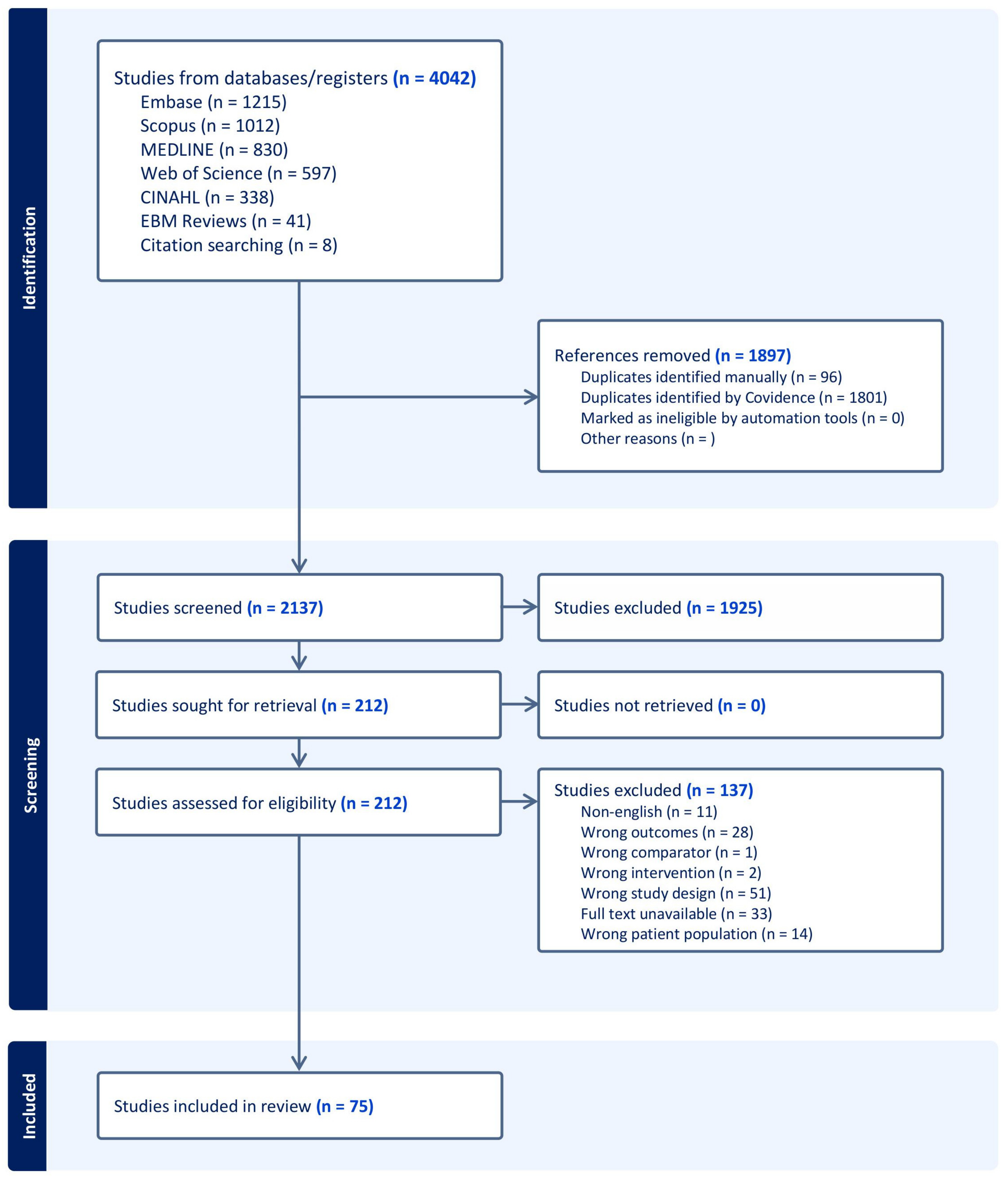
Figure 1. PRISMA-ScR search flowchart.
Jof 11 00260 g001
Figure 2. Geographical and temporal distribution of studies on oral yeast in OSCC. The map (top) illustrates the geographical distribution of studies, highlighting India as the leading contributor, followed by other regions such as Europe, North America, South America, and the Asia–Pacific. The graph (bottom) depicts the temporal distribution of studies, showing a gradual increase in publications from the 1970s to the 2020s, with a significant rise in recent years.
Jof 11 00260 g002
Figure 3. Techniques used for oral yeast identification (left) and speciation (right) across studies. Culture- and histologic-based methods dominate identification, while phenotype-based and PCR-based approaches are most common for speciation.
Jof 11 00260 g003
Disclaimer/Publisher’s Note: The statements, opinions and data contained in all publications are solely those of the individual author(s) and contributor(s) and not of MDPI and/or the editor(s). MDPI and/or the editor(s) disclaim responsibility for any injury to people or property resulting from any ideas, methods, instructions or products referred to in the content.