Article

Occurrence and Diversity of Fungi and Their Mycotoxin Production in Common Edible and Medicinal Substances from China

1
College of Food Science, South China Agricultural University, Guangzhou 510640, China
2
National Health Commission Science and Technology Innovation Platform for Nutrition and Safety of Microbial Food, Guangdong Provincial Key Laboratory of Microbial Safety and Health, State Key Laboratory of Applied Microbiology Southern China, Guangdong Detection Center of Microbiology, Institute of Microbiology, Guangdong Academy of Sciences, Guangzhou 510070, China
*
Author to whom correspondence should be addressed.
J. Fungi 2025, 11(3), 212; https://doi.org/10.3390/jof11030212
Submission received: 6 January 2025 / Revised: 28 February 2025 / Accepted: 1 March 2025 / Published: 10 March 2025

Abstract

:
Edible and medicinal substances can be contaminated by fungi during harvesting, processing, and storage, leading to mycotoxin production and quality deterioration. The distribution of mycotoxigenic fungi in edible and medicinal substances was investigated in this study. Fungi and mycotoxins were detected in 163 commercially available edible and medicinal substances using standard microbiological techniques and high-performance liquid chromatography. A total of 92.0% of samples contained fungi (0.5–5.3 lg colony-forming units (CFU)·g−1); 208 fungal strains belonging to 16 genera were identified, predominantly Aspergillus and Penicillium. Aspergillus section Nigri (30.3%) produced fumonisin B2, which was distributed mainly in radix and rhizome samples. Thirteen samples had mycotoxins, of which ochratoxin A was the most common, followed by aflatoxins and zearalenone (ZEN). One Nelumbinis semen sample contained 10.75 μg·kg−1 AFB1, and one Raisin tree semen sample contained 484.30 μg·kg−1 ZEN, which exceeded regulatory limits in Europe and China. These findings highlight the potential risks associated with fungal contamination and mycotoxins in edible and medicinal substances. Enhanced quality control measures are essential to reduce contamination during harvesting, processing, and storage. Expanded mycotoxin screening, improved preservation techniques, and stricter regulatory standards need to be implemented to ensure consumer safety.

1. Introduction

Edible and medicinal substances are essential for healthcare globally and are traditionally used for disease prevention and treatment [1,2,3]. Approximately 80% of the global population depends on herbal remedies as their primary healthcare solution, and the global herbal supplement market will reach USD 560 billion by 2024 [4]. At present, 106 kinds of medicinal materials have been incorporated into the list of edible and medicinal substances in China [5,6]. Edible and medicinal substances such as Ganoderma, with various diverse secondary metabolites (e.g., polysaccharides and triterpenoids), are widely utilized for their immunomodulatory, antioxidant, and antitumor properties [7]. Based on the World Health Organization’s recognition of the value of traditional Chinese medicine in fighting the coronavirus disease (COVID-19) epidemic, edible and medicinal substances are favored globally. However, microbial contamination of edible and medicinal substances has become increasingly prevalent, especially with respect to the presence of mycotoxigenic fungi and mycotoxins, which directly impact the quality, stability, and safety of these preparations [8,9]. For instance, highly nutritious edible and medicinal substances such as Nelumbinis semen (lotus seed), Codonopsis radix (Codonopsis tangshen), Scutellariae radix (Baikal skullcap root), and Angelicae sinensis radix (Danggui) are easily contaminated by filamentous fungi because of factors such as poor storage conditions, improper harvest and drying practices, and the use of contaminated soil and water [10,11]. In addition, fluctuations in temperature and humidity during storage and transportation can further promote fungal growth, leading to potential mycotoxin biosynthesis. To ensure the safety and quality of edible and medicinal substances, regulatory agencies have established threshold limits for fungal contamination. For example, the European Pharmacopoeia recommends that the total fungal load in herbal materials should not exceed 104 colony-forming units (CFU)/g [12].
Aspergillus, Penicillium, Fusarium, and Alternaria are the most predominant genera in edible and medicinal substances, with Aspergillus being the most frequently isolated in previous studies [13,14,15,16,17,18,19]. The prevalence of these genera is concerning owing to their ability to produce mycotoxins. For instance, A. flavus is among the most important producers of aflatoxins, whereas A. niger produces ochratoxin and fumonisin [20]. The presence of these mycotoxin-producing fungi in edible and medicinal substances poses a considerable health risk to humans and animals and has detrimental effects on the quality and safety of food products.
Mycotoxins are secondary metabolites that can cause acute and chronic toxic effects, including carcinogenicity, mutagenicity, and teratogenicity [21,22,23]. Mycotoxins are stable molecules that are difficult to remove or eradicate during processing and most likely remain in the final product. Humans are exposed to mycotoxins by various means, including consumption of contaminated food and beverages, inhalation of airborne mold spores, and dermal contact with surfaces contaminated with mycotoxin-producing fungi [24,25]. In addition, people may ingest products from livestock that consume feed contaminated with mycotoxins [26]. Long-term consumption of edible and medicinal substances contaminated with mycotoxins inevitably increases the occurrence of adverse events and harms human health. Understanding the prevalence and distribution of these fungal species is essential for formulating strategies to prevent and manage the fungal contamination of edible and medicinal substances.
Considering the growing popularity of high-quality edible and medicinal substances and inherent risks posed by fungal contamination, investigating the occurrence and levels of mycotoxigenic fungi and mycotoxins in different types of edible and medicinal substances in specific production areas is crucial. Therefore, this study aimed to: (1) ascertain fungal and mycotoxin levels within edible and medicinal substances, (2) determine the predominant genera using polyphasic taxonomy and analyze the distribution of fungal species, and (3) verify the potential for mycotoxin production.

2. Materials and Methods

2.1. Edible and Medicinal Substances

For this study, 163 samples from the most used edible and medicinal substances, encompassing 40 distinct varieties divided into six types (animal, edible fungi, flos et folium, fructus, radix et rhizoma, and semen), were selected from specific production regions in China (Table 1). All samples were commercially available products purchased from large Chinese herbal medicine wholesale markets. Each sample was placed in a sterile sampling bag and labeled with a unique identifier corresponding to its origin, variety, and collection date. All samples were transported to the laboratory and identified according to the first part of the Chinese Pharmacopoeia (2020) [27].

2.2. Fungal Isolation and Enumeration

The total combined yeast and mold count (TYMC) was determined using the plate-count method, with at least two counts for each medium; counts were averaged. Samples were prepared by homogenizing 25 g of the test sample with 225 mL of a buffered sodium chloride–peptone solution (pH 7.0 ± 0.2), in accordance with the microbiological examination methods specified in the fourth part of the Chinese Pharmacopoeia [27]. Four sequential dilutions were performed for each sample, with dilution factors of 1:10, 1:100, 1:1000, and 1:10,000. Serial decimal dilutions (1 mL each) were inoculated onto a 9-cm diameter Petri dish. Subsequently, 15–20 mL of Sabouraud dextrose agar (SDA) (Guangdong Huankai Microbial Sci & Tech. Co. Ltd., Guangzhou, China) at approximately 45 °C was poured into the dishes. All plates were subsequently incubated at 25 °C for 5–7 d. After incubation, the CFUs were enumerated. The fungal load level was expressed as log TYMC and calculated using Equation (1):
log TYMC = log10 (N × D)
where N represents the number of colony-forming units (CFU) per plate and D denotes the dilution factor. When the TYMC was <10 (below the detection limit), log TYMC was recorded as 0.5 [28].

2.3. Morphological Identification

Three-point inoculations of the isolates were conducted to examine their macromorphology on malt extract agar (MEA) and Czapek yeast extract agar (CYA) (Guangdong Huankai Microbial Sci & Tech. Co. Ltd., Guangzhou, China) following incubation at 25 °C in the dark for 3–7 d [29,30,31]. Colony texture, obverse and reverse colony colors, degree of sporulation, exudates, and pigments were recorded [20]. The microscopic morphology of the fungi was examined using a biological microscope (CX21FS1C; Olympus Ltd., Tokyo, Japan) accompanied by lactophenol cotton blue staining [32]. One to two drops of lactophenol cotton blue stain (Qingdao Hope Bio-Technolongy Co. Ltd., Qingdao, China) were placed on clean microscope slides along with a small amount of spore bearing hyphae from the fungal colony. The hyphae were carefully dispersed, and a cover slip was gently placed over the sample to ensure that no air bubbles were trapped. The sample was then observed under a microscope, starting with a low-power (20×) objective and switching to a high-power (40×) objective if necessary.

2.4. Molecular Identification

Fungal DNA was extracted from pure isolates using genomic DNA kits (Omega BioTek Inc., Norcross, GA, USA). The quality and concentration of the extracted DNA were assessed using a NanoDrop 2000 spectrophotometer (Thermo Fisher Scientific, Waltham, MA, USA). Polymerase chain reaction (PCR) assays were performed using the internally transcribed spacer (ITS) region and β-tubulin gene. The ITS region was amplified using primers ITS1 (5′-TCC GTA GGT GAA CCT GCG G-3′) and ITS4 (5′-TCC TCC GCT TAT TGA TAT GC-3′); the BenA gene encoding β-tubulin was amplified using primers Bt2a (5′-GGT AAC CAA ATC GGT GCT GCT TTC-3′) and Bt2b (5′-ACC CTC AGT GTA GTG ACC CTT GGC-3′). The PCR protocols are described by Visagie et al. [33]. The sequences were compared with known fungal sequences using the Basic Local Alignment Search Tool (https://blast.ncbi.nlm.nih.gov/Blast.cgi, accessed on 1 December 2024) to determine the closest matches and confirm the identities of the isolated fungi. Following identification, a phylogenetic analysis was conducted to further elucidate the evolutionary relationships among the different fungal species.

2.5. Mycotoxin-Producing Abilities of Aspergillus and Penicillium Strains

One hundred and seventeen strains, comprising 90 Aspergillus and 27 Penicillium strains, were inoculated into CYA using the method described by Silva et al. [34]. Three small pieces of mycelia (each 9 mm in diameter) were excised from the central portion of the colony using a sterile pipette tip (9 mm caliber) to ensure precision and consistency in the sample size. Mycotoxins were then extracted using 1 mL of 70% methanol in an ultrasonic bath for 30 min, followed by centrifugation at 10,000× g for 10 min. Subsequently, the supernatant was transferred to a clean 15 mL centrifuge tube and diluted with ultrapure water to a final volume of 3 mL. The liquid was purified using a Poly-Sery HLB Pro SPE Cartridge (ANPEL Laboratory Technologies Inc., Shanghai, China). Mycotoxins were qualitatively detected by ultra-performance liquid chromatography coupled with tandem mass spectrometry (UPLC–MS/MS) using an LC20 UPLC system paired with a 5500+ mass spectrometer (AB SCIEX, Framingham, MA, USA).
UPLC analysis was performed using a Waters CORTECS C18 column (2.1 × 100 mm, 1.6 μm; Waters Corp., Milford, MA, USA). The column temperature was set at 40 °C. The elution gradient was: 0 min, 95% B; 5 min, 10% B; maintained at 10% B to 8 min; 8.1 min, reverted to 95% B; and held at 95% B to 10 min. The flow rate was set to 0.3 μL·min−1. For the analysis of aflatoxins B1, B2, G1, and G2, ochratoxin A (OTA), fumonisin B1, B2, B3, and zearalenone (ZEN), the mobile phase was composed of acetonitrile (phase A) and 0.1% acetic acid (phase B). In the case of patulin (PAT), the mobile phase was water (phase A) and acetonitrile (phase B). For citrinin (CTN), the mobile phase comprised acetonitrile (phase A) and 5 mM ammonium acetate (phase B).
Mass analysis was performed using electrospray ionization (ESI) in the positive and negative ion modes, and detection conditions were set as: curtain gas (CUR), 25 psi; GS1, 60 psi; GS2, 60 psi; ion spray voltage (IS), 5500 V (positive mode) and −4500 V (negative mode); and source temperature, 550 °C. The optimal multiple reaction monitoring transitions, dwell times, declustering potentials, and collision energies are summarized in Table S1.

2.6. Mycotoxin Determination in Edible and Medicinal Substances

Five grams of each sample were homogenized with 50 mL of a 70% methanol solution, and the resulting mixture was sonicated for 30 min. Following centrifugation at 6000 rpm for 10 min, 10 mL of the supernatant was diluted with 20 mL of ultrapure water, and 3 mL of the diluted liquid was purified using a Poly-Sery HLB Pro SPE Cartridge. The eluate was dried using nitrogen at 50 °C, and the residue was subsequently dissolved in 1 mL of 50% acetonitrile. After vortex-mixing for 30 s, the solution was passed through a 0.22 μm microporous membrane and subjected to qualitative analysis with UPLC–MS/MS. Furthermore, positive samples were confirmed by high-performance liquid chromatography combined with fluorescence detection (HPLC–FLD; LC-20AT with RF-20A fluorescence detector; Shimadzu, Kyoto, Japan) to accurately detect mycotoxin concentration in accordance with the National Food Safety Standards of China methods [35,36,37]. HPLC separation was performed using an Eclipse Plus C18 column (150 × 4.6 mm, 5.0 μm; Agilent, Santa Clara, CA, USA).
Aflatoxin B1, B2, G1, and G2 were analyzed at a column temperature of 40 °C and constant flow rate of 1 mL·min−1. The mobile phase consisted of water (A) and acetonitrile: methanol (50:50) (B). The gradient elution program was 68% A and 32% B. Excitation and emission wavelengths were set to 360 and 440 nm, respectively, for fluorescence detection. The aflatoxins were determined using a post-column photochemical derivative reactor. For OTA detection, the column temperature was 35 °C, and the mobile phase was acetonitrile–water–acetic acid (96:102:2) at a flow rate of 1.0 mL·min−1. For fluorescence detection, the excitation and emission wavelengths were 333 and 460 nm, respectively. ZEN was detected at a column temperature of 25 °C. The mobile phase was acetonitrile–water–methanol (46:46:8) at a flow rate of 1.0 mL·min−1. For fluorescence detection, the excitation and emission wavelengths were set to 274 and 440 nm, respectively.
Mycotoxins were quantified by comparing their peak areas with calibration curves obtained using standard solutions. All standard curves exhibited excellent linearity, with correlation coefficients (R²) ranging from 0.9992 to 0.9999. The limits of detection (LOD) and quantification (LOQ) were 0.03–5 and 0.1–17 μg·kg−1, respectively (Table S2). In the absence of certified reference materials, the accuracy of the method was assessed through recovery studies by spiking known concentrations of analytes into blank matrices [38,39]. Recovery experiments were conducted at three spiking levels in six different blank matrices. The recovery rates for target mycotoxin ranged from 79.9% to 106.7%, accompanied by relative standard deviations (RSDs) below 5.92% (Table S3).

2.7. Statistical Analysis

IBM SPSS (v26) was used for descriptive data analysis and to plot histograms. For inferential statistics, one-way analysis of variance was conducted to determine significant differences among the groups. Gene sequences were aligned, and an evolutionary tree was constructed with the neighbor-joining method using MEGA 7.0.26 [40] and ChiPlot (https://www.chiplot.online/, accessed on 7 February 2024).

3. Results and Discussion

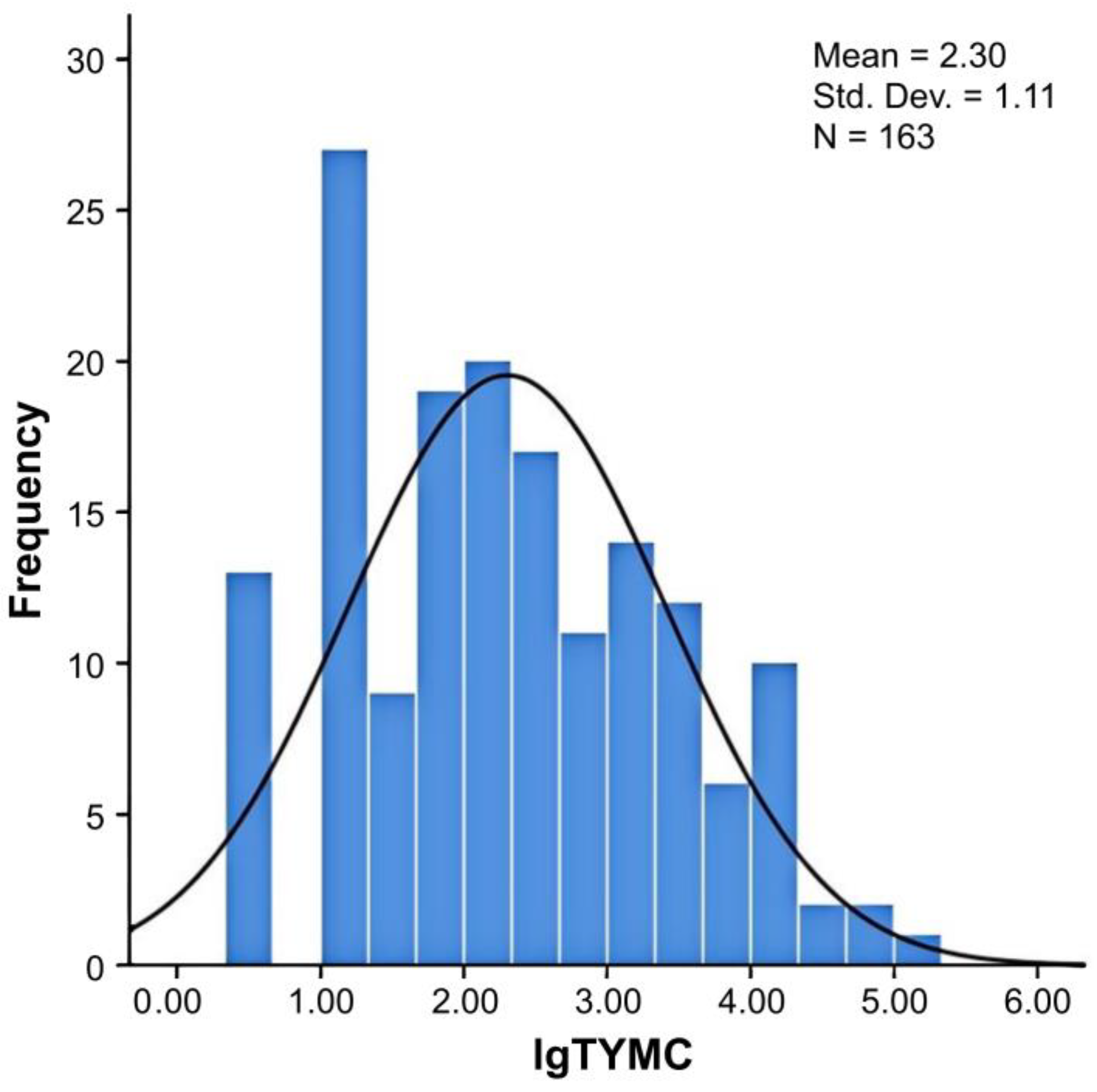
3.1. Fungal Contamination in Edible and Medicinal Substances

Approximately 92.0% (150 of 163) of samples tested positive for fungal contamination. Fungal load per sample was 0.5–5.28 log CFU·g−1. The value of log TYMC for edible and medicinal substances was skewed (Figure 1), and only 7.9% (13/163) of the samples exceeded the contamination limit (104 CFU·g−1 = 4 lgCFU·g−1) recommended by the European Pharmacopoeia [12]. These samples originated from nine edible and medicinal substances: Acanthopanacis senticosi radix et rhizoma seu caulis (Acanthopanacis senticosi), Cuscutae semen, Euryales semen, Flammulina velutipes, Glycyrrhizae radix et rhizoma (licorice), Leonuri herba, Mori folium, Polygonati rhizoma, and Puerariae lobatae radix (kudzu vine).
Elevated fungal counts may suggest processing failure or recontamination during transportation, storage, or marketing, likely due to increased moisture content. A previous study indicated that the log TYMC ranged from 1 to 7 in Chinese herbal medicines from Shanghai and Beijing [41,42]. Recent studies have focused on fungal levels within a single or limited types of edible or medicinal substrates [43,44,45], often lacking a comprehensive analysis across various herbal medicines or substrates. This may hinder a holistic understanding of the variability and significance of log TYMC levels in Chinese herbal medicines.
Based on the plant parts used, samples were classified into six types: animal, edible fungi, flos et folium, fructus, radix et rhizoma, and semen. The detectable log TYMC values differed by type (Table 2). The log TYMC for radix et rhizoma ranged from 0.5 to 4.80, indicating that the fungal contamination of radix et rhizoma varied greatly, and the quality of the samples was unstable. The roots and rhizomes of medicinal plants are prone to mechanical damage during harvesting, which exposes the tissues and provides conditions for microbial invasion and proliferation [46]. This variability emphasizes the need for stricter quality control measures to ensure the safety and stability of these products.
The higher microbial loads in flos et folium and edible fungi may be attributed to their structural characteristics and moisture content [47], which can create favorable conditions for fungal growth. In contrast, the relatively low microbial loads in animal-based products and fructus may be due to their low moisture content or natural antimicrobial properties [48,49].
The effect of processing methods on microbial contamination is evident. Directly cut and sun-dried slices exhibited higher microbial loads, likely because these methods did not effectively reduce the microbial populations [50]. In contrast, heat processing methods such as boiling, roasting, and steaming substantially reduced microbial contamination, as observed in products such as Asini corii colla and Mume fructus. This highlights the importance of adopting appropriate processing techniques to minimize the microbial risks in edible and medicinal substances.

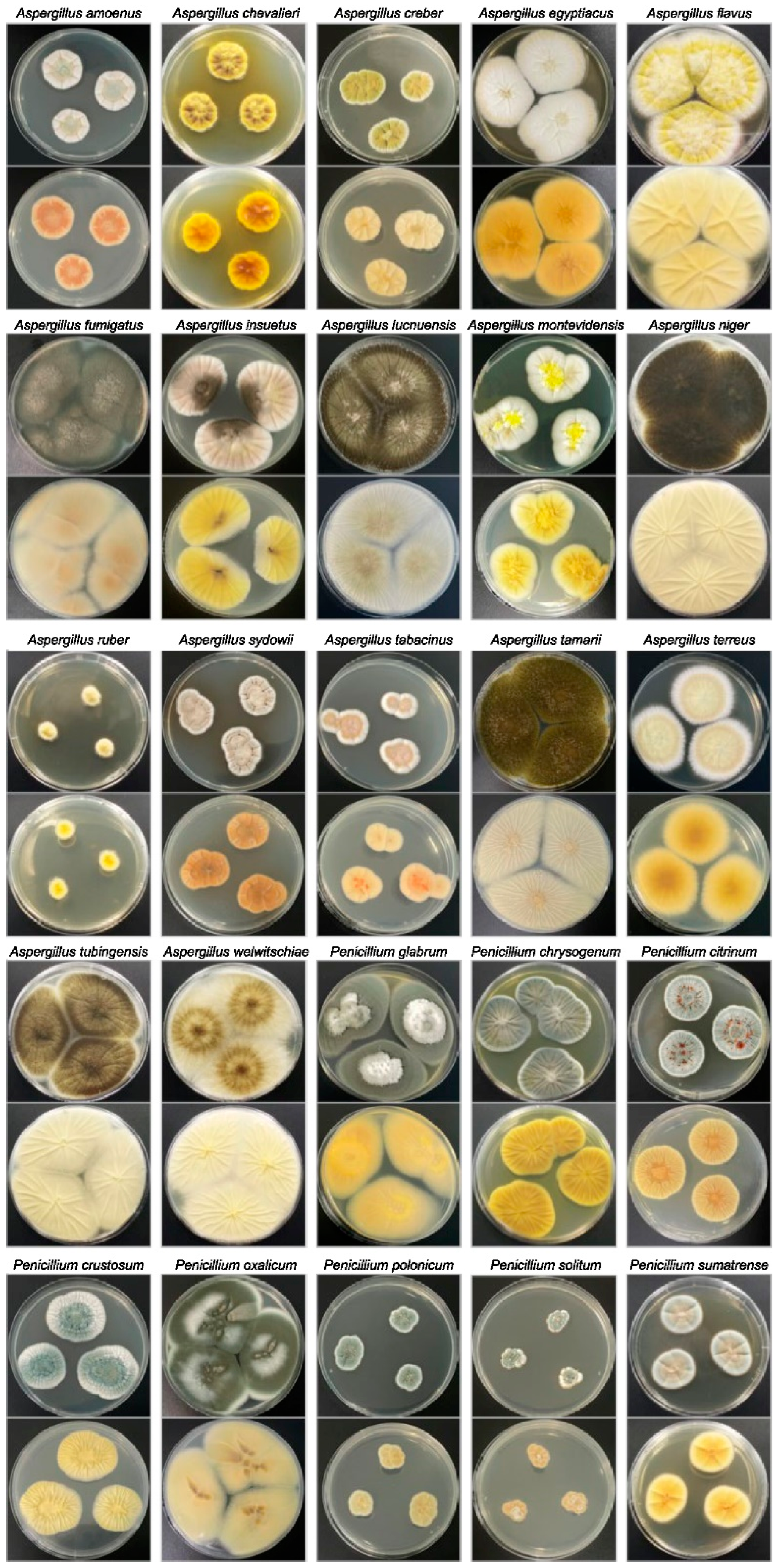
3.2. Fungal Genera and Species Diversity

A total of 208 strains (196 mold and 12 yeast strains) were isolated from edible and medicinal substances. Preliminary morphological identification indicated the presence of 90 strains of Aspergillus; 27 of Penicillium; 24 of Mucor; 23 of Rhizopus; 11 of Trichoderma; four of Alternaria; four of Chaetomium; three of Talaromyces; two of Epicoccum; two of Cladosporium; one each of Aplosporella, Byssochlamys, Curvularia, Dichotomopilus, Neurospora, and Periconia; and 12 unclassified yeasts. Among the isolates, the Aspergillus genus was the most prevalent (43.3%), followed by Penicillium (13.0%). Silva et al. [34] found that Aspergillus was predominant in 30 yerba mates. Yu [51] analyzed the fungal diversity on the surfaces of six herbal materials using high-throughput sequencing and observed the prevalence of Aspergillus, Rhizopus, and Penicillium over other genera. Aspergillus and Penicillium are common storage fungi used for various edible and medicinal substances, which was consistent with our research [18,52]. Although Wei et al. [18] observed a certain proportion of Fusarium (5.17%) in herbal medicines using high-throughput sequencing, traditional culture methods failed to obtain Fusarium isolates from 240 samples. A similar response was observed in the present study; we did not isolate Fusarium (generally classified as field fungi). This result implies that field fungi can be removed or reduced following processing of edible plant materials, such as washing, drying, or fumigation. Fusarium spp. do not usually develop under low humidity, which explains their predominant occurrence in field environments [53]. The low Fusarium contents of edible and medicinal substances make them difficult to isolate and identify.
Molecular identification by genetic characterization was used to accurately assign 90 Aspergillus and 27 Penicillium strains to the species. The colony morphologies of the primary strains (identified at the species level) are shown in Figure 2. Figure 3 shows the phylogenetic tree of the sequences compared with the standard and type strain reference sequences available in GenBank. The 90 Aspergillus isolates were divided into six distinct sections: Nigri, Terrei, Flavus, Versicolores, Aspergillus (formerly Eurotium), and Fumigati. Aspergillus section Nigri was the most isolate-rich section, with 48 isolates, including 33 of A. niger, seven A. welwitschiae, five A. tubingensis, and three A. luchuensis.
Aspergillus niger was the dominant species among the Aspergillus isolates, followed by A. flavus; A. welwitschiae from the A. niger taxon is present in dried fruits, grapes, coffee beans, cocoa, onions, and in rhizosphere soil [15]. In agriculture, A. welwitschiae in sisal substances has been implicated as the causative agent of maize ear rot and bole rot disease [54,55]. These infections can lead to considerable crop losses and economic burden for farmers. Furthermore, A. welwitschiae is a pathogen in human infections, particularly otomycosis (ear canal infections). This species is often resistant to nystatin, posing challenges in its treatment [56].
A total of 27 Penicillium isolates were identified as seven species belonging to three distinct sections (Furcatum, Aspergilloides, and Penicillium). The dominant species were P. chrysogenum (37.0%) and P. citrinum (22.2%). These species are the potential producers of CTN.

3.3. Mycotoxin-Producing Abilities of Aspergillus and Penicillium Isolates

Aflatoxins and ochratoxins are among the most important mycotoxins, and their producers are predominantly found within the genus Aspergillus, particularly in Aspergillus section Flavi [20]. However, Aspergillus section Nigri species, such as A. niger and A. welwitschiae, produce ochratoxins and fumonisin [15]. Penicillium is a major PAT- and CTN-producing genus [57,58]. Therefore, 117 isolates (90 Aspergillus and 27 Penicillium strains) were analyzed for their ability to produce 11 mycotoxins using UPLC–MS/MS.
Eighteen strains produced mycotoxins (Table 3). Approximately 17.6% of the isolates could produce B-type aflatoxins (specifically aflatoxins B1 and B2; Figure S1), which were isolated from three samples: Angelicae dahuricae radix 03-3A and Nelumbinis semen 26–3A and 26–5B. Nelumbinis semen, commonly known as the lotus seed or lotus seed kernel, is the dried mature seed of Nelumbo nucifera and is a commonly used edible substance with important medicinal and edible functions. This herb exhibits various health benefits, including spleen strengthening for diarrhea prevention, kidney nourishment and essence preservation, leukorrhea alleviation, and heart soothing to promote tranquility [59]. Nelumbinis semen is rich in protein and is predominantly produced in the warm and humid regions of southern China. It is highly vulnerable to fungal infections, posing considerable risks to its quality [60].
Aflatoxin-producing fungi in Nelumbinis semen samples produced in Zhejiang Province were successfully isolated. These findings highlight the potential for aflatoxin contamination of improperly-managed Nelumbinis semen. Some studies have indicated that Nelumbinis semen is highly likely contaminated with aflatoxins [61,62]. To address this issue, the 2020 version of the Chinese Pharmacopoeia lists 24 varieties of medicinal substances, including Nelumbinis semen, as potentially containing aflatoxins, with a permissible limit of 10 µg·kg−1. The regulatory measures are designed to guarantee the safety and quality of medicinal materials by setting permissible limits for aflatoxin contamination, thereby safeguarding consumers from potential health risks associated with the ingestion of contaminated products.
None of the A. flavus isolates in this study produced G-type aflatoxins. Aspergillus flavus is well-known for its ability to produce aflatoxins, particularly B-type aflatoxins (AFB1 and AFB2). This is consistent with the study by Adelusi et al. [63], who reported that A. flavus strains isolated from smallholder dairy cattle feeds in South Africa predominantly produced B-type aflatoxins, with no detection of G-type aflatoxins. This selective production is likely due to genetic differences in the aflatoxin biosynthesis gene cluster, where certain genes required for G-type aflatoxin production may be absent or non-functional in these strains.
Of the 33 strains of A. niger, 10 were FB2 producers (Figure S3), isolated from Atractylodis macrocephalae rhizoma 06–2A; Acanthopanacis senticosi radix et rhizoma seu caulis 01-3A; Salviae miltiorrhizae radix et rhizoma 37–1B and 37–3C; Rhodiolae crenulatae radix et rhizoma 35-4A; Rosae laevigatae fructus 36–1B; Dioscoreae rhizoma 13–2D; Crataegi fructus 10–2A; Leonuri herba 22–3B; and Polygonati rodorati rhizoma 32–1B. One A. niger isolate (from Rhodiolae crenulatae radix et rhizoma 35–4A) also produced OTA (Figure S2). Only A. niger strains isolated from Alismatis rhizoma 02–2A and Gardeniae fructus 17–1B could produce OTA (Figure S2).
Aspergillus niger is common in tropical and subtropical food sources [17], including edible and medicinal substances. The incidence of strains within this species that produce FB2 is high. Our observation that a few A. niger strains produced ochratoxin A (OTA) aligns with findings from other studies. Aspergillus niger is recognized as a potential producer of OTA, although not all strains exhibit this capability. Antonia et al. [64] demonstrated that the presence of OTA biosynthetic genes (e.g., ota1, ota2, ota3, ota4, and ota5) was crucial for OTA production. In their study, OTA-nonproducing strains of A. niger were found to lack these genes, indicating that the genetic potential for OTA production is a key determinant. The current results suggest that the A. niger strains producing OTA possessed the necessary biosynthetic genes, whereas the non-producing strains may lack them. In contrast, none of the seven strains of A. welwitschiae analyzed in the current study were found to produce either OTA or FB2. This indicates that the A. welwitschiae strains isolated from edible and medicinal substances in the current study do not possess mycotoxin-producing capabilities. These findings elucidate the mycotoxin-producing potential of Aspergillus species in medicinal and edible substances, emphasizing the importance of genetic factors in determining mycotoxin production.
CTN with renal toxicity was detected in three strains of P. citrinum (Lablab semen album 21–2A and Nelumbinis semen 26–1C and 26–5D; Figure S4). Penicillium citrinum can colonize almost all substrates, including grains, fruits, vegetables, and tea [65,66]. Despite the low isolation rate of P. citrinum from edible and medicinal substances, all identified strains produced CTN. Furthermore, we not only isolated CTN-producing strains from Nelumbinis semen but also found aflatoxin-producing strains, indicating that Nelumbinis semen is at risk of contamination with mycotoxin mixtures. Mycotoxin mixtures in food products pose a considerable health risk owing to their potential synergistic or additive toxic effects [67].
Mycotoxins, such as aflatoxins, ochratoxins, and fumonisins, are secondary metabolites produced by fungi that contaminate various foods and medicinal substances. For instance, the co-occurrence of aflatoxin B1 and ochratoxin A exacerbates liver and kidney damage in animal studies [68]. Furthermore, mycotoxin mixtures complicate risk assessment and regulatory control because current safety guidelines often focus on individual mycotoxins rather than their combined effects. This highlights the need for comprehensive monitoring and stricter regulations to address the risks associated with mycotoxin mixtures in food and medicinal products. However, CTN and FB are not listed in the monitoring directory of food standards or pharmacopoeia. Therefore, some edible and medicinal substances and related products may be contaminated with these mycotoxins because of a lack of official legislation. To ensure food safety and protect consumer interests, extensive surveys of CTN and FB residues in edible and medicinal substances must be conducted to establish a scientific foundation for permissible limits of mycotoxins.
Not all strains produce mycotoxins. The 18 mycotoxigenic strains obtained were distributed across a variety of edible and medicinal substances, including radix et rhizoma, semen, fructus, and flos et folium. Half of these mycotoxigenic strains were isolated from radix et rhizoma samples. Mycotoxins, particularly aflatoxins and fumonisins, are common in herbs containing radix et rhizoma [19]. Mycotoxin production is influenced by mycotoxigenic strains and environmental factors (e.g., host, light, temperature, and water activity) [69,70,71]. The presence of mycotoxin-synthesizing gene clusters in the genomes of these strains is a fundamental cause of mycotoxin production. Environmental factors are transmitted to mycotoxigenic strains through signal transduction. The expression of mycotoxin synthesis gene clusters can be activated or inhibited, thereby affecting mycotoxin synthesis [72]. Therefore, cool (≤20 °C) and dry (average relative humidity <40% at 20 °C) storage of the above edible and medicinal substances is recommended (Chinese Pharmacopeia Commission [27]; United States Pharmacopeia Commission [73]). Producers should ensure optimal storage conditions, including temperature, humidity, and ventilation, to minimize the proliferation of potentially mycotoxigenic fungi [74]. Regular monitoring of these conditions is essential to determine the appropriate storage duration and mitigate the risk of mycotoxin accumulation. Decisions regarding the storage period and timely sale of products should be based on these monitoring results [75].

3.4. Mycotoxin Content in Edible and Medicinal Substances

Comprehensive analysis of 11 mycotoxins in 163 edible and medicinal substances indicated that 8.0% of samples exceeded the detection limits (Table 4; Figure S5). Mycotoxin contamination was identified in Angelicae dahuricae radix, Chrysanthemi flos, Glycyrrhizae radix et rhizoma, Mori fructus, Nelumbinis semen, and Raisin tree semen. The predominant mycotoxins detected were AFB1, AFB2, OTA, and ZEN. No traces of the seven other mycotoxins, namely AFG1, AFG2, FB1, FB2, FB3, PAT, or CTN, were found in any of the samples. The levels of mycotoxins found in these samples ranged from 0.07 to 484.30 μg·kg−1, with OTA being the most prevalent (detected in 4.3% of samples). AFB1 was detected in 2.5% of samples, whereas ZEN and AFB2 were detected in 1.8% and 0.6% of samples, respectively.
The legal threshold for AFB1 in food products is typically 2–10 μg·kg−1, whereas that for ZEN is 50–200 μg·kg−1 [76,77]. One batch of Nelumbinis semen had 10.75 μg·kg−1 AFB1 and one batch of Raisin tree semen contained 484.30 μg·kg−1 ZEN. The concentrations detected in these batches far exceeded these limits, indicating a potential failure in quality control measures. Furthermore, AFB1- and AFB2-producing A. flavus strains were isolated from Nelumbinis semen and Angelicae dahuricae radix, suggesting a cumulative risk of aflatoxin exposure to these particular edible and medicinal substances. Although fumonitoxin-producing A. niger was detected in various edible and medicinal substances, fumonitoxin was not detected. The absence of detectable mycotoxins in some samples, despite the presence of potentially toxigenic fungi, may have been influenced by optimal storage, transport, or processing conditions.
Appropriate storage (e.g., controlled temperature and humidity), efficient transport practices, and effective processing methods (e.g., drying, cleaning, and sterilization) significantly substantially inhibits fungal growth and mycotoxin production [78]. The fungi may have been dormant or may not have been exposed to conditions conducive to toxin production at sampling. Alternatively, the detection methods used may not have been sensitive enough to detect low levels of fumonitoxin. Further studies on the effect of storage and processing conditions on mycotoxin production using these materials will provide valuable insights.
Mycotoxins in edible and medicinal substances pose a direct threat to consumer health and highlight the need for stringent quality control measures during production and storage. Adherence to competent agricultural practices and effective post-harvest handling procedures are imperative for minimizing fungal growth and mycotoxin production. Regulatory bodies should regularly monitor edible and medicinal substances to ensure compliance with safety standards and protect public health. Further research is required to develop more sensitive and specific detection methods for mycotoxins in edible and medicinal substances and to explore the potential for mycotoxin reduction through processing and treatment methods.

4. Conclusions

In this study, the levels of fungi and prevalence of mycotoxigenic fungi in 40 different types of popular edible and medicinal substances from China were systematically evaluated. The study indicated low concentrations of fungi in several edible and medicinal substances, suggesting that most samples were within acceptable fungal contamination limits. The fungal flora of edible and medicinal substances was diverse, with Aspergillus being the most prevalent genus, followed by Penicillium, which was consistent with the results of previous studies. The ability of the prevalent isolates to produce mycotoxins was also assessed, with 18 strains testing positive for the production of five different mycotoxins. Ochratoxin was of particular concern because strains of A. niger produce fumonisin B2 and OTA. Strains producing the two mycotoxins were obtained from Nelumbinis semen and R. crenulatae radix et rhizoma, which may increase the risk of mycotoxin residues. Mycotoxin content analysis of edible and medicinal substances indicated high levels of ZEN in Raisin tree semen. Aflatoxin B was detected in Angelicae dahuricae radix and Nelumbinis semen, and aflatoxin-producing Aspergillus spp. were isolated from these samples. These findings emphasize the need for stringent quality control measures during the production, processing, and storage of edible and medicinal substances to minimize fungal contamination and mycotoxin production. Regular monitoring and the establishment of limits for mycotoxins in edible and medicinal substances are crucial for ensuring consumer safety. Further research is warranted to develop more sensitive detection methods for mycotoxins and explore potential reduction strategies through processing and treatment. In addition, the establishment and improvement of a mycotoxigenic strain resource bank will support research on the biological characteristics, production mechanisms, detection methodologies, and control strategies of mycotoxin-producing fungi.

Supplementary Materials

The following supporting information can be downloaded at: https://www.mdpi.com/article/10.3390/jof11030212/s1, Table S1. Optimal multiple reaction monitoring transitions, declustering potential, dwell time (ms), collision energies of mass analysis performed using electrospray ionization; Table S2. Linearity equations, correlation coefficients, limits of detection (LODs), and limits of quantification (LOQs) of the method (LODs and LOQs were measured using standard solutions); Table S3. The recoveries (Rs) and relative standard deviations (RSDs) for six different edible and medicinal substances with mycotoxins; Table S4. Isolates from China examined in this study. Figure S1. Ability of fungi to produce aflatoxins. (a–d) Standard solution: AFB1; AFB2; AFG1; AFG2 (e,f) Aspergillus flavus; Figure S2. Ability of fungi to produce ochratoxin A. (a) Standard solution: OTA, (b) Aspergillus niger; Figure S3. Ability of fungi to produce fumonisin. (a–c) Standard solution: FB1; FB2; FB3, (d) Aspergillus niger; Figure S4. Ability of fungi to produce citrinin. (a) Standard solution: citrinin, (b) Penicillium citrinum; Figure S5. HPLC–FLD chromatograms for (a) AFB1, AFB2, AFG1, and AFG2 standard (AFB1, AFG1 = 10 ng·mL−1; AFB2, AFG2 = 3 ng·mL−1); (b) AFB1-, AFB2-positive sample (Angelicae dahuricae radix 03-2); (c) OTA standard (OTA = 10 ng·mL−1); (d) OTA-positive sample (Chrysanthemi flos 09-1); (e) ZEN standard (ZEN = 200 ng·mL−1); and (f) ZEN-positive sample (Raisin tree semen 34-1).

Author Contributions

Conceptualization, Q.W. and J.Z.; Methodology, L.C. and J.W.; Software, X.L.; Validation, M.Z., W.C., and M.D.; Formal Analysis, W.G.; Investigation, J.W. and X.L; Data Curation, Z.L.; Writing—Original Draft Preparation, L.C.; Writing—Review and Editing, S.Z.; Visualization, L.C.; Supervision, J.Z.; Funding Acquisition, Q.W. All authors have read and agreed to the published version of the manuscript.

Funding

This work was supported by the National Key Research and Development Program of China (2022YFF1100700) and Natural Science Foundation of Guangdong Province, China (2023A1515012578).

Institutional Review Board Statement

Not applicable.

Informed Consent Statement

Not applicable.

Data Availability Statement

The original contributions presented in the study are included in the article/Supplementary Materials. Further inquiries can be directed to the corresponding authors.

Conflicts of Interest

The authors declare no conflicts of interest.

Abbreviations

The following abbreviations are used in this manuscript:
AFaflatoxin
CTNcitrinin
CYACzapek yeast extract agar
Ffumonisin
LODlimit of detection
LOQlimit of quantification
MEAmalt extract agar
OTAochratoxin A
PATpatulin
TYMCtotal combined yeast and mold count
UPLC–MS/MSultra-performance liquid chromatography coupled with tandem mass spectrometry
ZENzearalenone

References

  1. Giblette, J. Production of Chinese medicinal herbs in North America: Challenge and reconciliation. China Med. 2022, 5, 234–240. [Google Scholar] [CrossRef]
  2. Liu, H.P.; Tang, J.F.; Chen, T.J.; Zhu, P.P.; Sun, D.D.; Wang, W.Y. Assessment of heavy metals contamination and human health risk assessment of the commonly consumed medicinal herbs in China. Environ. Sci. Pollut. Res. Int. 2023, 30, 7345–7357. [Google Scholar] [CrossRef]
  3. Nakhaee, S.; Kooshki, A.; Hormozi, A.; Akbari, A.; Mehrpour, O.; Farrokhfall, K. Cinnamon and cognitive function: A systematic review of preclinical and clinical studies. Nutr. Neurosci. 2024, 27, 132–146. [Google Scholar] [CrossRef]
  4. Global Industry Analysts Inc. Herbal Supplements and Remedies: A Global Strategic Business Report MCP-1081. 2024. Available online: http://www.strategyr.com/Herbal_Supplements_and_Remedies_Market_Report.asp (accessed on 1 February 2025).
  5. National Health Commission of the People’s Republic of China; State Administration for Market Regulation. Announcement on Rehmannia glutinosa Libosch. and Other Four Substances Listed in Edible and Medicinal Substances. 2024. Available online: http://www.nhc.gov.cn/sps/s7890/202408/01f4b82eff294f4182102cf43e2e898e.shtml (accessed on 26 August 2024).
  6. Xue, H.K.; Wang, W.L.; Bian, J.Y.; Gao, Y.C.; Hao, Z.T.; Tan, J.Q. Recent advances in medicinal and edible homologous polysaccharides: Extraction, purification, structure, modification, and biological activities. Int. J. Biol. Macromol. 2022, 222, 1110–1126. [Google Scholar] [CrossRef]
  7. Xiao, C.; Wu, Q.; Zhang, J.; Xie, Y.; Cai, W.; Tan, J. Antidiabetic activity of Ganoderma lucidum polysaccharides f31 down-regulated hepatic glucose regulatory enzymes in diabetic mice. J. Ethnopharmacol. 2017, 196, 47–57. [Google Scholar] [CrossRef]
  8. Oztekin, S.; Dikmetas, D.N.; Devecioglu, D.; Acar, E.G.; Karbancioglu-Guler, F. Recent insights into the use of antagonistic yeasts for sustainable biomanagement of postharvest pathogenic and mycotoxigenic fungi in fruits with their prevention strategies against mycotoxins. J. Agric. Food Chem. 2023, 71, 9923–9950. [Google Scholar] [CrossRef]
  9. Rocha-Miranda, F.; Venâncio, A. Mycotoxigenic fungi in plant-based supplements and medicines. Curr. Opin. Food Sci. 2019, 30, 27–31. [Google Scholar] [CrossRef]
  10. Chen, L.; Guo, W.P.; Zheng, Y.Q.; Zhou, J.Z.; Liu, T.T.; Chen, W.; Liang, D.Q.; Zhao, M.P.; Zhu, Y.D.; Wu, Q.P.; et al. Occurrence and characterization of fungi and mycotoxins in contaminated medicinal herbs. Toxins 2020, 12, 30. [Google Scholar] [CrossRef]
  11. Zhang, Y.H.; Tie, X.Y.; Yang, X.C.; Cheng, F.; Li, Y. Study on the quality change and validity of Chinese herbal medicine and Chinese herbal decoction pieces. J. Trad. Chin. Vet. 2021, 40, 44–48. [Google Scholar]
  12. European Pharmacopoeia Commission. European Pharmacopoeia, 10th ed.; European Directorate for the Quality of Medicines & Healthcare: Strasbourg, France, 2019; pp. 68–97. [Google Scholar]
  13. Chen, A.J.; Huang, L.F.; Wang, L.Z.; Tang, D.; Cai, F.; Gao, W.W. Occurrence of toxigenic fungi in ochratoxin A contaminated liquorice root. Food Addit. Contam. Part A Chem. Anal. Control Expo. Risk Assess. 2011, 28, 1091–1097. [Google Scholar] [CrossRef]
  14. Han, Z.; Ren, Y.P.; Liu, X.S.; Luan, L.J.; Wu, Y.J. A reliable isotope dilution method for simultaneous determination of fumonisins B1, B2 and B3 in traditional Chinese medicines by ultra-high-performance liquid chromatography–tandem mass spectrometry. J. Sep. Sci. 2010, 33, 2723–2733. [Google Scholar] [CrossRef]
  15. Jiang, W.; Guo, M.; Yu, J.; Fan, C.; Yang, M.; Pang, X. Variations of the fungal microbiome in Corydalis rhizoma with different collection areas, processing methods, and storage conditions. Food Res. Int. 2024, 180, 114045. [Google Scholar] [CrossRef]
  16. Massi, F.P.; Sartori, D.; de Souza Ferranti, L.D.S.; Iamanaka, B.T.; Taniwaki, M.H.; Vieira, M.L.C.; Fungaro, M.H.P. Prospecting for the incidence of genes involved in ochratoxin and fumonisin biosynthesis in Brazilian strains of Aspergillus niger and Aspergillus welwitschiae. Int. J. Food Microbiol. 2016, 221, 19–28. [Google Scholar] [CrossRef]
  17. Susca, A.; Moretti, A.; Stea, G.; Villani, A.; Haidukowski, M.; Logrieco, A.; Munkvold, G. Comparison of species composition and fumonisin production in Aspergillus section Nigri populations in maize kernels from USA and Italy. Int. J. Food Microbiol. 2014, 188, 75–82. [Google Scholar] [CrossRef]
  18. Wei, G.F.; Guo, X.T.; Liang, Y.C.; Liu, C.S.; Zhang, G.Z.; Liang, C.L.; Huang, Z.X.; Zheng, Y.Q.; Chen, S.L.; Dong, L.L. Occurrence of fungi and mycotoxins in herbal medicines and rapid detection of toxin-producing fungi. Environ. Pollut. 2023, 333, 122082. [Google Scholar] [CrossRef]
  19. Zhao, X.S.; Ying, G.Y.; Wei, J.H.; Sun, H.; Yang, M.H. Overview of aflatoxin B1 contamination in traditional Chinese medicine. Chin. J. Pharmacovigilance 2018, 15, 608–616, 622. [Google Scholar]
  20. Frisvad, J.C.; Hubka, V.; Ezekiel, C.N.; Hong, S.B.; Nováková, A.; Chen, A.J.; Arzanlou, M.; Larsen, T.O.; Sklenář, F.; Mahakarnchanakul, W.; et al. Taxonomy of Aspergillus section Flavi and their production of aflatoxins, ochratoxins, and other mycotoxins. Stud. Mycol. 2019, 93, 1–63. [Google Scholar] [CrossRef]
  21. Hamad, G.M.; Mehany, T.; Simal-Gandara, J.; Abou-Alella, S.; Esua, O.J.; Abdel-Wahhab, M.A.; Hafez, E.E. A review of recent innovative strategies for controlling mycotoxins in foods. Food Control 2023, 144, 109350. [Google Scholar] [CrossRef]
  22. Schulz, M.C.; Schumann, L.; Rottkord, U.; Humpf, H.U.; Gekle, M.; Schwerdt, G. Synergistic action of the nephrotoxic mycotoxins ochratoxin A and citrinin at nanomolar concentrations in human proximal tubule-derived cells. Toxicol. Lett. 2018, 291, 149–157. [Google Scholar] [CrossRef]
  23. Wang, J.; Gan, C.; Qi, X.; Lebre, M.C.; Schinkel, A.H. Human organic anion transporting polypeptide (OATP) 1B3 and mouse OATP1A/1B affect liver accumulation of ochratoxin A in mice. Toxicol. Appl. Pharmacol. 2020, 401, 115072. [Google Scholar] [CrossRef]
  24. Ehsanifar, M.; Rajati, R.; Gholami, A.; Reiss, J.P. Mold and mycotoxin exposure and brain disorders. J. Integr. Neurosci. 2023, 22, 137. [Google Scholar] [CrossRef]
  25. Eivazzadeh-Keihan, R.; Pashazadeh, P.; Hejazi, M.; de la Guardia, M.; Mokhtarzadeh, A. Recent advances in nanomaterial-mediated bio and immune sensors for detection of aflatoxin in food products. TrAC Trends Anal. Chem. 2017, 87, 112–128. [Google Scholar] [CrossRef]
  26. Lach, M.; Kotarska, K. Negative effects of occurrence of mycotoxins in animal feed and biological methods of their detoxification: A review. Molecules 2024, 29, 4563. [Google Scholar] [CrossRef]
  27. Chinese Pharmacopeia Commission. Pharmacopoeia of the People’s Republic of China; Chinese Pharmacopeia Commission: Beijing, China, 2020. [Google Scholar]
  28. US Food and Drug Administration. Bacteriological Analytical Manual (BAM). Available online: https://www.fda.gov/food/laboratory-methods-food/bacteriological-analytical-manual-bam (accessed on 30 December 2024).
  29. Kirk, P.; Cannon, P.; Minter, D.; Stalpers, J. Ainsworth and Bisby’s Dictionary of the Fungi, 10th ed.; CABI Publishing: Wallingford, UK, 2008. [Google Scholar]
  30. Kong, H.Z. Flora fungorum sinicorum. In Penicillium et Teleomorphi Cognati; Science Press: Beijing, China, 2007; Volume 35. [Google Scholar]
  31. Qi, Z.T. Flora fungorum sinicorum. In Aspergillus et Teleomorphi Cognati; Science Press: Beijing, China, 1997; Volume 5. [Google Scholar]
  32. Huang, T.S.; Wang, K.V.; Ye, X.Y.; Chen, C.S.; Chang, F.C. Attention-guided transfer learning for identification of filamentous fungi encountered in the clinical laboratory. Microbiol. Spectr. 2023, 11, e0461122. [Google Scholar] [CrossRef]
  33. Visagie, C.M.; Houbraken, J.; Frisvad, J.C.; Hong, S.B.; Klaassen, C.H.W.; Perrone, G.; Seifert, K.A.; Varga, J.; Yaguchi, T.; Samson, R.A. Identification and nomenclature of the genus, Penicillium. Stud. Mycol. 2014, 78, 343–371. [Google Scholar] [CrossRef]
  34. Silva, J.J.; Puel, O.; Lorber, S.; Ferranti, L.S.; Ortiz, L.F.; Taniwaki, M.H.; Iamanaka, B.T.; Fungaro, M.H.P. Occurrence and diversity of Aspergillus in commercial yerba mate elaborated for the Brazilian beverage, “chimarrão”. Food Res. Int. 2019, 121, 940–946. [Google Scholar] [CrossRef]
  35. National Health Commission of the People’s Republic of China; National Food and Medical Products Administration. GB 5009.22-2016; Determination of Aflatoxin B and G in Foods of National Food Safety Standards; China Standard Press: Beijing, China, 2016; pp. 11–17. [Google Scholar]
  36. National Health Commission of the People’s Republic of China; National Food and Medical Products Administration. GB 5009.96-2016; Determination of Ochratoxin A in Foods of National Food Safety Standards; China Standard Press: Beijing, China, 2016; pp. 1–6. [Google Scholar]
  37. National Health Commission of the People’s Republic of China; National Food and Medical Products Administration. GB 5009.209-2016; Determination of Zearalenone in Foods of National Food Safety Standards; China Standard Press: Beijing, China, 2016; pp. 2–5. [Google Scholar]
  38. Lu, Q.; Ruan, H.N.; Sun, X.Q.; Luo, J.Y.; Yang, M.H. Contamination status and health risk assessment of 31 mycotoxins in six edible and medicinal plants using a novel green defatting and depigmenting pretreatment coupled with LC–MS/MS. LWT 2022, 161, 113401. [Google Scholar] [CrossRef]
  39. Huang, X.J.; Feng, R.; Hu, Q.; Mao, X.H.; Zhou, H. Contamination status and health risk assessment of 73 mycotoxins in four edible and medicinal plants using an optimized QuEChERS pretreatment coupled with LC–MS/MS. Toxins 2025, 17, 52. [Google Scholar] [CrossRef]
  40. Kumar, S.; Stecher, G.; Tamura, K. MEGA7: Molecular Evolutionary Genetics Analysis version 7.0 for bigger datasets. Mol. Biol. Evol. 2016, 33, 1870–1874. [Google Scholar] [CrossRef]
  41. Fan, Y.L.; Li, Q.Q.; Fang, L.; Yang, M.C. A survey of microbial contamination for ten processed pieces of Chinese materia medica in Shanghai. Chin. Trad. Herb. Drugs 2015, 46, 1908–1913. [Google Scholar]
  42. Zhang, G.H.; Wang, S.J.; Jiang, Z.J.; Gao, C. Investigation of microbial contamination for 10 kinds of TCM decoction pieces in Beijing area. China Pharm. 2018, 29, 1940–1944. [Google Scholar]
  43. Deng, H.Y.; Gong, Y.X.; Li, L.F.; Deng, X.D.; Tang, S.; Kang, S.H.; Jiang, Z.Y. Research progress on microbial contamination of decoction pieces and identification of typical bacteria. Chin. Trad. Herb. Drugs 2019, 50, 2242–2250. [Google Scholar]
  44. Gao, X.Z.; Liu, Z.; Wang, Y.; Wang, Y.H. Investigation and research on current situation of microbial contamination of medicinal and edible Chinese medicine prepared pieces. Food Drug 2019, 21, 70–74. [Google Scholar]
  45. Lin, T.H.; Zhang, F.; Zhu, H.M.; Hong, J.W. Investigation and risk analysis of microbial contamination in the Chinese herbal medicine Angelicae sinensis radix in Guangdong Province. Chin. J. Pharm. Anal. 2024, 44, 859–865. [Google Scholar]
  46. Li, K.; Wang, H.Y.; Guo, X.Z.; Zhang, C.C.; Wang, Y.F.; Guo, L.P. Advances in research and application of Trichoderma for inducing resistance against root rot diseases in root and rhizome of Chinese medicinal materials. Zhongguo Zhong Yao Za Zhi 2023, 48, 4942–4949. [Google Scholar] [CrossRef]
  47. Tian, B.; Pan, Y.; Wang, J.; Cai, M.; Ye, B.; Yang, K.; Sun, P. Insoluble dietary fibers from by-products of edible fungi industry: Basic structure, physicochemical properties, and their effects on energy intake. Front. Nutr. 2022, 9, 851228. [Google Scholar] [CrossRef]
  48. Xu, X.X.; Li, X.J.; Pan, K.W.; Deng, L.; Xia, S.B.; Dong, J.W. Microbial transformation of geniposide in Gardeniae fructus under the fermentation with Aspergillus niger DQWM-G11. Nat. Prod. Res. 2024, 1–7. [Google Scholar] [CrossRef]
  49. Gupta, A.; Updahyay, H.; Pandey, S. Evaluation of Microbial Contamination in Herbal Drugs. IJSR 2023, 12, 2333–2336. [Google Scholar] [CrossRef]
  50. Li, Q.C.; Jiang, Y.F.; Lai, S.; Li, W.J.; Lin, T.H. Study on the whole chain microbial load in the production of one traditional Chinese medicine pills. Chin. J. Pharm. Anal. 2024, 44, 1394–1399. Available online: https://ywfx.nifdc.org.cn/EN/10.16155/j.0254-1793.2024-0192 (accessed on 1 February 2025).
  51. Yu, J.S. Fungal Identification on the Surface of Chinese Herbal Materials Through High Throughput Sequencing—Case Studies of Six Herbal Materials; Peking Union Medical College: Beijing, China, 2022. [Google Scholar]
  52. Moorthy, K.; Prasanna, I.; Thajuddin, N.; Arjunan, S.; Gnanendra, T.S.; Zahir Hussain, M.I. Occurrence of myco population in spices and herbal drugs. Int. J. Biosci. Technol. 2010, 1, 6–14. [Google Scholar]
  53. Brito, V.D.; Achimón, F.; Zunino, M.P.; Zygadlo, J.A.; Pizzolitto, R.P. Fungal diversity and mycotoxins detected in maize stored in silo-bags: A review. J. Sci. Food Agric. 2022, 102, 2640–2650. [Google Scholar] [CrossRef]
  54. Nikolic, M.; Savić, I.; Nikolic, A.; Stevanović, M.; Kandić, V.; Stanković, G.; Stankovic, S. First report of Aspergillus welwitschiae causing maize ear rot in Serbia. Plant Dis. 2023, 108, 209. [Google Scholar] [CrossRef]
  55. Quintanilha-Peixoto, G.; Marone, M.P.; Raya, F.T.; José, J.; Oliveira, A.; Fonseca, P.L.C.; Tomé, L.M.R.; Bortolini, D.E.; Kato, R.B.; Araújo, D.S.; et al. Phylogenomics and gene selection in Aspergillus welwitschiae: Possible implications in the pathogenicity in Agave sisalana. Genomics 2022, 114, 110517. [Google Scholar] [CrossRef]
  56. Halvaeezadeh, M.; Jalaee, G.A.; Fatahinia, M.; Mahmoudabadi, A.Z. Aspergillus welwitschiae; an otomycosis predominant agent, new epidemiological and antifungal susceptibility data from Iran. Microb. Pathog. 2023, 181, 106180. [Google Scholar] [CrossRef]
  57. Li, H.F.; Zhao, L.N.; Zheng, X.F.; Wang, Y.; Diao, J.W.; Zhang, X.Y.; Zhang, H.Y. Identification of a Penicillium expansum strain producing citrinin and optimization of culture conditions for citrinin production. Food Sci. 2018, 39, 162–167. [Google Scholar]
  58. Pillay, Y.; Nagiah, S.; Tiloke, C.; Phulukdaree, A.; Chuturgoon, A.A. mir-27b inhibition contributes to cytotoxicity in patulin-exposed HEK293 cells. Toxicon 2022, 210, 58–65. [Google Scholar] [CrossRef]
  59. Mallela, V.J.; Rudrapal, M.; Prasanth, D.S.N.B.K.; Pasala, P.K.; Bendale, A.R.; Bhattacharya, S.; Aldosari, S.M.; Khan, J. Lotus seed (Nelumbinis semen) extract: Anticancer potential and chemoprofiling by in vitro, in silico, and GC–MS studies. Front. Chem. 2024, 12, 1505272. [Google Scholar] [CrossRef]
  60. Arooj, M.; Imran, S.; Inam-Ur-Raheem, M.; Rajoka, M.S.R.; Sameen, A.; Siddique, R.; Sahar, A.; Tariq, S.; Riaz, A.; Hussain, A.; et al. Lotus seeds (Nelumbinis semen) as an emerging therapeutic seed: A comprehensive review. Food Sci. Nutr. 2021, 9, 3971–3987. [Google Scholar] [CrossRef]
  61. Jo, S.A.; Lee, S.D.; Kim, D.G.; Lee, H.K.; Jung, K. A survey of aflatoxin contamination in medicinal herbs for food and medicine. Korean J. Pharmacogn. 2014, 45, 154–160. [Google Scholar]
  62. Liu, L.N.; Jin, H.Y.; Sun, L.; Ma, S.C. General investigation and preliminary risk assessment of aflatoxins in traditional Chinese medicines and their preparations. Chin. Pharm. J. 2015, 50, 1541–1546. [Google Scholar]
  63. Adelusi, O.A.; Gbashi, S.; Adebo, J.A.; Aasa, A.O.; Oladeji, O.M.; Kah, G.; Adebo, O.A.; Changwa, R.; Njobeh, P.B. Seasonal and geographical impact on the mycotoxigenicity of Aspergillus and Fusarium species isolated from smallholder dairy cattle feeds and feedstuffs in Free State and Limpopo provinces of South Africa. Toxins 2023, 15, 128. [Google Scholar] [CrossRef]
  64. Susca, A.; Proctor, R.H.; Morelli, M.; Haidukowski, M.; Gallo, A.; Logrieco, A.F.; Moretti, A. Variation in Fumonisin and ochratoxin production associated with differences in biosynthetic gene content in Aspergillus niger and A. welwitschiae isolates from multiple crop and geographic origins. Front. Microbiol. 2016, 7, 1412. [Google Scholar] [CrossRef]
  65. Houbraken, J.A.M.P.; Frisvad, J.C.; Samson, R.A. Taxonomy of Penicillium citrinum and related species. Fungal Divers. 2010, 44, 117–133. [Google Scholar] [CrossRef]
  66. Xu, W.; Xiang, L.; Chen, Y.; Zhao, Y.; Liao, S.; Li, Y.; Li, Y.; Tunyaluk, B.; Lin, L. Risk assessment of citrinin in Chinese dark tea and inhibitory effects of tea polyphenols on citrinin production. LWT Food Sci. Technol. 2024, 205, 116527. [Google Scholar] [CrossRef]
  67. Chhaya, R.S.; Nag, R.; Cummins, E. Human health risk from co-occurring mycotoxins in dairy: A feed-to-fork approach. Food Control 2025, 168, 110954. [Google Scholar] [CrossRef]
  68. Abdullah, R.; Kamarozaman, N.S.; Ab Dullah, S.S.A.; Aziz, M.Y.; Aziza, H.B.A. Health risks evaluation of mycotoxins in plant-based supplements marketed in Malaysia. Sci. Rep. 2025, 15, 1244. [Google Scholar] [CrossRef]
  69. Liu, L.N.; Jin, H.Y.; Sun, L.; Ma, S.C.; Lin, R.C. Determination of aflatoxins in medicinal herbs by high-performance liquid chromatography-tandem mass spectrometry. Phytochem. Anal. 2012, 23, 469–476. [Google Scholar] [CrossRef]
  70. Reverberi, M.; Ricelli, A.; Zjalic, S.; Fabbri, A.A.; Fanelli, C. Natural functions of mycotoxins and control of their biosynthesis in fungi. Appl. Microbiol. Biotechnol. 2010, 87, 899–911. [Google Scholar] [CrossRef]
  71. Zhang, X.M.; Jiao, X.L.; Su, C.Y.; Luo, Y.; Zhang, L.; Gao, W.W. Research progresses in and management strategies for aflatoxin contamination of fruits and seeds medicinal materials in China. Plant Prot. 2023, 49, 118–126. [Google Scholar]
  72. Vinacour, M.; Moiana, M.; Forné, I.; Jung, K.; Bertea, M.; Valdayo, C.P.M.; Nikel, P.I.; Imhof, A.; Palumbo, M.C.; Do Porto, F.D.; et al. Genetic dissection of the degradation pathways for the mycotoxin fusaric acid in Burkholderia ambifaria T16. Appl. Environ. Microbiol. 2023, 89, e0063023. [Google Scholar] [CrossRef]
  73. United States Pharmacopeia Commission. United States Pharmacopeia. 2017, 38, 43. Available online: https://www.uspnf.com (accessed on 1 February 2025).
  74. Mafe, A.N.; Büsselberg, D. Mycotoxins in food: Cancer risks and strategies for control. Foods 2024, 13, 3502. [Google Scholar] [CrossRef] [PubMed]
  75. Daba, H.G.; Delele, M.A.; Fanta, S.W.; Habtu, N.G.; Abera, M.K. Mycotoxigenic fungal growth within stored maize (Zea mays L.) at different storage altitudes and durations. J. Stored Prod. Res. 2024, 108, 102383. [Google Scholar] [CrossRef]
  76. European Commission. Commission Regulation (EC) no 1881/2006 of 19 December 2006 setting maximum levels for certain contaminants in foodstuffs. Off. J. Eur. Union 2006, 364, 5–24. [Google Scholar]
  77. National Health Commission of the People’s Republic of China; National Food and Medical Products Administration. National Food Safety Standard-Limits of Mycotoxins in Foods: GB 2761–2017; Standards Press of China: Beijing, China, 2017. [Google Scholar]
  78. Stakheev, A.A.; Taliansky, M.; Kalinina, N.O.; Zavriev, S.K. RNAi-based approaches to control mycotoxin producers: Challenges and perspectives. J. Fungi 2024, 10, 682. [Google Scholar] [CrossRef]
Figure 1. Histogram of log TYMC of edible and medicinal substances. Log TYMC: log total combined yeast and mold count.
Jof 11 00212 g001
Figure 2. Colony morphology (top and reverse) of Aspergillus and Penicillium species isolated in this study after 5–10 d of incubation at 25 °C in the dark on Czapek Yeast Extract Agar (CYA).
Jof 11 00212 g002
Figure 3. Neighbor-joining tree based on sequence data of internally transcribed spacer (ITS) and β-tubulin from 117 isolates (Aspergillus and Penicillium) using MEGA 7.0.26. Bootstrap values are shown in the nodes according to 1000 replications. “T” type strain; AF, aflatoxin; OTA, ochratoxin A; FB, fumonisin; CTN, citrinin.
Jof 11 00212 g003
Table 1. Forty different species of edible and medicinal substances.
Scientific Name *Type of Edible and Medicinal SubstancesNumber of SamplesSample NameProducing Regions
Acanthopanacis senticosi radix et rhizoma seu caulisradix et rhizoma401-1; 01-2; 01-3; 01-4Heilongjiang
Alismatis rhizomaradix et rhizoma302-1; 02-2; 02-3Sichuan
Angelicae dahuricae radixradix et rhizoma303-1; 03-2; 03-3Sichuan
Angelicae sinensis radixradix et rhizoma404-1; 04-2; 04-3; 04-4Gansu
Asini corii collaanimal305-1; 05-2; 05-3Shandong
Atractylodis macrocephalae rhizomaradix et rhizoma506-1; 06-3; 06-4; 06-5Zhejiang
06-2Sichuan
Auricularia corneaedible fungi507-1; 07-5Jiangsu
07-2; 07-3; 07-4Zhejiang
Cassiae semensemen508-1Hebei
08-2; 08-3; 08-4; 08-5Anhui
Chrysanthemi flosflos et folium509-1; 09-2; 09-3; 09-4; 09-5Zhejiang
Crataegi fructusfructus810-1; 10-2; 10-3; 10-4; 10-5; 10-6; 10-7;10-8Shandong
Cuscutae semensemen411-1; 11-2; 11-3; 11-4Inner Mongolia
Dictyophora inausiataedible fungi612-1; 12-2; 12-3; 12-4; 12-5; 12-6Zhejiang
Dioscoreae rhizomaradix et rhizoma313-1; 13-2; 13-3Henan
Euryales semensemen614-1; 14-3; 14-4; 14-5; 14-6Jiangxi
14-2Guangdong
Flammulina velutipesedible fungi415-1; 15-2; 15-3; 15-4Zhejiang
Ganoderma lingzhiedible fungi416-1; 16-3Shaanxi
16-2Zhejiang
16-4Sichuan
Gardeniae fructusfructus317-1Zhejiang
17-2; 17-3Jiangxi
Ginseng radix et rhizomaradix et rhizoma518-1; 18-2; 18-3; 18-4; 18-5Jilin
Glycyrrhizae radix et rhizomaradix et rhizoma519-1; 19-2; 19-3; 19-4; 19-5Inner Mongolia
Juglandis semensemen320-1Yunnan
20-2; 20-3Hebei
Lablab semen albumsemen421-1; 21-2; 21-3; 21-4Yunnan
Leonuri herbaflos et folium422-1; 22-4Guangdong
22-2Hubei
22-3Hebei
Mori foliumflos et folium823-1; 23-2; 23-3; 23-6; 23-7; 23-8Guangdong
23-4; 23-5Henan
Mori fructusfructus424-1Guangdong
24-2; 24-3Zhejiang
24-4Guangxi
Mume fructusfructus325-1; 25-2; 25-3Sichuan
Nelumbinis semensemen526-1; 26-2; 26-3; 26-4; 26-5Zhejiang
Ophiopogonis radixradix et rhizoma327-1; 27-2; 27-3Sichuan
Ostreae conchaanimal328-1Guangdong
28-2; 28-3Shandong
Panacis quinquefolii radixradix et rhizoma329-1; 29-2Liaoning
29-3Beijing
Platycodonis radixradix et rhizoma330-1; 30-2Shaanxi
30-3Inner Mongolia
Polygonati rhizomaradix et rhizoma431-1; 31-2; 31-3; 31-4Zhejiang
Polygonati rodorati rhizomaradix et rhizoma432-1; 32-2; 32-3; 32-4Hunan
Puerariae lobatae radixradix et rhizoma333-1; 33-2; 33-3Anhui
Raisin tree semensemen334-1Zhejiang
34-2; 34-3Shaanxi
Rhodiolae crenulatae radix et rhizomaradix et rhizoma435-1; 35-2; 35-3; 35-4Tibet
Rosae laevigatae fructusfructus336-1Anhui
36-2; 36-3Jiangxi
Salviae miltiorrhizae radix et rhizomaradix et rhizoma437-1; 37-2; 37-3; 37-4Shandong
Schisandrae chinensis fructusfructus338-1; 38-2; 38-3Jilin
Scrophulariae radixradix et rhizoma439-1; 39-2; 39-3Hebei
39-4Zhejiang
Sennae foliumflos et folium340-1; 40-2; 40-3Yunnan
* Common names in this column are in italics.
Table 2. Fungal load in six types of edible and medicinal substances from China.
Type of Edible and Medicinal SubstanceNumber of SamplesLog TYMCMean of Log TYMCMedian of Log TYMC
Animal60.50~4.002.182.15
Edible fungi190.50~4.282.713.00
Flos et folium201.85~5.282.982.83
Fructus240.50~3.781.581.30
Radix et rhizoma640.50~4.802.212.21
Semen300.50~4.082.402.45
Log TYMC: log total combined yeast and mold count.
Table 3. Isolated Aspergillus and Penicillium strains and mycotoxigenic strains.
Number of IsolatesNumber of Mycotoxigenic Fungi
Aflatoxins B1 and B2Ochratoxin AFumonisin B2Citrinin
Aspergillus amoenus1----
Aspergillus chevalieri3----
Aspergillus creber1----
Aspergillus egyptiacus1----
Aspergillus flavus173---
Aspergillus fumigatus2----
Aspergillus insuetus1----
Aspergillus luchuensis3----
Aspergillus montevidensis3----
Aspergillus niger33-3 a10-
Aspergillus ruber2----
Aspergillus sydowii5----
Aspergillus tabacinus3----
Aspergillus tamarii2----
Aspergillus terreus1----
Aspergillus tubingensis5----
Aspergillus welwitschiae7----
Penicillium glabrum1----
Penicillium chrysogenum10----
Penicillium citrinum6---3
Penicillium crustosum2----
Penicillium oxalicum1----
Penicillium polonicum2----
Penicillium solitum1----
Penicillium sumatrense1----
unclassified Penicillium3----
Total11733 a103
- Number of mycotoxigenic fungal isolates was zero. a; one of the three Aspergillus niger isolates was an ochratoxin A and fumonisin B2 producer. All isolates were negative for six mycotoxins (aflatoxins, G1 and G2; fumonisins, B1 and B3; patulin; and zearalenone).
Table 4. Mycotoxin content of edible and medicinal substances.
Number of SampleMycotoxin Content (μg·kg−1)
Aflatoxins B1Aflatoxins B2Ochratoxin AZearalenone
Angelicae dahuricae radix03–10.16 ± 0.00---
03–21.35 ± 0.010.07 ± 0.01--
Chrysanthemi flos09–10.76 ± 0.01-1.29 ± 0.01-
09–3--3.48 ± 0.02-
Glycyrrhizae radix et rhizoma19–1--2.54 ± 0.07-
19–4--2.84 ± 0.01-
19–5--8.82 ± 0.05-
Mori fructus24–2--3.33 ± 0.02-
24–4--0.98 ± 0.02-
Nelumbinis semen26–110.75 ± 0.16 a1.31 ± 0.04--
Raisin tree semen34–1---484.30 ± 6.18 b
34–2---38.23 ± 2.48
34–3---35.05 ± 10.42
Note: a is excessive (according to Pharmacopoeia of the People’s Republic of China, 2020, AFB1 may not exceed 5.0 μg·kg−1 in Nelumbinis semen); b is excessive (according to GB 2761-2017 [76], ZEN may not exceed 60 μg·kg−1 in wheat and corn); AF, aflatoxin; ZEN, zearalenone.
Disclaimer/Publisher’s Note: The statements, opinions and data contained in all publications are solely those of the individual author(s) and contributor(s) and not of MDPI and/or the editor(s). MDPI and/or the editor(s) disclaim responsibility for any injury to people or property resulting from any ideas, methods, instructions or products referred to in the content.

