Article

Botrytis cinerea PMT4 Is Involved in O-Glycosylation, Cell Wall Organization, Membrane Integrity, and Virulence

by
Verónica Plaza
1,
Alice Pasten
1,
Luz A. López-Ramírez
2,
Héctor M. Mora-Montes
2,
Julia Rubio-Astudillo
3,
Evelyn Silva-Moreno
4 and
Luis Castillo
1,*
1
Laboratorio de Biología Molecular y Bioquímica, Departamento de Biología, Universidad de La Serena, La Serena 1700000, Chile
2
Departamento de Biología, División de Ciencias Naturales y Exactas, Universidad de Guanajuato, Guanajuato 36050, Mexico
3
Instituto de Ciencias Biomédicas, Universidad Autónoma de Chile, Santiago 7500912, Chile
4
GEMA Center for Genomics, Ecology and Environment, Faculty of Interdisciplinary Studies, Universidad Mayor, Santiago 7510041, Chile
*
Author to whom correspondence should be addressed.
J. Fungi 2025, 11(1), 71; https://doi.org/10.3390/jof11010071
Submission received: 8 November 2024 / Revised: 19 December 2024 / Accepted: 19 December 2024 / Published: 17 January 2025
(This article belongs to the Special Issue Growth and Virulence of Plant Pathogenic Fungi)

Abstract

:
Proteins found within the fungal cell wall usually contain both N- and O-oligosaccharides. N-glycosylation is the process where these oligosaccharides (hereinafter: glycans) are attached to asparagine residues, while in O-glycosylation the glycans are covalently bound to serine or threonine residues. The PMT family is grouped into PMT1, PMT2, and PMT4 subfamilies. Using bioinformatics analysis within the Botrytis cinerea genome database, an ortholog to Saccharomyces cerevisiae Pmt4 and other fungal species was identified. The aim of this study was to assess the relevance of the bcpmt4 gene in B. cinerea glycosylation. For this purpose, the bcpmt4 gene was disrupted by homologous recombination in the B05.10 strain using a hygromycin B resistance cassette. Expression of bcpmt4 in S. cerevisiae ΔScpmt4 or ΔScpmt3 null mutants restored glycan levels like those observed in the parental strain. The phenotypic analysis showed that Δbcpmt4 null mutants exhibited significant changes in hyphal cell wall composition, including reduced mannan levels and increased amounts of chitin and glucan. Furthermore, the loss of bcpmt4 led to decreased glycosylation of glycoproteins in the B. cinerea cell wall. The null mutant lacking PMT4 was hypersensitive to a range of cell wall perturbing agents, antifungal drugs, and high hydrostatic pressure. Thus, in addition to their role in glycosylation, the PMT4 is required to virulence, biofilm formation, and membrane integrity. This study adds to our knowledge of the role of the B. cinerea bcpmt4 gene, which is involved in glycosylation and cell biology, cell wall formation, and antifungal response.

1. Introduction

Botrytis cinerea is an anamorphic, phytopathogenic fungus responsible for causing gray mold disease on a wide variety of dicotyledonous plants, including fruits, vegetables, and ornamental species [1,2,3,4]. Fungicides with different modes of action are currently used to control B. cinerea in agricultural industries, such as Phenylpyrroles (Fludioxonil), Hydroxyanilides (Fenhexamid), Dicarboxamides (Iprodione), Succinate Dehydrogenase inhibitors (Boscalid), DeMethylation inhibitors (Tebuconazole), and methionine biosynthesis inhibitors (Pyrimethanil) [4]. On the other hand, the food industry employs heat or high hydrostatic pressure (HHP) to control filamentous fungi like B. cinerea to extend the shelf life of food products [5,6,7].
The fungal cell wall (FCW) is a highly dynamic structure that protects against physical and chemical stressors and plays crucial roles during infection. In many species, the FCW is composed of an inner skeletal layer made up of various polysaccharides, including β-glucans (e.g., β-1,3-, mixed β-1,3-/β-1,4-, β-1,6-glucans), α-glucans, galactomannans, and chitin, which is surrounded by a layer that is densely packed with glycoproteins that are also vital to fungi [8,9,10,11,12,13,14].
The glycoproteins are attached to the wall by covalent and non-covalent linkages. The outer layer is removed using detergents or chaotropic agents, while the inner layer is extracted by degrading the polysaccharides with hydrolytic enzymes. The two types of covalently linked proteins that have been found are glycosyl phosphatidylinositol (GPI-dependent wall proteins) and PIR proteins (proteins with internal repeats) [14,15,16,17]. Glycoproteins usually contain both N- and O-linked oligosaccharides. The N-linked glycans are attached to asparagine residue, while O-linked glycans are attached to serine or threonine protein residue. However, the number of genes involved in both glycosylation processes appears to be higher in yeast than in filamentous fungi. The genome of B. cinerea contains more than 100 different genes encoding putative glycoproteins that can be anchored to the cell wall through GPI anchor to β1,3-glucan or β1,6-glucan or the plasma membrane, in line with other fungal species where a similar gene number has been found [18,19]. PIR proteins are highly O-glycosylated and are attached directly to β1,3-glucan via an ester linkage to a deamidated glutamine residue in the repeat sequence, possible through a transglutaminase-type of reaction [20,21]. In silico analysis of the B. cinerea genome showed that two PIR proteins contain the conserved PIR-repeat sequence (Q[IV]XDGQ[IVP]Q) [19].
This analysis also found conserved family genes involved in N- and O-glycosylation. The protein-O-mannosyltransferases are involved in coupling of the first mannose residue to the hydroxyl side groups of serine and threonine amino acids, involving two conserved gene families: the PMT and KRE2/MNT1 families. The PMT family is further divided into three subfamilies, namely PMT1, PMT2, and PMT4, with the number of members in each subfamily differing across species. Only three PMT genes are found in B. cinerea, one in each of the subgroups [19,22,23,24]. The lower number of PMT genes in Botrytis is similar to other filamentous ascomycetes, such as Aspergillus nidulans, Neurospora crassa, and Fusarium gramineum [23,25,26]. In some fungi, PMT2 is essential for growth [27], and crucial for cell wall integrity [26,28]. Both PMT1 and PMT4 are induced during cell wall regeneration [29,30], and are required for normal cell wall composition and virulence [22,31,32,33]. In filamentous fungi, deletion of individual PMTs has been reported. Deletions of Trichoderma reesei pmtI, A. fumigatus pmt1, A. fumigatus pmt2, A. nidulans pmtA, and A. awamori pmtA were not lethal but affected hyphal growth, cell wall integrity, and development [23,24,34,35,36,37]. Evidence for the relevance of O-glycosylation in B. cinerea has come from a study on pmt genes, which encode proteins that function in the O-linked glycosylation pathway and were shown to be required for fungal pathogenicity [38]. This evidence suggests that Pmt4 plays a pivotal role in fungal pathogenesis, cell wall integrity, and plant penetration [22,31,38]. Nevertheless, certain aspects of cell wall structure and its role in conferring resistance to chemical antifungals or maintaining membrane integrity in B. cinerea have yet to be fully elucidated.
In this study, we describe the importance of the cell surface O-glycosylation and its role in the B. cinerea cell wall. To establish the role of the B. cinerea bcpmt4 gene in the O-glycosylation of this fungus, we characterized single null mutants and performed functional complementation analyses of this gene in glycosylation mutants of Saccharomyces cerevisiae.

2. Materials and Methods

2.1. B. cinerea and S. cerevisiae Strains, Growth Conditions, and Genomic DNA Isolation

Strain B05.10 of B. cinerea was used as a wild-type (WT) strain and recipient for genetic modifications [39,40]. The S. cerevisiae strains that were used in this study (relevant genotypes are indicated) are as follows: BY4742 (WT), YOR321W (Δpmt3), YOR321W- pVTU260 (Δpmt3), YOR321W-pVTU260-bcpmt4pmt3/bcpmt4), YJR143C (Δpmt4), YJR143C-pVTU260 (Δpmt4), and YJR143C-pVTU260-bcpmt4pmt4/bcpmt4). The mutant strains are isogenic to and derived from WT strain BY4742 (MATα his3Δ1 leu2Δ0 lys2Δ0 ura3Δ0). The S. cerevisiae strains were obtained from the EUROSCARF gene deletion collection (http://www.euroscarf.de/index.php?name=Description, accessed on 12 December 2015). The fungal strains were kept at −80 °C in a preservation tube containing 25% glycerol.
B. cinerea strains were grown on malt agar (MEA) (Merck) or in synthetic minimal medium (MM) modified by Cotoras, Folch [41]. Conidia production was measured from cultures on MEA, and each strain was seeded with 10 μL of conidia suspensions (2.5 × 10⁵ conidia/mL); the isolate was incubated in MEA plates for 2 weeks at 20 °C, under 24 h photoperiod (12 h light/12 h darkness). A conidial suspension of each strain was prepared in sterile water and filtered through a sterile 5 mL pipette tip containing glass wool. The conidia were collected and filtered through Miracloth (Merck, Rahway, NJ, USA), resuspended in 10 mL sterile water, and counted with a Neubauer camera [42].
S. cerevisiae strains were grown in yeast extract peptone dextrose (YPD), extract peptone galactose YPG, or yeast nitrogen (YNB) base without amino acid (Difco, Beirut, Lebanon) and supplements as needed. Cultures were grown at 30 °C for 24 h [42].
For DNA isolation, B. cinerea mycelium was harvested, submerged into liquid nitrogen, and ground into a powder. Genomic DNA extraction was performed using standard protocols [42]. DNA pellets were dissolved in 50 μL of TE (10 mM Tris-HCl [pH 8.0], 1 mM EDTA) and quantified using a Nanodrop spectrophotometer (Thermo Fisher Scientific, Carlsbad, CA, USA).

2.2. Generation of a B. cinerea Δpmt4 Strain

Plasmid pLOB7 was generously provided by Dr. Jan van Kan (Wageningen University, The Netherlands). The plasmid pUC18-hph was constructed according to [42]. The selection cassette was amplified by PCR using primers hph-fw and hph-rv (Figure S1A) and cloned into the BamHI and SalI restriction sites of plasmid pUC18 to generate pUC18-hph. The primers used in this study are listed in Table S1. The disruption cassette pUC18-hph-pmt4 was generated by fusing two 0.5 kb PCR-generated fragments, carrying part of the PMT4 ORF and part of its terminator, respectively, to hph in the pUC18-hph plasmid. The entire replacement cassette was amplified using primers P1fw and P4rv and used to transform B. cinerea strain B05.10.
Protoplast generation and transformation were carried out as previously described [43]. The homologous integration cassette was verified by PCR. To check the integration, the flanking region upstream of bcpmt4 and part of the hph marker gene were amplified using primers P1fw and P2rv. The 3’ fusion point was checked by amplifying a fragment using primers P3fw and P4rv. Homologous integration into the correct locus occurred in two out of the three transformants analyzed (Figure S1B,C). Determination of the copy number of the hygromicin B resistance gene was carried out as previously described by Aguayo, Riquelme [44], with a single confirmed integration event at the target locus (Table S2).

2.3. Real-Time PCR

The qPCR amplification and analysis were performed using a Light Cycler 96 (Roche, Diagnostics, Indianapolis, IN, USA) instrument, 2× KAPA SYBR kit (Sigma-Aldrich, Burlington, MA, USA), and the CT was determined by relative method, in a total volume of 20 µL, following the manufacturer’s recommendations. The following two-step qPCR conditions were employed: activation at 95 °C for 2 min; 40 cycles of denaturation for 5 s at 95 °C; and annealing and elongation for 30 s at 60 °C. Melting curve analysis was performed at the end of the amplification. Serial dilutions of both plasmids were used to generate a standard curve from 2.0 × 101 to 2.0 × 10⁹ copies/µL. Each standard dilution was analyzed in triplicate, followed by plotting of CT values, thus generating a standard curve by linear regression. PCR amplification efficiency (E) was calculated according to Rasmussen [45] and compared using Lightcycler 96 software data (Table S2) [46].

2.4. Construction of bcpmt4 Complementation Strain

A complementation strategy following [42,47] was used. Briefly, a DNA fragment containing bcpmt4, including the 1500 bp upstream and 300 bp downstream of the coding region, was amplified using primers Pattb1 and Pattb2. The purified DNA fragment was recombined with the pNR4 plasmid (generously provided by Dr. Jan van Kan) in BP reactions (Invitrogen, Waltham, MA, USA) in the appropriate concentration [47]. The resulting plasmid was used for B. cinerea protoplast transformation. Homologous integration of the pmt4 locus and its expression were verified by PCR and RT-PCR (Figure S1B,C).

2.5. Construction of bcpmt4 Expression Plasmid for S. cerevisiae Complementation Assays

To obtain the coding region of bcpmt4 without intron sequences, the total RNA from B. cinerea strain B05.10 was isolated using a total RNA kit (Omega, USA, Norwalk, CT, USA). cDNA was generated by reverse transcription–polymerase chain reaction (RT-PCR), using a SuperScript One-Step RT-PCR System (Invitrogen, Waltham, MA, USA). The bcpmt4 coding region was amplified from the cDNA by PCR using Expand High Fidelity Taq and Pfu polymerases (Roche, Indianapolis, IN, USA) and primers Pex-fw and P4rv. The bcpmt4 gene was cloned in the NcoI and BamHI restriction sites of plasmid pVTU260 (URA3 auxotrophic marker (EUROCARF, Stockholm, Sweden)), which contains the yeast ADH1 promoter for gene expression, yielding plasmid pVTU260-bcpmt4.
The pVTU260-bcpmt4 plasmid, as well as the empty vector, were transformed into S. cerevisiae ΔScpmt3 or ΔScpmt4 null mutant, respectively, following [48]. Yeast transformants were selected on YNB without amino acid containing 2% glucose without uracil, and were used for phenotypic cell wall-related assays, following [48].

2.6. Quantification of Cell Wall Carbohydrates

The strains were cultivated at 20 °C in 2% [wt/vol] Malt Extract Broth (MEB) medium for 24 h with shaking at 200 rpm. The cells were disrupted using glass beads in a FastPrep machine (MP Biomedicals, Santa Ana, CA, USA), and the resulting homogenate was centrifuged at 8500× g for 10 min to isolate the pellet containing cell debris and walls. This pellet was washed with 1 M NaCl, resuspended in a buffer containing 500 mM Tris-HCl (pH 7.5), 2% [wt/vol] SDS, 0.3 M β-mercaptoethanol, and 1 mM EDTA, then boiled for 10 min and freeze-dried. For glucose and mannose quantification, the cell walls were hydrolyzed in 2 M trifluoroacetic acid and boiled for 3 h. Chitin content was determined by hydrolyzing the cell walls in 6N HCl at 100 °C for 17 h [49]. Quantification of sugar monomers from acid-hydrolyzed walls was performed using an HPAEC-PAD with a Dionex Bio-LC system [49].

2.7. Sensitivity to Cell Wall Perturbing Agents

The sensitivity of B. cinerea to cell wall perturbing agents was performed according to [42]. Conidia were collected and washed with sterile water in a concentration of 2.5 × 10⁵ conidia/mL. Aliquots of 100 μL of 2.5 × 10⁵ conidia/mL were added to sterile polystyrene 96-well microtiter plates (JetBiofil, Guangzhou, China) with MEB containing Congo red (CR, 500 μg/mL), Calcofluor white (CFW, 500 μg/mL), or caffeine (500 μg/mL). The plates were incubated at 20 °C for 72 h, and the extent of growth was quantified at 590 nm (Victor X3 Perkin Elmer 2030 workstation, Perkin Elmer, Shelton, CT, USA). The sensitivity of cells to drugs were confirmed by testing the effects of a serial dilution on plate agar. Ten-fold serial dilutions were prepared and 3 μL of each dilution was spotted onto MEA (control) and MEA containing CR (900 μg/mL), CFW (800 μg/mL) or caffeine (1000 μg/mL). All experiments were performed in triplicate.
The sensitivity to Zymolyase was determined using a modified version of a previously described assay [42,50].
The sensitivity of S. cerevisiae strains towards the addition of CFW or CR in the growth medium was tested in a spot assay similar to that described for B. cinerea [42]. Cells of each strain were resuspended at OD600 = 1.0, and a series of ten-fold dilutions were prepared and spotted on YPD agar containing either CFW (200 μg/mL) or CR (200 μg/mL) [51]. Petri dishes were incubated for 24 h at 28 °C.
To evaluate the antifungal susceptibility, we tested three chemical groups of antifungals, namely Boscalid, Iprodione, and Fenhexamid, as previously described by [4]. Aliquots containing 100 μL of 2.5 × 10⁵ conidia/mL were inoculated in plastic 96 wells with MEB, Boscalid (19.2 mg/L, 9.6 mg/L, 4.8 mg/L, 2.4 mg/L, 1.2 mg/L, and 0.6 mg/L), Iprodione (7.2 mg/L, 3.6 mg/L, 1.8 mg/L, 0.9 mg/L, 0.45 mg/L, and 0.225 mg/L), and Fenexamid (23.04 mg/L 11.5 mg/L, 5.76 mg/L, 2.88 mg/L, 1.44 mg/L and 0.72 mg/L). The plates were incubated at 20 °C for 72 h and quantified at 590 nm (Victor X3 Perkin Elmer 2030 workstation).

2.8. Plant Infection Tests

Leaf infection tests were performed on tomato and apple fruits following Doehlemann, Berndt [52]. Fruit tissues were punctured with a 21G syringe, sterilized in 75% ethanol, and inoculated with 5 µL of 2.5 × 10⁵ conidia/mL suspension. After 4 days at 20 °C, lesion sizes were measured and internal damage was assessed by halving the apple fruits.

2.9. Membrane Integrity Assay

A suspension of 5 × 10⁵ onidia/mL was collected by centrifuging at 8500× g for 10 min at 25 °C, then washed twice with 50 mM sodium phosphate buffer (pH 7.0) and centrifuged at 8500× g for 2 min. It was stained with 10 µg/mL propidium iodide (PI) for 5 min at 30 °C, washed again to remove excess dye, and observed under a light microscope with an epifluorescence system (Eclipse E-200, Nikon, Nishioi, Shinagawa-ku, Tokyo) [53].

2.10. Biofilm Formation and Quantification

Aliquots of 100 µL of 2.5 × 10⁵ conidia/mL were incubated in 96-well plates at 20 °C for 72 h. After washing off unbound cells, 0.1% methyl violet was added for 5 min to stain the adhered biofilm. The wells were washed with PBS and measured at 590 nm, (Victor X3 PerkinElmer 2030 workstation, Perkin Elmer, Shelton, CT, USA) with OD values reflecting the biofilm’s hyphae and extracellular polymeric content [42,53].

2.11. High Hydrostatic Pressure (HHP) Assay

High hydrostatic pressure was performed following [54]. A concentration of 1 × 10⁴ conidia/mL of B05.10 or bcpmt4 mutants or reintegrant B. cinerea were used in this experiment. The samples were packed in flexible polyethylene bags and processed in HHP equipment (capacity 2 L cylindrical container, Avure Technologies Incorporated, Kent, WA, USA). Pressure level and pressurization time were controlled automatically, and the samples were pressurized at 150, 250, and 400 MPa for 2 and 4 min, respectively, according to each trial, at room temperature (20 ± 2 °C). The control sample was an unpressurized sample at atmospheric pressure (0.1 MPa) and room temperature.
Microbiological analysis of different samples treated with HHP was performed following [55]. To mold enumeration 1.0 mL of the initial (10−1) dilution was spread-plated on three different plates of Dichloran Rose Bengal Chloramphenicol (DRBC) agar (Difco, Detroit, MI, USA), and 0.1 mL of each subsequent dilution was spread on individual DRBC plates. The inoculated plates were then incubated at 20 °C for 5 days, and plates with 15–300 colonies were counted.

2.12. Preparation of Wall and Soluble Fractions

The purified cell wall was prepared according to the procedure described by [20]. Fungal cells were collected and washed twice with distilled water, suspended in 1 mM phenylmethylsulphonyl fluoride (PMSF), and broken with glass beads in a FastPrep-24 instrument (MP Biomedicals, Santa Ana, CA, USA) (5 mg beads/mg cells). The cell walls were washed four times with chilled distilled water, then boiled for 10 min (twice) with 2% SDS in distilled water, and finally washed six times more with chilled 1.0 mM PMSF in distilled water. Isolated walls were freeze-dried (frozen at −20 °C, vacuum pressure 0.120 mbar and condensing temperature −50 °C) using FreeZone 2.5 Liter Benchtop Freeze Dryers (Labconco, London, UK), and stored at −20 °C until further use.
The cell walls were suspended in 2.5 mL of a solution of 0.01 M ammonium acetate, pH 6.3, containing 2% v/v of β-2-mercaptoethanol, shaken for 3 h at 28 °C, and then pelleted. The supernatant was concentrated by freeze-drying and the total protein contents of the samples were determined by the Bradford method using bovine serum albumin as standard.

2.13. Gel Electrophoresis and Glycoprotein Stain

The proteins were separated by SDS–PAGE onto a precast, gradient, 4–20% polyacrylamide gel using Tris-Glycine-SDS running buffer. The same gel was processed with the Pro-Q Emerald 300 Glycoprotein stain kit (Molecular Probes, Invitrogen, Waltham, MA, USA), as described by [56]. The Glycoprotein bands were visualized under a UV light system (modelo U1000 Labnet, Edison, NJ, USA). To verify equal sample loading, the same gel was subsequently stained with Coomassie Blue (Thermo Fisher Scientific, Waltham, MA, USA).

2.14. Statistical Analysis

All the experiments were repeated at least three times. One-way analysis of variance (ANOVA) (Statgraphics Plus® 5.1 software, Statistical Graphics Corp., Herndon, VA, USA) was used to demonstrate significant differences among samples. Significance testing was performed using Fisher’s least significant difference (LSD) test and Mann–Whitney U test differences were accepted with p-values < 0.05 [53].

3. Results

3.1. The B. cinerea PMT4

The 2307 bp bcpmt4 (769 aa) open reading frame (GenBankTM/EBI accession ID XP_024546940) was predicted before as orthologs of ScPMT4 [19,22]. In addition, Pmt4 is predicted to be an integral membrane protein with multiple transmembrane domains, the hydropathy profiles with those of S. cerevisiae PMTs is shown in Figure 1 [57,58].

3.2. The bcpmt4 Complements a S. cerevisiae Δpmt4 Null Mutant

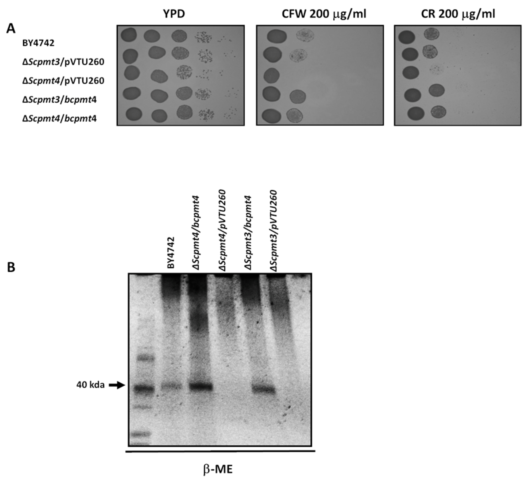
We carried out genetic complementation to find out whether the function of bcpmt4 is similar to that of S. cerevisiae PMT4. The 2307 bp cDNA fragment, containing the entire bcpmt4 coding sequence was cloned into the yeast expression vector pVTU260. The resulting construction and the empty vector were used to transform the S. cerevisiae Δpmt3 and Δpmt4 mutants, which are defective for O-glycosylation [28]. S. cerevisiae Δpmt4 mutant is sensitive to CFW and CR, but the expression of B. cinerea bcpmt4 in this mutant background restored the phenotype to WT levels (Figure 2A).
To confirm these results, cell wall analysis in S. cerevisiae BY4742, pmt3Δ, pmt4Δ, and complemented strains (Scpmt4Δ + bcpmt4, Scpmt3Δ + bcpmt4) were carried out. In the β-ME cell wall extracts, high molecular weight highly polydispersed material of 150–200 kDa was evident in BY4742 strain and complement strains, together a 40 kDa band, except in the null mutants Scpmt3Δ and Scpmt4Δ, indicating a significant decrease in glycosylation of cell wall glycoproteins, but the expression of bcpmt4 in these mutants restores the WT phenotype (Figure 2B). Our findings suggest that B. cinerea bcpmt4 is a functional ortholog of S. cerevisiae PMT3 and PMT4.

3.3. Deletion of B. cinerea bcpmt4 Led to Alterations in Hyphal Wall Composition and Glycosylation Deficiencies

To ascertain whether O-glycosylation plays a role in the B. cinerea cell wall, a simple deletion mutant of putative bcpmt4 genes was obtained. The strategy used was a targeted mutagenesis using the gene replacement method described by Kars, McCalman [43] and B05.10 as WT strain. Later, the protoplasts transformed with a gene replacement construct, containing a hygromycin B selection marker cassette flanked by bcpmt4 sequences was obtained, and a single conidia isolation was used to confirm the targeted deletions (Table S1, Figure S1A). The analyses performed were those described by Aguayo, Riquelme [44], with a single confirmed integration event at the target locus (Figure S1B,C).
The deletion of bcpmt4 from the haploid B05.10 strain showed a significant alteration in its hyphal wall composition. A reduction of 88% in mannan levels in Δbcpmt4 mutant, respectively. Also, chitin and glucan levels increased in the mutant cells (Table 1). Our data are consistent with other studies, showing a drop in mannan and an increase in wall chitin and glucan content [59]. To determine whether mannan changes may be related to the cell wall glycosylation, the cell walls of B. cinerea strains were treated with hot SDS, and then with β-2-mercaptoethanol. The released glycoproteins were separated by SDS–PAGE and detected with Pro-Q Emerald 300 Glycoprotein stain. Figure 3 shows that the mutant showed reduced glycan levels in the glycoprotein when compared to the B05.10 or complemented strains, indicating that the bcpmt4 gene could be involved in O-glycosylation on B. cinerea.

3.4. Cell Wall Defects of the Δbcpmt4 Mutant

It has been demonstrated that S. cerevisiae strains with defects in cell wall biosynthesis are hypersensitive to Calcofluor White (a reagent known to bind to β-1,4-linked polysaccharides, specifically chitin and cellulose), Congo red (a compound that binds to glucan, interfering with proper cell wall construction), caffeine, and Zymolyase (which affects cell wall integrity as a result of the presence of a main β-1,3-glucanase activity and a residual protease activity). These agents interfere with cell wall construction and stress response mechanisms [26,27,60,61,62].
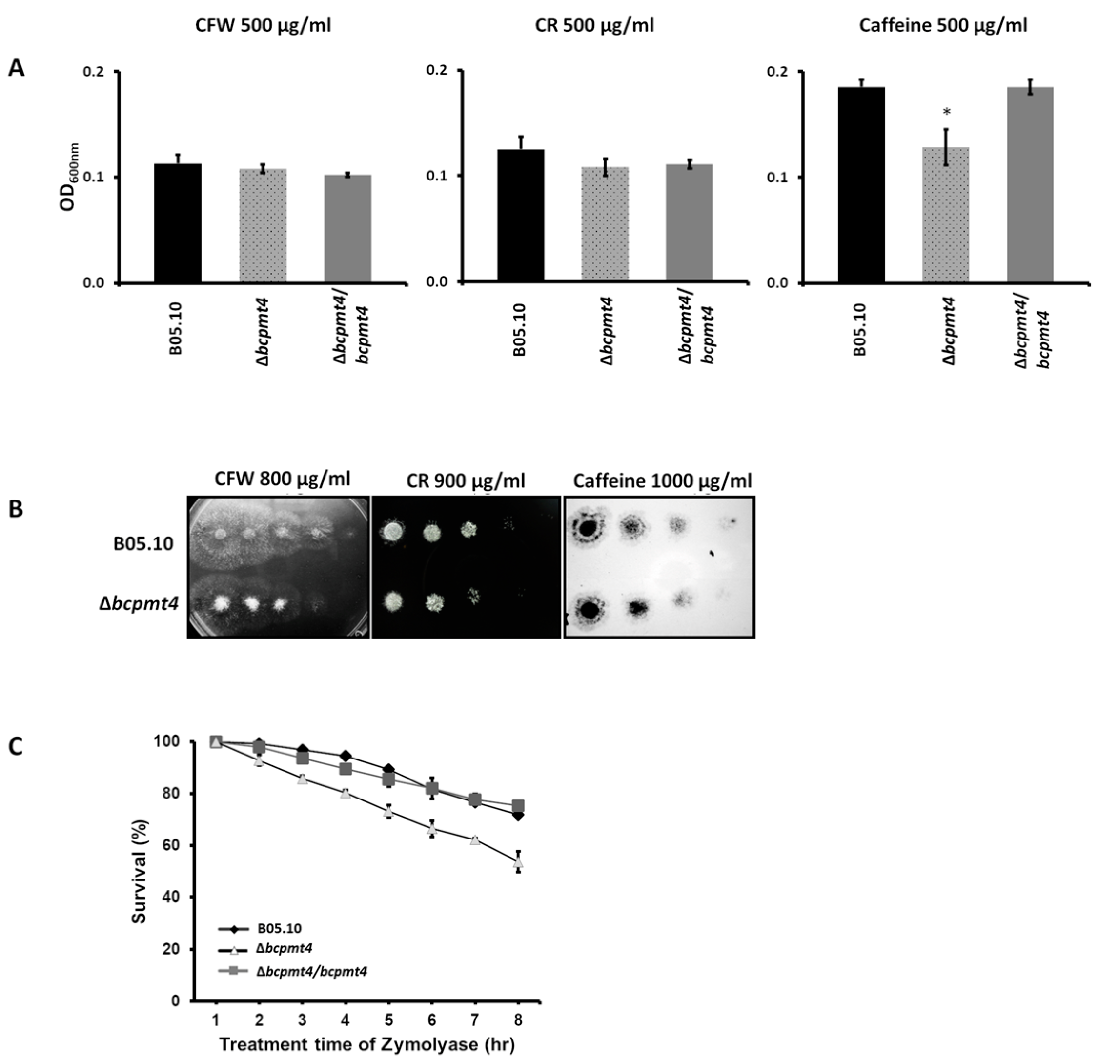
To verify the cell wall defect in the B. cinerea Δpmt4 mutants, the WT and mutants were grown on solid and liquid media containing CFW, CR, or caffein. As shown in Figure 4A, the Δbcpmt4 mutant was sensitive to caffeine when compared to the B05.10 or complemented strains (Figure 4A). In addition, upon treatment with Zymolyase, only 54% Δbcpmt4 conidia survived after 7 h of treatment, whereas the survival of the WT control strain or complemented strains was more than 80% (Figure 4B). These results demonstrate that the Δbcpmt4 mutant is hypersensitive to Zymolyase, indicating a defect in its cell wall structure. These results show that deletion of Δbcpmt4 in B. cinerea causes cell wall defects.

3.5. Membrane Integrity Defects in the Δbcpmt4 Mutant

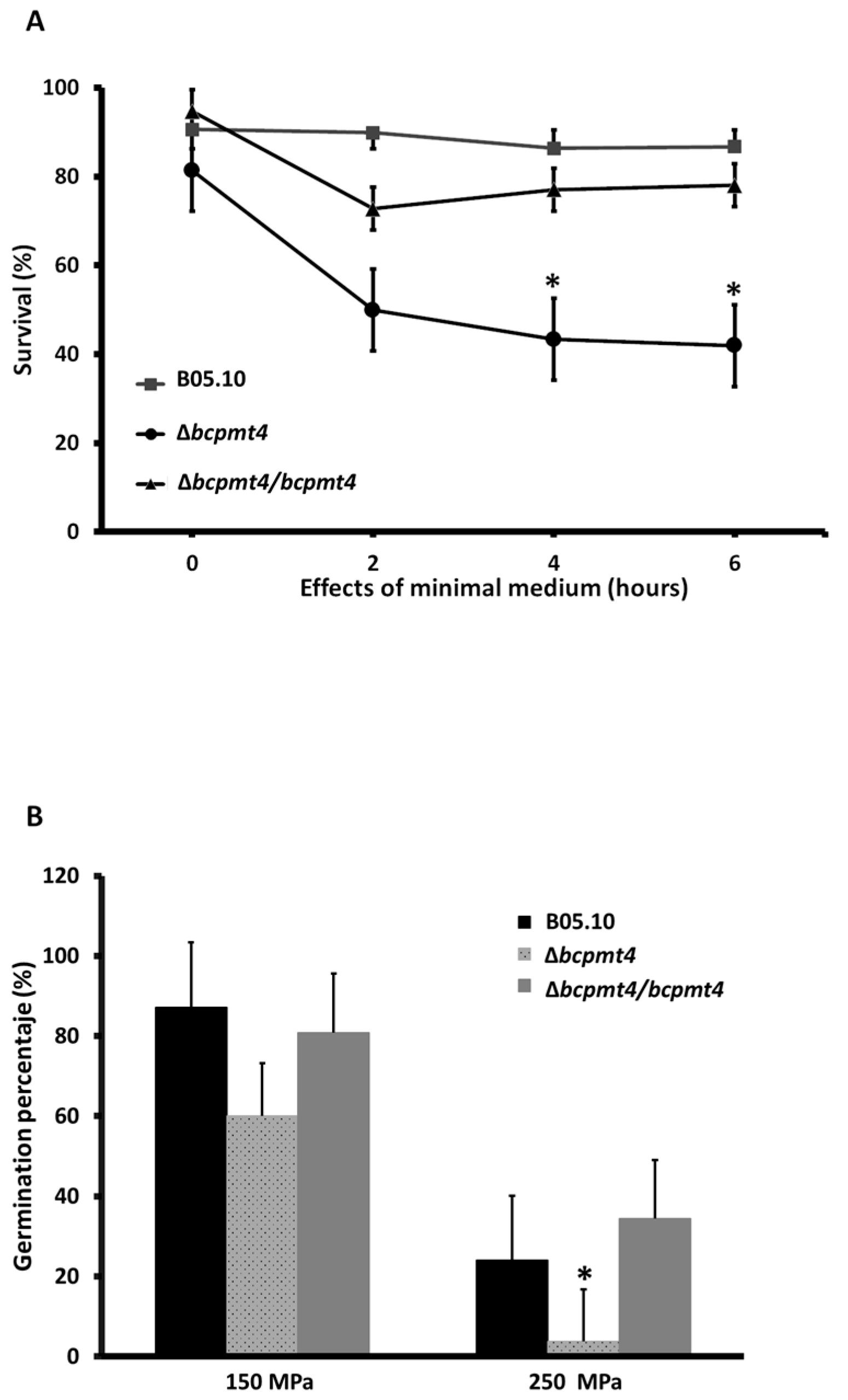
To characterize the Δbcpmt4 mutant phenotypes in more detail, we studied the effect of glycosylation reduction on membrane integrity loss in B. cinerea, by fluorescent staining with propidium iodide (PI) and High hydrostatic pressure (HHP) stress response. Cellular membrane disruption allows PI to enter the cell, where it binds to DNA, and consequently, cells are fluorescent under fluorescence microscopy. According to the data in Figure 5A, the plasmatic membrane of conidia of B. cinerea Δbcpmt4 mutant was markedly damaged, in comparison to the parental strain B05.10 or complemented strains in minimal medium, where the integrity of the membrane declined after 3 h and 4 h of incubation.
HHP exerts effects upon yeast or bacterial cells, interfering with cell membranes or cellular architecture [63,64]. To determine whether glycosylation is involved in membrane stability during HHP stress, the parental and Δbcpmt4 mutant strains were subjected to high-pressure treatments at 100 and 200 MPa. Figure 5B shows the immediate effect of both treatments. From 100 MPa or 200 MPa, lower growth of B. cinerea Δbcpmt4 mutant was observed, compared to the control or complemented strain. These data show that glycosylation is involved in cell membrane stability on HHP stress.

3.6. Deletion of bcpmt4 in B. cinerea Leads to Hypersensitivity to Antifungal Drugs

Fungicides with various modes of action are currently employed to control B. cinerea; however, no information has been associated with genes involved in glycosylation. To assess whether the cell wall defect in the B. cinerea Δpmt4 mutants can change the sensitivity to these fungicides, the strains were incubated with different concentrations of Hydroxyanilides (Fenhexamid), Dicarboxamides (Iprodione), and Succinate Dehydrogenase inhibitors (Boscalid). The results show that the B. cinerea Δpmt4 mutant was highly sensitivity to Iprodione from 2 up to 7 mg/L, compared to the parental strain or complemented strains. A similar result was found in Fenhexamid up to 20 mg/L. However, for Boscalid, a significative difference was only observed at 20 mg/L (Figure 6). These results show a possible correlation between cell wall integrity and hypersensitivity to these antifungal drugs.

3.7. Lack of bcpmt4 Affects Conidia and Biofilm Formation Capacity

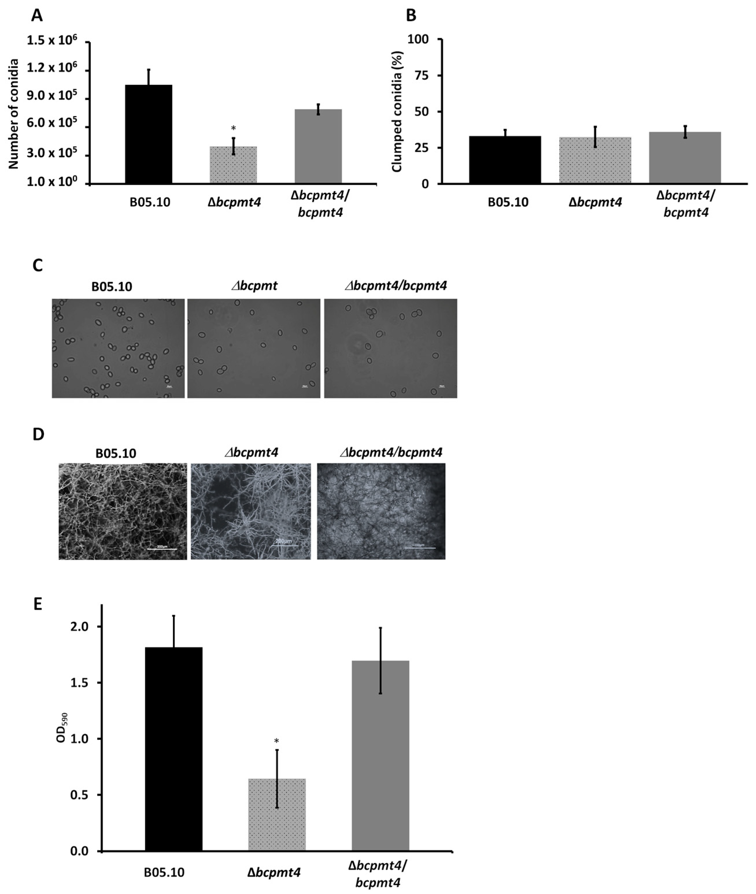
For the assessment of conidia production, Δbcpmt4 mutant and WT Botrytis strains were incubated in MAE at 20 °C under a regular photoperiod for 24 h (12 h light/12 h darkness) to induce conidiation. As shown in Figure 7A,C, the mutant produced viable conidia; but the conidia number was reduced on average by 62% when compared to the WT strain or complemented strains. Complementary to this, the conidia produced by the Δbcpmt4 mutant did not show aggregation (Figure 7B), suggesting that cell surface alterations are not involved in this process.
Regarding biofilm formation, this is considered an important virulence factor for pathogenic fungi. It has been described that yeasts and filamentous fungi can stick to biotic and abiotic surfaces, and the morphological patterns can be affected by environmental parameters. According to the studies carried out in C. albicans mutants, glycosylation is essential for proper biofilm formation [65]. Therefore, we analyzed the biofilm-forming capacity of the mutant generated in this study. The WT and Δbcpmt4 mutant mycelia were growing and analyzed in polystyrene microtiter plates. In the Δbcpmt4 mutant strain, we observed a 66% reduction in biofilm formation when compared to WT or complemented strains (Figure 7D,E). These observations suggested that the deletion of bcpmt4 caused alteration in conidia and biofilm formation capacity.

3.8. O-Glycosylation Is Required for Virulence on Fruit

The glycoproteins in the cell wall play a key role in the host–fungus interaction and the outcome during the infection in C. albicans, U. maydis, B. cinerea, Candida glabrata, C. parapsilosis, and A. fumigatus [10,22,31,34,38,59,66,67]. To confirm the results obtained by Gonzalez, Brito [38], indicating that bcpmt4 is involved in the pathogenicity of B. cinerea, tomato and apple fruits were infected by inoculating 5 μL of 2.5 × 10⁵ conidia/mL of Δbcpmt4 and WT strains. Four days post-inoculation (dpi), disease development was measured as the diameter of the expanding lesions. The deletion of bcpmt4 displayed a statistically significant decrease in radial lesion sizes in comparison with WT in tomato and apple fruits (Figure 8A,B). Based on the findings obtained in this research and the obtained by Gonzalez, Brito [38], it can be suggested that O-glycosylation contributes significantly to the virulence of B. cinerea.

4. Discussion

In S. cerevisiae or C. albicans, the loss of genes involved in glycosylation results in hypersensitivity to the cell wall-perturbing agents CW and CR [28,68]. The phenotypic severity of the mutant suggests that missing carbohydrate chains are required for normal cell growth [69]. The glycoprotein profile in the ΔScpmt4 and ΔScpmt3 mutants released by β-ME reflects changes in glycan content, where a missing protein with molecular weight around 40 kDa was observed, described previously by [20] PIR4. Further, the expression of the B. cinerea bcpmt4 gene into ΔScpmt4 or ΔScpmt3 mutant restores glycan levels in the glycoprotein, with the presence of PIR4, suggesting that this enzyme is involved in O-glycosylation in B. cinerea.
The loss of PMTs in S. cerevisiae, C. albicans, A. fumigatus, and Ustilago maydis cause several defects, such as abnormal morphology and hyphal growth, and cellular aggregation [26,27,61]. The B. cinerea Δbcpmt4 null mutant has characteristic phenotypes associated with disruption of protein mannosylation, including defects in the cell wall composition with reduction in mannan and an increase in the amount of chitin [42,66,70,71,72,73]. The reduction in mannan was confirmed in the glycoprotein releases by β-2-mercaptoethanol in cell wall mutants. An increase in the amount of chitin in the cell wall is often observed in mutants that are more sensitive to CFW and CR, which is viewed as part of a mechanism induced by fungi to compensate for the loss of cell wall integrity [49,59,66,73,74,75]. The increased susceptibility to the cell-wall perturbing agent observed here could be an effect of cell wall damage in the mutant. This has been confirmed by an increased zymolyase sensitivity—a typical response of mutants in S. cerevisiae defect in glucan component—or by a decreased amount of mannoprotein [76,77,78,79]. Similar results were obtained by Gonzalez, Brito [38], using different bcpmt mutants in B. cinerea. However, cell wall alteration may also explain the reduction in the adhesion on polystyrene surfaces by B. cinerea Δbcpmt4 null mutants [80,81]. Doss, Potter [82,83] described that B. cinerea conidia adhere to the host and can secrete a sheath fungal and adhesive material composed, in part, of glucose, protein, and polysaccharide. This mucilage could allow Botrytis to adhere to the tomato cuticle immediately upon conidia hydration. Equally important, a growing number of fungal cell wall proteins have been demonstrated to act as cell surface adhesins and confer binding to diverse ligands, and the majority of fungal adhesins are glycoproteins [80,84,85]. Therefore, glycosylation in B. cinerea seems to be required for fungal binding to polystyrene surfaces or on plant surfaces, thereby playing a pivotal role in pathogenesis [38].
Most glycoproteins are plasma membrane-associated cell wall and secreted proteins and several environmental stresses, such as temperature, ethanol, oxidative, and osmotic stress, as well as HHP, cause alterations in the physical properties of the membrane lipids in the cells [86,87,88]. In S. cerevisiae, HHP induces change in the cell membrane and the gene HSP12 is induced when the cells are exposed to 200 MPa, this gene has been associated with cell wall plasticizer and membrane destabilization [63]. The inactivation of yeast and filamentous fungi like B. cinerea at higher pressures (generally > 250 to 600 MPa) is required, wherein the ascospores are generally inactivated. However, it appears that reduced glycosylation in the B. cinerea mutant was made more susceptible to HHP at 100 or 200 MPa, which could be associated with the alteration of the membrane permeability in this mutant. This is the first time that O-glycosylation is related to HHP stress response in B. cinerea.
Our data suggest that the increased hypersensitivity to Iprodione or Fenhexamid observed in the Δbcpmt4 null mutant could be an associated with cell membrane or cell wall damage in the mutant. It is well known that the Iprodione damages the integrity of the mycelial cell wall and cell membrane in Bipolaris maydis by inhibiting enzymes involved in cell wall production [89]. On the other hand, Fenhexamid inhibits C3-ketoreductase, which is involved in ergosterol biosynthesis [90]. This cell wall or membrane alteration may also explain the reduced adhesion or HHP stress response in the mutant.
The survival of fungal species partly depends on their ability to produce large quantities of viable conidia, a process regulated by genetic, nutritional, and environmental factors. Recent studies have also shown that N-glycosylated proteins play a role during the vegetative growth stage, being involved in nutrient utilization, cell wall biogenesis, and redox processes. Moreover, at the conidium formation stage, the N-glycosylated proteins were involved in glycogen storage, cell wall biogenesis, and at the appressorium formation stage [91], which is consistent with our data showing that deletion of bcpmt4 in B. cinerea caused reduction conidia production. Attenuated virulence has been documented in mutants defective in N- and O-linked glycosylation; coincidently, some mutants have also been affected in adhesion [68,72,81,92]. Furthermore, our results have shown that the B. cinerea Δbcpmt4 mutant had significantly decreased virulence in tomato and apple fruits, with similar observations made by Gonzalez, Brito [38]. This phenotype could be associated with low adhesion and cell wall alteration in the mutants. Defects in glycosylation may compromise protein functions relevant to host–pathogen interactions and virulence [80,85,91].

5. Conclusions

In summary, the B. cinerea bcpmt4 is the functional ortholog of S. cerevisiae PMT4. In addition, the loss of bcpmt4 results in reduced glycosylation of the glycoprotein present in the B. cinerea cell wall, as well as cell wall alterations, which represent a reduction in adhesion and membrane integrity. These phenotypes could be responsible for the reduction in conidia production, hypersensitivity to HHP or synthetic fungicides, and pathogenicity in the B. cinerea mutant. These studies expand the current knowledge about the role of the B. cinerea bcpmt4 in O-glycosylation, cell biology, cell wall formation, and antifungal response.

Supplementary Materials

The following supporting information can be downloaded at: https://www.mdpi.com/article/10.3390/jof11010071/s1, Table S1. Primers used in this study. Table S2. Estimated hygromycin copy number (HCN) was determined by relative quantification qPCR. The mean fold change in expression of the target gene was calculated using ΔΔCT value. Figure S1. Schematic representation of the bcpmt4 disruption event. (A) Schematic representation of the disruption event (B). Diagnostic PCR analysis of genomic DNA isolated from WT and two independent Δbcpmt4 mutants using primer pairs P1fw+P4rv (lane 1–3), Pro-fw+hphint-rev (lane 4), Pro-fw/hphint-rv and Hphin-fw+ter-rv (lane 5 and 6 respectively) (C). RT-PCR analysis using primers P1fw/P2rv (lane 1, 2 and 3) and P3fw/P4rv (lane 4, 5 and 6) of the Δbcpmt4/bcpmt4 strain showed that was able to express bcpmt4 gene. (Primers sequences are listed in Table S1).

Author Contributions

Conceptualization, V.P., E.S.-M. and L.C.; methodology, V.P., A.P., L.A.L.-R., H.M.M.-M., J.R.-A. and L.C.; validation, V.P., A.P., L.A.L.-R., H.M.M.-M., J.R.-A. and L.C.; formal analysis, V.P., E.S.-M., H.M.M.-M. and L.C.; investigation, V.P. and L.C.; resources, L.C.; data curation, V.P.; writing—original draft preparation, V.P. and L.C.; writing—review and editing, V.P., H.M.M.-M. and L.C.; visualization, L.C.; supervision, H.M.M.-M., E.S.-M. and L.C.; project administration, L.C.; funding acquisition, H.M.M.-M. and L.C. All authors have read and agreed to the published version of the manuscript.

Funding

This study was supported by grants from FIC-R 30485880-0 [GORE-Coquimbo], and the Research Department of Universidad de La Serena [DIDULS] PT17122 and PT13121, La Serena, Chile to L.C. H.M.M.-M. is supported by CONAHCYT-Mexico (Ciencia de Frontera 2019-6380 and CBF2023-2024-655).

Institutional Review Board Statement

Not applicable.

Informed Consent Statement

Not applicable.

Data Availability Statement

The original contributions presented in the study are included in the article/Supplementary Material, further inquiries can be directed to the corresponding author.

Acknowledgments

We are grateful to Universidad de La Serena and La Dirección de Investigación y Desarrollo to support for this investigation.

Conflicts of Interest

The authors declare no conflicts of interest.

References

  1. Elad, Y.; Williamson, B.; Tudzynski, P.; Delen, N. Botrytis spp. and diseases they cause in agricultural systems—An introduction. In Botrytis: Biology, Pathology and Control; Kluwer Academic Publishers: Dordrecht, The Netherlands, 2004. [Google Scholar]
  2. Fillinger, S.; Elad, Y. Botrytis—The Fungus, the Pathogen and Its Management in Agricultural Systems; Springer International Publishing: New York, NY, USA, 2016. [Google Scholar]
  3. Jarvis, W. Botryotinia and Botrytis Species: Taxonomy, Physiology and Pathogenicity—A Guide to the Literature; Agriculture Canada: Ottawa, ON, Canada, 1977. [Google Scholar]
  4. Notte, A.-M.; Plaza, V.; Marambio-Alvarado, B.; Olivares-Urbina, L.; Poblete-Morales, M.; Silva-Moreno, E.; Castillo, L. Molecular identification and characterization of Botrytis cinerea associated to the endemic flora of semi-desert climate in Chile. Curr. Res. Microb. Sci. 2021, 2, 100049. [Google Scholar] [CrossRef]
  5. Daryaei, H.; Yousef, A.E.; Balasubramaniam, V.M. Microbiological Aspects of High-Pressure Processing of Food: Inactivation of Microbial Vegetative Cells and Spores. In High Pressure Processing of Food: Principles, Technology and Applications; Balasubramaniam, V.M., Barbosa-Cánovas, G.V., Lelieveld, H.L.M., Eds.; Springer: New York, NY, USA, 2016; pp. 271–294. [Google Scholar]
  6. De Simone, N.; Pace, B.; Grieco, F.; Chimienti, M.; Tyibilika, V.; Santoro, V.; Capozzi, V.; Colelli, G.; Spano, G.; Russo, P. Botrytis cinerea and Table Grapes: A Review of the Main Physical, Chemical, and Bio-Based Control Treatments in Post-Harvest. Foods 2020, 9, 1138. [Google Scholar] [CrossRef] [PubMed]
  7. Mukhopadhyay, S.; Sokorai, K.; Ukuku, D.; Fan, X.; Juneja, V. Effect of high hydrostatic pressure processing on the background microbial loads and quality of cantaloupe puree. Food Res. Int. 2017, 91, 55–62. [Google Scholar] [CrossRef] [PubMed]
  8. Adams, D.J. Fungal cell wall chitinases and glucanases. Microbiology 2004, 150, 2029–2035. [Google Scholar] [CrossRef] [PubMed]
  9. de Boer, A.D.; de Groot, P.W.J.; Weindl, G.; Schaller, M.; Riedel, D.; Diez-Orejas, R.; Klis, F.M.; de Koster, C.G.; Dekker, H.L.; Gross, U.; et al. The Candida albicans cell wall protein Rhd3/Pga29 is abundant in the yeast form and contributes to virulence. Yeast 2010, 27, 611–624. [Google Scholar] [CrossRef]
  10. De Las Penas, A.; Pan, S.J.; Castano, I.; Alder, J.; Cregg, R.; Cormack, B.P. Virulence-related surface glycoproteins in the yeast pathogen Candida glabrata are encoded in subtelomeric clusters and subject to RAP1- and SIR-dependent transcriptional silencing. Genes Dev. 2003, 17, 2245–2258. [Google Scholar] [CrossRef] [PubMed]
  11. Gelis, S.; de Groot, P.W.J.; Castillo, L.; Moragues, M.D.; Sentandreu, R.; Gomez, M.M.; Valentin, E. Pga13 in Candida albicans is localized in the cell wall and influences cell surface properties, morphogenesis and virulence. Fungal Genet. Biol. 2012, 49, 322–331. [Google Scholar] [CrossRef] [PubMed]
  12. Hoyer, L.L.; Green, C.B.; Oh, S.H.; Zhao, X.M. Discovering the secrets of the Candida albicans agglutinin-like sequence (ALS) gene family—A sticky pursuit. Med. Mycol. 2008, 46, 1–15. [Google Scholar] [CrossRef]
  13. Laforet, L.; Moreno, I.; Sanchez-Fresneda, R.; Martinez-Esparza, M.; Martinez, J.P.; Arguelles, J.C.; de Groot, P.W.J.; Valentin-Gomez, E. Pga26 mediates filamentation and biofilm formation and is required for virulence in Candida albicans. FEMS Yeast Res. 2011, 11, 389–397. [Google Scholar] [CrossRef] [PubMed]
  14. Ruiz-Herrera, J.; Elorza, M.V.; Valentin, E.; Sentandreu, R. Molecular organization of the cell wall of Candida albicans and its relation to pathogenicity. FEMS Yeast Res. 2006, 6, 14–29. [Google Scholar] [CrossRef] [PubMed]
  15. Pardo, M.; Monteoliva, L.; Vazquez, P.; Martinez, R.; Molero, G.; Nombela, C.; Gil, C. PST1 and ECM33 encode two yeast cell surface GPI proteins important for cell wall integrity. Microbiology 2004, 150, 4157–4170. [Google Scholar] [CrossRef] [PubMed]
  16. Ruiz-Herrera, J.; Garcia-Maceira, P.; Castillo-Barahona, L.C.; Valentin, E.; Sentandreu, R. Cell wall composition and structure of Yarrowia lipolytica transposon mutants affected in calcofluor sensitivity. Antonie Van Leeuwenhoek 2003, 84, 229–238. [Google Scholar] [CrossRef] [PubMed]
  17. Russo, P.; Kalkkinen, N.; Sareneva, H.; Paakkola, J.; Makarow, M. A heat shock gene from Saccharomyces cerevisiae encoding a secretory glycoprotein. Proc. Natl. Acad. Sci. USA 1992, 89, 3671–3675. [Google Scholar] [CrossRef]
  18. De Groot, P.W.; Hellingwerf, K.J.; Klis, F.M. Genome-wide identification of fungal GPI proteins. Yeast 2003, 20, 781–796. [Google Scholar] [CrossRef] [PubMed]
  19. De Groot, P.W.; Martinez, A.I.; Castillo, L. A Genomic Inventory of Cell Wall Biosynthesis in the Ubiquitous Plant Pathogen Botrytis cinerea. In The Fungal Cell Wall; Mora-Montes, H.M., Ed.; Nova Biomedical: Hauppauge, NY, USA, 2013. [Google Scholar]
  20. Castillo, L.; Martinez, A.I.; Garcera, A.; Elorza, M.V.; Valentin, E.; Sentandreu, R. Functional analysis of the cysteine residues and the repetitive sequence of Saccharomyces cerevisiae Pir4/Cis3: The repetitive sequence is needed for binding to the cell wall beta-1,3-glucan. Yeast 2003, 20, 973–983. [Google Scholar] [CrossRef] [PubMed]
  21. Ecker, M.; Deutzmann, R.; Lehle, L.; Mrsa, V.; Tanner, W. Pir proteins of Saccharomyces cerevisiae are attached to beta-1,3-glucan by a new protein-carbohydrate linkage. J. Biol. Chem. 2006, 281, 11523–11529. [Google Scholar] [CrossRef] [PubMed]
  22. Fernandez-Alvarez, A.; Elias-Villalobos, A.; Ibeas, J.I. The O-Mannosyltransferase PMT4 Is Essential for Normal Appressorium Formation and Penetration in Ustilago maydis. Plant Cell 2009, 21, 3397–3412. [Google Scholar] [CrossRef]
  23. Kriangkripipat, T.; Momany, M. Aspergillus nidulans Protein O-Mannosyltransferases Play Roles in Cell Wall Integrity and Developmental Patterning. Eukaryot. Cell 2009, 8, 1475–1485. [Google Scholar] [CrossRef]
  24. Zhou, H.; Hu, H.Y.; Zhang, L.J.; Li, R.Y.; Ouyang, H.M.; Ming, J.; Jin, C. O-mannosyltransferase 1 in Aspergillus fumigatus (AfPmt1p) is crucial for cell wall integrity and conidium morphology, especially at an elevated temperature. Eukaryot. Cell 2007, 6, 2260–2268. [Google Scholar] [CrossRef]
  25. Mouyna, I.; Kniemeyer, O.; Jank, T.; Loussert, C.; Mellado, E.; Aimanianda, V.; Beauvais, A.; Wartenberg, D.; Sarfati, J.; Bayry, J.; et al. Members of protein O-mannosyltransferase family in Aspergillus fumigatus differentially affect growth, morphogenesis and viability. Mol. Microbiol. 2010, 76, 1205–1221. [Google Scholar] [CrossRef]
  26. Willer, T.; Brandl, M.; Sipiczki, M.; Strahl, S. Protein O-mannosylation is crucial for cell wall integrity, septation and viability in fission yeast. Mol. Microbiol. 2005, 57, 156–170. [Google Scholar] [CrossRef]
  27. Prill, S.K.H.; Klinkert, B.; Timpel, C.; Gale, C.A.; Schroppel, K.; Ernst, J.F. PMT family of Candida albicans: Five protein mannosyltransferase isoforms affect growth, morphogenesis and antifungal resistance. Mol. Microbiol. 2005, 55, 546–560. [Google Scholar] [CrossRef] [PubMed]
  28. Gentzsch, M.; Tanner, W. The PMT gene family: Protein O-glycosylation in Saccharomyces cerevisiae is vital. Embo J. 1996, 15, 5752–5759. [Google Scholar] [CrossRef] [PubMed]
  29. Castillo, L.; Caivo, E.; Martinez, A.I.; Ruiz-Herrera, J.; Valentin, E.; Lopez, J.A.; Sentandreu, R. A study of the Candida albicans cell wall proteome. Proteomics 2008, 8, 3871–3881. [Google Scholar] [CrossRef] [PubMed]
  30. Castillo, L.; Martinez, A.I.; Garcera, A.; Garcia-Martinez, J.; Ruiz-Herrera, J.; Valentin, E.; Sentandreu, R. Genomic response programs of Candida albicans following protoplasting and regeneration. Fungal Genet. Biol. 2006, 43, 124–134. [Google Scholar] [CrossRef]
  31. Olson, G.M.; Fox, D.S.; Wang, P.; Alspaugh, J.A.; Buchanan, K.L. Role of protein O-mannosyltransferase Pmt4 in the morphogenesis and virulence of Cryptococcus neoformans. Eukaryot. Cell 2007, 6, 222–234. [Google Scholar] [CrossRef] [PubMed]
  32. Rouabhia, M.; Schaller, M.; Corbucci, C.; Vecchiarelli, A.; Prill, S.K.; Giasson, L.; Ernst, J.F. Virulence of the fungal pathogen Candida albicans requires the five isoforms of protein mannosyltransferases. Infect. Immun. 2005, 73, 4571–4580. [Google Scholar] [CrossRef]
  33. Willger, S.D.; Ernst, J.F.; Alspaugh, J.A.; Lengeler, K.B. Characterization of the PMT gene family in Cryptococcus neoformans. PLoS ONE 2009, 4, e6321. [Google Scholar] [CrossRef] [PubMed]
  34. Fang, W.; Ding, W.; Wang, B.; Zhou, H.; Ouyang, H.; Ming, J.; Jin, C. Reduced expression of the O-mannosyltransferase 2 (AfPmt2) leads to deficient cell wall and abnormal polarity in Aspergillus fumigatus. Glycobiology 2010, 20, 542–552. [Google Scholar] [CrossRef] [PubMed]
  35. Gorka-Niec, W.; Pniewski, M.; Kania, A.; Perlinska-Lenart, U.; Palamarczyk, G.; Kruszewska, J.S. Disruption of Trichoderma reesei gene encoding protein O-mannosyltransferase I results in a decrease of the enzyme activity and alteration of cell wall composition. Acta Biochim. Pol. 2008, 55, 251–259. [Google Scholar] [CrossRef] [PubMed]
  36. Oka, T.; Hamaguchi, T.; Sameshima, Y.; Goto, M.; Furukawa, K. Molecular characterization of protein O-mannosyltransferase and its involvement in cell-wall synthesis in Aspergillus nidulans. Microbiology 2004, 150, 1973–1982. [Google Scholar] [CrossRef] [PubMed]
  37. Oka, T.; Sameshima, Y.; Koga, T.; Kim, H.; Goto, M.; Furukawa, K. Protein O-mannosyltransferase A of Aspergillus awamori is involved in O-mannosylation of glucoamylase I. Microbiology 2005, 151 Pt 11, 3657–3667. [Google Scholar] [CrossRef] [PubMed]
  38. Gonzalez, M.; Brito, N.; Frias, M.; Gonzalez, C. Botrytis cinerea Protein O-Mannosyltransferases Play Critical Roles in Morphogenesis, Growth, and Virulence. PLoS ONE 2013, 8, e65924. [Google Scholar] [CrossRef] [PubMed]
  39. Buttner, P.; Koch, F.; Voigt, K.; Quidde, T.; Risch, S.; Blaich, R.; Bruckner, B.; Tudzynski, P. Variations in Ploidy among Isolates of Botrytis cinerea—Implications for Genetic and Molecular Analyses. Curr. Genet. 1994, 25, 445–450. [Google Scholar] [CrossRef] [PubMed]
  40. Quidde, T.; Osbourn, A.E.; Tudzynski, P. Detoxification of alpha-tomatine by Botrytis cinerea. Physiol. Mol. Plant Pathol. 1998, 52, 151–165. [Google Scholar] [CrossRef]
  41. Cotoras, M.; Folch, C.; Mendoza, L. Characterization of the antifungal activity on Botrytis cinerea of the natural diterpenoids kaurenoic acid and 3beta-hydroxy-kaurenoic acid. J. Agric. Food Chem. 2004, 52, 2821–2826. [Google Scholar] [CrossRef] [PubMed]
  42. Plaza, V.; Lagues, Y.; Carvajal, M.; Perez-Garcia, L.A.; Mora-Montes, H.M.; Canessa, P.; Larrondo, L.F.; Castillo, L. bcpmr1 encodes a P-type Ca2+/Mn2+-ATPase mediating cell-wall integrity and virulence in the phytopathogen Botrytis cinerea. Fungal Genet. Biol. 2015, 76, 36–46. [Google Scholar] [CrossRef] [PubMed]
  43. Kars, I.; McCalman, M.; Wagemakers, L.; Van Kan, J.A.L. Functional analysis of Botrytis cinerea pectin methylesterase genes by PCR-based targeted mutagenesis: Bcpme1 and Bcpme2 are dispensable for virulence of strain B05.10. Mol. Plant Pathol. 2005, 6, 641–652. [Google Scholar] [CrossRef]
  44. Aguayo, C.; Riquelme, J.; Valenzuela, P.D.T.; Hahn, M.; Silva Moreno, E. Bchex virulence gene of Botrytis cinerea: Characterization and functional analysis. J. Gen. Plant Pathol. 2011, 77, 230–238. [Google Scholar] [CrossRef]
  45. Rasmussen, R. Quantification on the LightCycler. In Rapid Cycle Real-Time PCR: Methods and Applications; Meuer, S., Wittwer, C., Nakagawara, K.-I., Eds.; Springer: Berlin/Heidelberg, Germany, 2001; pp. 21–34. [Google Scholar]
  46. Livak, K.J.; Schmittgen, T.D. Analysis of relative gene expression data using real-time quantitative PCR and the 2−ΔΔCT method. Methods 2001, 25, 402–408. [Google Scholar] [CrossRef] [PubMed]
  47. Zhang, L.S.; Thiewes, H.; van Kan, J.A.L. The D-galacturonic acid catabolic pathway in Botrytis cinerea. Fungal Genet. Biol. 2011, 48, 990–997. [Google Scholar] [CrossRef] [PubMed]
  48. Gietz, R.D.; Schiestl, R.H. High-efficiency yeast transformation using the LiAc/SS carrier DNA/PEG method. Nat. Protoc. 2007, 2, 31–34. [Google Scholar] [CrossRef] [PubMed]
  49. Mora-Montes, H.M.; Bates, S.; Netea, M.G.; Diaz-Jimenez, D.F.; Lopez-Romero, E.; Zinker, S.; Ponce-Norla, P.; Kullberg, B.J.; Brown, A.J.P.; Odds, F.C.; et al. Endoplasmic reticulum alpha-glycosidases of Candida albicans are required for N glycosylation, cell wall integrity, and normal host-fungus interaction. Eukaryot. Cell 2007, 6, 2184–2193. [Google Scholar] [CrossRef]
  50. van der Vaart, J.M.; Caro, L.H.; Chapman, J.W.; Klis, F.M.; Verrips, C.T. Identification of three mannoproteins in the cell wall of Saccharomyces cerevisiae. J. Bacteriol. 1995, 177, 3104–3110. [Google Scholar] [CrossRef] [PubMed]
  51. Yadav, J.; Muend, S.; Zhang, Y.; Rao, R. A phenomics approach in yeast links proton and calcium pump function in the Golgi. Mol. Biol. Cell 2007, 18, 1480–1489. [Google Scholar] [CrossRef] [PubMed]
  52. Doehlemann, G.; Berndt, P.; Hahn, M. Different signalling pathways involving a Galpha protein, cAMP and a MAP kinase control germination of Botrytis cinerea conidia. Mol. Microbiol. 2006, 59, 821–835. [Google Scholar] [CrossRef] [PubMed]
  53. Torres-Ossandon, M.J.; Vega-Galvez, A.; Salas, C.E.; Rubio, J.; Silva-Moreno, E.; Castillo, L. Antifungal activity of proteolytic fraction (P1G10) from (Vasconcellea cundinamarcensis) latex inhibit cell growth and cell wall integrity in Botrytis cinerea. Int. J. Food Microbiol. 2019, 289, 7–16. [Google Scholar] [CrossRef]
  54. Torres-Ossandon, M.J.; Castillo, L.; Uribe, E.; Bilbao-Sainz, C.; Ah-Hen, K.S.; Vega-Galvez, A. Combined Effect of High Hydrostatic Pressure and Proteolytic Fraction P1G10 from Vasconcellea cundinamarcensis Latex against Botrytis cinerea in Grape Juice. Foods 2023, 12, 3400. [Google Scholar] [CrossRef] [PubMed]
  55. Reyes, J.E.; Tabilo-Munizaga, G.; Perez-Won, M.; Maluenda, D.; Roco, T. Effect of high hydrostatic pressure (HHP) treatments on microbiological shelf-life of chilled Chilean jack mackerel (Trachurus murphyi). Innov. Food Sci. Emerg. Technol. 2015, 29, 107–112. [Google Scholar] [CrossRef]
  56. Davis, M.R., Jr.; Goldberg, J.B. Purification and visualization of lipopolysaccharide from Gram-negative bacteria by hot aqueous-phenol extraction. J. Vis. Exp. 2012, 63, 3916. [Google Scholar] [CrossRef]
  57. Strahl-Bolsinger, S.; Scheinost, A. Transmembrane topology of pmt1p, a member of an evolutionarily conserved family of protein O-mannosyltransferases. J. Biol. Chem. 1999, 274, 9068–9075. [Google Scholar] [CrossRef]
  58. Krogh, A.; Larsson, B.; von Heijne, G.; Sonnhammer, E.L. Predicting transmembrane protein topology with a hidden Markov model: Application to complete genomes. J. Mol. Biol. 2001, 305, 567–580. [Google Scholar] [CrossRef]
  59. Bates, S.; Hughes, H.B.; Munro, C.A.; Thomas, W.P.H.; MacCallum, D.M.; Bertram, G.; Atrih, A.; Ferguson, M.A.J.; Brown, A.J.P.; Odds, F.C.; et al. Outer chain N-glycans are required for cell wall integrity and virulence of Candida albicans. J. Biol. Chem. 2006, 281, 90–98. [Google Scholar] [CrossRef] [PubMed]
  60. Arroyo, J.; Bermejo, C.; Garcia, R.; Rodriguez-Pena, J.M. Genomics in the detection of damage in microbial systems: Cell wall stress in yeast. Clin. Microbiol. Infect. 2009, 15 (Suppl. 1), 44–46. [Google Scholar] [CrossRef] [PubMed]
  61. Proszynski, T.J.; Simons, K.; Bagnat, M. O-glycosylation as a sorting determinant for cell surface delivery in yeast. Mol. Biol. Cell 2004, 15, 1533–1543. [Google Scholar] [CrossRef]
  62. Ram, A.F.; Wolters, A.; Ten Hoopen, R.; Klis, F.M. A new approach for isolating cell wall mutants in Saccharomyces cerevisiae by screening for hypersensitivity to calcofluor white. Yeast 1994, 10, 1019–1030. [Google Scholar] [CrossRef]
  63. Bravim, F.; de Freitas, J.M.; Fernandes, A.A.; Fernandes, P.M. High hydrostatic pressure and the cell membrane: Stress response of Saccharomyces cerevisiae. Ann. N. Y. Acad. Sci. 2010, 1189, 127–132. [Google Scholar] [CrossRef] [PubMed]
  64. Prieto-Calvo, M.; Prieto, M.; Lopez, M.; Alvarez-Ordonez, A. Effects of high hydrostatic pressure on Escherichia coli ultrastructure, membrane integrity and molecular composition as assessed by FTIR spectroscopy and microscopic imaging techniques. Molecules 2014, 19, 21310–21323. [Google Scholar] [CrossRef] [PubMed]
  65. Peltroche-Llacsahuanga, H.; Goyard, S.; d’Enfert, C.; Prill, S.K.; Ernst, J.F. Protein O-mannosyltransferase isoforms regulate biofilm formation in Candida albicans. Antimicrob. Agents Chemother. 2006, 50, 3488–3491. [Google Scholar] [CrossRef] [PubMed]
  66. Bates, S.; Hall, R.A.; Cheetham, J.; Netea, M.G.; MacCallum, D.M.; Brown, A.J.; Odds, F.C.; Gow, N.A. Role of the Candida albicans MNN1 gene family in cell wall structure and virulence. BMC Res. Notes 2013, 6, 294. [Google Scholar] [CrossRef] [PubMed]
  67. Pinchai, N.; Juvvadi, P.R.; Fortwendel, J.R.; Perfect, B.Z.; Rogg, L.E.; Asfaw, Y.G.; Steinbach, W.J. The Aspergillus fumigatus P-Type Golgi Apparatus Ca2+/Mn2+ ATPase PmrA Is Involved in Cation Homeostasis and Cell Wall Integrity but Is Not Essential for Pathogenesis. Eukaryot. Cell 2010, 9, 472–476. [Google Scholar] [CrossRef]
  68. Bates, S.; MacCallum, D.M.; Bertram, G.; Munro, C.A.; Hughes, H.B.; Buurman, E.T.; Brown, A.J.P.; Odds, F.C.; Gow, N.A.R. Candida albicans Pmr1p, a secretory pathway P-type Ca2+/Mn2+-ATPase, is required for glycosylation and virulence. J. Biol. Chem. 2005, 280, 23408–23415. [Google Scholar] [CrossRef] [PubMed]
  69. Garcia-Rodriguez, N.; Manzano-Lopez, J.; Munoz-Bravo, M.; Fernandez-Garcia, E.; Muniz, M.; Wellinger, R.E. Manganese redistribution by calcium-stimulated vesicle trafficking bypasses the need for P-type ATPase function. J. Biol. Chem. 2015, 290, 9335–9347. [Google Scholar] [CrossRef] [PubMed]
  70. Cortes, J.C.; Katoh-Fukui, R.; Moto, K.; Ribas, J.C.; Ishiguro, J. Schizosaccharomyces pombe Pmr1p is essential for cell wall integrity and is required for polarized cell growth and cytokinesis. Eukaryot. Cell 2004, 3, 1124–1135. [Google Scholar] [CrossRef] [PubMed]
  71. Fujikawa, T.; Kuga, Y.; Yano, S.; Yoshimi, A.; Tachiki, T.; Abe, K.; Nishimura, M. Dynamics of cell wall components of Magnaporthe grisea during infectious structure development. Mol. Microbiol. 2009, 73, 553–570. [Google Scholar] [CrossRef] [PubMed]
  72. Navarro-Arias, M.J.; Defosse, T.A.; Dementhon, K.; Csonka, K.; Mellado-Mojica, E.; Dias Valerio, A.; Gonzalez-Hernandez, R.J.; Courdavault, V.; Clastre, M.; Hernandez, N.V.; et al. Disruption of Protein Mannosylation Affects Candida guilliermondii Cell Wall, Immune Sensing, and Virulence. Front. Microbiol. 2016, 7, 1951. [Google Scholar] [CrossRef]
  73. Popolo, L.; Gilardelli, D.; Bonfante, P.; Vai, M. Increase in chitin as an essential response to defects in assembly of cell wall polymers in the ggp1delta mutant of Saccharomyces cerevisiae. J. Bacteriol. 1997, 179, 463–469. [Google Scholar] [CrossRef] [PubMed]
  74. Kapteyn, J.C.; Ram, A.F.; Groos, E.M.; Kollar, R.; Montijn, R.C.; Van Den Ende, H.; Llobell, A.; Cabib, E.; Klis, F.M. Altered extent of cross-linking of beta1,6-glucosylated mannoproteins to chitin in Saccharomyces cerevisiae mutants with reduced cell wall beta1,3-glucan content. J. Bacteriol. 1997, 179, 6279–6284. [Google Scholar] [CrossRef]
  75. Mora-Montes, H.M.; Ponce-Noyola, P.; Villagomez-Castro, J.C.; Gow, N.A.; Flores-Carreon, A.; Lopez-Romero, E. Protein glycosylation in Candida. Future Microbiol. 2009, 4, 1167–1183. [Google Scholar] [CrossRef]
  76. Agaphonov, M.O.; Packeiser, A.N.; Chechenova, M.B.; Choi, E.S.; Ter-Avanesyan, M.D. Mutation of the homologue of GDP-mannose pyrophosphorylase alters cell wall structure, protein glycosylation and secretion in Hansenula polymorpha. Yeast 2001, 18, 391–402. [Google Scholar] [CrossRef] [PubMed]
  77. Boone, C.; Sommer, S.S.; Hensel, A.; Bussey, H. Yeast KRE genes provide evidence for a pathway of cell wall beta-glucan assembly. J. Cell Biol. 1990, 110, 1833–1843. [Google Scholar] [CrossRef] [PubMed]
  78. Brown, J.L.; Bussey, H. The yeast KRE9 gene encodes an O glycoprotein involved in cell surface beta-glucan assembly. Mol. Cell. Biol. 1993, 13, 6346–6356. [Google Scholar] [CrossRef]
  79. de Nobel, J.G.; Klis, F.M.; Priem, J.; Munnik, T.; van den Ende, H. The glucanase-soluble mannoproteins limit cell wall porosity in Saccharomyces cerevisiae. Yeast 1990, 6, 491–499. [Google Scholar] [CrossRef]
  80. Lipke, P.N. What We Do Not Know about Fungal Cell Adhesion Molecules. J. Fungi 2018, 4, 59. [Google Scholar] [CrossRef]
  81. Munro, C.A.; Bates, S.; Buurman, E.T.; Hughes, H.B.; MacCallum, D.M.; Bertram, G.; Atrih, A.; Ferguson, M.A.J.; Bain, J.M.; Brand, A.; et al. Mnt1p and Mnt2p of Candida albicans are partially redundant alpha-1,2-mannosyltransferases that participate in O-linked mannosylation and are required for adhesion and virulence. J. Biol. Chem. 2005, 280, 1051–1060. [Google Scholar] [CrossRef]
  82. Doss, R.P.; Potter, S.W.; Chastagner, G.A.; Christian, J.K. Adhesion of Nongerminated Botrytis cinerea Conidia to Several Substrata. Appl. Environ. Microbiol. 1993, 59, 1786–1791. [Google Scholar] [CrossRef] [PubMed]
  83. Doss, R.P.; Potter, S.W.; Soeldner, A.H.; Christian, J.K.; Fukunaga, L.E. Adhesion of germlings of Botrytis cinerea. Appl. Environ. Microbiol. 1995, 61, 260–265. [Google Scholar] [CrossRef]
  84. de Groot, P.W.; Bader, O.; de Boer, A.D.; Weig, M.; Chauhan, N. Adhesins in human fungal pathogens: Glue with plenty of stick. Eukaryot. Cell 2013, 12, 470–481. [Google Scholar] [CrossRef]
  85. Dranginis, A.M.; Rauceo, J.M.; Coronado, J.E.; Lipke, P.N. A biochemical guide to yeast adhesins: Glycoproteins for social and antisocial occasions. Microbiol. Mol. Biol. Rev. 2007, 71, 282–294. [Google Scholar] [CrossRef]
  86. Los, D.A.; Murata, N. Membrane fluidity and its roles in the perception of environmental signals. Biochim. Biophys. Acta 2004, 1666, 142–157. [Google Scholar] [CrossRef] [PubMed]
  87. Marin-Menguiano, M.; Moreno-Sanchez, I.; Barrales, R.R.; Fernandez-Alvarez, A.; Ibeas, J.I. N-glycosylation of the protein disulfide isomerase Pdi1 ensures full Ustilago maydis virulence. PLoS Pathog. 2019, 15, e1007687. [Google Scholar] [CrossRef] [PubMed]
  88. Scarlata, S. The effect of hydrostatic pressure on membrane-bound proteins. Braz. J. Med. Biol. Res. 2005, 38, 1203–1208. [Google Scholar] [CrossRef]
  89. Sun, J.Z.; Pang, C.Y.; Cheng, X.; Yang, B.Y.; Jin, B.B.; Jin, L.; Qi, Y.X.; Sun, Y.; Chen, X.; Liu, W.D.; et al. Investigation of the antifungal activity of the dicarboximide fungicide iprodione against Bipolaris maydis. Pestic. Biochem. Phys. 2023, 190, 105319. [Google Scholar] [CrossRef]
  90. Fillinger, S.; Leroux, P.; Auclair, C.; Barreau, C.; Al Hajj, C.; Debieu, D. Genetic analysis of fenhexamid-resistant field isolates of the phytopathogenic fungus Botrytis cinerea. Antimicrob. Agents Chemother. 2008, 52, 3933–3940. [Google Scholar] [CrossRef] [PubMed]
  91. Chen, X.L.; Liu, C.; Tang, B.; Ren, Z.; Wang, G.L.; Liu, W. Quantitative proteomics analysis reveals important roles of N-glycosylation on ER quality control system for development and pathogenesis in Magnaporthe oryzae. PLoS Pathog. 2020, 16, e1008355. [Google Scholar] [CrossRef] [PubMed]
  92. Perez-Garcia, L.A.; Csonka, K.; Flores-Carreon, A.; Estrada-Mata, E.; Mellado-Mojica, E.; Nemeth, T.; Lopez-Ramirez, L.A.; Toth, R.; Lopez, M.G.; Vizler, C.; et al. Role of Protein Glycosylation in Candida parapsilosis Cell Wall Integrity and Host Interaction. Front. Microbiol. 2016, 7, 306. [Google Scholar] [CrossRef] [PubMed]
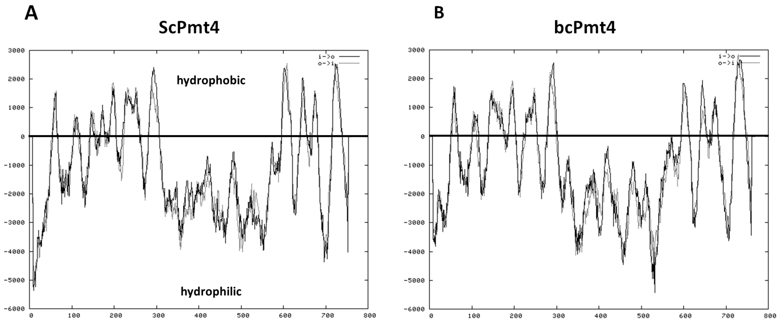
Figure 1. Scheme of transmembrane domains predicted in S. cerevisiae and B. cinerea Pmt4. The predictions of transmembrane segments were performed using the TMPRED programs. (A,B) represent the hydropathy profiles of ScPmt4 and bcPmt4, respectively. Hydropathy analysis of B. cinerea Pmt4 showed a similar profile compared to the S. cerevisiae protein.
Jof 11 00071 g001
Figure 2. B. cinerea bcpmt4 encodes a functional ortholog of S. cerevisiae PMT4. (A) Spot assays showing sensitivities of a WT S. cerevisiae strain (BY4742), Δpmt3 and Δpmt4 mutants transformed with empty pVTU260 plasmid (Y0R321W/pVTU260 and YJR143C/pVTU260), Δpmt3 and Δpmt4 mutants transformed with pVTU260/bcpmt4 cDNA (Y0R321W/bcpmt4 and YJR143C/bcpmt4). Control (YPD without drugs), CFW 200 μg/mL (YPD with 200 μg/mL of Calcofluor white), and CR 200 μg/mL (YPD with 200 μg/mL of Congo red). (B) Analysis by SDS–PAGE of the materials extracted by β-2-mercaptoethanol (β-ME) from the cell walls of S. cerevisiae pmt3Δ and pmt4Δ mutants and complemented strains. Gel stained with Pro-Q Emerald 300 Glycoprotein stain.
Jof 11 00071 g002
Figure 3. Analysis by SDS–PAGE of the glycoproteins present in B. cinerea strains. Glycoproteins extracted by β-2-mercaptoethanol from the cell walls of B. cinerea B05.10, Δbcpmt4 and complemented strain were electrophoresed at a constant current. Gel stained with Pro-Q Emerald 300 Glycoprotein stain.
Jof 11 00071 g003
Figure 4. The B. cinerea O-glycosylation mutant is hypersensitive to cell wall perturbing agents and Zymolyase. (A) Sensitivity to CFW, CR, or caffeine of the B05.10 WT, Δbcpmt4 mutant, and the complemented strains (Δbcpmt4 + bcpmt4). Cells were incubated in malt extract in the presence or absence of 500 µg/mL CFW, CR, or caffeine for 72 h at 20 °C. (B) Sensitivity to CFW, CR, or caffeine of the B05.10 WT strain and mutants on agar plates. (C) Zymolyase sensitivity. Conidia (2.5 × 10⁵ conidia/mL) of the B05.10 WT strain, mutants, and the complemented strain were incubated with 200 μg/mL Zymolyase 20T at 25 °C, and the decrease in OD590 nm was monitored over time (represented as conidia surviving the treatment; survival %). The data represents the mean ± SD from three independent experiments. The asterisk indicates significant differences (p < 0.05, Mann–Whitney U test) compared with the parental strain (* WT vs. Δbcpmt4).
Jof 11 00071 g004
Figure 5. Glycosylation is important for membrane integrity. (A) Effects of the minimal medium on plasma membrane integrity of conidia of B. cinerea strains. (B) Survival percentage of B. cinerea after treatment of high hydrostatic pressure (150 MPa/4 min, 250 MPa/4 min) in solid media incubation for 48 h at 20 °C. The asterisk indicates significant differences (p < 0.05, Mann–Whitney U test) compared with the parental strain (* WT vs. Δbcpmt4).
Jof 11 00071 g005
Figure 6. The bcpmt4 mutants display sensitivity to synthetic fungicides. The sensitivity of the B05.10 WT, Δbcpmt4, and complemented strains was determined quantitatively by the broth dilution method. The agents to which the Δbcpmt4 mutant demonstrated hypersensitivity are shown (Boscalid, Iprodione, and Fenexamid). The asterisk indicates significant differences (p < 0.05, Mann–Whitney U test) compared with the parental strain (* WT vs. Δbcpmt4).
Jof 11 00071 g006
Figure 7. Deletion of bcpmt4 affects conidia and biofilm formation. (A) Quantification of conidia production in the B05.10 WT, Δbcpmt4, and complemented strains grown for two weeks on MEA at 20 °C under a 24 h photoperiod (12 h light/12 h darkness). (B) Quantification of conidial clumping. Percentages of conidia present in groups of two or more clumped conidia together were counted. Bars represent the mean ± SD. (C) Light microscopic (40×) examination of conidial suspensions indicated that Δbcpmt4 conidia did not exhibit clumping (the white line represent 50 mm). (D) Biofilm formation of the B05.10 WT, Δbcpmt4, and complemented strains on polystyrene after incubation for 72 h at 20 °C. Bound mycelia were stained with Crystal violet and visualized by fluorescence microscopy. (E) Quantification of biofilm formation by measuring OD590. Bars represent the mean ± SD of three independent experiments measured in triplicate. The asterisk indicates significant differences (p < 0.05, Mann–Whitney U test) compared with the parental strain (* WT vs. Δbcpmt4).
Jof 11 00071 g007
Figure 8. Virulence of the B. cinerea Δbcpmt4 in comparison to the wild-type B05.10. (A,B) The tissues (apple or tomato) were wounded with a pinprick, followed by the application of 5 µL droplets (containing 2.5 × 10⁵ conidia/mL) for inoculation. Lesion sizes were measured at 4 days post-inoculation (dpi) at 20 °C. Bars represent the mean lesion diameter ± SD. The asterisk indicates significant differences (p < 0.05, Mann–Whitney U test) compared to the parental strain (* WT vs. Δbcpmt4).
Jof 11 00071 g008
Table 1. Chemical analysis of mutant cell wall.
StrainGlucoseMannoseGlucosamine
B05.100.469 ± 0.03 (46.9 ± 3%) *0.340 ± 0.01 (34.0 ± 1%) *0.007 ± 0.00 (7.0%) *
Δbcpmt40.771 ± 0.04 (77.1 ± 4%) *0.039 ± 0.06 (39.0 ± 6%) *0.035 ± 0.02 (3.5%) *
The values are measured in mg/mg of cell wall dry weight ± S.D. (n = 3). * Data are shown as percentage of cell wall dry weight.
Disclaimer/Publisher’s Note: The statements, opinions and data contained in all publications are solely those of the individual author(s) and contributor(s) and not of MDPI and/or the editor(s). MDPI and/or the editor(s) disclaim responsibility for any injury to people or property resulting from any ideas, methods, instructions or products referred to in the content.