Molecular Phylogeny and Morphology Reveal Four New Species of Conocybe (Bolbitiaceae, Agaricales) from the Qinghai-Xizang Plateau, China

 , , ,
, , ,  ,
, 
Abstract
1. Introduction
2. Materials and Methods
2.1. Samplings and Morphological Study
2.2. DNA Extraction, PCR and Sequencing
2.3. Phylogenetic Analyses
3. Results
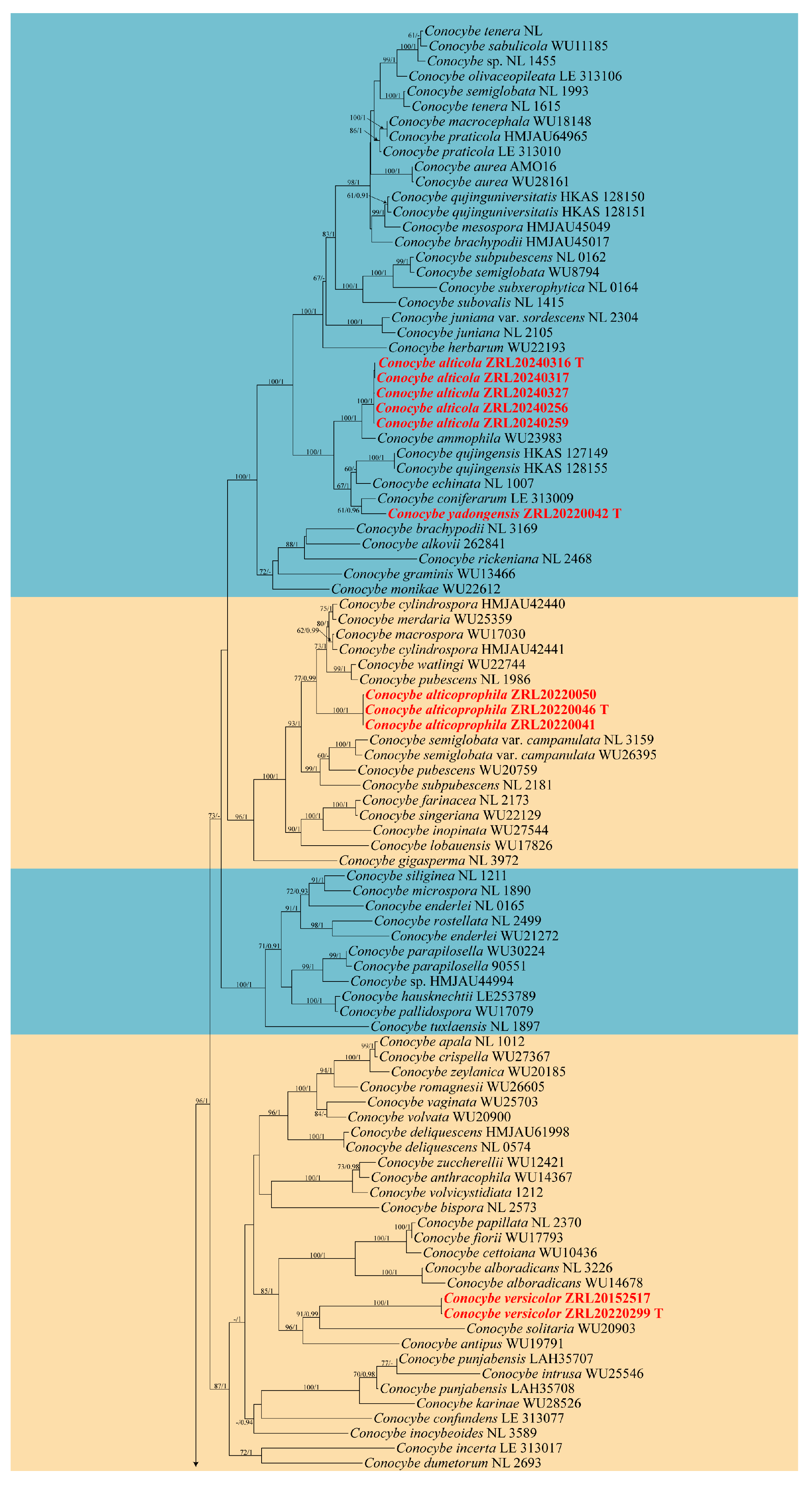
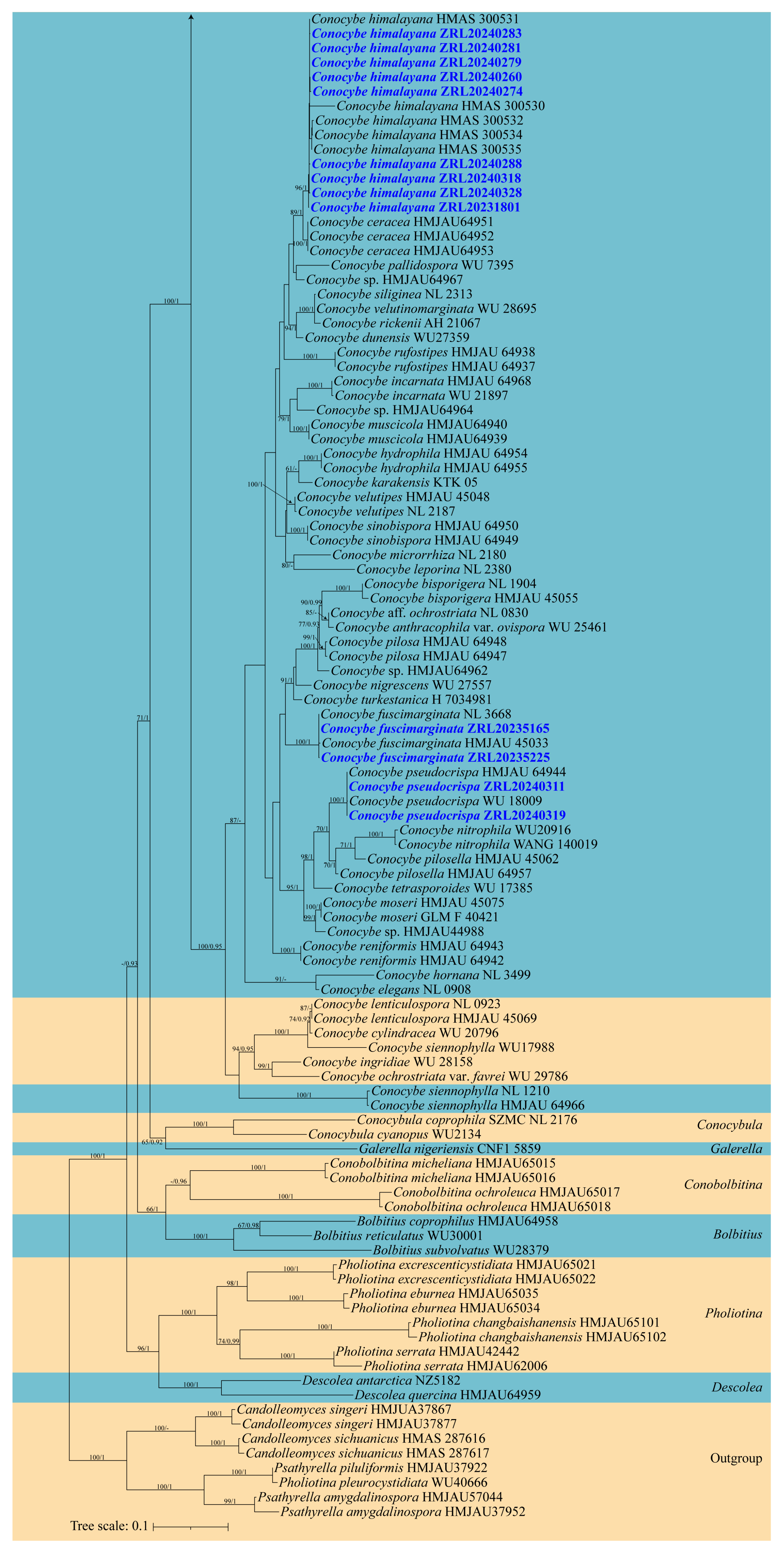
3.1. Phylogeny
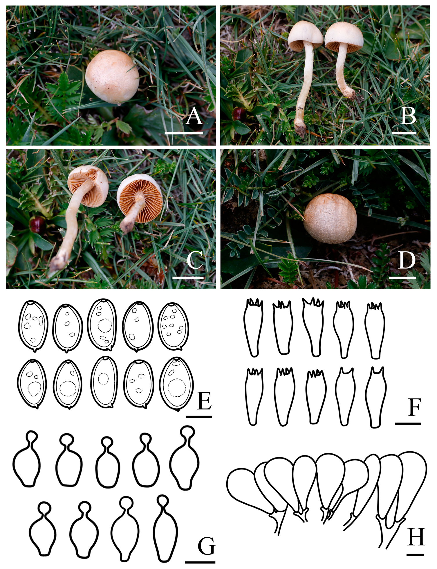
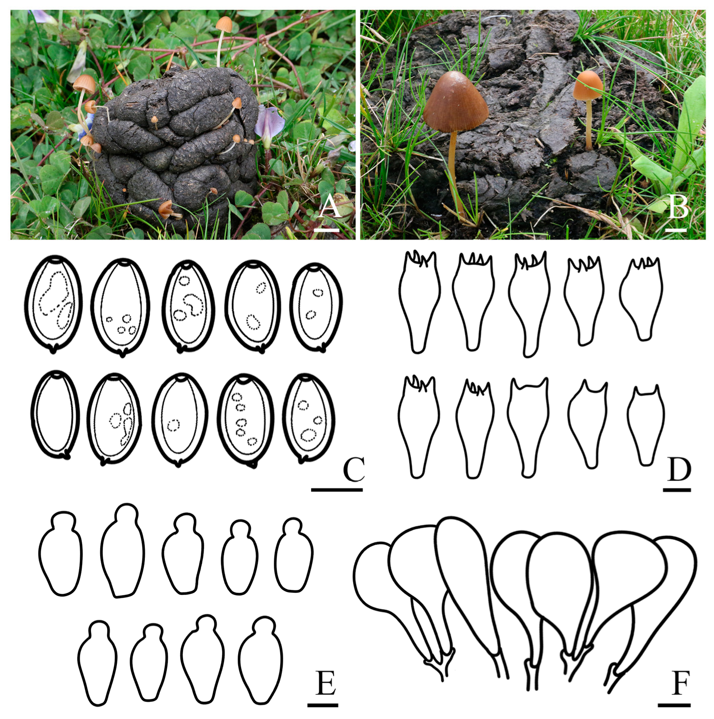
3.2. Taxonomy
4. Discussion
Author Contributions
Funding
Institutional Review Board Statement
Informed Consent Statement
Data Availability Statement
Acknowledgments
Conflicts of Interest
References
- Zhu, Q.; Chen, H.; Peng, C.; Liu, J.; Piao, S.; He, J.-S.; Wang, S.; Zhao, X.; Zhang, J.; Fang, X. An early warning signal for grassland degradation on the Qinghai-Tibetan Plateau. Nat. Commun. 2023, 14, 6406. [Google Scholar] [CrossRef]
- Mao, K.S.; Wang, Y.; Liu, J.Q. Evolutionary origin of species diversity on the Qinghai–Tibet Plateau. J. Syst. Evol. 2021, 59, 1142–1158. [Google Scholar] [CrossRef]
- Renner, S.S. Available data point to a 4-km-high Tibetan Plateau by 40 Ma, but 100 molecular-clock papers have linked supposed recent uplift to young node ages. J. Biogeogr. 2016, 43, 1479–1487. [Google Scholar] [CrossRef]
- Yang, M.X.; Werth, S.; Wang, L.S.; Scheidegger, C. Phylogeographic analyses of an epiphytic foliose lichen show multiple dispersal events westward from the Hengduan Mountains of Yunnan into the Himalayas. Ecol. Evol. 2022, 12, e9308. [Google Scholar] [CrossRef] [PubMed]
- Yu, H.; Deane, D.C.; Sui, X.; Fang, S.; Chu, C.; Liu, Y.; He, F. Testing multiple hypotheses for the high endemic plant diversity of the Tibetan Plateau. Glob. Ecol. Biogeogr. 2019, 28, 131–144. [Google Scholar] [CrossRef]
- Ding, L.; Kapp, P.; Cai, F.; Garzione, C.N.; Xiong, Z.; Wang, H.; Wang, C. Timing and mechanisms of Tibetan Plateau uplift. Nat. Rev. Earth Environ. 2022, 3, 652–667. [Google Scholar] [CrossRef]
- Deng, T.; Wu, F.; Zhou, Z.; Su, T. Tibetan Plateau: An evolutionary junction for the history of modern biodiversity. Sci. China Earth Sci. 2020, 63, 172–187. [Google Scholar] [CrossRef]
- Liu, H.; Wang, W.; Song, G.; Qu, Y.; Li, S.-H.; Fjeldså, J.; Lei, F. Interpreting the process behind endemism in China by integrating the phylogeography and ecological niche models of the Stachyridopsis ruficeps. PLoS ONE 2012, 7, e46761. [Google Scholar] [CrossRef]
- Yang, T.; Adams, J.M.; Shi, Y.; He, J.S.; Jing, X.; Chen, L.; Tedersoo, L.; Chu, H. Soil fungal diversity in natural grasslands of the Tibetan Plateau: Associations with plant diversity and productivity. New Phytol. 2017, 215, 756–765. [Google Scholar] [CrossRef] [PubMed]
- Zhang, T.; Wang, N.; Yu, L. Soil fungal community composition differs significantly among the Antarctic, Arctic, and Tibetan Plateau. Extremophiles 2020, 24, 821–829. [Google Scholar] [CrossRef]
- Li, Y.; Zhou, Y.; Liu, F.; Liu, X.; Wang, Q. Diversity patterns of wetland angiosperms in the Qinghai-Tibet Plateau, China. Diversity 2022, 14, 777. [Google Scholar] [CrossRef]
- Li, Q.; He, G.; Wen, T.; Zhang, D.; Liu, X. Distribution pattern of soil fungi community diversity in alpine meadow in Qilian Mountains of eastern Qinghai-Tibetan Plateau. Ecol. Indic. 2022, 141, 109054. [Google Scholar] [CrossRef]
- Phurbu, D.; Huang, J.-E.; Song, S.; Ni, Z.; Zhou, X.; Li, S.; Cai, L.; Liu, F. Diversity of culturable fungi in six Tibetan Plateau lakes, with descriptions of eight new taxa. Mycology 2024, 1–20. [Google Scholar] [CrossRef]
- Yang, N.; Li, X.; Liu, D.; Zhang, Y.; Chen, Y.; Wang, B.; Hua, J.; Zhang, J.; Peng, S.; Ge, Z. Diversity patterns and drivers of soil bacterial and fungal communities along elevational gradients in the Southern Himalayas, China. Appl. Soil Ecol. 2022, 178, 104563. [Google Scholar] [CrossRef]
- Han, X.; Liu, D.; Zhang, M.; He, M.; Li, J.; Zhu, X.; Wang, M.; Thongklang, N.; Zhao, R.; Cao, B. Macrofungal diversity and distribution patterns in the primary forests of the shaluli mountains. J. Fungi 2023, 9, 491. [Google Scholar] [CrossRef]
- Wang, K.; Liu, S.-L.; Liu, X.-Z.; Hong, P.; Wei, H.-W.; Wang, Y.; Phurbu, D.; Zhou, L.-W.; Wei, T.-Z. Catalogue of fungi in China 3. New taxa of macrofungi from southern Xizang, China. Mycology 2024, 1–33. [Google Scholar] [CrossRef]
- He, M.-Q.; Cao, B.; Liu, F.; Boekhout, T.; Denchev, T.T.; Schoutteten, N.; Denchev, C.M.; Kemler, M.; Gorjón, S.P.; Begerow, D. Phylogenomics, divergence times and notes of orders in Basidiomycota. Fungal Divers. 2024, 126, 127–406. [Google Scholar] [CrossRef]
- Song, H.-B.; Bau, T. Conocybe section Pilosellae in China: Reconciliation of taxonomy and phylogeny reveals seven new species and a new record. J. Fungi 2023, 9, 924. [Google Scholar] [CrossRef] [PubMed]
- Liu, J.; Bau, T. New species and new records in the genus Conocybe (Bolbitaceae) from China. Phytotaxa 2018, 357, 261–274. [Google Scholar] [CrossRef]
- Amandeep, K.; Atri, N.; Munruchi, K. Diversity of species of the genus Conocybe (Bolbitiaceae, Agaricales) collected on dung from Punjab, India. Mycosphere 2015, 6, 19–42. [Google Scholar] [CrossRef]
- Tóth, A.; Hausknecht, A.; Krisai-Greilhuber, I.; Papp, T.; Vágvölgyi, C.; Nagy, L.G. Iteratively refined guide trees help improving alignment and phylogenetic inference in the mushroom family Bolbitiaceae. PLoS ONE 2013, 8, e56143. [Google Scholar] [CrossRef]
- Malysheva, E.F. Five new species of Conocybe (Agaricomycetes, Bolbitiaceae) from Russia. Mycol. Prog. 2017, 16, 625–636. [Google Scholar] [CrossRef]
- He, M.Q.; Wang, M.Q.; Chen, Z.H.; Deng, W.Q.; Li, T.H.; Vizzini, A.; Jeewon, R.; Hyde, K.D.; Zhao, R.L. Potential benefits and harms: A review of poisonous mushrooms in the world. Fungal Biol. Rev. 2022, 42, 56–68. [Google Scholar] [CrossRef]
- Pu, C.-J.; Peng, Y.-L.; Li, Z.-H.; He, J.; Huang, R.; Feng, T.; Liu, J.-K. Two highly oxygenated ergosterols from cultures of the basidiomycete Conocybe siliginea. Nat. Prod. Res. 2019, 33, 3037–3043. [Google Scholar] [CrossRef] [PubMed]
- Luo, H.; Hallen-Adams, H.E.; Walton, J.D. Processing of the phalloidin proprotein by prolyl oligopeptidase from the mushroom Conocybe albipes. J. Biol. Chem. 2009, 284, 18070–18077. [Google Scholar] [CrossRef] [PubMed]
- He, J.; Pu, C.-J.; Wang, M.; Li, Z.-H.; Feng, T.; Zhao, D.-K.; Liu, J.-K. Conosiligins A–D, ring-rearranged tremulane sesquiterpenoids from Conocybe siliginea. J. Nat. Prod. 2020, 83, 2743–2748. [Google Scholar] [CrossRef]
- Erritzoe, D.; Barba, T.; Greenway, K.T.; Murphy, R.; Martell, J.; Giribaldi, B.; Timmermann, C.; Murphy-Beiner, A.; Jones, M.B.; Nutt, D. Effect of psilocybin versus escitalopram on depression symptom severity in patients with moderate-to-severe major depressive disorder: Observational 6-month follow-up of a phase 2, double-blind, randomised, controlled trial. eClinicalMedicine 2024, 76, 102799. [Google Scholar] [CrossRef]
- Griffiths, R.R.; Johnson, M.W.; Carducci, M.A.; Umbricht, A.; Richards, W.A.; Richards, B.D.; Cosimano, M.P.; Klinedinst, M.A. Psilocybin produces substantial and sustained decreases in depression and anxiety in patients with life-threatening cancer: A randomized double-blind trial. J. Psychopharmacol. 2016, 30, 1181–1197. [Google Scholar] [CrossRef]
- Wu, F.; Zhou, L.W.; Yang, Z.L.; Bau, T.; Li, T.H.; Dai, Y.C. Resource diversity of Chinese macrofungi: Edible, medicinal and poisonous species. Fungal Divers. 2019, 98, 1–76. [Google Scholar] [CrossRef]
- Hallen, H.E.; Watling, R.; Adams, G.C. Taxonomy and toxicity of Conocybe lactea and related species. Mycol. Res. 2003, 107, 969–979. [Google Scholar] [CrossRef]
- Matheny, P.B.; Curtis, J.M.; Hofstetter, V.; Aime, M.C.; Moncalvo, J.-M.; Ge, Z.-W.; Yang, Z.-L.; Slot, J.C.; Ammirati, J.F.; Baroni, T.J. Major clades of Agaricales: A multilocus phylogenetic overview. Mycologia 2006, 98, 982–995. [Google Scholar] [CrossRef]
- Hausknecht, A.; Contu, M. Interesting species of Conocybe (Agaricales, Bolbitiaceae) from Gallura (NE Sardinia, Italy). Österr. Z. Pilzk 2007, 16, 157–166. [Google Scholar]
- Watling, R. The genus Conocybe subgenus Pholiotina II. Some European exannulate species and North American annulate species. Persoonia-Mol. Phylogeny Evol. Fungi. 1971, 6, 313–339. [Google Scholar]
- Hausknecht, A. A monograph of the genera Conoybe Fayod, Pholiotina Fayod in Europe. Fungi Eur. 2009, 11, 969. [Google Scholar]
- Prydiuk, M.P. Some rare and interesting Conocybe found in Vyzhnytsia National Nature Park (Ukrainian Carpathians). Mycobiota 2014, 4, 1–24. [Google Scholar] [CrossRef]
- Lu, W.; Suwannarach, N.; Lumyong, S.; Elgorban, A.M.; Dai, D.-Q.; Dutta, A.K.; Han, L.-H.; Tibpromma, S.; Karunarathna, S.C. Molecular phylogeny and morphology reveal two new species of Conocybe (Bolbitiaceae, Agaricales) from southwest China. N. Z. J. Bot. 2024, 62, 501–518. [Google Scholar] [CrossRef]
- Ullah, T.; Ullah, K.; Saba, M.; Shah, F.H. Conocybe karakensis sp. nov.(Bolbitiaceae, Agaricales) from Pakistan. Phytotaxa 2023, 584, 135–148. [Google Scholar] [CrossRef]
- Largent, D. How to Identify Mushrooms to Genus Vol. I. Macroscopic Features; Mad River Press: Eureka, CA, USA, 1986; Volume 166. [Google Scholar]
- Bau, T.; Yan, J.Q. Two new rare species of Candolleomyces with pale spores from China. MycoKeys 2021, 80, 149–161. [Google Scholar] [CrossRef] [PubMed]
- Kornerup, A.; Wanscher, J.H. Methuen Handbook of Colour; Methuen & Co. Ltd.: London, UK, 1967. [Google Scholar]
- Gardes, M.; Bruns, T.D. ITS primers with enhanced specificity for basidiomycetes-application to the identification of mycorrhizae and rusts. Mol. Ecol. 1993, 2, 113–118. [Google Scholar] [CrossRef] [PubMed]
- Hopple Jr, J.S.; Vilgalys, R. Phylogenetic relationships in the mushroom genus Coprinus and dark-spored allies based on sequence data from the nuclear gene coding for the large ribosomal subunit RNA: Divergent domains, outgroups, and monophyly. Mol. Phylogenetics Evol. 1999, 13, 1–19. [Google Scholar] [CrossRef] [PubMed]
- Örstadius, L.; Ryberg, M.; Larsson, E. Molecular phylogenetics and taxonomy in Psathyrellaceae (Agaricales) with focus on psathyrelloid species: Introduction of three new genera and 18 new species. Mycol. Prog. 2015, 14, 25. [Google Scholar] [CrossRef]
- Li, J.X.; Cao, B.; Phurbu, D.; He, M.Q.; Zhu, X.Y.; Parra, L.A.; Zhao, R.L. The revision of the taxonomic system of Lycoperdaceae. Mycosphere 2024, 15, 4919–5016. [Google Scholar] [CrossRef]
- Mou, G.-F.; Bau, T. Asproinocybaceae fam. nov. (Agaricales, Agaricomycetes) for Accommodating the Genera Asproinocybe and Tricholosporum, and Description of Asproinocybe sinensis and Tricholosporum guangxiense sp. nov. J. Fungi 2021, 7, 1086. [Google Scholar] [CrossRef] [PubMed]
- Han, X.X.; Phurbu, D.; Ma, G.F.; Li, Y.Z.; Mei, Y.J.; Liu, D.M.; Lin, F.C.; Zhao, R.L.; Thongklang, N.; Cao, B. A Taxonomic Study of Candolleomyces Specimens from China Revealed Seven New Species. J. Fungi 2024, 10, 499. [Google Scholar] [CrossRef] [PubMed]
- Edgar, R.C. MUSCLE: Multiple sequence alignment with high accuracy and high throughput. Nucleic Acids Res. 2004, 32, 1792–1797. [Google Scholar] [CrossRef] [PubMed]
- Hall, T.A. BioEdit: A user-friendly biological sequence alignment editor and analysis program for Windows 95/98/NT. Nucleic Acids Symp. Ser. 1999, 41, 95–98. [Google Scholar]
- Zhang, D.; Gao, F.; Jakovlić, I.; Zou, H.; Zhang, J.; Li, W.X.; Wang, G.T. PhyloSuite: An integrated and scalable desktop platform for streamlined molecular sequence data management and evolutionary phylogenetics studies. Mol. Ecol. Resour. 2020, 20, 348–355. [Google Scholar] [CrossRef] [PubMed]
- Silvestro, D.; Michalak, I. raxmlGUI: A graphical front-end for RAxML. Org. Divers. Evol. 2012, 12, 335–337. [Google Scholar] [CrossRef]
- Lanfear, R.; Frandsen, P.B.; Wright, A.M.; Senfeld, T.; Calcott, B. PartitionFinder 2: New methods for selecting partitioned models of evolution for molecular and morphological phylogenetic analyses. Mol. Biol. Evol. 2017, 34, 772–773. [Google Scholar] [CrossRef] [PubMed]
- Ronquist, F.; Teslenko, M.; Van Der Mark, P.; Ayres, D.L.; Darling, A.; Höhna, S.; Larget, B.; Liu, L.; Suchard, M.A.; Huelsenbeck, J.P. MrBayes 3.2: Efficient Bayesian phylogenetic inference and model choice across a large model space. Syst. Biol. 2012, 61, 539–542. [Google Scholar] [CrossRef]
- Letunic, I.; Bork, P. Interactive Tree of Life (iTOL) v6: Recent updates to the phylogenetic tree display and annotation tool. Nucleic Acids Res. 2024, 52, gkae268. [Google Scholar] [CrossRef] [PubMed]
- Izhar, A.; Bashir, H.; Khalid, A.N. A new species of Conocybe (Bolbitaceae) from Punjab, Pakistan. Phytotaxa 2019, 402, 251–258. [Google Scholar] [CrossRef]
- Malysheva, E. Conocybe (Bolbitiaceae, Agaricomycetes) in the Russian Far East: New species and new section. Микoлoгия И Фитoпатoлoгия 2012, 46, 232–242. [Google Scholar]
- Malysheva, E.F. Conocybe hausknechtii, a new species of sect. Pilosellae from the Western Caucasus, Russia. Mycotaxon 2013, 121, 159–163. [Google Scholar] [CrossRef]
- Liu, J. Taxonomy and Molecular Phylogeny of Bolbitiaceae in Northeast China; Jilin Agricultural University: Changchun, China, 2018. [Google Scholar]
- Wang, Y.-W.; Tzean, S.-S. Dung-associated, potentially hallucinogenic mushrooms from Taiwan. Taiwania 2015, 60, 160–168. [Google Scholar]
- Siquier, J.; Salom, J. Contributo alla conoscenza dei Generi Conocybe (II) e Pholiotina (II) delle Isole Baleari (Spagna). Conocybe parapilosella sp. nov. Riv. Micol. 2021, 31, 131–155. [Google Scholar]
- Siquier, J.; Salom, J. Contributo alla conescenza del genere Conocybe nelle Isole Baleari (Spagna). I. Riv. Di Micol. 2018, 61, 35–77. [Google Scholar]
- Hausknecht, A.; Broussal, M. Conocybe volvicystidiata, a new species of the section Singerella. Osterr. Z. Für Pilzkd. 2016, 25, 191–199. [Google Scholar]
- Song, H.B.; Bau, T. Resolving the polyphyletic origins of Pholiotina s.l. (Bolbitiaceae, Agaricales) based on Chinese materials and reliable foreign sequences. Mycosphere 2024, 15, 1595–1674. [Google Scholar] [CrossRef]
- Peintner, U.; Bougher, N.L.; Castellano, M.A.; Moncalvo, J.M.; Moser, M.M.; Trappe, J.M.; Vilgalys, R. Multiple origins of sequestrate fungi related to Cortinarius (Cortinariaceae). Am. J. Bot. 2001, 88, 2168–2179. [Google Scholar] [CrossRef]
- Hausknecht, A.; Krisai-Greilhuber, I. Pholiotina pleurocystidiata (Bolbitiaceae), eine neue Art mit Pleuro-zystiden Pholiotina pleurocystidiata (Bolbitiaceae), a new species with pleuro-cystidia. Osterr. Z. Für Pilzkd. 2020, 28, 13–18. [Google Scholar]
- Yan, J.Q.; Bau, T. The Northeast Chinese species of Psathyrella (Agaricales, Psathyrellaceae). MycoKeys 2018, 33, 85–102. [Google Scholar] [CrossRef] [PubMed]
- Bau, T.; Yan, J.-Q. A new genus and four new species in the/Psathyrella sl clade from China. MycoKeys 2021, 80, 115. [Google Scholar] [CrossRef] [PubMed]
- Hausknecht, A.; Krisai-Greilhuber, I.; Voglmayr, H. Type studies in north American species of Bolbitiaceae belonging to the genera Conocybe and Pholiotina. Osterr. Z. Für Pilzkd. 2004, 13, 153–235. [Google Scholar]
- Hausknecht, A.; Kalamees, K.; Knudsen, H.; Mukhin, V. The genera Conocybe and Pholiotina (agaricomycotina, Bolbitiaceae) in temperate Asia. Folia Cryptogam. Est. 2009, 45, 23–47. [Google Scholar]
- Karun, N.; Sridhar, K. Elephant dung-inhabiting macrofungi in the Western Ghats. Curr. Res. Environ. Appl. Mycol. 2015, 5, 60–69. [Google Scholar] [CrossRef]
- Hausknecht, A. Beitrage zur kenntnis der Bolbitiaceae 3. Europaische Conocybe-arten mit wurzelndem oder tief im substrat eingesenktem stiel. Osterr Z Pilzk. 1996, 5, 161–202. [Google Scholar]
- Thomas, K.A.; Hausknecht, A.; Manimohan, P. Bolbitiaceae of Kerala State, India: New species and new and noteworthy records. Osterr. Z. Für Pilzkd. 2001, 10, 87–114. [Google Scholar]
- Prydiuk, M.P. New records of Conocybe species from Ukraine. II. The section Conocybe. Czech Mycol. 2007, 59, 39. [Google Scholar] [CrossRef]
- Sakamoto, Y. Influences of environmental factors on fruiting body induction, development and maturation in mushroom-forming fungi. Fungal Biol. Rev. 2018, 32, 236–248. [Google Scholar] [CrossRef]
- Krah, F.S.; Hagge, J.; Schreiber, J.; Brandl, R.; Müller, J.; Bässler, C. Fungal fruit body assemblages are tougher in harsh microclimates. Sci. Rep. 2022, 12, 1633. [Google Scholar] [CrossRef] [PubMed]
- Sun, X.Y.; Miao, L.Z.; Feng, X.K.; Zhan, X.X. Snow Disaster Risk Assessment Based on Long-Term Remote Sensing Data: A Case Study of the Qinghai-Tibet Plateau Region in Xizang. Remote Sens. 2024, 16, 1661. [Google Scholar] [CrossRef]
- Kong, H.J.; Wang, J.G.; Cai, L.; Cao, J.X.; Zhou, M.; Fan, Y.D. Surface Solar Radiation Resource Evaluation of Xizang Region Based on Station Observation and High-Resolution Satellite Dataset. Remote Sens. 2024, 16, 1405. [Google Scholar] [CrossRef]
- Mao, X.L. Economic Fungi of China; Science and Technology Press: Beijing, China, 1998; pp. 1–762. [Google Scholar]
- Bau, T.; Li, Y. Study on fungal flora diversity in Daqinggou Nature Reserve. Biodivers. Sci. 2000, 8, 73–80. [Google Scholar] [CrossRef]
- Mao, X.L. Alpine macrofungi of East Himalaya and their adaptive characteristics. Mt Res. 1985, 3, 299–307. [Google Scholar]
- Mao, X.L. The resources of macrofungi from the Mt. Namjagbarwa region in Xizang (Tibet), China. Acta Mycol. Sin. 1985, 4, 197–207. [Google Scholar]
- Mao, X.L. The Macrofungi in China; Henan Science and Technology Press: Zhengzhou, China, 2000; Volume 118. [Google Scholar]
- Yuan, M.S.; Sun, P.Q. Mushrooms of Sichuan; Sichuan Science and Technology Press: Chengdu, China, 1995; pp. 1–737. [Google Scholar]
- Bau, T.; Li, Y. Notes on Fungi in Tibet of China. Bull. Bot. Res. 2001, 2, 191–194. [Google Scholar]
- Yuan, M.; Sun, P. The Pictorial Book of Mushrooms of China; Sichuan Science and Technology Press: Chengdu, China, 2007; pp. 1–552. [Google Scholar]
- Zhuang, W.Y. Fungi of Northwestern China; Mycotaxon: Ithaca, NY, USA, 2005; pp. 1–430. [Google Scholar]

| Taxon Name | Voucher Number | ITS | nLSU | tef1 | Country of Origin | References |
|---|---|---|---|---|---|---|
| Conocybe aff. ochrostriata | NL-0830 | JX968236 | JX968354 | JX968447 | Hungary | [54] |
| Conocybe alboradicans | NL-3226 | JX968219 | JX968336 | JX968435 | Hungary | [21] |
| Conocybe alboradicans | WU14678 | JX968220 | JX968337 | – | Hungary | [54] |
| Conocybe alkovii | 262841 | JQ247196 | – | – | Russia | [55] |
| Conocybe alticola | ZRL20240256 | PQ699268 | PQ699292 | PQ836630 | China | This study |
| Conocybe alticola | ZRL20240259 | PQ699269 | PQ699293 | PQ836631 | China | This study |
| Conocybe alticola | ZRL20240316 | PQ699270 | PQ699294 | PQ836632 | China | This study |
| Conocybe alticola | ZRL20240317 | PQ699271 | PQ699295 | PQ836633 | China | This study |
| Conocybe alticola | ZRL20240327 | PQ699272 | PQ699296 | PQ836634 | China | This study |
| Conocybe alticoprophila | ZRL20220041 | PQ699273 | PQ699297 | PQ836635 | China | This study |
| Conocybe alticoprophila | ZRL20220046 | PQ699274 | PQ699298 | PQ836636 | China | This study |
| Conocybe alticoprophila | ZRL20220050 | PQ699275 | PQ699299 | PQ836637 | China | This study |
| Conocybe ammophila | WU23983 | JX968197 | JX968313 | JX968416 | Mongolia | [19] |
| Conocybe anthracophila | WU14367 | JX968212 | JX968329 | JX968430 | Italy | [21] |
| Conocybe anthracophila var. ovispora | WU 25461 | JX968237 | JX968355 | – | Italy | [19] |
| Conocybe antipus | WU19791 | JX968215 | JX968332 | JX968432 | Austria | [21] |
| Conocybe apala | NL-1012 | JX968209 | JX968326 | JX968427 | Hungary | [21] |
| Conocybe aurea | AMO16 | MG663237 | MT237453 | – | USA | Unpublished |
| Conocybe aurea | WU28161 | JX968184 | JX968300 | JX968407 | Italy | [21] |
| Conocybe bispora | NL-2573 | JX968203 | JX968320 | JX968423 | Hungary | [21] |
| Conocybe bisporigera | HMJAU 45055 | OP526418 | – | – | China | [18] |
| Conocybe bisporigera | NL-1904 | JX968235 | JX968353 | JX968446 | Hungary | [19] |
| Conocybe brachypodii | HMJAU45017 | MH141423 | – | – | China | [19] |
| Conocybe brachypodii | NL-3169 | JX968199 | JX968316 | JX968419 | Hungary | [19] |
| Conocybe ceracea | HMJAU64951 | OQ758110 | OQ758218 | OQ758305 | China | [18] |
| Conocybe ceracea | HMJAU64952 | OQ758111 | OQ758219 | OQ758306 | China | [18] |
| Conocybe ceracea | HMJAU64953 | OQ758112 | OQ758220 | – | China | [18] |
| Conocybe cettoiana | WU10436 | JX968218 | JX968335 | – | Italy | [21] |
| Conocybe confundens | LE 313077 | NR_155032 | – | – | Russia | [22] |
| Conocybe coniferarum | LE 313009 | NR_155030 | – | – | Russia | [22] |
| Conocybe crispella | WU27367 | JX968208 | JX968325 | JX968426 | Austria | [21] |
| Conocybe cylindracea | WU 20796 | JX968240 | JX968358 | JX968449 | Italy | [21] |
| Conocybe cylindrospora | HMJAU42440 | MG250375 | OQ758203 | – | China | [18,19] |
| Conocybe cylindrospora | HMJAU42441 | MG250377 | – | – | China | [19] |
| Conocybe deliquescens | HMJAU61998 | OP373403 | OQ758204 | OQ758292 | China | [18] |
| Conocybe deliquescens | NL-0574 | JX968210 | JX968327 | JX968428 | Hungary | [19] |
| Conocybe dumetorum | NL-2693 | JX968201 | JX968318 | JX968421 | Sweden | [21] |
| Conocybe dunensis | WU27359 | JX968227 | JX968345 | – | Spain | [21] |
| Conocybe echinata | NL-1007 | JX968196 | JX968312 | – | Hungary | [19] |
| Conocybe elegans | NL-0908 | JX968223 | JX968341 | JX968437 | Sweden | [21] |
| Conocybe enderlei | NL-0165 | JX968161 | JX968277 | JX968389 | Sweden | [21] |
| Conocybe enderlei | WU21272 | JX968163 | JX968279 | – | Italy | [21] |
| Conocybe farinacea | NL-2173 | JX968167 | JX968283 | – | Hungary | [21] |
| Conocybe fiorii | WU17793 | JX968217 | JX968334 | JX968434 | Italy | [21] |
| Conocybe fuscimarginata | HMJAU 45033 | OQ780310 | OQ758208 | OQ758296 | China | [18] |
| Conocybe fuscimarginata | NL-3668 | JX968238 | JX968356 | JX968448 | Sweden | [21] |
| Conocybe fuscimarginata | ZRL20235165 | PQ699276 | PQ699300 | PQ821725 | China | This study |
| Conocybe fuscimarginata | ZRL20235225 | PQ699277 | PQ699301 | PQ821726 | China | This study |
| Conocybe gigasperma | NL-3972 | JX968179 | JX968295 | JX968403 | Slovakia | [21] |
| Conocybe graminis | WU13466 | JX968195 | JX968311 | – | Austria | [21] |
| Conocybe hausknechtii | LE253789 | JQ247194 | – | – | Russia | [56] |
| Conocybe herbarum | WU22193 | JX968193 | JX968309 | – | Austria | [21] |
| Conocybe himalayana | HMAS 300530 | PQ099839 | PP968802 | PP992938 | China | [16] |
| Conocybe himalayana | HMAS 300531 | PQ099840 | PP968801 | PP992937 | China | [16] |
| Conocybe himalayana | HMAS 300532 | PQ099841 | PP968800 | PP992939 | China | [16] |
| Conocybe himalayana | HMAS 300534 | PQ099842 | PP968799 | PP992936 | China | [16] |
| Conocybe himalayana | HMAS 300535 | PQ099843 | PP968803 | PP992940 | China | [16] |
| Conocybe himalayana | ZRL20231801 | PQ699278 | PQ699302 | PQ821727 | China | This study |
| Conocybe himalayana | ZRL20240260 | PQ699279 | PQ699303 | PQ821728 | China | This study |
| Conocybe himalayana | ZRL20240274 | PQ699280 | PQ699304 | PQ821729 | China | This study |
| Conocybe himalayana | ZRL20240279 | PQ699281 | PQ699305 | PQ821730 | China | This study |
| Conocybe himalayana | ZRL20240281 | PQ699282 | PQ699306 | PQ821731 | China | This study |
| Conocybe himalayana | ZRL20240283 | PQ699283 | PQ699307 | PQ821732 | China | This study |
| Conocybe himalayana | ZRL20240288 | PQ699284 | PQ699308 | PQ821733 | China | This study |
| Conocybe himalayana | ZRL20240318 | PQ699285 | PQ699309 | PQ821734 | China | This study |
| Conocybe himalayana | ZRL20240328 | PQ699286 | PQ699310 | PQ821735 | China | This study |
| Conocybe hornana | NL-3499 | JX968178 | JX968294 | JX968402 | Slovakia | [21] |
| Conocybe hydrophila | HMJAU 64954 | OQ758116 | OQ758232 | OQ758313 | China | [18] |
| Conocybe hydrophila | HMJAU 64955 | OQ758117 | OQ758233 | OQ758314 | China | [18] |
| Conocybe incarnata | HMJAU 64968 | OQ780316 | – | – | China | [18] |
| Conocybe incarnata | WU 21897 | JX968229 | JX968347 | JX968441 | Finland | [21] |
| Conocybe incerta | LE 313017 | NR_155031 | – | – | Russia | [22] |
| Conocybe ingridiae | WU 28158 | JX968244 | JX968361 | JX968451 | Italy | [21] |
| Conocybe inocybeoides | NL-3589 | JX968202 | JX968319 | JX968422 | Hungary | [21] |
| Conocybe inopinata | WU27544 | JX968165 | JX968281 | JX968392 | Italy | [19] |
| Conocybe intrusa | WU25546 | JX968211 | JX968328 | JX968429 | Hungary | [21] |
| Conocybe juniana | NL-2105 | JX968191 | JX968307 | JX968413 | Hungary | [21] |
| Conocybe juniana var. sordescens | NL-2304 | JX968192 | JX968308 | JX968414 | Hungary | [21] |
| Conocybe karakensis | KTK-05 | ON392730 | – | – | Pakistan | [37] |
| Conocybe karinae | WU28526 | JX968151 | JX968268 | JX968384 | Hungary | [21] |
| Conocybe lenticulospora | HMJAU 45069 | OQ780317 | – | – | China | [18] |
| Conocybe lenticulospora | NL-0923 | JX968242 | JX968359 | JX968450 | Sweden | [21] |
| Conocybe leporina | NL-2380 | JX968177 | JX968293 | JX968401 | Hungary | [21] |
| Conocybe lobauensis | WU17826 | JX968176 | JX968292 | JX968400 | Italy | [19] |
| Conocybe macrocephala | WU18148 | JX968182 | JX968298 | – | Austria | [19] |
| Conocybe macrospora | WU17030 | JX968175 | JX968291 | – | Germany | [19] |
| Conocybe merdaria | WU25359 | JX968174 | JX968290 | – | Austria | [19] |
| Conocybe mesospora | HMJAU45049 | MH141419 | – | – | China | [57] |
| Conocybe microrrhiza | NL-2180 | JX968222 | JX968340 | JX968436 | Hungary | [21] |
| Conocybe microspora | NL-1890 | JX968160 | JX968276 | – | Hungary | [21] |
| Conocybe monikae | WU22612 | JX968200 | JX968317 | JX968420 | Hungary | [21] |
| Conocybe moseri | GLM-F 40421 | MK412354 | – | – | Germany | [18] |
| Conocybe moseri | HMJAU 45075 | OQ780309 | OQ758207 | – | China | [18] |
| Conocybe muscicola | HMJAU64939 | OQ758113 | OQ758223 | OQ758309 | China | [18] |
| Conocybe muscicola | HMJAU64940 | OQ758115 | OQ758224 | OQ758310 | China | [18] |
| Conocybe nigrescens | WU 27557 | JX968234 | JX968352 | JX968445 | Italy | [21] |
| Conocybe nitrophila | WANG 140019 | KR998384 | – | – | China | [58] |
| Conocybe nitrophila | WU20916 | JX968233 | JX968351 | JX968444 | India | [21] |
| Conocybe ochrostriata var. favrei | WU 29786 | JX968245 | JX968362 | JX968452 | Italy | [21] |
| Conocybe olivaceopileata | LE 313106 | NR_155028 | – | – | Russia | [22] |
| Conocybe pallidospora | WU 7395 | JX968239 | JX968357 | – | Austria | [21] |
| Conocybe pallidospora | WU17079 | JX968158 | – | – | Hungary | [21] |
| Conocybe papillata | NL-2370 | JX968216 | JX968333 | JX968433 | Hungary | [21] |
| Conocybe parapilosella | 90551 | NR_176713 | – | – | Spain | [59] |
| Conocybe parapilosella | WU30224 | MN872706 | – | – | Spain | [59] |
| Conocybe pilosa | HMJAU 64947 | OQ758122 | OQ758222 | OQ758307 | China | [18] |
| Conocybe pilosa | HMJAU 64948 | OQ758123 | OQ758221 | OQ758308 | China | [18] |
| Conocybe pilosella | HMJAU 45062 | OQ780305 | OQ758205 | OQ758294 | China | [18] |
| Conocybe pilosella | HMJAU 64957 | OQ780306 | OQ758206 | OQ758295 | China | [18] |
| Conocybe praticola | HMJAU64965 | OQ780303 | – | – | China | [18] |
| Conocybe praticola | LE 313010 | KY614060 | – | – | Russia | [22] |
| Conocybe pseudocrispa | HMJAU 64944 | OQ780308 | OQ758211 | OQ758298 | China | [18] |
| Conocybe pseudocrispa | WU 18009 | JX968230 | JX968348 | JX968442 | Austria | [21] |
| Conocybe pseudocrispa | ZRL20240311 | PQ699287 | PQ699311 | PQ821736 | China | This study |
| Conocybe pseudocrispa | ZRL20240319 | PQ699288 | PQ699312 | PQ821737 | China | This study |
| Conocybe pubescens | NL-1986 | JX968173 | JX968289 | JX968399 | Hungary | [21] |
| Conocybe pubescens | WU20759 | JX968170 | JX968286 | JX968396 | Italy | [21] |
| Conocybe punjabensis | SKP066 | MH981969 | – | – | Pakistan | [54] |
| Conocybe punjabensis | SKP069 | MH981968 | – | – | Pakistan | [54] |
| Conocybe qujingensis | HKAS 127149 | OQ755415 | – | – | China | [36] |
| Conocybe qujingensis | HKAS 128155 | OQ755416 | – | – | China | [36] |
| Conocybe qujinguniversitatis | HKAS 128150 | OQ755413 | – | – | China | [36] |
| Conocybe qujinguniversitatis | HKAS 128151 | OQ755414 | – | – | China | [36] |
| Conocybe reniformis | HMJAU 64942 | OQ758108 | OQ758229 | OQ758311 | China | [18] |
| Conocybe reniformis | HMJAU 64943 | OQ758109 | OQ758228 | OQ758312 | China | [18] |
| Conocybe rickeniana | NL-2468 | JX968198 | JX968315 | JX968418 | Hungary | [51] |
| Conocybe rickenii | AH 21067 | MF142238 | – | – | Spain | [60] |
| Conocybe romagnesii | WU26605 | JX968206 | JX968323 | JX968424 | Italy | [19] |
| Conocybe rostellata | NL-2499 | JX968162 | JX968278 | JX968390 | Sweden | [21] |
| Conocybe rufostipes | HMJAU 64937 | OQ758120 | OQ758227 | OQ758317 | China | [18] |
| Conocybe rufostipes | HMJAU 64938 | OQ758121 | OQ758226 | OQ758318 | China | [18] |
| Conocybe sabulicola | WU11185 | JX968186 | JX968302 | JX968409 | Italy | [19] |
| Conocybe semiglobata | NL-1993 | JX968181 | JX968297 | JX968405 | Hungary | [19] |
| Conocybe semiglobata | WU8794 | JX968188 | JX968304 | – | Austria | [21] |
| Conocybe semiglobata var. campanulata | NL-3159 | JX968168 | JX968284 | JX968394 | Austria | [21] |
| Conocybe semiglobata var. campanulata | WU26395 | JX968169 | JX968285 | JX968395 | Germany | [19] |
| Conocybe siennophylla | HMJAU 64966 | OQ780312 | OQ758210 | OQ758297 | China | [18] |
| Conocybe siennophylla | NL-1210 | JX968246 | JX968363 | JX968453 | Hungary | [21] |
| Conocybe siennophylla | WU17988 | JX968243 | JX968360 | – | Hungary | [21] |
| Conocybe siliginea | NL-1211 | JX968159 | JX968275 | – | Sweden | [21] |
| Conocybe siliginea | NL-2313 | JX968225 | JX968343 | JX968438 | Sweden | [21] |
| Conocybe singeriana | WU22129 | JX968166 | JX968282 | JX968393 | Austria | [21] |
| Conocybe sinobispora | HMJAU 64949 | OQ758118 | OQ758230 | OQ758315 | China | [18] |
| Conocybe sinobispora | HMJAU 64950 | OQ758119 | OQ758231 | OQ758316 | China | [18] |
| Conocybe solitaria | WU20903 | JX968214 | JX968331 | JX968431 | China | [21] |
| Conocybe sp. | HMJAU44988 | OQ749737 | – | OQ758302 | China | [18] |
| Conocybe sp. | HMJAU64962 | OQ749739 | – | OQ758303 | China | [18] |
| Conocybe sp. | HMJAU64967 | OQ749741 | – | – | China | [18] |
| Conocybe sp. | HMJAU64964 | OQ749742 | – | – | China | [18] |
| Conocybe sp. | HMJAU44994 | OQ749735 | – | – | China | [18] |
| Conocybe sp. | NL-1455 | JX968194 | JX968310 | JX968415 | Hungary | [21] |
| Conocybe subovalis | NL-1415 | JX968190 | JX968306 | JX968412 | Hungary | [19] |
| Conocybe subpubescens | NL-0162 | JX968189 | JX968305 | JX968411 | Sweden | [19] |
| Conocybe subpubescens | NL-2181 | JX968171 | JX968287 | JX968397 | Hungary | [19] |
| Conocybe subxerophytica | NL-0164 | JX968187 | JX968303 | JX968410 | Sweden | [19] |
| Conocybe tenera | NL- | JX968185 | JX968301 | JX968408 | Hungary | [21] |
| Conocybe tenera | NL-1615 | JX968180 | JX968296 | JX968404 | Hungary | [19] |
| Conocybe tetrasporoides | WU 17385 | JX968232 | JX968350 | – | New Zealand | [21] |
| Conocybe turkestanica | H 7034981 | MH055382 | – | – | Turkestan | [19] |
| Conocybe tuxlaensis | NL-1897 | JX968164 | JX968280 | JX968391 | Hungary | [21] |
| Conocybe vaginata | WU25703 | JX968204 | JX968321 | – | Sri Lanka | [21] |
| Conocybe velutinomarginata | WU 28695 | JX968226 | JX968344 | JX968439 | Germany | [21] |
| Conocybe velutipes | HMJAU 45048 | OQ780311 | OQ758209 | – | China | [18] |
| Conocybe velutipes | NL-2187 | JX968228 | JX968346 | JX968440 | Hungary | [21] |
| Conocybe versicolor | ZRL20152517 | PQ699289 | PQ699313 | PQ836638 | China | This study |
| Conocybe versicolor | ZRL20220299 | PQ699290 | PQ699314 | PQ836639 | China | This study |
| Conocybe volvata | WU20900 | JX968205 | JX968322 | – | India | [19] |
| Conocybe volvicystidiata | 1212 | KY346827 | – | – | France | [61] |
| Conocybe watlingi | WU22744 | JX968172 | JX968288 | JX968398 | Finland | [21] |
| Conocybe yadongensis | ZRL20220042 | PQ699291 | PQ699315 | PQ836640 | China | This study |
| Conocybe zeylanica | WU20185 | JX968207 | JX968324 | JX968425 | La Re’union | [21] |
| Conocybe zuccherellii | WU12421 | JX968213 | JX968330 | – | Italy | [21] |
| Bolbitius coprophilus | HMJAU64958 | OQ780315 | OQ758216 | – | China | [18] |
| Bolbitius reticulatus | WU30001 | JX968249 | JX968366 | JX968455 | Hungary | [21] |
| Bolbitius subvolvatus | WU28379 | JX968248 | JX968365 | JX968454 | Italy | [21] |
| Conobolbitina micheliana | HMJAU65015 | OR995677 | OR994080 | PP000869 | China | [62] |
| Conobolbitina micheliana | HMJAU65016 | OR995678 | OR994081 | PP000870 | China | [62] |
| Conobolbitina ochroleuca | HMJAU65017 | OR995679 | OR994082 | PP000871 | China | [62] |
| Conobolbitina ochroleuca | HMJAU65018 | OR995680 | OR994083 | PP000872 | China | [62] |
| Conocybula coprophila | SZMC-NL-2176 | JX968156 | JX968273 | – | Hungary | [21] |
| Conocybula cyanopus | WU2134 | JX968157 | JX968274 | JX968388 | Austria | [21] |
| Galerella nigeriensis | CNF1/5859 | JX968251 | JX968368 | JX968457 | Nigeria | [21] |
| Descolea antarctica | NZ5182 | AF325647 | – | – | USA | [63] |
| Descolea quercina | HMJAU64959 | OQ780313 | OQ758213 | OQ758299 | China | [18] |
| Pholiotina changbaishanensis | HMJAU65101 | OR995689 | OR994092 | PP000881 | China | [62] |
| Pholiotina changbaishanensis | HMJAU65102 | OR995690 | OR994093 | PP000882 | China | [62] |
| Pholiotina serrata | HMJAU42442 | MG250376 | – | – | China | [19] |
| Pholiotina serrata | HMJAU62006 | OP538570 | OQ758217 | OQ758301 | China | [18] |
| Pholiotina eburnea | HMJAU65034 | OR995693 | OR994096 | PP000885 | China | [62] |
| Pholiotina eburnea | HMJAU65035 | OR995694 | OR994097 | PP000886 | China | [62] |
| Pholiotina excrescenticystidiata | HMJAU65021 | OR995695 | OR994098 | PP000887 | China | [21] |
| Pholiotina excrescenticystidiata | HMJAU65022 | OR995696 | OR994099 | PP000888 | China | [62] |
| Pholiotina pleurocystidiata | WU40666 | NR_176740 | – | – | Austria | [64] |
| Candolleomyces sichuanicus | HMAS 287616 | PP734617 | PP734628 | PP729330 | China | [46] |
| Candolleomyces sichuanicus | HMAS 287617 | PP734618 | PP734629 | PP729331 | China | [46] |
| Candolleomyces singeri | HMJUA37867 | MG734718 | MW301088 | MW314077 | China | [65] |
| Candolleomyces singeri | HMJAU37877 | MW301073 | MW301091 | MW314080 | China | [11] |
| Psathyrella piluliformis | HMJAU37922 | MG734716 | MW413364 | MW411001 | China | [65] |
| Psathyrella amygdalinospora | HMJAU37952 | MW405104 | MW413361 | MW410999 | China | [66] |
| Psathyrella amygdalinospora | HMJAU57044 | MW405105 | – | – | China | [66] |
Disclaimer/Publisher’s Note: The statements, opinions and data contained in all publications are solely those of the individual author(s) and contributor(s) and not of MDPI and/or the editor(s). MDPI and/or the editor(s) disclaim responsibility for any injury to people or property resulting from any ideas, methods, instructions or products referred to in the content. |
© 2025 by the authors. Licensee MDPI, Basel, Switzerland. This article is an open access article distributed under the terms and conditions of the Creative Commons Attribution (CC BY) license (https://creativecommons.org/licenses/by/4.0/).
Share and Cite
Han, X.-X.; Phurbu, D.; Cao, B.; Li, J.-X.; Zhu, X.-Y.; Liu, L.-H.; Thongklang, N.; Hyde, K.D.; Zhao, R.-L. Molecular Phylogeny and Morphology Reveal Four New Species of Conocybe (Bolbitiaceae, Agaricales) from the Qinghai-Xizang Plateau, China. J. Fungi 2025, 11, 45. https://doi.org/10.3390/jof11010045
Han X-X, Phurbu D, Cao B, Li J-X, Zhu X-Y, Liu L-H, Thongklang N, Hyde KD, Zhao R-L. Molecular Phylogeny and Morphology Reveal Four New Species of Conocybe (Bolbitiaceae, Agaricales) from the Qinghai-Xizang Plateau, China. Journal of Fungi. 2025; 11(1):45. https://doi.org/10.3390/jof11010045
Chicago/Turabian StyleHan, Xi-Xi, Dorji Phurbu, Bin Cao, Jia-Xin Li, Xin-Yu Zhu, Lin-Hui Liu, Naritsada Thongklang, Kevin D. Hyde, and Rui-Lin Zhao. 2025. "Molecular Phylogeny and Morphology Reveal Four New Species of Conocybe (Bolbitiaceae, Agaricales) from the Qinghai-Xizang Plateau, China" Journal of Fungi 11, no. 1: 45. https://doi.org/10.3390/jof11010045
APA StyleHan, X.-X., Phurbu, D., Cao, B., Li, J.-X., Zhu, X.-Y., Liu, L.-H., Thongklang, N., Hyde, K. D., & Zhao, R.-L. (2025). Molecular Phylogeny and Morphology Reveal Four New Species of Conocybe (Bolbitiaceae, Agaricales) from the Qinghai-Xizang Plateau, China. Journal of Fungi, 11(1), 45. https://doi.org/10.3390/jof11010045