Abstract
Myxomycetes are an important component of terrestrial ecosystems, and in order to understand their diversity and phylogenetic relationships, taxonomic issues need to be addressed. In our 1985–2021 biodiversity investigations in Shaanxi Province, Jilin Province, the Inner Mongolia Autonomous Region, Hubei Province, and Henan Province, China, Diderma samples were observed on rotten leaves, rotten branches, and dead wood. The samples were studied, based on morphological features coupled with multigene phylogenetic analyses of nSSU, EF-1α, and COI sequence data, which revealed two new species (Diderma shaanxiense sp. nov. and D. clavatocolumellum sp. nov.) and two known species (D. radiatum and D. globosum). In addition, D. radiatum and D. globosum were newly recorded in Henan Province and the Inner Mongolia Autonomous Region, respectively. The paper includes comprehensive descriptions, detailed micrographs, and the outcomes of phylogenetic analyses for both the newly discovered and known species. Additionally, it offers morpho-logical comparisons between the new species and similar ones.
1. Introduction
Myxomycetes are a unique group of small eukaryotic organisms found in a wide range of terrestrial ecosystems. They are one of the few protist groups that exhibit sufficient morphological characteristics to enable the application of the morphological species concept. These organisms belong to the Eumycetozoa within the Amoebozoa [1,2].
The genus Diderma was established by Persoon in the 18th century (1794) to accommodate species characterized by the absence of lime knots connecting the capillitium, dark brown or black spores in mass, and a peridium with two or three layers covered by calcareous or cartilaginous material [3,4]. Diderma globosum Pers. was designated as the type species [3,4,5,6,7,8]. Rostafinski placed the genus Diderma in the family Didymiaceae in 1873 [9,10], a classification adopted by subsequent mycologists [3]. Historically, this genus has been primarily identified based on morphological characteristics.
In recent years, the advancement of molecular biology has enabled researchers to conduct phylogenetic studies on the genus Diderma using sequences such as SSU and EF-1A [6,7,8]. These studies, which also utilized COI sequences, have demonstrated that Diderma forms a monophyletic clade within the family Didymiaceae.
Currently, over 1100 species of myxomycetes are known [11] but only 87 species of Diderma have been identified worldwide, with 30 species recorded in China [12,13,14,15,16]. In this study, we investigate the species diversity of Diderma and report the first occurrences of two species, D. shaanxiense and D. clavatocolumellum. Additionally, we document two new provincial records: D. globosum in the Inner Mongolia Autonomous Region and D. radiatum in Henan Province. We provide a morphological description of the new species.
2. Materials and Methods
2.1. Sampling and Morphological Studies
Habitat details of fresh myxomycetes were recorded at the time of collection. To prevent mixing or crushing, each specimen was wrapped in a single box. Fresh specimens were air-dried in a cool place shortly after returning from the field. Both macroscopic and micromorphological descriptions were based on dried materials. The color terminology used in our descriptions follows the Flora of British Fungi: Colour Identification Chart [17].
Macroscopic characteristics were observed using a Zeiss dissecting microscope (Axio Zoom V16, Carl Zeiss Microscopy GmbH, Göttingen, Germany), and photographs were taken with a Leica stereoscopic dissector (Leica M165, Wetzlar, Germany). The prepared specimens were observed using a Lab A1 microscope (Carl Zeiss AG, Jena, Germany) and documented with photographs using ZEN 2.3 software (Carl Zeiss AG).
Twenty mature spores from each species were measured in side view. Spore dimensions are presented as (a) b–c (d), where the b–c range encompasses at least 90% of the measured values, while “a” represents the smallest length and “d” represents the largest value, if present. Additionally, specimens for scanning electron microscopy (SEM) were mounted on copper stubs with double-sided tape or a thin layer of nail polish, sputter-coated with gold, and then observed and photographed using a Hitachi S-4800 SEM (Hitachi, Tokyo, Japan) to examine the ultrastructure of spores and capillitium. The examined collections have been deposited in the Herbarium of Mycology at Jilin Agriculture University (HMJAU), Jilin, China.
2.2. DNA Extraction, PCR Amplification, and Sequencing
DNA extraction from dried sporocarps was performed using the TIANamp Micro DNA Kit (TianGen Biotech Co., Ltd., Beijing, China) according to the manufacturer’s instructions. The polymerase chain reaction (PCR) mixture consisted of 1 μL (10 mmol/L) of each primer, 3 μL of DNA template, 12.5 μL of 2X SanTaq PCR Mix (Sangon Biotech, Shanghai, China), and water added to a final volume of 25 μL. The primers utilized for amplifying the small subunit ribosomal RNA (nSSU), translation elongation factor 1-alpha (EF-1α), and mitochondrial cytochrome c oxidase subunit I (COI) were SSU101F/P1R [18], PB1F/PB1R [19], and COMF/COMRs [20,21,22], respectively.
The PCR programs for amplifying the nSSU region were as follows: initial pre-denaturation at 95 °C for 6 min, followed by 32 cycles of denaturation at 94 °C for 1 min, annealing at 52 °C for 1 min, an extension at 72 °C for 1 min, and a final extension at 72 °C for 10 min. For the amplification of EF-1α, the PCR program included an initial pre-denaturation at 95 °C for 5 min, followed by 36 cycles of denaturation at 95 °C for 30 s, annealing at 65.4 °C for 30 s, extension at 72 °C for 1 min, and a final extension at 72 °C for 10 min. Similarly, for COI, the PCR parameters consisted of an initial pre-denaturation at 95 °C for 5 min, followed by 36 cycles of denaturation at 95 °C for 30 s, annealing at 52 °C for 20 s, extension at 72 °C for 1 min, and a final extension at 72 °C for 10 min. The PCR products were sequenced using the Sanger method by Kumei Biotechnology Co., Ltd. (Changchun, China).
2.3. Data Analysis
The sequences from different loci were auto-strategy aligned using MAFFT 7.490 software [23] and manually edited with BioEdit 7.1.3 and Clustal X 1.81 [24,25] separately. Separate phylogenetic analyses were conducted for each marker gene, and the resulting topologies were compared to assess congruence through visual inspection. The partition homogeneity test was then performed using PAUP 4.0 [26] to evaluate the compatibility of the marker sequences. Based on the test results, which indicated sufficient congruence, the marker sequences were concatenated for the final phylogenetic analysis. Both the Bayesian Inference (BI) and maximum likelihood (ML) methods were employed for constructing phylogenetic trees. The best-fit substitution models were determined using Modelfinder [27]. For maximum likelihood (ML) analyses, the datasets we were subjected to analysis using IQTREE 1.6.12 software [28] with a rapid bootstrap comprising 1000 bootstrap (BS) replicates. For the Bayesian analysis, posterior probabilities (PPs) were computed using Markov Chain Monte Carlo sampling (MCMC) in MrBayes on XSEDE v. 3.2.7a [29,30], employing the estimated evolutionary models. Runs of 4,000,000 generations were conducted, with trees sampled every 1000 generations, using eight heated and one cold Markov chain(s), with the initial 25% of samples discarded as burn-in. The average standard deviation of split frequencies was 0.003222, indicating a significant difference. If the average standard deviation of split frequencies was above 0.01, ESS values were checked using Tracer v1.7.1 software [31], where values greater than 200 indicated convergence. The final trees were visualized using ITOL version 6.9 [32]. Detailed information regarding reference strains is provided in Table 1 and Table S1.
Table 1.
Information for the sequences produced in this study.
3. Results
3.1. Phylogenetic Analysis
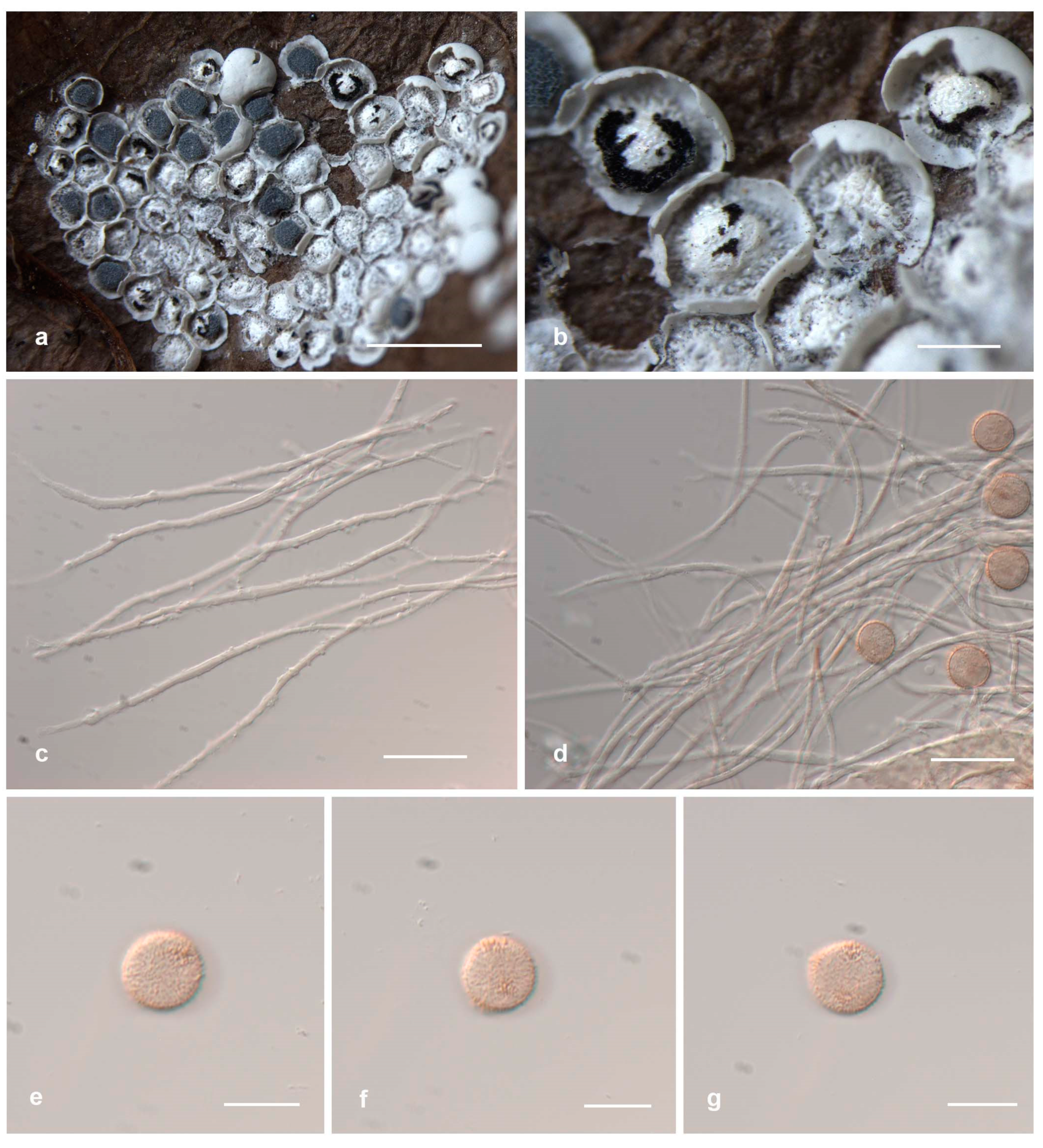
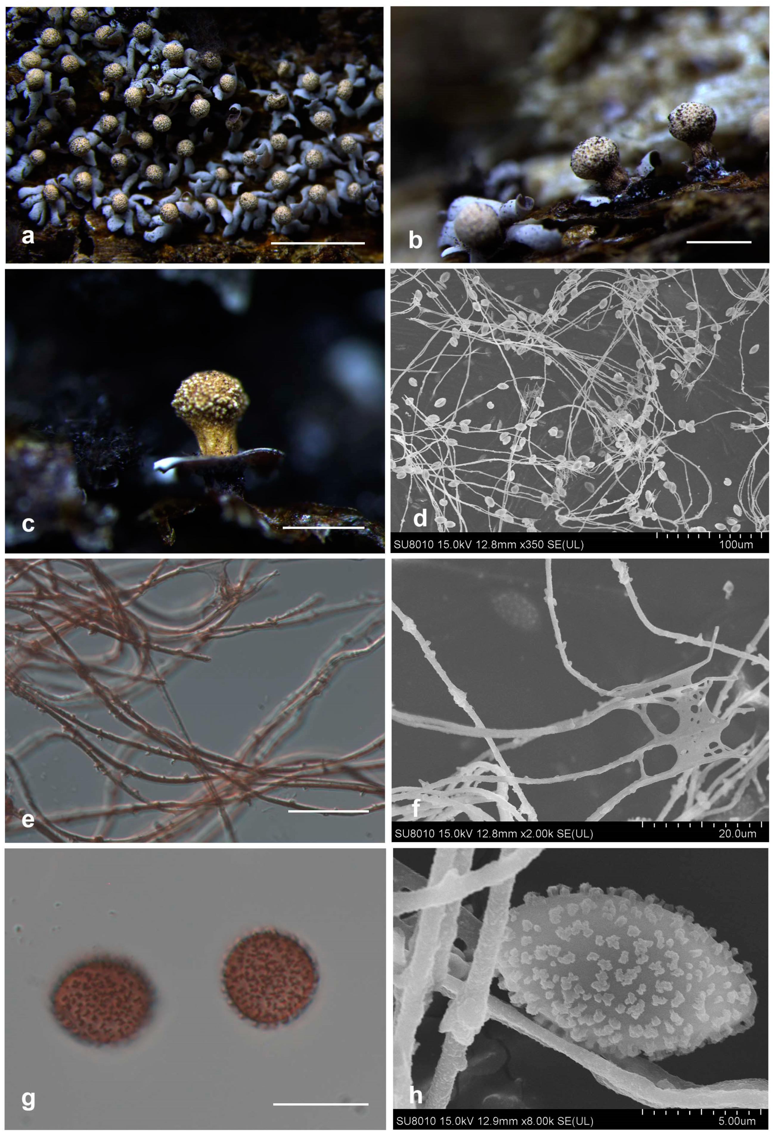
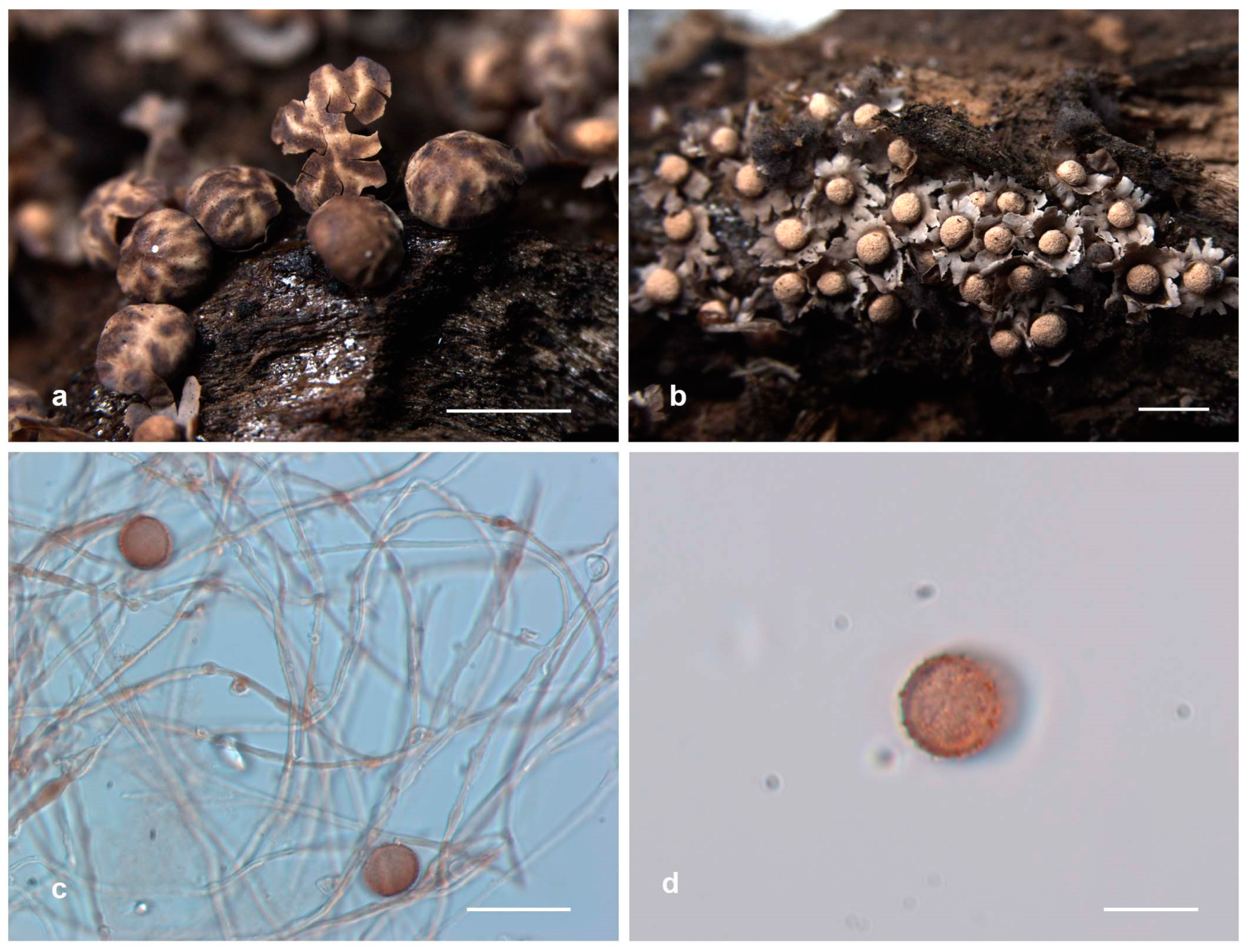
The topologies of individual marker sequences were examined through separate phylogenetic analyses for SSU, EF-1A, and COI genes. A visual inspection showed general congruence among the topologies. To quantitatively assess this congruence, a partition homogeneity test was performed using PAUP*. The results for the individual marker genes and the concatenated dataset are shown in Table S2.
The combined SSU-EF1A-COI dataset consisted of 88 nSSU, 70 EF-1α, and 16 COI sequences derived from 42 taxa representing 10 genera across three orders. This dataset encompassed 13 described species and 2 undescribed species of Diderma, with two sequences from Echinostelium serving as outgroups. In total, 18 new sequences were generated in this study, comprising 7 nSSU, 5 EF-1α, and 6 COI sequences (see Table 1). The final dataset comprised 6442 bp (including gaps), with 3980 bp (including gaps) from SSU, 1676 bp from EF-1α, and 786 bp from COI. The alignment was submitted to TreeBASE (http://purl.org/phylo/treebase/phylows/study/TB2:S31522, accessed on 15 July 2024).
The best nucleotide substitution models for the BI phylogeny were GTR+F+G4 for SSU and GTR+F+I+G4 for both EF-1α and COI. The Bayesian analysis ran for two million generations, yielding an average standard deviation of split frequencies of 0.003873. Subsequently, the same dataset and alignment underwent analysis using the ML method. In the ML analysis, the best nucleotide substitution model for SSU and EF-1A is TIM2e+I+G4, while the COI model is TIM2e+F+I+G4. A comparison of the overall topologies between the ML and BI trees indicated nearly identical results across all datasets. Given this congruence, the ML analysis was chosen as the representative phylogeny. Internal nodes of the tree are labeled with values representing ML and BI support. Branches display bootstrap values (BP) of ≥70% from the ML analysis and Bayesian posterior probabilities (BPPS) of ≥0.90 (Figure 1). The phylogenetic tree obtained revealed two distinct clades within Didymiaceae, representing Diderma and Didymium (Figure 1). Additionally, our findings revealed the division of the genus Diderma into three distinct clades. Among these, four sampled specimens constituted two separate clades indicative of the presence of two new species, namely D. shaanxiense and D. clavatocolumellum.
Figure 1.
Maximum likelihood phylogenetic tree generated from the combined nSSU, EF-1α, and COI dataset of the Diderma species. The display on internal nodes indicates maximum likelihood bootstrap (BP) and Bayesian posterior probability (BPPS) values. The study species are marked in bold (known species) and red (new species) font for easy identification. (Notes: the final dataset comprised 6442 bp, including gaps, with 3980 bp (including gaps) from SSU, 1676 bp from EF-1α, and 786 bp from COI.).
Diderma shaanxiense sequences formed a distinct branch with 100/1 support, closely related to D. globosum and D. europaeum. The phylogenetic distance between D. shaanxiense and D. floriforme must be very similar to that of D. aurantiacum and lower than that of D. tigrinum. These data indicate significant differences between them, supporting the classification of these two as independent species. Additionally, two sampled specimens, which clustered with D. radiatum and D. globosum with strong support, were confirmed as new records from Henan Province and the Inner Mongolia Autonomous Region, respectively.
3.2. Taxonomy
Diderma shaanxiense X.F. Li, B. Zhang and Y. Li, sp. nov.
MycoBank: MB 852952
Figure 2.
Habitat and microstructure of Diderma shaanxiense (HMJAU M20033 Holotype). (a,b) Sporangia and columella; (c,d) capillitium and some spores via TL; (e–g) spores via TL. Scale bars: (a) = 2 mm, (b) = 500 μm, (c,d) = 20 μm, (e–g) = 10 μm.
Diagnosis. Columella hemispherical, with a shining layer of film.
Holotype. China, Shaanxi Province, Baoji City, Meixian County, and Taibai Mountain National Forest Park National Forest Park; on the rotten leaves, 22 July 2014, B. Zhang, HMJAU M20033.
Etymology. The epithet “shaanxiense” refers to Shaanxi, the location of the holotype.
Description. Sporangia, sessile, spheroid, gregarious, mutual squeezing deformation. Peridium is double-layered; the outer layer is calcareous, white, smooth; the inner layer is membranous, grey, iridescent, sometimes wrinkled, and separated from the outer layer. Columella is developed, yellowish brown or white, calcareous, pulvinate, or hemispherical, with a shining layer of film. Hypothallus is inconspicuous. Capillitium is crude, branched, and anastomosing, with membranous enlargement, brown or colorless, occasionally with some darker thickenings. Spore-mass dark or dark brown, fawn under transmitted light (TL), 8–11 μm in diam., with warts.
Habitat. On the rotten leaves.
Distribution in China. Shaanxi Province.
Distribution in the world. China.
Additional specimens examined. China, Shaanxi Province, Baoji City, Meixian County, Taibai Mountain National Forest Park National Forest Park, on the rotten leaves, 22 July 2014, B. Zhang, HMJAU M20034, HMJAU M20050; China, Shaanxi Province, Niubeiliang National Nature Reserve, on the rotten leaves, 21 July 2014, B. Zhang, HMJAU M20051.
Notes. Diderma shaanxiense is characterized by its shining layer of film columella. Diderma shaanxiense shares morphological similarities with D. europaeum and D. globosum in terms of sessile and subglobose sporocarpic, two-layered peridium. However, it can be differentiated by its iridescent columella, spores with warts, and small size (8–11 μm) compared to D. europaeum, while D. europaeum has a pulvinate columella, spores with spines, and larger size (10–12 μm). D. shaanxiense can be distinguished from D. globosum based on its small and pulvinate shining columella. From the phylogenetic tree, we can see that D. shaanxiense is located between D. globosum and D. europaeum, and a separate branch is formed.
Diderma clavatocolumellum X.F. Li, B. Zhang and Y. Li, sp. nov.
MycoBank: MB 852955
Figure 3.
Habitat and microstructure of Diderma clavatocolumellum (HMJAU 11,084 Holotype). (a,b) Sporocarpous and columella; (c) columella and short-stalk sporocarpic; (d) capillitium and some spores via SEM; (e,f) capillitium with many dark-brown swollen knots via TL and SEM; (g,h) spores with warts and ridges via TL and SEM. Scale bars: (a) = 2 mm, (b,c) = 500 mm, (e,g) = 10 μm.
Diagnosis. Columella club-shaped, developed, with yellow-brown stalk.
Holotype. China, Jilin Province, Yanji Prefecture, Antu County, Erdaobaihe, on the dead woods, 5 October 2001, H.Z. Liu and Tolgor, HMJAU 11084.
Etymology. The epithet “clavatocolumellum” refers to the club-shaped columella.
Description. Sporophores sporocarpic, gregaria, sessile or short-stalk, oblate or hemispherical, grey. Peridium is two-layered; the outer layer is cartilaginous, covered with a layer of lime granules, crustose, white, light brown and light ochre, smooth; the inner layer is tightly attached together with the outer layer, calcareous, white or grey. Irregular dehiscence at the top, basal petaloid dehiscence and eversion. Columella present, either developed or conspicuous, clavate, close to the top of the columella, rough, ochre-orange or ivory, 0.5–0.7 mm. Hypothallus is inconspicuous, membranous, translucent, brown. Capillitium is abundant, radiantly extending from the columella, brown or colorless, branching and connecting, with many dark brown swollen knots. Spores dark in mass, purple-brown via transmitted light, subglobose or oval, 9–11 (11.5) μm in diam., with obvious warts, sparse.
Habitat. On the dead woods.
Distribution in China. Jilin Province.
Distribution in the world. China.
Additional specimens examined. China, Jilin Province, Yanji Prefecture, Antu County, Erdaobaihe, on the dead woods, 5 October 2001, H.Z. Liu and Tolgor, HMJAU M20120, HMJAU M20121.
Notes. Morphologically, D. clavatocolumellum is similar to D. alpinum (Meyl.) Meyl. and D. floriforme with its double-layer peridium, spore size, and capillitium having swelling. However, D. clavatocolumellum can be distinguished from D. alpinum based on its developed and conspicuous columella (clavate), two layers of peridium that are tightly connected, membranous and brown hypothallus, and spores with warts; meanwhile, D. alpinum has a pulvinate columella; two layers of peridium kept away; easily separated, calcareous, and white hypothallus; and spores with spines. Diderma clavatocolumellum can be distinguished from D. floriforme based on its sessile or short stalk, while D. floriforme has a long stalk. From the phylogenetic tree, we can clearly see that D. clavatocumullum is related to D. floriforme and they are similar, but the genetic distance between the two is relatively far.
Diderma globosum Pers., Neues Mag. Bot. 1:89 (1794).
Figure 4.
Habitat and microstructure of Diderma globosum (HMJAU M20016). (a) Sporocarpous; (b) columella; (c,d) capillitium and spores via TL; (e–g) spores with verrucose via TL. Scale bars: (a,b) = 2 mm, (c,d) = 20 μm, (e–g) = 10 μm.
Description. Sporophores sporocarpic, gregarious or densely crowded, mutual extrusion deformation, sessile, hemispheric to globose. Peridium is two-layered; the outer layer is limy, eggshell-like, smooth, white; the inner layer is membranous, grey-white, separate from the outer layer, iridescent. Columella is developed, hemispheric, white or pale cream, calcareous. Capillitium threads are slender, dense, pale-brown, sometimes with darker swellings, branched and anastomosed at the end, with membranous enlargement. Spores are dark in mass, reddish brown via transmitted light, with obvious warts, 9–11 μm in diam.
Habitat. On rotten branches and moss.
Distribution in China. Beijing City, Gansu Province, Hebei Province, Heilongjiang Province, Inner Mongolia Autonomous Region, Jilin Province, Liaoning Province, Shaanxi Province, and Yunnan Province.
Distribution in the world. Algeria, Australia, Austria, Canada, China, Costa Rica, Finland, France, Germany, Italy, Japan, Karelia, Korea, Latvia, Morocco, Nepal, the Netherlands, New Zealand, Norway, Peru, Russia, Spain, Sweden, Switzerland, the United States, the United Kingdom, Ukraine, and Venezuela.
Specimens examined. China, Inner Mongolia Autonomous Region, Genhe City, 207 management and protection station of forestry bureau, on the rotten branches, 4 September 2021, B. Zhang, HMJAU M20016; China, Hubei Province, Shiyan City, on the moss, 8 September 2013 B. Zhang, HMJAU M20062.
Notes. Diderma globosum is often confused with D. europaeum and D. crustaceum due to their similar morphology. They are all smooth, white, and hemispheric to globose sporocarp with a two-layered peridium. However, D. globose differs from D. europaeum and D. crustaceum due to its small spores with warts and developed and hemispheric columella.
Diderma radiatum (L.) Morgan, J. Cincinnati Soc. Nat. Hist. 16(4):151, 1894.
Figure 5.
Habitat and microstructure of Diderma radiatum (HMJAU M20026). (a) Sporocarpous; (b) columella; (c) capillitium and spores via TL; (d) Spores with verrucose via TL. Scale bars: (a,b) = 2 mm, (c) = 20 μm, (d) = 10 μm.
Description. Fructification sporocarps, scattered, spherical or oblate, concave below the umbilicus, light yellow-brown, with floral spots and lattice concavities, 0.6–1.58 mm in diam. Sessile or short-stalked, thick, yellow-brown, sulcate, 0.2–0.6 mm in diam. Columella developed and conspicuous and then calcareous, hemispherical, orange, ivory or auburn. Peridium is two-layered; the outer layer is cartilaginous, brown, latticed; the inner layer is membranous, light reddish brown, tightly fused with the outer layer, petaloid or radially dehiscent. Hypothallus is membranous, light brown, translucent. Capillitium is thin, dense, with few branches, brown. Spores are dark in mass, purple-brown via transmitted light, globose, 9–11 μm in diam., with warts, spores accumulated surround columella, separated from the inner layer of the peridium.
Habitat. On the rotten woods.
Distribution in China. Gansu Province, Hebei Province, Henan Province, Heilongjiang Province, Inner Mongolia Autonomous Region, Jilin Province, Qinghai Province, Shaanxi Province, Taiwan Province, and Xinjiang Uygur Autonomous Region.
Distribution in the world. Australia, Austria, Argentina, Canada, China, Denmark, Finland, France, Germany, India, Italy, Japan, Karelia, New Caledonia, New Zealand, Nepal, Norway, Pakistan, Portugal, Puerto Rico, Russia, Spain, Sweden, the United States, and the United Kingdom.
Specimens examined. China, Henan Province, Sanmenxia City, Ganshan National Forest Park, on rotten wood, 26 October 2012, B. Zhang, HMJAU M20026, HMJAU M20057, HMJAU M20058, HMJAU M20059, HMJAU M20060, HMJAU M20061; China, Inner Mongolia Autonomous Region, Hinggan League, Arxan, Motianling, on rotten wood, 15 August 1985, Y. Li and S.L. Chen, HMJAU 9136; China, Inner Mongolia Autonomous Region, Genhe City, on rotten wood, 1 September 1992, W.C. Gao, HMJAU 9963.
Notes. Diderma radiatum is easily confused with D. roanense (Rex) T. Macbr. due to their highly similar morphology, such as sporocarpic appearance with sessile or short-stalked structures, two-layered peridium, large and developed columella, dehiscence floriform, and spores with warts. However, D. radiatum can be differentiated from D. roanense by its yellow-brown stalk with limy features, brown capillitium, and smaller spores.
4. Discussion
The Diderma genus is widely distributed across China, yet its complete species diversity remains unclear. This study presents comprehensive descriptions of two new species, D. shaanxiense and D. clavatocolumellum, along with two previously undocumented species, D. globosum and D. radiatum, discovered in the Inner Mongolia Autonomous Region and Henan Province. The species most closely related in micromorphology to D. shaanxiense appears to be D. globosum. D. shaanxiense is characterized by small, pulvinate, and shining columella, whereas D. globosum has a hemispherical, halo-free columella, making it easier to differentiate between these two species. Diderma clavatocolumellum can be readily differentiated from D. alpinum by its distinct features, such as a clavate columella, spores with warts, and a brown hypothallus. On the phylogenetic tree, these two species are distinctly positioned on individual branches and separate from known species, thus confirming our morphological findings. Furthermore, the phylogenetic analysis also validates certain species previously documented.
Diderma is divided into three main branches. The first branch, led by D. effusum, D. verrucocapillitia, D. deplanatum, D. chondrioderma, D. acanthosporum, D. yucatanense, D. hemisphaericum, and D. pseudotestaceum, encompasses eight species. These species are distinguished by calcareous peridium extending from the sporangium to the synangium, spores with warts, and a columella that is not distinctly pulvinate or absent. Clade 2 comprises two species, D. niveum and D. alpinum, and is characterized by a double-layered peridium, with spores mainly spiny and living in high mountain areas. In 2024, Shchepin et al. also used partial sequences of 18SrDNA to clearly distinguish these two species from some other known species [33]. The third branch consists of D. globosum, D. europaeum, D. radiatum, D. velutinum, D. meyerae, D. floriforme, D. cattiense, D. subasteroides, D. gracile, D. tigrinum, D. aurantiacum, D.montanum, D. umbilicatum, D. microcarpum, D. kamchaticum, and two newly discovered species in this study, D. shaanxiense and D. clavatocolumellum, totaling seventeen species, and the main feature of this branch is that the columella is more prominent, hemispherical or clavate, and the spores are mainly warty.
In the past, morphology was predominantly used for classification and identification purposes. However, molecular barcoding and phylogeny have emerged as a reliable tool for verifying species. These approaches indicate that the species diversity of myxomycetes is not yet fully understood [34]. Phylogenetic trees constructed by incorporating molecular barcodes into numerous morphologically described species have revealed that these species are often complex, comprising several or even dozens of distinct species [6,21,35,36,37,38,39].
In order to evaluate the compatibility of the marker sequences, the partition homogeneity test was performed. The results indicated sufficient congruence among the marker sequences, justifying their concatenation. The balanced index values for the concatenated dataset suggest a robust and reliable phylogenetic inference, minimizing biases from individual markers. In our constructed phylogenetic tree, Physarum Pers., Badhamia Berk., and Fuligo Haller are sister branches, suggesting that the taxonomic status of Physarum and Badhamia is uncertain and that they may be polyphyletic. This finding is consistent with previous studies by Nandipati et al. [40], Cainelli et al. [41], Prikhodko et al. [7], and Ronikier et al. [42]. Prikhodko et al. [7] conducted a phylogenetic analysis of species within Didymiaceae (Myxomycetes) based on three independent genetic markers (18S rDNA gene, translation elongation factor 1-alpha, and cytochrome c oxidase), which supported taxonomic rearrangements in this family. Their study revealed that all studied species of Lepidoderma formed a distinct clade along with Diderma fallax, suggesting that they should be classified under the genus Polyschismium. Additionally, Lepidoderma tigrinum and two related species were transferred to the genus Diderma, leading to the invalidation of the genus Lepidoderma and an increase in species diversity within the genus Diderma.
The current study has expanded the known species diversity of the genus Diderma in China. However, ongoing debates persist regarding the taxonomic classification of this genus, largely due to limited species sampling and insufficient genetic variation in DNA loci. Therefore, additional evidence is required to enhance our understanding of this genus comprehensively. Despite recent discoveries of new Diderma species in China, the full extent of its species diversity remains uncertain, highlighting the need for a comprehensive systematic analysis.
Supplementary Materials
The following supporting information can be downloaded at: https://www.mdpi.com/article/10.3390/jof10080514/s1, Table S1: Information for the sequences from database used in this study [43,44,45,46,47,48,49,50,51,52,53,54]; Table S2: Partition homogeneity test.
Author Contributions
Conceptualization, Y.L. (Yu Li), X.L. (Xiao Li) and B.Z.; methodology, X.L.(Xuefei Li), Y.T., B.Z., D.D. and J.H.; software, X.L., J.H., F.L.S. and Y.T.; validation, X.L., F.L.S. and B.Z.; formal analysis, Y.L. (You Li) and X.L. (Xuefei Li); investigation, X.L. (Xuefei Li), M.L. and Y.G.; resources, X.L. (Xuefei Li), Y.T., J.H., M.L. and B.Z.; data curation, X.L. (Xuefei Li), F.L.S. and Y.L. (You Li).; writing—original draft preparation, X.L.; writing—review and editing, F.L.S., X.L. (Xiao Li) and B.Z.; visualization, X.L. (Xuefei Li), D.D. and M.L.; supervision, X.L. (Xiao Li)., B.Z. and Y.L. (Yu Li); project administration, B.Z.; funding acquisition, B.Z., X.L. (Xiao Li) and Y.L. (Yu Li). All authors have read and agreed to the published version of the manuscript.
Funding
This work was financed by the National Key R & D of the Ministry of Science and Technology (No. 2023YFD1201601-2), the Jilin Provincial key research and development program (20240304155SF), the Research on the Creation of Excellent Edible Mushroom Resources and High Quality and Efficient Ecological Cultivation Technology in Jiangxi Province (20212BBF61002), the 111 program (No. D17014), the Youth Doctoral Program of Zhejiang Normal University study on the species diversity of macrofungi in Baishanzu National Park (2023QB043), and the Zhejiang Normal University Doctoral Initiation Fund (YS304024921).
Institutional Review Board Statement
Not applicable.
Informed Consent Statement
Not applicable.
Data Availability Statement
The original contributions presented in the study are included in the article/Supplementary Material, further inquiries can be directed to the corresponding author/s.
Acknowledgments
We express our gratitude to Xiaolan He from the Sichuan Edible Fungi Research Institute and Haixia Ma from the Chinese Academy of Tropical Agricultural Sciences for their valuable assistance during the fieldwork.
Conflicts of Interest
The authors declare no conflicts of interest.
References
- Stephenson, S.L.; Schnittler, M.; Novozhilov, Y.K. Myxomycete diversity and distribution from the fossil record to the present. Biodivers. Conserv. 2008, 17, 285–301. [Google Scholar] [CrossRef]
- Adl, S.M.; Bass, D.; Lane, C.E.; Lukeš, J.; Schoch, C.L.; Smirnov, A.; Agatha, S.; Berney, C.; Brown, M.W.; Burki, F.; et al. Revisions to the classification, nomenclature, and diversity of eukaryotes. J. Eukaryot. Microbiol. 2019, 66, 4–119. [Google Scholar] [CrossRef] [PubMed]
- Martin, G.W.; Alexopoulos, C.J. The Myxomycetes. The Myxomycètes; University of Iowa Press: Iowa City, IA, USA, 1969; pp. ix + 560. [Google Scholar]
- García-Martín, J.M.; Zamora, J.C.; Lado, C. Multigene phylogeny of the order Physarales (Myxomycetes, Amoebozoa): Shedding light on the dark-spored clade. Persoonia 2023, 51, 89–124. [Google Scholar] [CrossRef] [PubMed]
- Nmf, D. Neues Magazin für die Botanik in Ihrem Ganzen Umfange: Band 1; Saraswati Press: Kolkata, India, 1794. [Google Scholar]
- Leontyev, D.V.; Schnittler, M.; Stephenson, S.L.; Novozhilov, Y.K.; Shchepin, O.N. Towards a phylogenetic classification of the Myxomycetes. Phytotaxa 2019, 399, 209–238. [Google Scholar] [CrossRef]
- Prikhodko, I.S.; Shchepin, O.N.; Bortnikova, N.A.; Novozhilov, Y.K.; Gmoshinskiy, V.I.; Moreno, G.; López-Villalba, N.; Stephenson, S.L.; Schnittler, M. A three-gene phylogeny supports taxonomic rearrangements in the family Didymiaceae (Myxomycetes). Mycol Prog. 2023, 22, 11. [Google Scholar] [CrossRef]
- Lado, C.; Eliasson, U. Taxonomy and systematics: Current knowledge and approaches on the taxonomic treatment of Myxomycetes: Updated version. In Myxomycetes, 2nd ed.; Rojas, C., Stephenson, S.L., Eds.; Academic Press: New York, NY, USA, 2022; pp. 269–324. [Google Scholar]
- Rostafiński, J.T. Versuch Eines Systems der Mycetozoen. F. Wolff 1873, 9–13. Available online: https://BiotaNZ.landcareresearch.co.nz/references/1cb0f8b9-36b9-11d5-9548-00d0592d548c (accessed on 19 July 2024).
- Chow, Z.H. Taxonomic information of Myxomycetes. Jilin Agricultural University, Institute of Microbiology; Chinese Academy of Sciences: Beijing, China, 1978; pp. 157–174. [Google Scholar]
- Lado, C. (2005–2024) An Online Nomenclatural Information System of Eumycezotozoa. Available online: http://www.nomen.eumycetozoa.com (accessed on 1 July 2024).
- Song, W.L.; Lin, D.; Li, M.; Du, Q.; Chen, S.L. Four new records of myxomycetes from China. Phytotaxa 2022, 567, 181–188. [Google Scholar] [CrossRef]
- Gao, Y.; Yan, S.Z.; Wang, G.W.; Chen, S.L. Two new species and two new records of myxomycetes from subtropical forests in China. Phytotaxa 2018, 350, 51–63. [Google Scholar] [CrossRef]
- Zhao, H.N.; Rao, G.; Yang, X.Y.; Li, X.; Yu, H.L.; Zhang, B.; Li, Y. Two new species of Diderma (Physarales, Didymiaceae) from northern China. Phytotaxa 2022, 572, 61–63. [Google Scholar] [CrossRef]
- Chen, S.L.; Yan, S.Z.; Li, Y. Species of the genus Diderma from China and their distributions. Mycosystema 2013, 32, 200–206. [Google Scholar]
- Liu, C.H.; Chang, J.H. Myxomycetes of Taiwan XXI. The genus Diderma. Taiwania 2011, 56, 118–124. [Google Scholar] [CrossRef]
- Royal, B.G. Edinburgh. Flora of British Fungi: Colour Identification Chart; HM Stationery Office: Edinburgh, UK, 1969. [Google Scholar]
- Wang, W.; Wang, W.; Wei, S.W.; Huang, W.; Qi, B.; Wang, Q.; Li, Y. Design of potentially universal SSU primers in myxomycetes using next-generation sequencing. J. Microbiol. Methods 2021, 184, 106203. [Google Scholar] [CrossRef]
- Novozhilov, Y.K.; Okun, M.V.; Erastova, D.A.; Shchepin, O.N.; Zemlyanskaya, I.V.; García-Carvajal, E.; Schnittler, M. Description, culture and phylogenetic position of a new xerotolerant species of Physarum. Mycologia 2013, 105, 1535–1546. [Google Scholar] [CrossRef] [PubMed]
- Liu, Q.S.; Yan, S.Z.; Chen, S.L. Further resolving the phylogeny of Myxogastria (slime molds) based on COI and SSU rRNA genes. Russ. J. Genet. 2015, 51, 46–53. [Google Scholar] [CrossRef]
- Feng, Y.; Schnittler, M. Sex or no sex? Group I introns and independent marker genes reveal the existence of three sexual but reproductively isolated biospecies in Trichia varia (Myxomycetes). Org. Divers. Evol. 2015, 15, 631–650. [Google Scholar] [CrossRef]
- Novozhilov, Y.K.; Prikhodko, I.S.; Shchepin, O.N. A new species of Diderma from Bidoup Nui Ba National Park (southern Vietnam). Protistology 2019, 13, 126–132. [Google Scholar] [CrossRef]
- Standley, D.M. MAFFT multiple sequence alignment software version 7: Improvements in performance and usability. Mol. Biol. Evol. 2013, 30, 772. [Google Scholar] [CrossRef]
- Thompson, J.D.; Gibson, T.J.; Plewniak, F. The Clustal–X windows interface: Flexible strategies for multiple sequence alignment aided by quality analysis tools. Nucleic Acids Res. 1997, 63, 215–228. [Google Scholar] [CrossRef]
- Hall, T. BioEdit: A user-friendly biological sequence alignment editor and analysis program for Windows 95/98/NT. Nucleic Acids Symp. Ser. 1999, 41, 95–98. [Google Scholar] [CrossRef]
- Goloboff, P.A.; Catalano, S.A.; Torres, A. Parsimony analysis of phylogenomic datasets (II): Evaluation of PAUP*, MEGA and MPBoot. Cladistics 2022, 38, 126–146. [Google Scholar] [CrossRef]
- Kalyaanamoorthy, S.; Minh, B.Q.; Wong, T.K.F.; Haeseler, A.V.; Jermiin, L.S. ModelFinder: Fast model selection for accurate phylogenetic estimates. Nat. Methods. 2017, 14, 587–589. [Google Scholar] [CrossRef] [PubMed]
- Nguyen, L.T.; Schmidt, H.A.; Haeseler, A.V.; Minh, B.Q. IQ-TREE: A fast and effective stochastic algorithm for estimating maximum-likelihood phylogenies. Mol. Biol. Evol. 2015, 32, 268–274. [Google Scholar] [CrossRef] [PubMed]
- Zhaxybayeva, O.; Gogarten, J.P. Bootstrap, Bayesian prob-ability and maximum likelihood mapping: Exploring newtools for comparative genome analyses. BMC Genom. 2002, 3, 1–15. [Google Scholar] [CrossRef] [PubMed]
- Huelsenbeck, J.P.; Ronquist, F. MRBAYES: Bayesian inference of phylogenetic trees. Bioinformatics 2001, 17, 754–755. [Google Scholar] [CrossRef] [PubMed]
- Rambaut, A.; Drummond, A.J.; Xie, D.; Baele, G.; Suchard, M.A. Posterior Summarization in Bayesian Phylogenetics Using Tracer 1.7. Syst Biol. 2018, 67, 901–904. [Google Scholar] [CrossRef]
- Bork, P. Interactive Tree of Life (iTOL): An online tool for phylogenetic tree display and annotation. Bioinformatics 2007, 23, 127–128. [Google Scholar] [CrossRef]
- Shchepin, O.N.; López, V.Á.; Inoue, M.; Prikhodko, I.S.; Erastova, D.A.; Okun, M.V.; Woyzichovski, J.; Yajima, Y.; Gmoshinskiy, V.I.; Moreno, G.; et al. DNA barcodes reliably differentiate between nivicolous species of Diderma (Myxomycetes, Amoebozoa) and reveal regional differences within Eurasia. Protist 2024, 175, 126023. [Google Scholar] [CrossRef] [PubMed]
- Leontyev, D.V.; Schnittler, M. The phylogeny and phylogenetically based classification of myxomycetes. In Myxomycetes: Biology, Systematics, Biogeography and Ecology; Rojas Alvarado, C., Stephenson, S.L., Eds.; Elsevier Academic Press: Amsterdam, The Netherlands, 2022; pp. 97–124. [Google Scholar] [CrossRef]
- Shchepin, O.; Novozhilov, Y.; Woyzichovski, J.; Bog, M.; Prikhodko, I.; Fedorova, N.; Gmoshinskiy, V.; Borg, D.M.; Dagamac, N.H.A.; Yajima, Y.; et al. Genetic structure of the protist Physarum albescens (Amoebozoa) revealed by multiple markers and genotyping by sequencing. Mol. Ecol. 2022, 31, 372–390. [Google Scholar] [CrossRef] [PubMed]
- Dagamac, N.H.A.; Rojas, C.; Novozhilov, Y.K.; Moreno, G.H.; Schlueter, R.; Schnittler, M. Speciation in progress? A phylogeographic study among populations of Hemitrichia serpula (Myxomycetes). PLoS ONE 2017, 12, e0174825. [Google Scholar] [CrossRef]
- Leontyev, D.V.; Schnittler, M.; Stephenson, S.L. A critical revision of the Tubifera ferruginosa-complex. Mycologia 2015, 107, 959–985. [Google Scholar] [CrossRef]
- Leontyev, D.V.; Buttgereit, M.; Kochergina, A.; Shchepin, O.N.; Schnittler, M. Two independent genetic markers support separation of the myxomycete Lycogala epidendrum into numerous biological species. Mycologia 2023, 115, 32–43. [Google Scholar] [CrossRef] [PubMed]
- Leontyev, D.V.; Ishchenko, Y.; Schnittler, M. Fifteen new species from the genus Lycogala (Myxomycetes). Mycologia 2023, 115, 524–560. [Google Scholar] [CrossRef] [PubMed]
- Nandipati, S.C.; Haugli, K.; Coucheron, D.H.; Haskins, E.F.; Johansen, S.D. Polyphyletic origin of the genus Physarum (Physarales, Myxomycetes) revealed by nuclear rDNA mini-chromosome analysis and group I intron synapomorphy. BMC Evol. Biol. 2012, 12, 166. [Google Scholar] [CrossRef]
- Cainelli, R.; Haan, M.; Meyer, M.; Bonkowski, M.; Fiore-Donno, A.M. Phylogeny of Physarida (Amoebozoa, Myxogastria) based on the small-subunit ribosomal RNA gene, redefinition of Physarum pusillum s. str. and reinstatement of P. gravidum Morgan. J. Eukaryot Microbiol. 2020, 67, 327–336. [Google Scholar] [CrossRef]
- Ronikier, A.; Janik, P.; Haan, M.; Kuhnt, A.; Zankowicz, M. Importance of type specimen study for understanding genus boundaries-taxonomic clarifications in Lepidoderma based on integrative taxonomy approach leading to resurrection of the old genus Polyschismium. Mycologia 2022, 114, 1008–1031. [Google Scholar] [CrossRef]
- García-Martín, J.M.; Mosquera, J.; Lado, C. Morphological and molecular characterization of a new succulenticolous Physarum (Myxomycetes, Amoebozoa) with unique polygonal spores linked in chains. Eur. J. Protistol. 2018, 63, 13–25. [Google Scholar] [CrossRef]
- Gmoshinskiy, V.I.; Prikhodko, I.S.; Bortnikov, F.M.; Shchepin, O.N.; Schnittler, M. Morphology and phylogeny of Diderma aurantiacum (myxomycetes)-A new species for Russia from the far east. Hobocmu Cucemamuku Hu3wux Oacmehuu 2023, 57, 27–42. [Google Scholar] [CrossRef]
- Novozhilov, Y.K.; Mitchell, D.W.; Okun, M.V.; Shchepin, O.N. New species of Diderma from Vietnam. Mycosphere 2014, 5, 554–564. [Google Scholar] [CrossRef]
- Hoppe, T.; Schnittler, M. Characterization of myxomycetes in two different soils by TRFLP- analysis of partial 18S rRNA gene sequences. Mushroom Research Foundation. Mycosphere 2015, 6, 216–227. [Google Scholar] [CrossRef]
- Novozhilov, Y.K.; Shchepin, O.N.; Prikhodko, I.; Schnittler, M. A new nivicolous species of Diderma (Myxomycetes) from Kamchatka, Russia. Nova Hedwig. Z. Fur Kryptogamenkd. 2022, 114, 181–196. [Google Scholar] [CrossRef]
- Bortnikov, F.M.; Shchepin, O.N.; Gmoshinskiy, V.I.; Prikhodko, I.S.; Novozhilov, Y.K. Diderma velutinum, a new species of Diderma (Myxomycetes) with large columella and triple peridium from Russia. Bot. Pacifica 2018, 7, 47–51. [Google Scholar] [CrossRef]
- Lado, C.; Treviño, Z.I.; García-Martín, J.M.; Basanta, D.W. Diachea mitchellii: A new myxomycete species from high elevation forests in the tropical Andes of Peru. Mycologia 2022, 114, 798–811. [Google Scholar] [CrossRef] [PubMed]
- Wikmark, O.G.; Haugen, P.; Lundblad, E.W.; Haugli, K.; Johansen, S.D. The molecular evolution and structural organization of group I introns at position 1389 in nuclear small subunit rDNA of myxomycetes. J. Eukaryot Microbiol. 2007, 54, 49–56. [Google Scholar] [CrossRef] [PubMed]
- Janik, P.; Lado, C.; Ronikier, A. Range-wide Phylogeography of a nivicolous protist Didymium nivicola Meyl. (Myxomycetes, Amoebozoa): Striking contrasts between the northern and the southern hemisphere. Protist 2020, 171, 125771. [Google Scholar] [CrossRef] [PubMed]
- Zhao, F.Y.; Liu, S.Y.; Stephenson, S.L.; Hsiang, T.; Qi, B.; Li, Z. Morphological and molecular characterization of the new aethaloid species Didymium yulii. Mycologia 2021, 113, 926–937. [Google Scholar] [CrossRef]
- Fiore-Donno, A.M.; Berney, C.; Pawlowski, J.; Baldauf, S.L. higher-order phylogeny of plasmodial slime molds (Myxogastria) based on elongation factor 1-A and small subunit rRNA gene sequences. J. Eukaryot Microbiol. 2005, 52, 201–210. [Google Scholar] [CrossRef]
- Borg, D.M.; Brejnrod, A.D.; Unterseher, M.; Hoppe, T.; Feng, Y.; Novozhilov, Y.; Sørensen, S.J.; Schnittler, M. Genetic barcoding of dark-spored myxomycetes (Amoebozoa)-Identification, evaluation and application of a sequence similarity threshold for species differentiation in NGS studies. Mol. Ecol. Resour. 2018, 18, 306–318. [Google Scholar] [CrossRef]
Disclaimer/Publisher’s Note: The statements, opinions and data contained in all publications are solely those of the individual author(s) and contributor(s) and not of MDPI and/or the editor(s). MDPI and/or the editor(s) disclaim responsibility for any injury to people or property resulting from any ideas, methods, instructions or products referred to in the content. |
© 2024 by the authors. Licensee MDPI, Basel, Switzerland. This article is an open access article distributed under the terms and conditions of the Creative Commons Attribution (CC BY) license (https://creativecommons.org/licenses/by/4.0/).