Article

Description and Pathogenicity of Colletotrichum kapreanum sp. nov, a Cherelle Wilt Pathogen Belonging to the Gigasporum Species Complex

by
Yoshiki Takata
1,
Celynne Ocampo-Padilla
1,2,
Mike Andre C. Malonzo
3,
Dan Charlie Joy Camara Pangilinan
1,
Shunsuke Nozawa
4 and
Kyoko Watanabe
1,4,*
1
Graduate School of Agriculture, Tamagawa University, Tamagawa-Gakuen 6-1-1, Machida 194-8610, Japan
2
Department of Crop Protection, Central Luzon State University, Science City of Muñoz 3120, Philippines
3
BaCaDM Project, College of Agriculture, Tamagawa University, Tamagawa-Gakuen 6-1-1, Machida 194-8610, Japan
4
College of Agriculture, Tamagawa University, Tamagawa-Gakuen 6-1-1, Machida 194-8610, Japan
*
Author to whom correspondence should be addressed.
J. Fungi 2024, 10(3), 204; https://doi.org/10.3390/jof10030204
Submission received: 24 January 2024 / Revised: 21 February 2024 / Accepted: 26 February 2024 / Published: 8 March 2024
(This article belongs to the Section Fungal Pathogenesis and Disease Control)

Abstract

:
Similar to cacao pod rot, cherelle wilt decreases production from cacao fields. Among all known fungal pathogens of the cacao, Colletotrichum spp. are common infectious agents that affect the cherelles and pods of cacao; thus, cacao diseases are often classified by stage. Therefore, knowing whether these pathogens are common in both fruit stages is necessary for implementing disease control measures. Symptoms of cherelle wilt were found in cacao plants in Pangasinan, Philippines, in 2022. The fungal strain obtained from the lesion was found to be pathogenic towards cherelles, but not towards pods. The strain was classified as an unknown species belonging to the gigasporum species complex, based on the morphological and molecular phylogenetic analyses of ITS, GAPDH, CHS1, ACT, and TUB2. We propose Colletotrichum kapreanum sp. nov. as a causal agent of cacao cherelle wilt, but not pod rot.

1. Introduction

Cacao (Theobroma cacao L.), the source of cacao beans used to make chocolate, is an important cash crop in tropical countries [1,2]. In the Philippines, the majority of the cacao farms are smallholdings owned and managed by farmers. According to Philippine Statistics Authority (PSA) data, the Philippines’ cacao production has been slowly but steadily increasing, by an average of 2743 hectares per year, from 2013 to 2020. Though production is increasing, the country cannot still meet the increasing market demand of cacao locally and globally. The low production of cacao in the Philippines is mainly due to the high mortality rate of planting materials and the lack of knowledge and training specifically on good agricultural practices (GAP) for cacao farmers, resulting in improper farm management, which can result in low yield and susceptibility of cacao plants to pests and diseases [3].
Cacao pods are classified by size, into either ‘cherelles’ and ‘pods.’ Cherelles are defined as pods with a length <10 cm, while pods are >10 cm. Cacao diseases are classified based on the pod stage or morphology. Cacao pod rot is caused by several pathogens; thus, it is difficult to identify based solely on its symptoms. However, some pathogens that cause diseases in pods have their own disease names; for example, black pod rot and brown pod rot are caused by Phytophthora spp.; frosty pod disease is caused by Moniliophthora roreri (Cif.) H.C. Evans, Stalpers, Samson & Benny; and anthracnose is caused by Colletotrichum spp., all of which are diseases that have generated research interest. However, 80% of cherelle wilt cases are thought to be caused by physiological disorders [2]; thus, little research has been conducted on the relevant infections. Therefore, cherelle wilt is a disease that farmers are unfamiliar with as a control target.
Anthracnose disease in cacao, caused by Colletotrichum spp., manifests as lesions on the leaves and pods [4,5]. To date, all recorded causative species belong to the Colletotrichum gloeosporioides species complex (CGLSC), which comprises C. gloeosporioides, C. siamense, C. theobromicola, C. tropicale, and C. aeschynomenes [4,6,7,8,9]. Among these, C. tropicale is pathogenic to cacao pods at different maturation stages [5], and C. theobromicola Delacr. has been isolated from both cherelle wilt and cacao pod rot [1]. There is no record of other species as common pathogens in cherelles and pods. Thus, there may be a low potential for pathogens in cherelles and pods to infect each other and cause anthracnose outbreaks in cacao-producing farms. Additionally, many diseases from Colletotrichum species are reported as pathogens causing disease on fruits and leaves, such as diseases associated with C. gloeosporioides (syn. Glomerella cingulate), which causes leaf spots on apples [10]; C. truncatum, which causes leaf spots on leaves of Citrus reticulata; [11] and C. acutatum, which causes almond anthracnose [12]. However, González et al. [10] found that some of the lineages of C. acutatum and C. gloeosporioides show host organ-specific pathogenicity. Some studies show that the latent phase of pathogenic Colletotrichum spp. in fruits, such as apples, strawberries, and citrus, is present in the leaves and twigs of the plant. In cacao, Rojas et al. [9] reported that fungi belonging to the CGLSC and gigasporum species complex (CGISC) have the potential to act not only as endophytes, but also as pathogens. Therefore, these pathogens are prospective targets for control of the disease.
This study aimed to identify the pathogen causing anthracnose in cacao cherelles and to determine its pathogenicity to cherelles, pods, and leaves, as well as to elucidate its life cycle for disease control strategies. We identified an isolate, PH22T135, obtained from a cherelle wilt at the species level based on morphological and molecular phylogenetic analysis, and tested its pathogenicity to cherelles, pods, and leaves of cacao. The strain was thus identified as a new species belonging to the CGISC and pathogenic only to cherelles.

2. Materials and Methods

2.1. Fungal Isolation

A cacao fruit sample showing “cherelle wilt” was collected in June 2022 from Pangasinan, Philippines. The fruit was blighted and dried with salmon-pink conidiomata (Figure 1A). Approximately 5 × 5 mm segments of symptomatic pod tissues were soaked into 0.6% (v/v) sodium hypochlorite for 30 s for surface sterilization, blot-dried using sterile tissue paper, and placed on water agar (agar 15 g/L) plates. Hyphae emerging from the tissues were transferred onto potato dextrose agar (PDA; 39 g/L; Eiken Kagaku, Tokyo, Japan) plates and maintained at room temperature (25 °C) to induce sporulation. A monoculture was obtained by isolating a single conidium from a conidial mass of the fungal colony. The isolate was maintained on a PDA plate. Our isolate was preserved in a tube with 10% sterile glycerol at −80 °C, as the ex-type living culture and type specimen were deposited at Central Luzon State University.

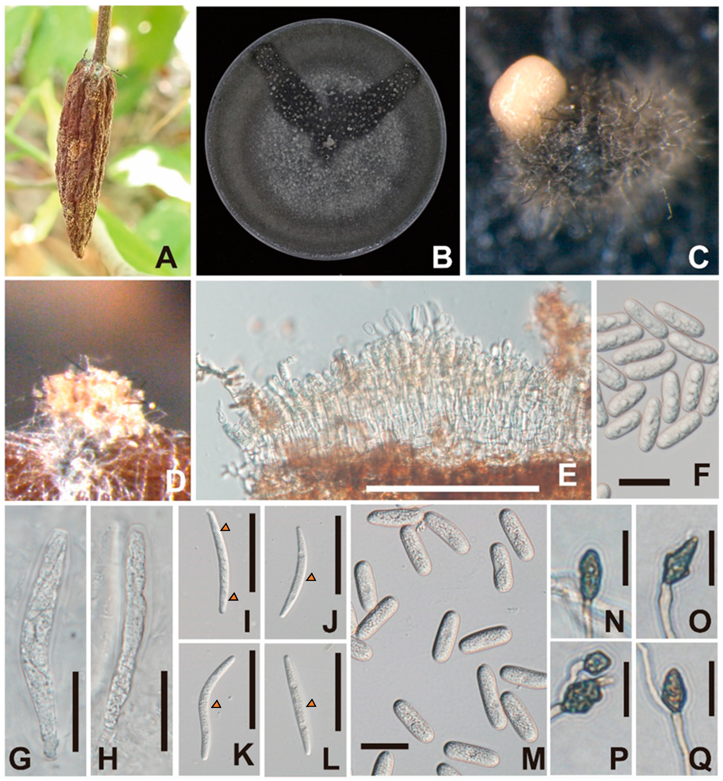
2.2. Morphological Observation

After a ten-day inoculation period, the characteristics of colonies on oatmeal agar (OA; oatmeal 20 g/L, agar 15 g/L) plate and synthetic nutrient-poor agar (SNA) [13] were observed at 25 °C in natural light conditions. Conidial masses produced on OA plates and pine needle placed on SNA plates were observed under an MDG-17 stereo microscope (Leica Microsystems GmbH, Wetzlar, Germany). Morphological characteristics of asci, ascospores, and conidia on OA plates or pine needles placed on SNA plates as well as the appressoria that formed on the bottom surface of the SNA plates were examined under a light microscope (BX51, Olympus, Tokyo, Japan). Each structure (n = 30) was measured using ImageJ software 1.53o (https://imagej.nih.gov/ij/index.html, accessed on 20 January 2022).

2.3. Genomic DNA Extraction, PCR, and Sequencing

DNA was extracted for polymerase chain reaction (PCR) using the cetyltrimethylammonium bromide method described by Doyle and Doyle [14]. Complete sequences of the internal transcribed spacer 1, 2 including 5.8S rRNA, and partial sequences of 18S rRNA and 28S rRNA regions (ITS), glyceraldehyde-3-phosphate dehydrogenase (GAPDH), chitin synthase 1 (CHS1), actin (ACT), and β-tubulin 2 (TUB2) genes were amplified using the primer pairs ITS1F [15]/ITS4 [16], GDF/GDR [17], CoCHS586F/CoCHS2089R [18], CoACTF/CoACTR [18], and CoTUB2F/CoTUB2R [18], respectively. The PCR conditions for amplifying these gene regions were in accordance with the report of Liu et al. [19]. PCR products were purified using ExoSAP-IT reagent (GE Healthcare, Tokyo, Japan) after detecting bands using electrophoresis. Cycle sequencing reactions were conducted using BigDye Terminator v3.1 (Applied Biosystems, Tokyo, Japan). Purified sequencing products obtained by ethanol precipitation were analyzed at FASMAC DNA Sequencing Services (Kanagawa, Japan).

2.4. Molecular Phylogenetic Analyses

To identify PH22T135, 18 sequences of strains belonging to 10 species under the CGISC were used to construct molecular phylogenetic trees (Table 1). Sequences from Colletotrichum acutatum (CBS112996) and C. gloeosporioides (ICMP17821) were used as outgroups. The sequences of ITS, GAPDH, CHS1, ACT, and TUB2 were aligned independently with ClustalW [20] and the gap-including sites at both ends were pruned using MEGA 10.2 software [21]. ML analyses were performed using MEGA 10.2 software. The best substitution model for ML analysis was decided based on the Akaike information criterion in the complete edition of the sequence using model search; the Tamura-Nei model + G was chosen for the substitution model. All gap-including sites were eliminated from the dataset, after which bootstrap (BS) analysis was performed in MEGA 10.2 software.
Bayesian probability (BP) analysis was performed using MrBayes (version 3.1.1) [22]. The best-fit models were separately searched for each DNA region using MrModeltest v.2.3 [23], and performed using PAUP* v.4.0b10 [24]. SYM + G, HKY + I, and HKY + G were selected for ITS, GAPDH, and CHS1, respectively, while SYM + I was selected for ACT and TUB2. Analysis of the two Markov and Monte Carlo chains of the four runs was run for 1,000,000 generations. Data were rated every 100 generations, and the first 6500 generations were burned-in, because the average standard deviation of the split frequency rate in these generations was less than 0.01.

2.5. Pathogenicity Test

Pathogenicity tests were conducted using a conidial suspension adjusted to 1 × 10⁵ conidia/mL. Twenty microliters of the conidial suspension were dropped onto three areas per pod, some of which had wounds, and some did not, on healthy young and mature cacao pods, and on young and old leaves. Sterile water was used as control. The samples were covered with plastic bags for five days to maintain high humidity. After 9–14 d, the inoculated samples were observed, and re-isolation was conducted from symptomatic pods and leaves.

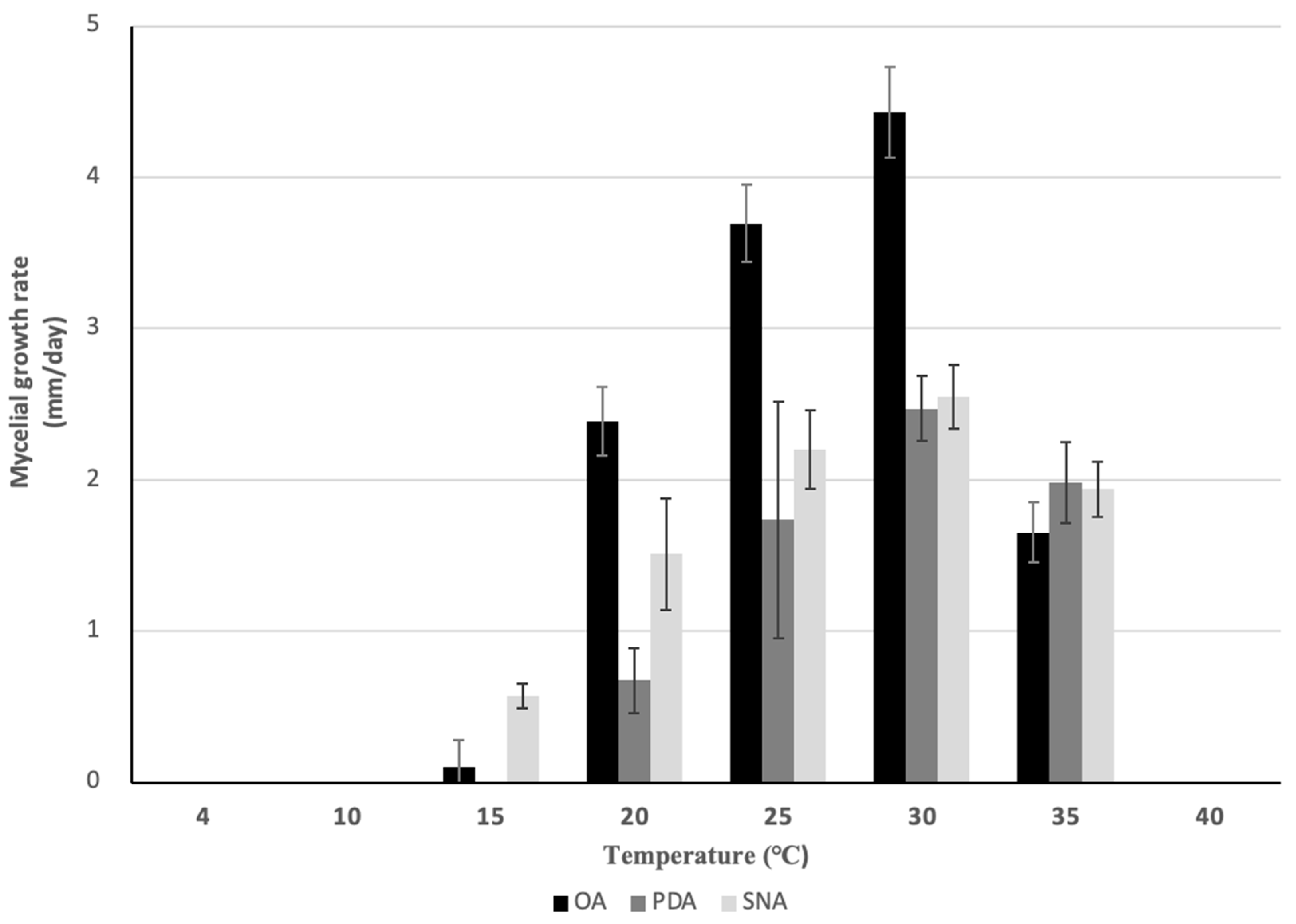
2.6. Mycelial Growth Test

To determine the optimal temperature for the growth of the isolate, a mycelial growth test was conducted twice with 5 replicates each. The mycelial plugs (φ6 mm) were obtained from the colonies of our isolate, which was grown for 7 d at 25 °C, and placed at the center of PDA, SNA, and OA plates (φ9 cm). The plates were incubated at 4, 10, 15, 20, 25, 30, 35, and 40 °C for 4 d. Five replications were conducted for each temperature. Mycelial growth was measured daily, and the average colony diameter at each temperature was calculated.

3. Results

3.1. Molecular Phylogenetic Analyses

To clarify the molecular phylogenetic position of our isolate, a BLAST search was performed in the NCBI database, and phylogenetic analyses were conducted. Partial nucleotide sequences of ITS, GAPDH, CHS1, ACT, and TUB2 were matched with C. gigasporum strains JS-0328 (query cover: 99%; 550 bp; identity: 100%; accession no. OM397116), CBS 132881 (query cover: 100%; 295 bp; identity: 99%; accession no. KF687829), JS-0367 (query cover: 100%; 727 bp; identity: 95%; accession no. CP077955), JS-0367 (query cover: 100%; identity 94%; accession no. CP077947), and CX34 (query cover 100%, 678 bp; identity 99%; accession no. ON420414). These species, which were identified using the nucleotide data of our isolate, belong to CGISC.
Identification of our isolate at the species level was conducted using the data of species in CGISC (Figure 2). The final dataset contained 1725 bp, including gaps, comprising 545, 286, 280, 228, and 391 positions from ITS, GAPDH, CHS1, ACT, and TUB2, respectively. As the ML and BP trees exhibited similar topologies, the ML tree shown in Figure 2 had the highest log likelihood score of −6928.70. Our isolate was independent from the other 11 clades, including all 9 ex-type strains belonging to CGISC (Figure 2), and most closely related to C. zahaoqingense, as evidenced by high BS values and BP (BS/BP: 99/1.0).

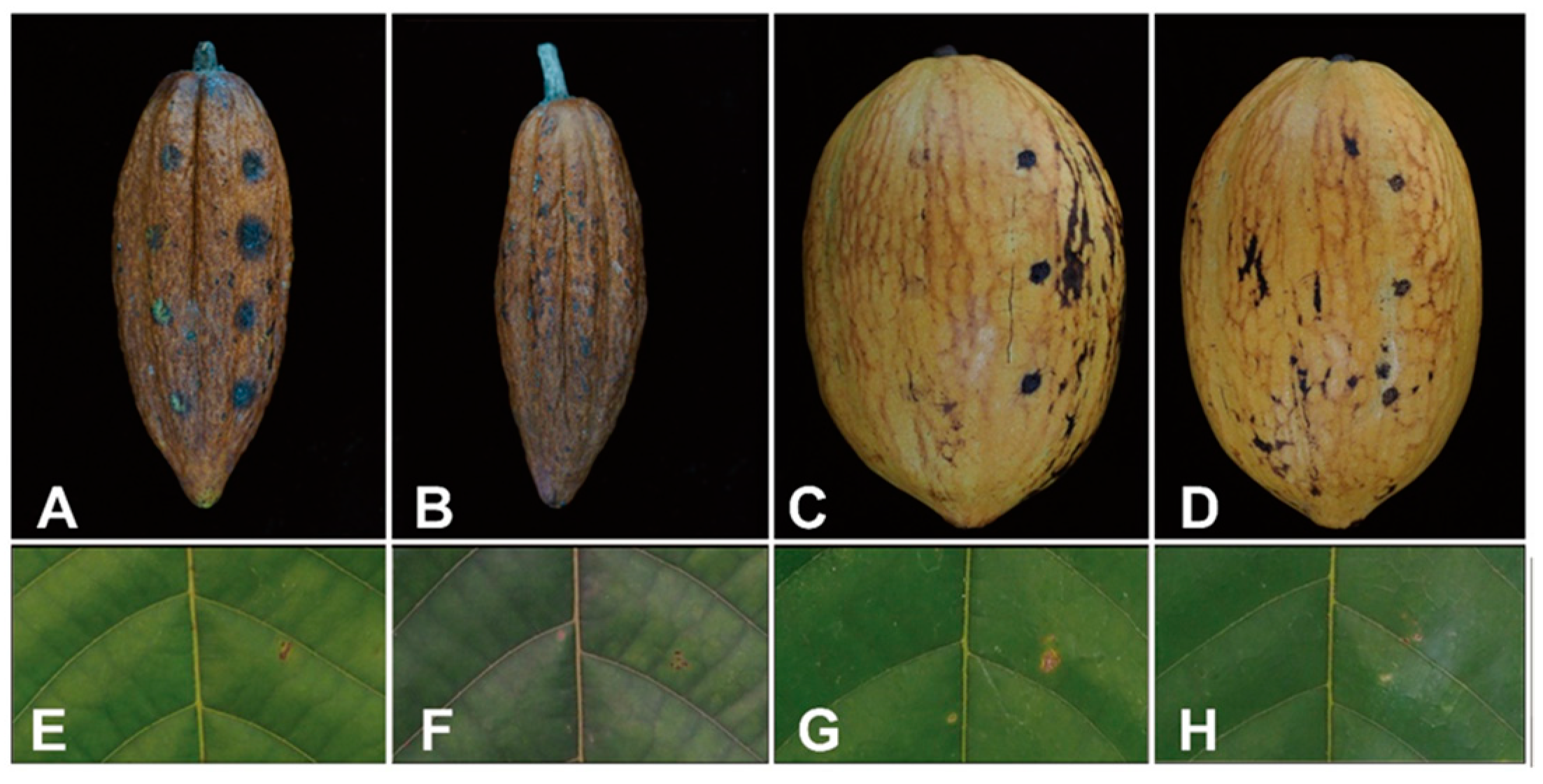
3.2. Pathogenicity Test

Our isolate produced symptoms in cherelles, resulting in black lesions in all inoculated areas four days after inoculation (Figure 3A). In mature pods, black spots appeared only in wounded areas (Figure 3C). These were slightly darker than those in control (Figure B), and no expansion of lesions was observed. No symptoms developed in the pods or leaves treated with sterile water as controls (Figure 3B,D,F,H). The inoculum was re-isolated from all symptomatic tissues. Regarding pathogenicity on wounded young and mature leaves, the isolate did not show pathogenicity as seen in cherelles (Figure 3E,G). However, over time, the inoculated parts were slightly bigger as lesions than the control one, and the inoculum was re-isolated from the lesion.

3.3. Taxonomy

Colletotrichum kapreanum Takata & Kyoko Watan., sp. nov. (Figure 1).
MycoBank no.: MB 852300.
Etymology: The species name was derived from the name ‘kapre’, a giant living on a tree based on Philippine mythology.
Type: Machida, Tokyo, Japan, Tamagawa University, isolated from a wilted cacao cherelle. Holotype PH22T135S, ex-type strain PH22T135.
Sexual morph: Sexual morphology was observed in colonies grown on OA plates, exclusively on black hyphae (Figure 1B). Ascomata were solitary, scattered, and few. Asci were unitunicate, eight-spored, broadly cylindrical to clavate, and truncate at apex (Figure 1G,H). Ascospores were hyaline, fusoid to cylindrical, mostly straight to slightly curved or simultaneously irrigate curved, rounded at both ends, aseptate or 1-septum (rarely 3 septa), septum median, and measured (44.5–)47.5–68.6(–69.5) × 5.2–7.0(–7.4) μm (ave. ± sd. 60.5 ± 6.5 × 6.0 ± 0.5) (Figure 1I–L). Asexual morph: Colony grew on OA plates at 15–35 °C with an optimum temperature of 30 °C (ave. ± sd. 4.4 ± 0.3 mm/day) in the dark (Figure 4), davis gray to sonic silver. Hyphae were hyaline, smooth-walled, septate, and branched. Setae were medium brown, verruculose, 6–8-septae, and 70–160 μm long. Conidiomata was pale orange (Figure 1C). Conidia were aseptate straight, cylindrical, guttulate, rounded at both ends, and measured 22.9–28.4 × 7.1–8.7 (ave. ± sd. 26.2 ± 1.7 × 8.1 ± 0.4) μm (L/W = 3.2) (Figure 1M). On PDA plates, hyphae grew at 20–35 °C, with an optimum temperature of 30 °C (ave. ± sd. 2.5 ± 0.2 mm/day) in the dark. Conidia were not observed. On SNA, hyphae grew at 15–35 °C, with an optimum temperature of 30 °C (ave. ± sd. 4.4 ± 0.3 mm/day) in the dark. Conidia were not observed. Appressoria were pale to medium brown, irregular or ovoid-shaped, and measured 4.9–9.6(–10.8) × (2.9–)3.1–5.1(–7.6) μm (ave. ± sd. 10.8 ± 1.5 × 4.1 ± 0.9) (L/W = 1.8) (Figure 1N–Q). On pine needles, conidiomata were pale orange and there were few (Figure 1D). The acervalus was lined with conidiogenous cells (Figure 1E). Setae were 6–8 septate, medium brown, verruculose, 6–8-setae, 80–140 μm long. Conidia were aseptate, straight, cylindrical, guttulate, rounded at both ends, and measured (18.2–)21.2–25.5 × 6.7–7.1 (ave. ± sd 22.2 ± 1.6 × 6.9 ± 0.4) μm (Figure 1F).
Note: C. kapreanum resembles C. zahaoqingense, a sister group of C. kapreanum, in the shape of conidia and appressoria, but not in conidial size, as those of C. kapreanum are larger than those of C. zahaoqingense (Table 2). Two species produce ascospores with septa in the CGISC, including C. gigasporum [18,25] and C. taiwanense [26]; however, the number of septa in C. taiwanense is larger than that of C. kapreanum (Table 3). The size of the conidia of C. kapreanum, however, is similar to that of C. gigasporum (Table 2). Meanwhile, the size of appressoria of C. gigasporum is larger than those of C. kapreanum (Table 4). Recorded sizes of ascospores of C. gigasporum and C. taiwanense were measured with PDA plates and rice straw, but PH22T135 asci and ascospores were not observed on PDA plates.

4. Discussion

4.1. Identification of Pathogenic Isolate

The PH22T135 strain extracted from cherelle wilt was identified as a new species pathogenic to cacao, belonging to the CGISC based on morphological and phylogenetic analyses; it was named Colletotrichum kapreanum. Although there are some reports of other pathogens belonging to the genus Colletorichum that affect cacao, C. kapreanum is independent of not only these species, including C. gloeosporioides, C. siamense, C. theobromicola, C. tropicale, and C. aeschynomenes [4,6,7,8,9], but also other reported species in the molecular phylogenetic tree, per morphological analysis. CGISC comprises 17 species, typically with longer conidia, characterized by a straight shape with a round end, than those of other Colletotrichum species, belonging to other species complexes. These species can also be distinguished from other species in other species complexes using phylogenetic analyses [19,20]. The anamorph of C. kapreanum is consistent with the morphology of species belonging to CGISC. Focusing on morphologies of conidia and appressoria, C. kapreanum is distinct from C. zhaoqingense in the size of conidia and the shape and size of appressoria. Although C. gigasporum produces similarly sized conidia to that of C. kapreanum, their appressoria are significantly different in size. Additionally, we morphologically compared C. kapreanum with C. taiwanense, the latter of which has no molecular data, because the anamorphs of C. taiwanense are alike those of the species in CGISC, and we found no significant difference in the size of conidia between C. kapreanum with C. taiwanense. However, these two species also are distinguished from each other in the size and shape of appressoria. Regarding morphologies of ascospores, C. kapreanum produces smaller ascospores than those of C. gigasporum, while those produced by C. taiwanense are similar in size to those of C. kapreanum. However, C. kapreanum differs from C. taiwanense in terms of the number of septa of ascospores. C. kapreanum produces ascospores with a septum in the middle (usually one septum, rarely three septa), while C. taiwanense produces 0–3 ascospores and, rarely, eight septa [26,27]. Therefore, we propose C. kapreanum as the new species belonging to CGISC. To the best of our knowledge, this is the third report of this species having ascospores with a septum, as belonging to the genus Colletotrichum.

4.2. Pathogenicity

The isolate was relatively pathogenic toward cherelles and produced symptoms in both wounded and unwounded parts of the organ. However, the isolate did not show pathogenicity to mature pods and leaves. C. tropicale is reported to cause anthracnose of cacao pods at different stages, including young, immature, and mature pods [5]. Paguntalan et al. [27] reported Colletotrichum sp. isolated from an anthracnose-infected pod through a postharvest survey in Mindanao, Philippines. This strain belongs to CGLSC, according to the results obtained from morphological data and the pictures in their article. There are no records of Colletotrichum spp., except for CGLSC, related to anthracnose in cacao. In addition to our results, cacao pods are affected by different pathogens depending on their developmental stage which is a point to be noted when managing the relevant diseases.
In the life cycle of the plant pathogens, latent infection must be considered because this information is crucial in disease management strategies. Some studies provide information on the importance of latent infection of Colletotrichum spp. to control the disease. For example, Peres et al. [12] investigated the life cycle of the pathogenic C. acutatum in citrus, strawberries, blueberries, and almonds, which was detected as the causal agent of latent infection stages in the tissues of leaves, twigs, and/or flowers. Eventually, these fungi produced fruit rot from acervuli that appeared on damaged tissues, infected twigs, flowers, and/or buds. Our isolate C. kapreanum produced symptoms on mature pods, and colonies could be reisolated, which indicates that, although our isolate can be regarded as a host organ-specific pathogen to cherelles, it may be able to infect leaves and mature pods quiescently. However, knowledge about the biotrophic and epiphytic phases of this pathogen is needed. Furthermore, the investigation of this species on several organs with/without symptoms in cacao trees is essential for clarifying the life cycle of the pathogen, and further investigation of pathogens causing cherelle wilt in cacao could help clarify whether C. kapreanum is the dominant pathogen of cacao cherelles.

5. Conclusions

A new fungal pathogen, Colletotrichum kapreanum sp. nov, was identified in cacao fields in Pangasinan, Philippines. This pathogen only affects cacao cherelles, not pods, and can lead to a reduction in cacao production. Removing diseased cherelles is crucial in mitigating its impact on cacao cultivation.

Author Contributions

Conceptualization, K.W.; methodology, K.W. and Y.T.; formal analysis, Y.T.; investigation, Y.T., M.A.C.M., S.N. and K.W.; pathogenicity test, Y.T. and D.C.J.C.P.; writing—original draft preparation, Y.T.; writing—review and editing, K.W., C.O.-P. and S.N.; supervision, K.W. All authors have read and agreed to the published version of the manuscript.

Funding

This research was funded by the Science and Technology Research Partnership for Sustainable Development (SATREPS) (grant no. JPMJSA2007) program of the Japan Science and Technology Agency (JST) and the Japan International Cooperation Agency (JICA).

Institutional Review Board Statement

Not applicable.

Informed Consent Statement

Not applicable.

Data Availability Statement

DNA data obtained in this study were submitted to the DNA data bank of Japan, and their accession numbers are found in Table 1.

Acknowledgments

We thank Rose Ann Fernandez-Grospe and John Troy Bagsic for preserving and taking care of the isolates. We are grateful to cacao farmers for letting us collect samples.

Conflicts of Interest

The authors declare no conflicts of interest.

References

  1. Bailey, B.A.; Meinhardt, L.W. Cherelle wilt of cacao: A physiological condition. In Cacao Diseases; Bryan, A.B., Lyndel, W.M., Eds.; Springer: Cham, Switzerland, 2016; pp. 483–499. [Google Scholar]
  2. Nair, K.P. Chapter 5 Cocoa (Theobroma Cacao L.). In Tree Crops; Margaret, D., Ed.; Springer International Publishing: Heidelberg, Germany, 2020; pp. 153–213. [Google Scholar]
  3. Philippine Cacao Industry Roadmap 2021–2025. Available online: https://www.da.gov.ph/wp-content/uploads/2023/05/Philippine-Cacao-Industry-Roadmap.pdf (accessed on 11 November 2023).
  4. Serrato-Diaz, L.M.; Mariño, Y.A.; Guadalupe, I.; Bayman, P.; Goenaga, R. First report of Lasiodiplodia pseudotheobromae and Colletotrichum siamense causing cacao pod rot, and first report of C. tropicale causing cacao pod rot in Puerto Rico. Plant Dis. 2020, 104, 592. [Google Scholar] [CrossRef]
  5. Shibata, A.; Minoshima, A.; Sugawara, Y.; Hirooka, K.Y.; Kagiwada, S. The first record of anthracnose on cacao pods caused by Colletotrichum tropicale in Japan. Annu. Rep. Kanto-Tosan Plant Prot. Soc. 2015, 62, 78–82. [Google Scholar]
  6. Huda-Shakirah, A.R.; Mohd, M.H. Morphology, Phylogeny, and Pathogenicity of Colletotrichum siamense associated with leaf blight and pod rot of Theobroma cacao in Malaysia. Trop. Plant Pathol. 2023, 48, 319–331. [Google Scholar] [CrossRef]
  7. James, R.S.; Ray, J.; Tan, Y.P.; Shivas, R.G. Colletotrichum siamense, C. theobromicola and C. queenslandicum from several plant species and the identification of C. asianum in the Northern Territory, Australia. Australas. Plant Dis. Notes 2014, 9, 138. [Google Scholar] [CrossRef]
  8. Nascimento, A.D.; Lima, M.O.; Feijó, F.M.; Júnior, J.H.; Sobrinho, R.R.; Assunção, I.P.; Lima, G.S.A. First report of Colletotrichum aeschynomenes causing anthracnose in cacao (Theobroma cacao) in Brazil. Plant Dis. 2019, 103, 3284. [Google Scholar] [CrossRef]
  9. Rojas, E.I.; Rehner, S.A.; Samuels, G.J.; Van Bael, S.A.; Herre, E.A.; Cannon, P.; Chen, R.; Pang, J.; Wang, R.; Zhang, Y.; et al. Colletotrichum gloeosporioides s.l. associated with Theobroma cacao and other plants in Panama: Multilocus phylogenies distinguish host-associated pathogens from asymptomatic endophytes. Mycologia 2010, 102, 1318–1338. [Google Scholar] [CrossRef]
  10. González, E.; Sutton, T.B.; Correll, J.C. Clarification of the etiology of Glomerella leaf spot and bitter rot of apple caused by Colletotrichum spp. based on morphology and genetic, molecular, and pathogenicity tests. Phytopathology 2006, 96, 982–992. [Google Scholar] [CrossRef]
  11. Cheng, B.P.; Huang, Y.H.; Peng, A.T.; Ling, J.F.; Song, X.B.; Chen, X. First report of leaf and fruit spot of Citrus reticulata Blanco cv. Nian Ju caused by Colletotrichum truncatum in China. Plant Dis. 2014, 98, 422. [Google Scholar] [CrossRef]
  12. Peres, N.A.; Timmer, L.W.; Adaskaveg, J.E.; Correll, J.C. Lifestyles of Colletotrichum acutatum. Plant Dis. 2005, 89, 784–796. [Google Scholar] [CrossRef]
  13. Réblová, M.; Gams, W.; Seifert, K.A. Monilochaetes and allied genera of the Glomerellales, and a reconsideration of families in the Microascales. Stud. Mycol. 2011, 68, 163–191. [Google Scholar] [CrossRef]
  14. Doyle, J.J.; Doyle, J.L. A rapid DNA isolation procedure for small quantities of fresh leaf tissue. Phytochem. Bull. 1987, 19, 11–15. [Google Scholar]
  15. Gardes, M.; Bruns, T.D. ITS Primers with enhanced specificity for basidiomycetes-application to the identification of mycorrhizae and rusts. Mol. Ecol. 1993, 2, 113–118. [Google Scholar] [CrossRef]
  16. White, T.J.; Bruns, T.; Lee, S.; Taylor, J. Amplification and direct sequencing of fungal ribosomal RNA genes for phylogenetics. In PCR Protocols A Guide to Methods and Applications; Innis, M.A., Gelfand, D.H., Sninsky, J.J., White, T.J., Eds.; Academic Press: San Diego, CA, USA, 1990; pp. 315–322. [Google Scholar]
  17. Templeton, M.D.; Rikkerink, E.H.; Solon, S.L.; Crowhurst, R.N. Cloning and molecular characterization of the glyceraldehyde-3-phosphate dehydrogenase-encoding gene and cDNA from the plant pathogenic fungus Glomerella cingulata. Gene 1992, 122, 225–230. [Google Scholar] [CrossRef]
  18. Takata, Y.; Komine, M.; Uchikawa, K.; Nozawa, S.; Watanabe, K. Pathogenicity of Colletotrichum species isolated from rotten fruit and asymptomatic flowers of loquat in Nagasaki Prefecture, Japan and characterization of C. nagasakiense Takata & Kyoko Watan., sp. nov. J. Gen. Plant Pathol. 2024, 90, 69–97. [Google Scholar] [CrossRef]
  19. Liu, F.; Cai, L.; Crous, P.W.; Damm, U. The Colletotrichum gigasporum species complex. Persoonia 2014, 33, 83–97. [Google Scholar] [CrossRef]
  20. Thompson, J.D.; Gibson, T.J.; Higgins, D.G. Multiple sequence alignment using ClustalW and ClustalX. Curr. Protoc. Bioinform. 2002, 1, 2–3. [Google Scholar] [CrossRef]
  21. Kumar, S.; Stecher, G.; Li, M.; Knyaz, C.; Tamura, K. MEGA X: Molecular evolutionary genetics analysis across computing platforms. Mol. Biol. Evol. 2018, 35, 1547–1549. [Google Scholar] [CrossRef] [PubMed]
  22. Ronquist, F.; Huelsenbeck, J.P. MrBayes 3: Bayesian phylogenetic inference under mixed models. Bioinformatics 2003, 19, 1572–1574. [Google Scholar] [CrossRef] [PubMed]
  23. Nylander, J.A.A. MrModeltest v2; Distributed by the Author; Evolutionary Biology Center, Uppsala University: Uppsala, Sweden, 2004. [Google Scholar]
  24. Swofford, D. PAUP*. Phylogenetic analysis using parsimony (*and other methods). Version 4.0b10. Phytotaxa 2002, 218, 227–240. [Google Scholar]
  25. Rakotoniriana, E.F.; Scauflaire, J.; Rabemanantsoa, C.; Urveg-Ratsimamanga, S.; Munaut, F. Colletotrichum gigasporum sp. nov., a new species of Colletotrichum producing long straight conidia. Mycol. Prog. 2012, 12, 403–412. [Google Scholar] [CrossRef]
  26. Sivanesan, A.; Hsieh, W.H. A new ascomycete, Glomerella septospora sp. nov. and its coelomycete anamorph, Colletotrichum taiwanense sp. nov. from Taiwan. Mycol. Res. 1993, 97, 1523–1529. [Google Scholar] [CrossRef]
  27. Paguntalan, D.P.; Aggangan, N.S.; Buot, I.E., Jr.; Sotto, R.C.; Pangga, I.B. Effect of AMF-cacao association on pod disease incidence in three-year-old cacao trees in an agroforest in Calauan, Laguna, Philippines. Philipp. J. Sci. 2022, 151, 383–395. [Google Scholar] [CrossRef]
Figure 1. Cacao cherelle with symptoms of cherelle wilt (A). Colony of pathogenic PH22T135 on OA plate (B). Mucilaginous conidial mass on an acervulus developed on an OA plate (C). Conidial mass and acervulus developed on pine needles (D). Longitudinal section of an acervus; conidia were produced from conidiogenesis cells lined inside the acervulus on a pine needle (E). Conidia on a pine needle (F). Ascus containing unmatured ascospores on an OA plate (G,H). Ascospores; arrowheads point to septa (IL). Conidia on an OA plate (M). Appressoria produced inside of the SNA plate (NQ). Scale bars: (E,GL) = 50 μm, (F,M) = 20 μm, (NQ) = 10 μm.
Jof 10 00204 g001
Figure 2. Maximum likelihood (ML) tree of the Colletotrichum gigasporum species complex based on combined data sets of ITS, GAPDH, CHS1, ACT, and TUB2 sequences (1725 bp including gaps). ML bootstrap values and BP analysis are shown at the nodes (BS/BP). BS > 80% and BP > 0.95 are shown. Asterisks indicate ex-type strains. C. kapreanum, written in red, is a new species proposed in the present study.
Jof 10 00204 g002
Figure 3. Cacao pods and leaves inoculated with the PH22T135 strain. Black spot lesions in (A) wounded (right) and unwounded (left) cherelle and (B) the control nine days after inoculation. (C) Black spots appeared in the mature wounded pod (right) whereas no spots were found in the unwounded pod (left) 14 days after inoculation or (D) control. No lesions were found in (E) wounded (right) and unwounded (left) or (F) control young leave, nor in (G) wounded or (H) control old leave at 14 days after inoculation.
Jof 10 00204 g003
Figure 4. Mycelial growth rate of C. kspresii PH22T135 on OA, PDA, and SNA at different temperatures in the dark for 7 days. The vertical lines on each bar indicate standard deviations.
Jof 10 00204 g004
Table 1. Details of strains included in the phylogenetic analyses.
SpeciesStrainSpecies ComplexHostGenBank Accession Number
ITSGAPDHCHS1ACTTUB2
C. arxiiCBS169.59GigasporumOncidium excavatumKF687717KF687824KF687781KF687784KF687868
C. arxiiPaphi 2-1 *GigasporumPaphiopedilum sp.KF687716KF687843KF687780KF687802KF687881
C. gigasporumCBS101881GigasporumSolanum betaceumKF687736KF687841KF687777KF687797KF687886
C. gigasporumCBS124947GigasporumTheobroma cacaoKF687731KF687828KF687763KF687786KF687871
C. gigasporumCBS125385GigasporumTheobroma cacaoKF687733KF687834KF687765KF687788KF687873
C. gigasporumCBS125730GigasporumTheobroma cacaoKF687735KF687840KF687770KF687793KF687878
C. gigasporumCBS181.52GigasporumTheobroma cacaoKF687734KF687838KF687775KF687799KF687885
C. magnisporumCBS398.84 *GigasporumUnknownKF687718KF687842KF687782KF687803KF687882
C. pseudomajusCBS571.88 *GigasporumCamellia sinensisKF687722KF687826KF687779KF687801KF687883
C. radicisCBS529.93 *GigasporumUnknownKF687719KF687825KF687762KF687785KF687869
C. subvariabileNN040649 *GigasporumUnknown plantMZ595883MZ664054MZ799343MZ664181MZ674001
C. variabileNN040656 *GigasporumUnknown plantMZ595884MZ664055MZ799344MZ664182MZ674002
C. vietnamenseCBS125477GigasporumCoffea sp.KF687720KF687831KF687768KF687791KF687876
C. vietnamenseLD16(L2) *GigasporumCoffea sp.KF687721KF687832KF687769KF687792KF687877
C. zhaoqingenseNN058985 *GigasporumDead petiole of palmMZ595905MZ664065MZ799304MZ664203MZ674023
C. zhaoqingenseNN071035GigasporumDead petiole of palmMZ595906MZ664066MZ799305MZ664204MZ674024
C. zhaoqingenseNN071036GigasporumDead petiole of palmMZ595907MZ664067MZ799306MZ664205MZ674025
C. kapreanumPH22T135 *GigasporumTheobroma cacao, cherelleLC750347LC750348LC750349LC750350LC750351
Colletotrichum sp.CBS159.50GigasporumDerris sp.KF687724KF687823KF687778KF687800KF687867
C. acutatumCBS112996 *AcutatumCarica papayaJQ005776JQ948677JQ005797JQ005839JQ005860
C. gloeosporioidesICMP17821 *GloeosporioidesCitrus sinensisJX010152JX010056JX009818JX009531JX010445
The new sequences obtained in this study are highlighted in bold. * Ex-type cultures.
Table 2. Conidial characteristics of Colletotrichum kapreanum and related species.
SpeciesStrainConidial Size (µm)Medium
Size (µm)Ave. ± SD (µm)L/W Ratio
C. kapreanumTAP22T13522.9–28.7 × 7.1–8.726.2 ± 1.7 × 8.1 ± 0.43.2OA
(18.2–)21.2–25.5 × 6.7–7.122.2 ±1.6 × 6.9 ± 0.43.0Pine needle
C. gigasporum aCBS133266(22–)25–29(–32) × (6–)7–9NRNRPDA
C. zhaoqingense bNN05898520–24 × 5.5–721.3 ± 0.9 × 6.3 ± 0.63.1Pine needle
C. taiwanense cIMI35302422–35(–45) × 5–8NRNRPDA
a Rakotoniriana et al. [25], b Liu et al. [18], c Sivanesan and Hsieh [26]. NR denotes data not reported.
Table 3. Ascospore characteristics of Colletotrichum kapreanum and related species.
SpeciesStrainAscosporesMedium
Size (µm)No. of Septa
C. kapreanumTAP22T135(44.5–)47.5–68.6(–69.5) × 5.2–7(–7.4)0–3 (mainly 1)OA
C. gigasporum aCBS133266(56–)60–84(–92) × 5–70–1PDA
C. taiwanense bIMI35302450–58 × 5–84–5Rice straw
a Rakotoniriana et al. [25], b Sivanesan and Hsieh [26].
Table 4. Appressorial characteristics of Colletotrichum kapreanum and related species.
SpeciesStrainAppressoria
Size (µm)ColorShape
C. kapreanumTAP22T1354.9–9.6(–10.8) × (2.9–)3.1–5.1(–7.6)pale to medium brownirregular, ovoid
C. gigasporum aCBS13326614–16 × 10–12pale to medium brownclavate, irregular
C. zhaoqingense bNN0589855.5–18 × 3.5–6.5pale brownglobose, subglobose, ovoid, irregular
C. taiwanense cIMI3530247.0–12.5 × 7.5–15.5brownglobose, clavate, irregular
a Rakotoniriana et al. [25], b Liu et al. [18], c Sivanesan and Hsieh [26].
Disclaimer/Publisher’s Note: The statements, opinions and data contained in all publications are solely those of the individual author(s) and contributor(s) and not of MDPI and/or the editor(s). MDPI and/or the editor(s) disclaim responsibility for any injury to people or property resulting from any ideas, methods, instructions or products referred to in the content.

