Article

Prevalence, Correlates, and Prognostic Significance of In-Hospital Transthoracic Echocardiography Use in Stable Acute Myocardial Infarction

1
Department of Cardiology, Rabin Medical Center, Petach Tikva 4910000, Israel
2
Gray Faculty of Medical and Health Sciences, Tel Aviv University, Tel Aviv 6997801, Israel
3
Division of Cardiovascular Medicine, Department of Medicine, Brigham and Women’s Hospital, Boston, MA 02115, USA
4
Cardiovascular Division, Department of Medicine, Harvard Medical School, Boston, MA 02115, USA
5
Department of Cardiology, Smidt Heart Institute, Cedars-Sinai Medical Center, Los Angeles, CA 90048, USA
6
David Geffen School of Medicine, University of California Los Angeles, Los Angeles, CA 90095, USA
7
Goldman Medical School, Faculty of Health Sciences, Ben-Gurion University of the Negev, Beer Sheva 8410501, Israel
8
Department of Nursing, Recanati School for Community Health Professions, Faculty of Health Sciences, Ben-Gurion University of the Negev, Beer Sheva 8410501, Israel
9
Department of Emergency Medicine, Soroka University Medical Center, Beer Sheva 8410101, Israel
*
Author to whom correspondence should be addressed.
J. Cardiovasc. Dev. Dis. 2026, 13(7), 322; https://doi.org/10.3390/jcdd13070322
Submission received: 24 May 2026 / Revised: 6 July 2026 / Accepted: 9 July 2026 / Published: 10 July 2026
(This article belongs to the Section Imaging)

Abstract

Little is known regarding in-hospital transthoracic echocardiography (TTE) utilization and its prognostic implications among stable patients with acute myocardial infarction (AMI). We aimed to explore patient and disease characteristics, treatment strategies, and mid-term outcome following uncomplicated AMI according to TTE use during the hospitalization phase. A single-center, retrospective analysis was conducted that included consecutive adult individuals admitted for AMI who did not develop cardiogenic shock and who survived the index hospitalization. Stratified by in-hospital TTE administration status, the cohort was evaluated for all-cause mortality at 1-year post-discharge. Overall, 15,971 subjects (mean age 66 ± 14 years, 69.8% males, 46.1% with ST-elevation myocardial infarction) were analyzed, of whom 12,610 (79.0%) underwent TTE. TTE use correlated with younger age, fewer comorbidities, greater odds of invasive revascularization and intensive coronary care unit management, and lengthier hospital stay. Ultimately, it was associated with a lower rate, cumulative incidence, and—independent of accompanying prognostic markers—risk of all-cause mortality (n = 1032/12,619, 8.2% vs. n = 804/3361, 23.9%, p < 0.001; Log-Rank p < 0.001; adjusted hazard ratio 0.75, 95% confidence interval 0.67–0.83, p < 0.001). Similar results were observed within a 6270-patient, 1-1 propensity score-matched sub-cohort. To conclude, in our experience, in-hospital TTE administered for stable AMI patients was associated with improved mid-term survival. Further research is needed to re-evaluate the present-day recommendation’s Level of Evidence C for its routine use.

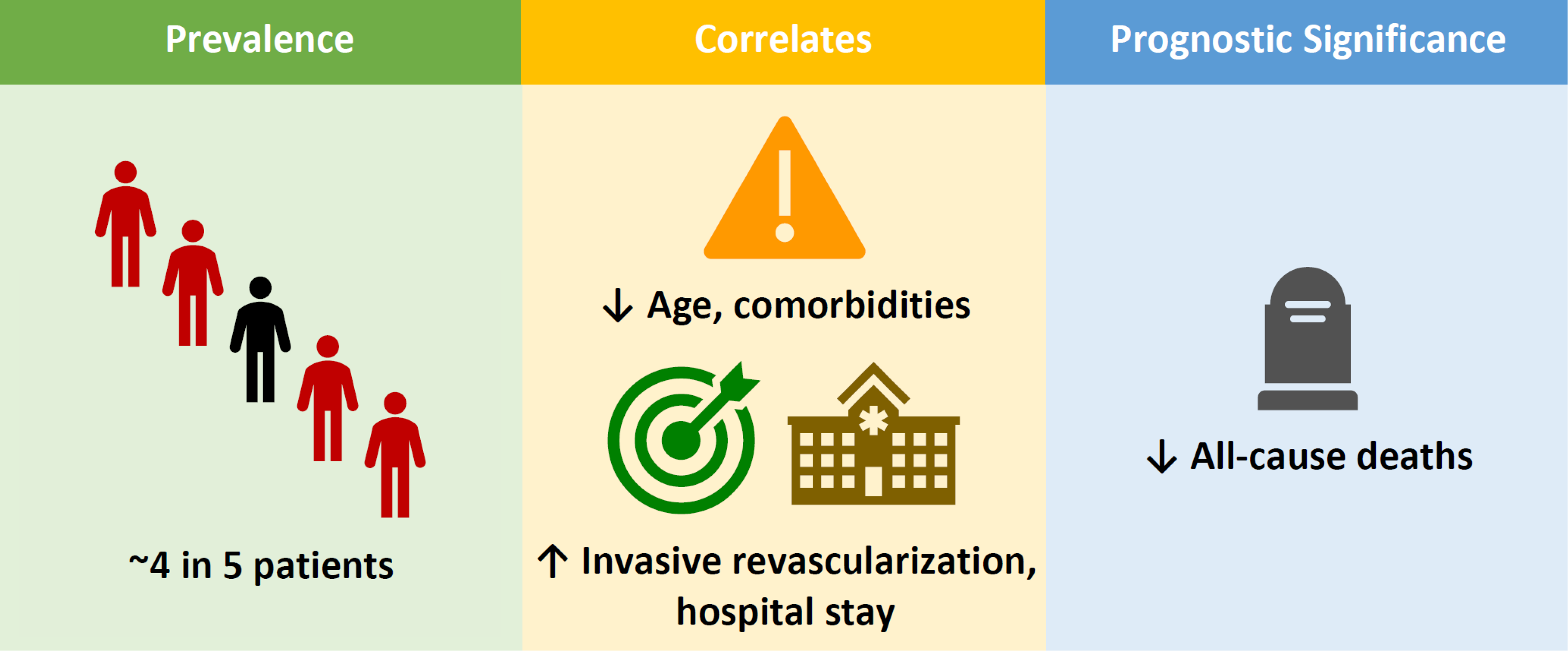
Graphical Abstract

1. Introduction

A simple, readily available, bedside non-invasive imaging modality, transthoracic echocardiography (TTE), is potentially advantageous in subjects with acute coronary syndromes, as it allows for the appreciation of cardiac structure and function as well as hemodynamic status, thus assisting treating teams in recognizing therapeutic targets, formulating management strategies and, ultimately, enhancing downstream patient course [1]. Nevertheless, currently there are only limited data to support the theoretical prognostic benefit associated with routinely performing TTE in stable acute myocardial infarction (AMI) patients during hospitalization, as reflected by a Level of Evidence C recommendation for such practice in the most recent European [2] and American [3] guidelines. To address this knowledge gap, and specifically test the hypothesis linking TTE administration with improved survival following uncomplicated AMI, we analyzed a large, contemporary database according to TTE use during the index admission.

2. Materials and Methods

2.1. Study Population and Outcome

Our study represents a sub-analysis of the previously described [4,5] Soroka Acute Myocardial Infarction (SAMI) registry. Included in the present, retrospective study were adult (i.e., ≥18-year-old) Israeli citizens admitted for AMI at Soroka University Medical Center, Israel, between 2002 and 2017 who did not develop cardiogenic shock and who survived the acute hospitalization phase. For subjects with multiple hospitalizations, we considered only the initial one. The study outcome was all-cause mortality during the first year after discharge.
Conforming to the Declaration of Helsinki, the project was approved by Soroka’s Institutional Review Board (approval number SOR-0319-16), and the need for informed consent was waived.

2.2. Data Collection and Definitions

Clinical data were retrieved from a web-based medical chart, in which baseline comorbidities were identified by International Classification of Diseases, Ninth Revision, Clinical Modification (ICD-9-CM) [6] codes, as documented in real-time by the treating physicians and according to accepted diagnostic criteria. Deaths were confirmed using the Israeli Ministry of the Interior Population Registry.
AMI diagnosis was based upon the constellation of ischemic signs and/or symptoms coupled with an abrupt rise and fall in cardiac biomarker levels consistent with acute myocardial injury, as dictated by the Universal Definition of Myocardial Infarction at the time [7]. Diagnosis of obstructive coronary artery disease required angiographic evidence of a ≥70% vessel diameter narrowing.
Echocardiographic diagnoses aligned with the American Society of Echocardiography guidelines. Accordingly, severe left ventricular dysfunction was defined by an ejection fraction of <30%, and pulmonary hypertension as an estimated pulmonary arterial systolic pressure of ≥37 mmHg.

2.3. Statistical Analysis

Both the entire cohort and a propensity score (PS)-matched sub-cohort were analyzed according to the administration of in-hospital TTE. Within the latter, pairing was dictated by the probability of undergoing TTE, using a nearest neighbor, 1-to-1 matching method, and a tolerance of ≤0.2. Included in the binary logistic regression model used for predicting this probability were baseline/presenting characteristics of known or perceived prognostic significance, all selected prior to the execution of descriptive analyses: increasing age, male sex, atherosclerotic risk factors, chronic obstructive pulmonary disease, chronic kidney disease, pre-existent chronic coronary syndrome, clinical heart failure, right heart failure, atrial fibrillation/flutter, and more recent year of event. Between-group balance for each of PS variable was assessed using absolute standardized differences, as depicted by a Love plot (Figure S1). In addition, discriminative ability of the score was checked using Harrell’s C-statistic, which proved acceptable (0.72, 95% CI 0.71–0.73, p < 0.001).
At all stages, variables were reported as frequencies and percentages or means and standard deviations and compared using Pearson’s Chi-Square or Student’s t tests, as appropriate.
The time-dependent probability and cumulative incidence of mortality as a function of TTE performance were assessed by the Kaplan–Meier method and compared using the Log-Rank test. Independent associations with the risk for all-cause death were evaluated by a Cox proportional hazard multivariable analysis and expressed using each’s unadjusted hazard ratio (HR)/adjusted HR (AdjHR) and the latter’s 95% confidence interval (CI). The model incorporated covariates exhibiting a p-value of <0.1 on the univariable stage. The proportional hazards assumption was evaluated qualitatively by checking for parallel orientation of the log-minus-log survival plots for all covariates. Model fit was assessed using the −2 log likelihood and likelihood ratio test.
Statistical significance required a two-sided p-value of <0.05. All analyses were performed using Statistical Package for the Social Sciences (SPSS), version 31 (IBM Corporation, Armonk, NY, USA).

3. Results

3.1. Baseline Characteristics of the Study Population

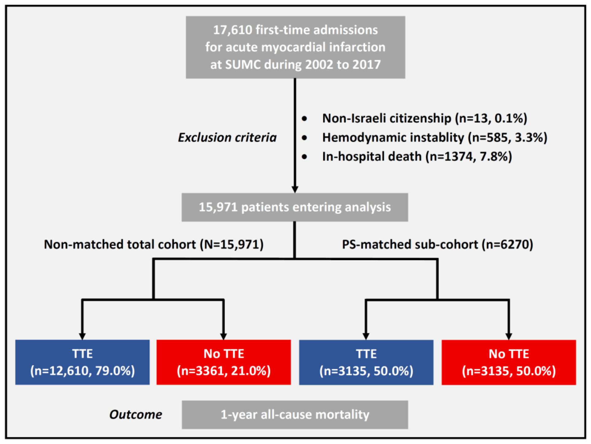
A total of 15,971 individuals qualified for analysis (Figure 1). Of them, 12,610 (79.0%) underwent TTE during their hospital stay. Notably, TTE utilization rose during the study recruitment period, from 74.2% in 2002 to 86.6% in 2017 (p for trend <0.001).
Compared to patients who were not scanned, those within the TTE group were recruited to the study at a later calendar year and were younger and more likely to be males of a non-Jewish minority background (Table 1). They also exhibited an overall higher burden of atherosclerotic cardiovascular risk factors but fewer comorbidities including overt heart failure. Echocardiograms demonstrated severe left ventricular dysfunction in 1316 (8.2%) out of the 12,610 cases referred for TTE, right ventricular dysfunction in 971 (7.7%), moderate and above mitral regurgitation in 670 (4.2%), moderate and above tricuspid regurgitation in 426 (2.7%), and pulmonary hypertension in 913 (5.7%).

3.2. Acute Event Aspects

Subjects who underwent TTE presented more commonly with ST elevation myocardial infarction (STEMI) (n = 6356, 50.4% vs. n = 1005, 29.9%, p < 0.001) and with a similarly low rate (0.3%) of cardiac arrest compared to those who did not undergo TTE (Table 2). The former were more likely to be assessed angiographically and to obtain definitive revascularization, leading to more intensive coronary care unit admissions/transfers and longer hospital stays. Also, their hospitalization courses were characterized by more frequent arrhythmic sequelae and fewer bleeding complications and sepsis. Specifically among patients who underwent both TTE and invasive revascularization and for whom data regarding respective timings were available (n = 10,356), TTE preceded revascularization by more than a day in 1129 (10.9%) cases, took place within a day of revascularization in 7238 (70.0%), or was performed more than a day after revascularization in 1989 (19.2%).

3.3. Outcome

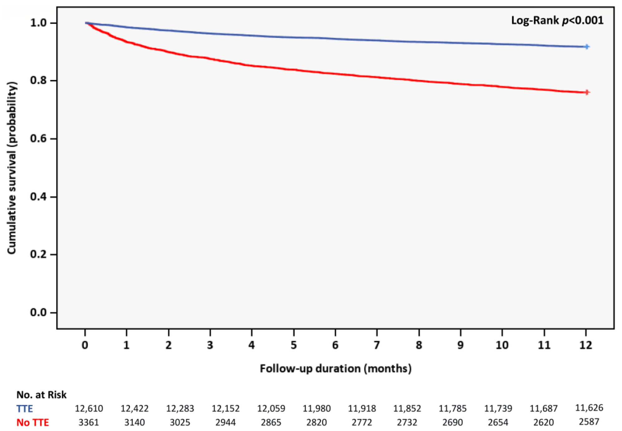
By 1 year after discharge, 1836 (11.5%) patients died. TTE performance was associated with a lower rate, cumulative incidence, and—independent of accompanying prognostic markers at baseline and during hospitalization—risk of all-cause mortality (n = 1032/12,619, 8.2% vs. n = 804/3361, 23.9%, p < 0.001; Log-Rank p < 0.001; unadjusted HR 0.31, 95% CI 0.28–0.34, p < 0.001; AdjHR 0.75, 95% CI 0.67–0.83, p < 0.001) (Figure 2 and Table 3).
Of note, within the TTE/invasive revascularization subgroup, death rate was highest among patients scanned after revascularization (n = 187/1989, 9.4%), intermediate among those scanned before revascularization (n = 48/1129, 4.3%), and lowest among those scanned surrounding revascularization (n = 284/7238, 3.9%) (p < 0.001).
Per exploratory analysis, the significant association between TTE performance and reduced mortality risk was confined to patients presenting with non-ST elevation MI (NSTEMI) (AdjHR 0.71, 95% CI 0.63–0.80, p < 0.001) and those managed non-invasively (AdjHR 0.72, 95% CI 0.64–0.81, p < 0.001), as opposed to patients with STEMI (AdjHR 0.92, 95% CI 0.74–1.13, p = 0.417) and those referred to invasive revascularization (AdjHR 0.88, 95% CI 0.69–1.13, p = 0.328).

3.4. Propensity Score Matching Analysis

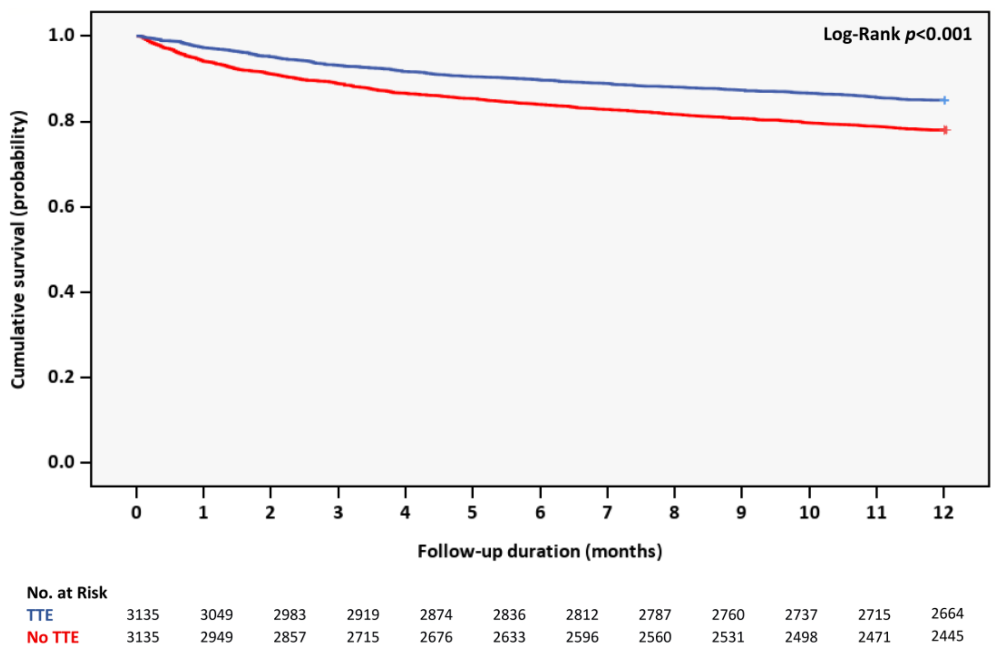
Within a 6270-case PS-matched sub-cohort, composed of 3135 patients who underwent TTE and 3135 matched controls who did not, most differences in baseline characteristics and acute event aspects became neutralized (Table 4 and Table 5). Still, ST elevation, angiographic assessment, invasive revascularization, and intensive care unit management were more common in the TTE group. Similar to the total unmatched cohort, patients who underwent TTE experienced fewer, more distant death events (n = 471, 15.0% vs. n = 690, 22.0%, p < 0.001; Log-Rank p < 0.001), and TTE utilization conferred a lower death risk (unadjusted HR 0.65, 95% CI 0.58–0.73, p < 0.001; AdjHR 0.77, 95% CI 0.68–0.88, p < 0.001) (Figure 3 and Table 6). Likewise, the prognostic significance of TTE administration was only observed among NSTEMI cases (AdjHR 0.75, 95% CI 0.65–0.86, p < 0.001) and non-invasively managed patients (AdjHR 0.75, 95% CI 0.65–0.86, p < 0.001), as opposed to STEMI scenarios (AdjHR 0.89, 95% CI 0.68–1.16, p = 0.369) and invasively treated subjects (AdjHR 0.90, 95% CI 0.67–1.21, p = 0.492).

4. Discussion

In this single-center, retrospective study, we examined the utilization of in-hospital TTE among 15,971 real-world patients admitted for non-fatal, stable AMI over a period of 15 years. Its results revealed the following: 1. TTE was performed more frequently in later years and in almost 80% of subjects overall. 2. Compared to patients who did not undergo TTE during the index hospitalization, those who did presented at a younger age and with fewer comorbidities, were more often managed invasively and within the intensive coronary care unit, and exhibited a lengthier hospital stay. 3. TTE performance was associated with improved 1-year survival, independently conferring a mortality risk that was 1.3 times lower than that observed within the no-TTE group.
The current literature concerning the prevalence, correlates, and prognostic significance of in-hospital TTE administration in AMI is limited. In the largest report prior to ours, covering almost 100,000 admissions across nearly 400 centers in the United States, TTE was performed in 74% of cases and a more liberal use of TTE correlated with higher costs and longer hospitalizations without affecting inpatient mortality nor 3-month readmissions [8]. Importantly, the authors relied on an administrative, billing code-focused database that spanned merely one year of practice (2014) and which included a highly heterogenous population treated in centers of varying capabilities (with some not offering percutaneous coronary intervention services at all). Moreover, the study’s analyses revolved around inter-center comparisons rather than patient-level, actual administration of TTE per se, all while considering stable/unstable patients and first-time/repeat hospitalizations as one. An earlier study (2001–2011) examining the use of echocardiography in various medical conditions identified enhanced in-hospital survival among AMI patients who underwent TTE [9]. However, echocardiography was performed in only 7% of cases and no adjustment was made for deaths occurring prior to the performance of TTE. Bearing in mind these previous works’ setbacks, we designed the present study which focused on mid-term survival post-AMI, presumably providing a more accurate reflection of TTE utilization.
Our observations have two clinically relevant implications. The first is that performing TTE during hospitalization for AMI was linked to better prognosis following discharge. It could be that earlier detection of actionable conditions known to affect survival, such as systolic dysfunction, diastolic impairment, valvular disorders, and structural/thrombotic complications allowed for a more comprehensive management (that is, beyond mere revascularization) within the TTE group that in turn led to improved outcomes. Consistent with this assumption were: a. the lower death rates reported among revascularized patients who underwent TTE at an earlier stage; and b. the confinement of the reduced death hazard associated with TTE use to patients presenting with NSTEMI or who were managed non-invasively, both of whom are traditionally less exposed to prognostically meaningful interventions and may thus derive added benefit from TTE. Alternatively, the administration of TTE may have simply served as a surrogate of stricter guideline-directed therapy adherence and hence higher standards of care. This hypothesis fits well with the observation linking TTE performance with greater utilization of invasive revascularization and intensive care environment and more advanced year of event, although the latter did not alter the independent prognostic value of TTE use (and, furthermore, intensive care unit stay and event year were not associated with survival on their own). As our registry lacked information regarding full TTE reports, their impact on management strategy, death causes, and clinical pathways leading to TTE use in the first place, the exact mechanism(s) underlying our findings remain speculative. Accordingly, and as much as the study’s extensive multivariable analyses implied the independent predictive role of TTE use, further, preferentially prospective validation is needed.
The second message offered by our study is that TTE was relatively underutilized during the hospitalization phase surrounding AMI diagnosis, much in the same manner reported by prior publications [10]. In light of the observed intergroup differences in patient characteristics and revascularization approach, it could be that TTE was perceived as lower-yield in those subjects who were deemed too high risk or unfit for interventional therapies a priori. Technical obstacles, too, may have contributed to the low usage of TTE in persons presenting with numerous comorbidities, in whom hand-held echocardiography could constitute a similarly accurate and more feasible alternative [11]. Once again, we were unable to identify causality, thus we are leaving it for future studies to better define barriers for the implementation of TTE in this setting.

Limitations

First, our study stemmed from a single-center, retrospective analysis that did not employ randomization nor external adjudication, potentially leading to selection bias and hampering the generalizability of the results. However, we relied on a large cohort that represented a population of close to 1 million and whose baseline characteristics resembled those of prior registries. Also, randomized-controlled trials of TTE utilization among stable AMI patients are hardly pragmatic, arguably making real-world exploration as our the next best alternative. Secondly, and again reflecting the study’s retrospective design, statistical precision and associated clinical relevance of our findings may be questionable. Yet, based on the sample size, exposure (i.e., TTE use) distribution, event rate, and Cox model utilized, and assuming a two-sided α of 0.05 and 80% power, we estimated the minimum detectable effect size as HR for all-cause mortality of approximately 0.90, a figure which is generally considered clinically meaningful in cardiovascular outcome research, particularly in settings where imaging may influence downstream decision-making rather than exert direct therapeutic effects. Thirdly, aiming to focus on patients for whom current practice guidelines provide expert opinion-based recommendation only, and in view of the study’s objective to explore the 1-year trajectory of these individuals, we deliberately excluded in-hospital fatalities, hence introducing survivor bias. This could also mitigate the possibility of immortal time bias and in essence led to a modest 7.8% exclusion rate (Figure 1). Fourthly, the protracted timeframe of enrollment may have led to inconsistencies in medical definitions and treatment approaches, which also could have affected the interpretation of the findings. Nevertheless, both study groups were exposed to similarly evolving diagnostic criteria and practices, and admission year did not possess an independent prognostic capacity nor did it undermine the significant association demonstrated between TTE performance and 1-year mortality. Fifthly, in addition to death causes, we were also unaware of the exact TTE scope (complete vs. focused), and—in some patients—timing, as well as MI types (e.g., type 2) and extent (e.g., Killip class, serum biomarker levels), medical therapies other than blood transfusion, and reason for re-admissions. Consequently, we could not comment on the pathophysiologic basis accounting for our observations nor rule out confounding by non-coronary-related phenomena. Lastly, and in view of the evaluation of multiple covariates by a Cox regression model, chance findings could not be entirely excluded, necessitating cautious interpretation which takes into consideration effect size (and the possibility of smaller effect sizes than the one estimated above), confidence intervals, and their clinical plausibility.

5. Conclusions

In our large, single-center experience, in-hospital TTE was utilized in close to 80% of subjects with stable, non-fatal AMI and was associated with a lower-risk patient profile, a higher rate of invasive coronary interventions and intensive care management, and a more favorable 1-year survival. These findings call for further assessment of the routine use of TTE in such scenarios.

Supplementary Materials

The following supporting information can be downloaded at https://www.mdpi.com/article/10.3390/jcdd13070322/s1. Figure S1: Love plot for balance in propensity score variables.

Author Contributions

Conceptualization, A.S. (Alon Shechter); methodology, A.S. (Alon Shechter) and Y.P.; formal analysis, Y.P.; data curation, H.G. and Y.P.; writing—original draft preparation, A.S. (Alon Shechter); writing—review and editing, A.S. (Alon Shechter), A.S. (Arthur Shiyovich), R.J.S., O.M., H.G. and Y.P. All authors have read and agreed to the published version of the manuscript.

Funding

This research received no external funding.

Institutional Review Board Statement

The study was conducted in accordance with the Declaration of Helsinki and approved by the Soroka Medical Center Institutional Review Board (initial approval SOR-0319-16, granted 19 September 2016, extended up to 13 December 2022).

Informed Consent Statement

Patient consent was waived due the study’s retrospective nature.

Data Availability Statement

The data underlying this article will be shared upon reasonable request to the corresponding author.

Conflicts of Interest

The authors declare no conflicts of interest.

Abbreviations

The following abbreviations are used in this manuscript:
AMIAcute myocardial infarction
TTETransthoracic echocardiography

References

  1. Mollema, S.A.; Nucifora, G.; Bax, J.J. Prognostic Value of Echocardiography After Acute Myocardial Infarction. Heart 2009, 95, 1732–1745. [Google Scholar] [CrossRef] [PubMed]
  2. Byrne, R.A.; Rossello, X.; Coughlan, J.J.; Barbato, E.; Berry, C.; Chieffo, A.; Claeys, M.J.; Dan, G.A.; Dweck, M.R.; Galbraith, M.; et al. 2023 ESC Guidelines for the Management of Acute Coronary Syndromes. Eur. Heart J. 2023, 44, 3720–3826. [Google Scholar] [CrossRef] [PubMed]
  3. Rao, S.V.; O’Donoghue, M.L.; Ruel, M.; Rab, T.; Tamis-Holland, J.E.; Alexander, J.H.; Baber, U.; Baker, H.; Cohen, M.G.; Cruz-Ruiz, M.; et al. 2025 ACC/AHA/ACEP/NAEMSP/SCAI Guideline for the Management of Patients with Acute Coronary Syndromes: A Report of the American College of Cardiology/American Heart Association Joint Committee on Clinical Practice Guidelines. Circulation 2025, 151, e771–e862. [Google Scholar] [CrossRef] [PubMed]
  4. Plakht, Y.; Shiyovich, A.; Weitzman, S.; Fraser, D.; Zahger, D.; Gilutz, H. Soroka Acute Myocardial Infarction (SAMI) Score Predicting 10-Year Mortality Following Acute Myocardial Infarction. Int. J. Cardiol. 2013, 167, 3068–3070. [Google Scholar] [CrossRef] [PubMed]
  5. Shechter, A.; Shiyovich, A.; Skalsky, K.; Gilutz, H.; Plakht, Y. Interaction between Anemia and Renal Dysfunction in Relation to Long-Term Survival Following Acute Myocardial Infarction. Clin. Res. Cardiol. 2024, 113, 1692–1706. [Google Scholar] [CrossRef] [PubMed]
  6. Centers for Disease Control. 2014. Available online: https://www.cms.gov/Medicare/Coding/ICD9ProviderDiagnosticCodes/codes (accessed on 1 August 2025).
  7. Thygesen, K.; Alpert, J.S.; Jaffe, A.S.; Chaitman, B.R.; Bax, J.J.; Morrow, D.A.; White, H.D. Executive Group on behalf of the Joint European Society of Cardiology (ESC)/American College of Cardiology (ACC)/American Heart Association (AHA)/World Heart Federation (WHF) Task Force for the Universal Definition of Myocardial Infarction. Fourth Universal Definition of Myocardial Infarction (2018). Circulation 2018, 138, e618–e651. [Google Scholar] [CrossRef] [PubMed]
  8. Pack, Q.R.; Priya, A.; Lagu, T.; Pekow, P.S.; Schilling, J.P.; Hiser, W.L.; Lindenauer, P.K. Association Between Inpatient Echocardiography Use and Outcomes in Adult Patients with Acute Myocardial Infarction. JAMA Intern. Med. 2019, 179, 1176–1185. [Google Scholar] [CrossRef] [PubMed]
  9. Papolos, A.; Narula, J.; Bavishi, C.; Chaudhry, F.A.; Sengupta, P.P. U.S. Hospital Use of Echocardiography: Insights from the Nationwide Inpatient Sample. J. Am. Coll. Cardiol. 2016, 67, 502–511. [Google Scholar] [CrossRef] [PubMed]
  10. Santolucito, P.A.; Tighe, D.A.; Lessard, D.; Ismailov, R.M.; Gore, J.M.; Yarzebski, J.; Goldberg, R.J. Changing Trends in the Evaluation of Ejection Fraction in Patients Hospitalized with Acute Myocardial Infarction: The Worcester Heart Attack Study. Am. Heart J. 2008, 155, 485–493. [Google Scholar] [CrossRef] [PubMed]
  11. Geers, J.; Balfour, A.; Molek, P.; Barron, P.; Botezatu, S.; Joshi, S.S.; White, A.; Buchwald, M.; Everett, R.; McCarley, J.; et al. Systematic Hand-Held Echocardiography in Patients Hospitalized with Acute Coronary Syndrome. Eur. Heart J. Cardiovasc Imaging 2024, 25, 1441–1450. [Google Scholar] [CrossRef] [PubMed]
Figure 1. Study flow chart. SUMC = Soroka University Medical Center; TTE = transthoracic echocardiography.
Jcdd 13 00322 g001
Figure 2. Cumulative survival according to transthoracic echocardiography use. TTE = transthoracic echocardiography.
Jcdd 13 00322 g002
Figure 3. Cumulative survival according to transthoracic echocardiography use within the propensity score-matched sub-cohort. TTE = transthoracic echocardiography.
Jcdd 13 00322 g003
Table 1. Baseline clinical characteristics.
Total Cohort
(N = 15,971)
Transthoracic
Echocardiography Use
p-Value
Yes
(n = 12,610)
No
(n = 3361)
Demographic details
Age <0.001
Continuous (years)66 ± 1464 ± 1372 ± 14
<65 years7926 (49.6)6959 (55.2)967 (28.8)
65–74 years3623 (22.7)2836 (22.5)787 (23.4)
≥75 years4422 (27.7)2815 (22.3)1607 (47.8)
Sex male11,152 (69.8)9165 (72.7)1987 (59.1)<0.001
Non-Jewish minority2651 (16.6)2213 (17.5)438 (13.0)<0.001
Atherosclerotic
cardiovascular risk factors
Diabetes mellitus6444 (40.3)4912 (39.0)1532 (45.6)<0.001
Dyslipidemia12,975 (81.2)10,556 (83.7)2419 (72.0)<0.001
Hypertension8280 (51.8)6438 (51.1)1842 (54.8)<0.001
Obesity3579 (22.4)3033 (24.1)546 (16.2)<0.001
Smoking history6743 (42.2)5879 (46.6)864 (25.7)<0.001
Family history of
ischemic heart disease
1494 (9.4)1360 (10.8)134 (4.0)<0.001
Cardiovascular
comorbidities
Chronic coronary syndrome12,496 (78.2)10,473 (83.1)2023 (60.2)<0.001
History of
myocardial infarction
1786 (11.2)1320 (10.5)466 (13.9)<0.001
Prior revascularization
Percutaneous
coronary intervention
1907 (11.9)1493 (11.8)414 (12.3)0.448
Coronary artery
bypass grafting
1243 (7.8)889 (7.0)354 (10.5)<0.001
Peripheral arterial disease1799 (11.3)1337 (10.6)462 (13.7)0.266
Atrial fibrillation/flutter2447 (15.3)1738 (13.8)709 (21.1)<0.001
Atrioventricular block592 (3.7)470 (3.7)122 (3.6)0.791
Clinical heart failure2556 (16.0)1870 (14.8)686 (20.4)<0.001
Non-cardiovascular
comorbidities
Chronic obstructive
pulmonary disease
1232 (7.7)880 (7.0)352 (10.5)<0.001
Stage ≥III
chronic kidney disease
5157 (32.3)3749 (29.7)1408 (41.9)<0.001
Anemia8498 (53.2)6437 (51.0)2061 (61.3)<0.001
Neurological disorders2567 (16.1)1710 (13.6)857 (25.5)<0.001
Malignancy612 (3.8)422 (3.3)190 (5.7)<0.001
Substance use disorder340 (2.1)284 (2.3)56 (1.7)0.036
Psychotic disorders258 (1.6)177 (1.4)81 (2.4)<0.001
Data are presented as number (percent) or mean ± standard deviation. Figures in bold denote statistical significance.
Table 2. Acute event aspects.
Total Cohort
(N = 15,971)
Transthoracic
Echocardiography Use
p-Value
Yes
(n = 12,610)
No
(n = 3361)
Clinical presentation
ST elevation
myocardial infarction
7361 (46.1)6356 (50.4)1005 (29.9)<0.001
Cardiac arrest52 (0.3)43 (0.3)9 (0.3)0.508
Right heart failure1251 (7.8)971 (7.7)280 (8.3)0.227
Angiographic parameters
Angiogram performed11,038 (69.1)10,356 (82.1)682 (20.3)<0.001
Vessels
significantly involved
<0.001
0507 (4.6)446 (4.3)61 (8.9)
13076 (27.9)2913 (28.1)163 (23.9)
23092 (28.0)2913 (28.1)179 (26.2)
3/Left main4363 (39.5)4084 (39.4)279 (40.9)
Hospital course
Revascularization approach <0.001
None/
conservative treatment
4207 (26.3)2087 (16.6)2120 (63.1)
Percutaneous
coronary intervention
9720 (60.9)8753 (69.4)967 (28.8)
Coronary artery bypass grafting2044 (12.8)1770 (14.0)274 (8.2)
Intensive coronary care unit stay11,029 (69.1)10,119 (80.2)910 (27.1)<0.001
Ventricular tachycardia378 (2.4)332 (2.6)46 (1.4)<0.001
Any form of pacing284 (1.8)269 (2.1)15 (0.4)<0.001
Mechanical ventilation548 (3.4)438 (3.5)110 (3.3)0.570
Gastrointestinal bleeding320 (2.0)242 (1.9)78 (2.3)0.140
Blood transfusion1887 (11.8)1449 (11.5)438 (13.0)0.014
Sepsis124 (0.8)85 (0.7)39 (1.2)0.004
Hospitalization length
Continuous (days)10.0 ± 9.010.3 ± 9.28.8 ± 8.1<0.001
≥7 days7538 (47.2)6207 (49.2)1331 (39.6)<0.001
Data are presented as number (percent) or mean ± standard deviation. Figures in bold denote statistical significance.
Table 3. Multivariable Cox proportional hazard model for the outcome of all-cause mortality at 1 year.
UnivariableMultivariable
HR
(95% CI)
p-ValueAdjHR
(95% CI)
p-Value
Year of admission
(continuous, per 1-year increase)
0.97 (0.96–0.98)<0.0011.00 (0.99–1.02)0.456
Demographic details
Age ≥ 65 vs. <65 years4.42 (3.78–5.18)<0.0012.26 (1.91–2.66)<0.001
Sex male0.52 (0.47–0.57)<0.0010.92 (0.83–1.02)0.102
Atherosclerotic
cardiovascular risk factors
Dyslipidemia0.46 (0.41–0.50)<0.0010.75 (0.68–0.83)<0.001
Obesity0.49 (0.42–0.56)<0.0010.77 (0.67–0.88)<0.001
Diabetes mellitus1.51 (1.42–1.70)<0.0011.18 (1.07–1.30)<0.001
Cardiovascular
comorbidities
Chronic coronary syndrome0.30 (0.27–0.33)<0.0010.81 (0.73–0.91)<0.001
Peripheral arterial disease2.35 (2.10–2.62)<0.0011.49 (1.32–1.67)<0.001
Atrial fibrillation/flutter2.62 (2.38–2.90)<0.0011.28 (1.16–1.42)<0.001
Clinical heart failure2.75 (2.49–3.03)<0.0011.31 (1.18–1.46)<0.001
Non-cardiovascular
comorbidities
Chronic obstructive
pulmonary disease
2.55 (2.25–2.88)<0.0011.59 (1.40–1.81)<0.001
Stage ≥ III
chronic kidney disease
3.36 (3.06–3.69)<0.0011.42 (1.28–1.58)<0.001
Anemia3.13 (2.81–3.49)<0.0011.56 (1.39–1.75)<0.001
Neurological disorders3.22 (2.93–3.54)<0.0011.57 (1.42–1.74)<0.001
Malignancy3.88 (3.37–4.47)<0.0012.32 (2.00–2.68)<0.001
Substance use disorder2.19 (1.69–2.83)<0.0011.83 (1.37–2.46)<0.001
Psychotic disorders2.55 (2.25–2.88)<0.0011.46 (1.12–1.88)0.004
Clinical presentation
ST elevation (vs. non-ST elevation) myocardial infarction0.46 (0.42–0.51)<0.0011.01 (0.90–1.13)0.880
Right heart failure2.64 (2.34–2.98)<0.0011.39 (1.23–1.58)<0.001
Hospital course
Invasive revascularization vs. conservative approach0.15 (0.14–0.17)<0.0010.45 (0.39–0.52)<0.001
Mechanical ventilation2.50 (2.10–2.98)<0.0011.34 (1.12–1.61)0.001
Transthoracic
echocardiography use
0.31 (0.28–0.34)<0.0010.75 (0.67–0.83)<0.001
Figures in bold denote statistical significance. AdjHR = adjusted hazard ratio; CI = confidence interval; HR = hazard ratio.
Table 4. Baseline clinical characteristics of the propensity score-matched sub-cohort.
Propensity Score-Matched
Sub-Cohort
(N = 6270)
Transthoracic
Echocardiography Use
p-Value
Yes
(n = 3135)
No
(n = 3135)
Demographic details
Age
Continuous (years)71.42 ± 13.0471.4 ± 12.5971.41 ± 13.480.938
<65 years1953 (31.1)986 (31.5)967 (30.8)0.711
65–74 years1533 (24.4)753 (24.0)780 (24.9)
≥75 years2784 (44.4)1396 (44.5)1388 (44.3)
Sex male3833 (61.1)1921 (61.3)1912 (61.0)0.816
Non-Jewish minority455 (7.3)31 (1.0)424 (13.5)<0.001
Atherosclerotic
cardiovascular risk factors
Diabetes mellitus2808 (44.8)1379 (44.0)1429 (45.6)0.204
Dyslipidemia4736 (75.5)2380 (75.9)2356 (75.2)0.481
Hypertension3448 (55.0)1720 (54.9)1728 (55.1)0.839
Obesity1098 (17.5)557 (17.8)541 (17.3)0.595
Smoking history1696 (27.0)837 (26.7)859 (27.4)0.532
Family history of
ischemic heart disease
323 (5.2)189 (6.0)134 (4.3)0.002
Cardiovascular
comorbidities
Chronic coronary syndrome4020 (64.1)2000 (63.8)2020 (64.4)0.598
History of
myocardial infarction
732 (11.7)270 (8.6)462 (14.7)<0.001
Prior revascularization
Percutaneous
coronary intervention
716 (11.4)303 (9.7)413 (13.2)<0.001
Coronary artery
bypass grafting
588 (9.4)235 (7.5)353 (11.3)<0.001
Peripheral arterial disease801 (12.8)369 (11.8)432 (13.8)0.017
Atrial fibrillation/flutter1304 (20.8)658 (21.0)646 (20.6)0.709
Atrioventricular block281 (4.5)163 (5.2)118 (3.8)0.006
Clinical heart failure1253 (20.0)631 (20.1)622 (19.8)0.776
Non-cardiovascular
comorbidities
Chronic obstructive
pulmonary disease
589 (9.4)263 (8.4)326 (10.4)0.006
Stage ≥ III
chronic kidney disease
2556 (40.8)1266 (40.4)1290 (41.1)0.537
Anemia3741 (59.7)1848 (58.9)1893 (60.4)0.247
Neurological disorders1432 (22.8)716 (22.8)716 (22.8)1.000
Malignancy320 (5.1)149 (4.8)171 (5.5)0.207
Substance use disorder108 (1.7)54 (1.7)54 (1.7)1.000
Psychotic disorders149 (2.4)70 (2.2)79 (2.5)0.456
Data are presented as number (percent) or mean ± standard deviation. Figures in bold denote statistical significance.
Table 5. Acute event aspects within the propensity score-matched sub-cohort.
Propensity Score-Matched
Sub-Cohort
(N = 6270)
Transthoracic
Echocardiography Use
p-Value
Yes
(n = 3135)
No
(n = 3135)
Clinical presentation
ST elevation
myocardial infarction
2327 (37.1)1363 (43.5)964 (30.7)<0.001
Cardiac arrest17 (0.3)10 (0.3)7 (0.2)0.466
Right heart failure569 (9.1)291 (9.3)278 (8.9)0.568
Angiographic parameters
Angiogram performed2723 (43.4)2042 (65.1)681 (21.7)<0.001
Vessels
significantly involved
0.198
0197 (7.2)136 (6.7)61 (9.0)
1640 (23.5)477 (23.4)163 (23.9)
2758 (27.8)579 (28.4)179 (26.3)
3/Left main1128 (41.4)850 (41.6)278 (40.8)
Hospital course
Revascularization approach <0.001
None/
conservative treatment
2948 (47.0)1052 (33.6)1896 (60.5)
Percutaneous
coronary intervention
2699 (43.0)1734 (55.3)965 (30.8)
Coronary artery bypass grafting623 (9.9)349 (11.1)274 (8.7)
Intensive coronary care unit stay2969 (47.4)2062 (65.8)907 (28.9)<0.001
Ventricular tachycardia107 (1.7)61 (1.9)46 (1.5)0.144
Any form of pacing103 (1.6)88 (2.8)15 (0.5)<0.001
Mechanical ventilation227 (3.6)124 (4.0)103 (3.3)0.156
Gastrointestinal bleeding149 (2.4)80 (2.6)69 (2.2)0.362
Blood transfusion823 (13.1)416 (13.3)407 (13.0)0.736
Sepsis57 (0.9)23 (0.7)34 (1.1)0.143
Hospitalization length
Continuous (days)9.93 ± 9.1011.03 ± 9.688.82 ± 8.32<0.001
≥7 days2973 (47.4)1724 (55.0)1249 (39.8)<0.001
Data are presented as number (percent) or mean ± standard deviation. Figures in bold denote statistical significance.
Table 6. Multivariable Cox proportional hazard model for the outcome of all-cause mortality at 1 year within the propensity score-matched sub-cohort.
UnivariableMultivariable
HR
(95% CI)
p-ValueAdjHR
(95% CI)
p-Value
Year of admission
(continuous, per 1-year increase)
0.98 (0.97–0.99)<0.0011.01 (1.00–1.02)0.214
Demographic details
Age ≥ 65 vs. <65 years3.83 (3.01–4.87)<0.0012.27 (1.78–2.90)<0.001
Sex male0.68 (0.61–0.77)<0.0010.97 (0.86–1.10)0.633
Atherosclerotic
cardiovascular risk factors
Diabetes mellitus1.21 (1.08–1.36)0.0011.05 (0.93–1.19)0.408
Dyslipidemia0.58 (0.51–0.65)<0.0010.76 (0.67–0.86)<0.001
Obesity0.53 (0.44–0.64)<0.0010.78 (0.64–0.94)0.010
Cardiovascular
comorbidities
Chronic coronary syndrome0.44 (0.39–0.49)<0.0010.84 (0.73–0.96)0.009
Peripheral arterial disease1.96 (1.70–2.26)<0.0011.45 (1.25–1.68)<0.001
Atrial fibrillation/flutter1.86 (1.64–2.10)<0.0011.20 (1.06–1.37)0.005
Clinical heart failure2.06 (1.83–2.34)<0.0011.28 (1.13–1.46)<0.001
Non-cardiovascular
comorbidities
Chronic obstructive
pulmonary disease
2.14 (1.84–2.5)<0.0011.57 (1.34–1.85)<0.001
Stage ≥ III
chronic kidney disease
2.37 (2.11–2.67)<0.0011.33 (1.17–1.51)<0.001
Anemia2.40 (2.09–2.75)<0.0011.46 (1.27–1.68)<0.001
Neurological disorders2.39 (2.12–2.69)<0.0011.62 (1.44–1.83)<0.001
Malignancy3.04 (2.54–3.63)<0.0012.19 (1.83–2.62)<0.001
Psychotic disorders1.72 (1.27–2.32)<0.0011.39 (1.03–1.88)0.033
Clinical presentation
ST elevation (vs. non-ST elevation) myocardial infarction0.57 (0.50–0.65)<0.0011.04 (0.90–1.20)0.619
Right heart failure2.08 (1.78–2.44)<0.0011.35 (1.15–1.60)<0.001
Hospital course
Invasive revascularization vs. conservative approach0.21 (0.18–0.24)<0.0010.42 (0.35–0.51)<0.001
Mechanical ventilation2.34 (1.87–2.93)<0.0011.44 (1.14–1.82)0.002
Transthoracic
echocardiography use
0.65 (0.58–0.73)<0.0010.77 (0.68–0.88)<0.001
Figures in bold denote statistical significance. AdjHR = adjusted hazard ratio; CI = confidence interval; HR = hazard ratio.
Disclaimer/Publisher’s Note: The statements, opinions and data contained in all publications are solely those of the individual author(s) and contributor(s) and not of MDPI and/or the editor(s). MDPI and/or the editor(s) disclaim responsibility for any injury to people or property resulting from any ideas, methods, instructions or products referred to in the content.