Coronary CT Angiography in PCI Planning: Advances, Clinical Applications, and Challenges

Abstract
1. Introduction
1.1. The Historical Arc: From Plain Radiography to CTCA-Based PCI Planning
1.2. Limitations of Invasive Coronary Angiography as a Sole Planning Tool
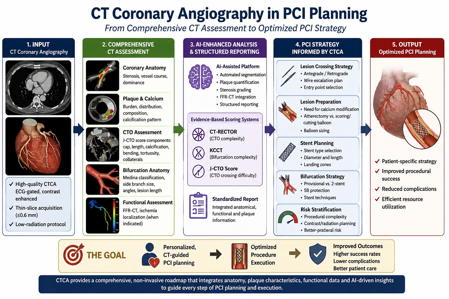
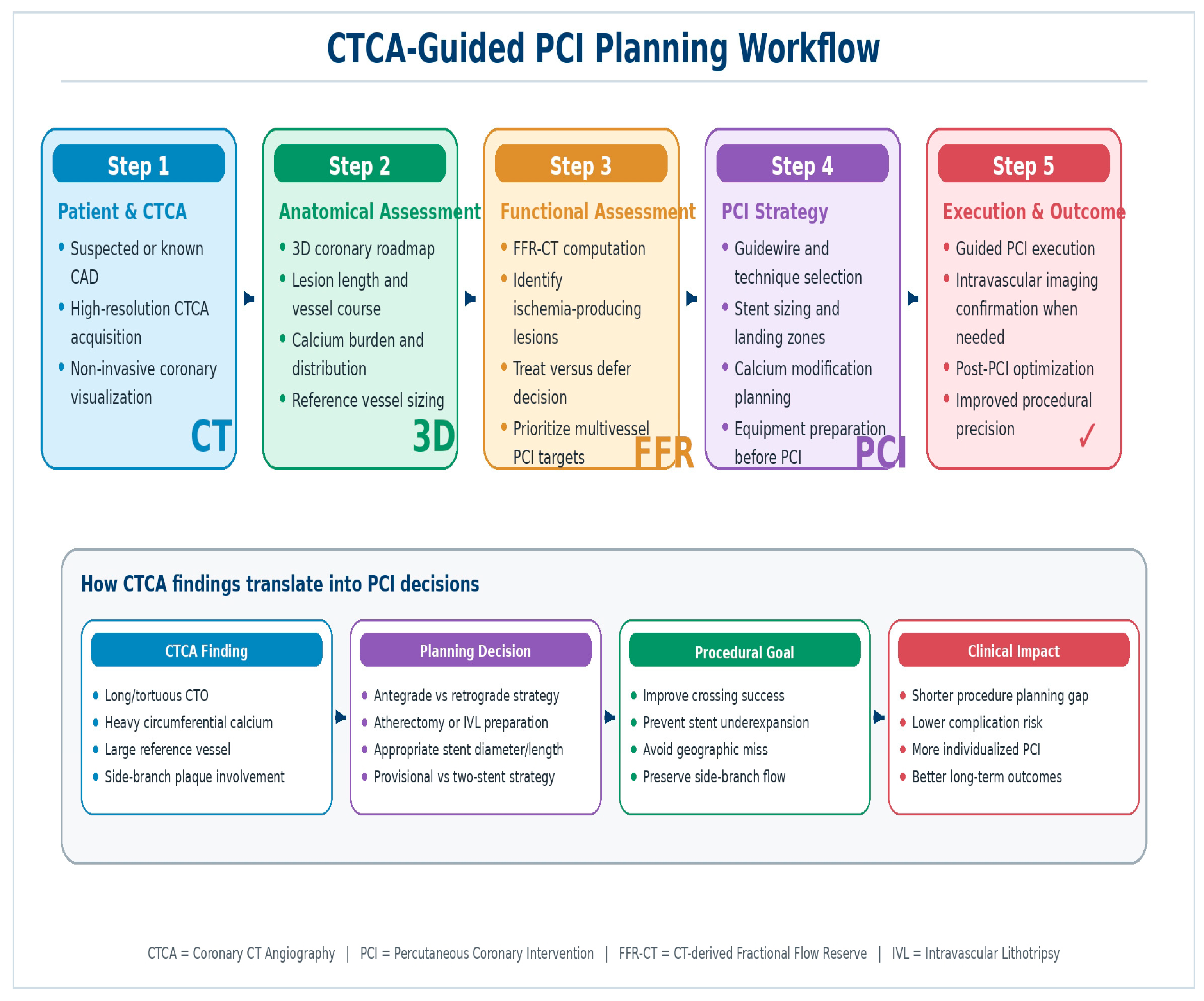
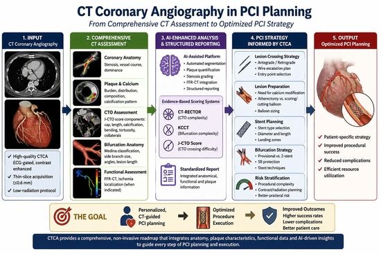
1.3. CTCA as a Pre-Procedural Planning Platform: The Emerging Paradigm
2. Technological Advances: FFR-CT, Artificial Intelligence, and Next-Generation Hardware
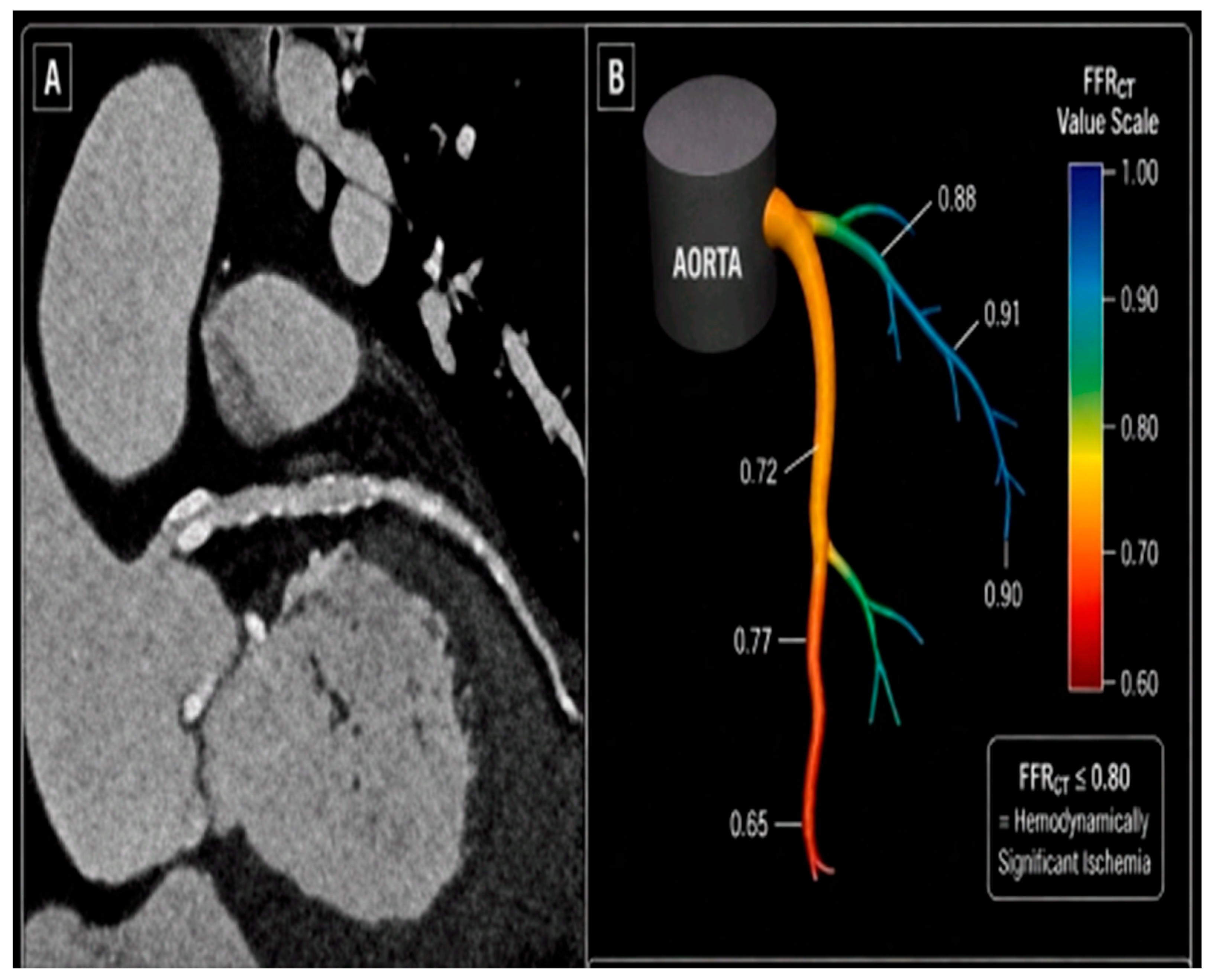
2.1. CT-Derived Fractional Flow Reserve (FFR-CT)
- Case Example 1: Left Main Stem PCI Guided by CTCA and FFR-CT
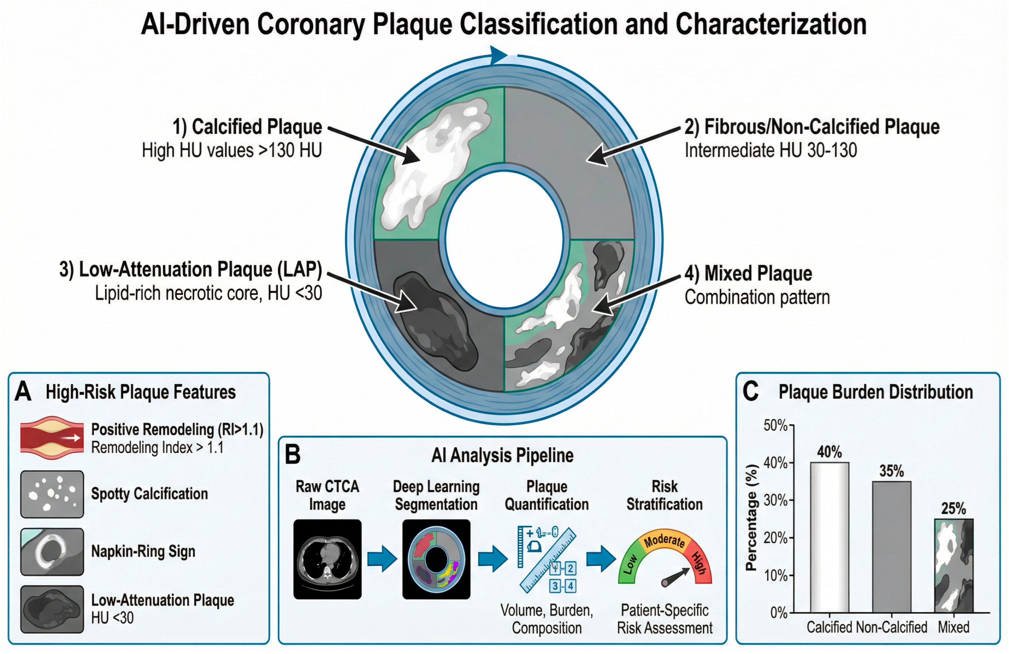
2.2. Artificial Intelligence in the CTCA Workflow
2.3. Next-Generation CT Hardware (Second Comment)
3. Clinical Applications in Complex PCI
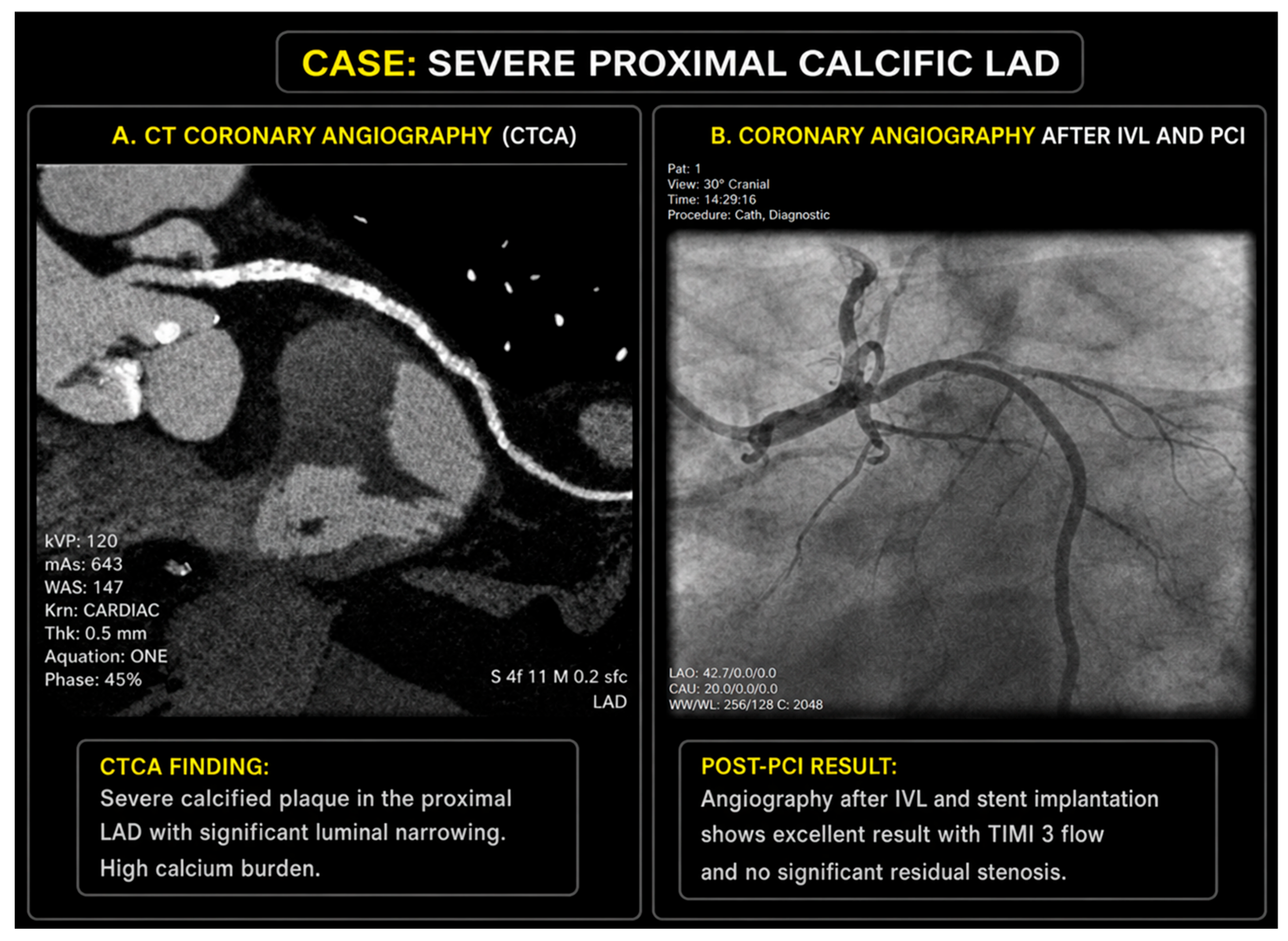
- Case Example 2: IVL-Assisted PCI for Severe Calcific Proximal LAD Disease
3.1. CTCA-Guided Lesion Preparation and PCI Optimization
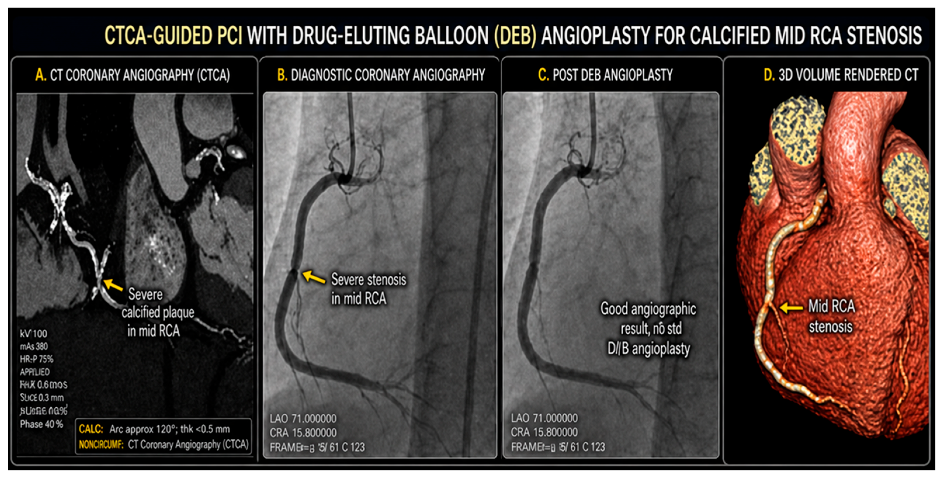
- Case Example 3: CTCA-Guided PCI with DEB Angioplasty for Calcified Mid-RCA Stenosis
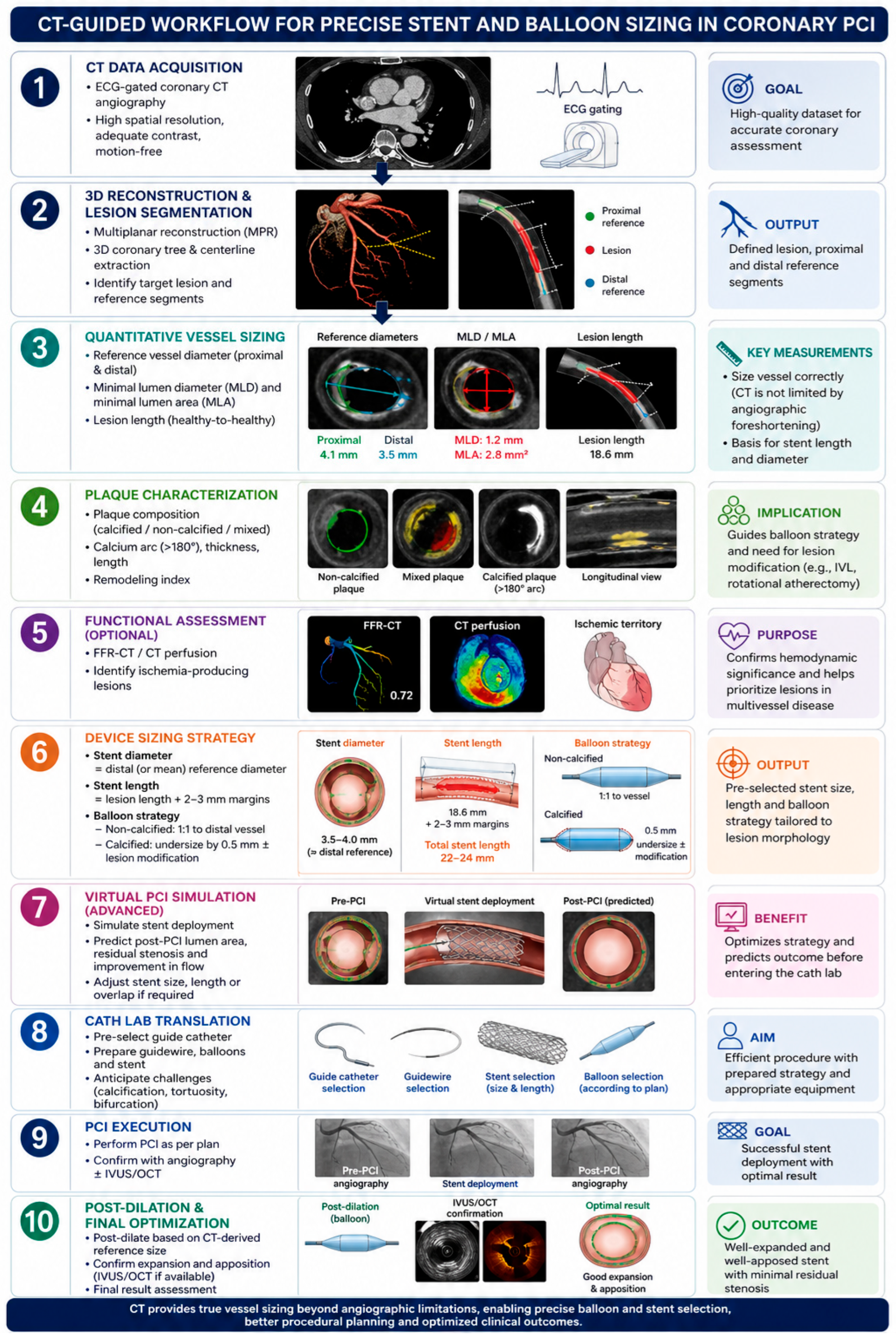
3.1.1. Precise Vessel Sizing and Stent Optimization
3.1.2. CTCA-Guided Calcium Modification Planning
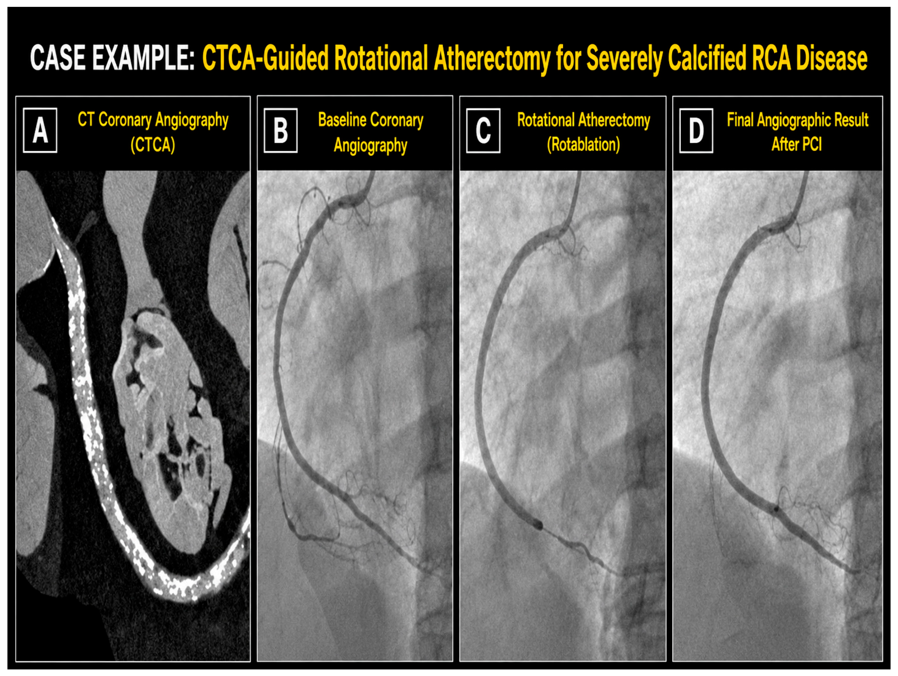
- Case Example 4: CTCA-Guided Rotablation for Severely Calcified RCA Disease
3.1.3. Multi-Vessel Disease and Revascularization Completeness
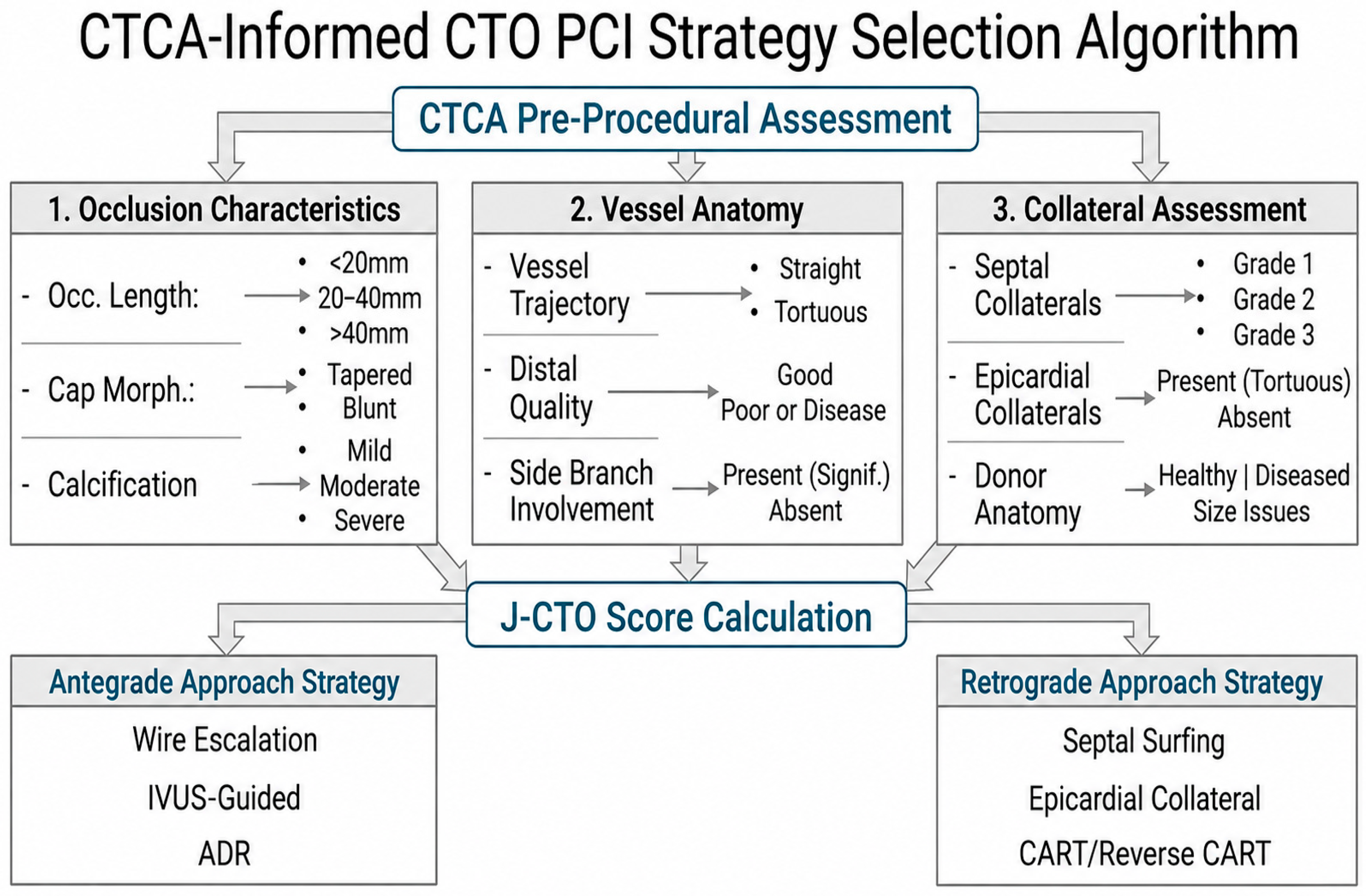
3.2. Chronic Total Occlusion PCI: CTCA-Guided Procedural Planning
3.3. Left Main and Bifurcation PCI: Three-Dimensional Planning for Two-Dimensional Complexity
4. Comparative Analysis: CTCA Versus Invasive Coronary Angiography in PCI Planning
5. Current Challenges and Emerging Solutions
6. Future Perspectives: Toward a New Era of Precision Interventional Cardiology
6.1. Photon-Counting Detector CT: The Next Hardware Revolution
6.2. Digital Twin Technology and Patient-Specific Simulation
6.3. Integration with Hybrid Catheterization Laboratories
7. Discussion
8. Conclusions
Author Contributions
Funding
Institutional Review Board Statement
Informed Consent Statement
Data Availability Statement
Conflicts of Interest
References
- Topol, E.J.; Nissen, S.E. Our preoccupation with coronary luminology. Circulation 1995, 92, 2333–2342. [Google Scholar] [CrossRef]
- Zir, L.M.; Miller, S.W.; Dinsmore, R.E.; Gilbert, J.P.; Harthorne, J.W. Interobserver variability in coronary angiography. Circulation 1976, 53, 627–632. [Google Scholar] [CrossRef]
- Leschka, S.; Alkadhi, H.; Plass, A.; Desbiolles, L.; Grünenfelder, J.; Marincek, B.; Wildermuth, S.; Schertler, T.; Scheffel, H.; Stolzmann, P.; et al. Accuracy of MSCT coronary angiography with 64-slice technology: First experience. Eur. Heart J. 2005, 26, 1482–1487. [Google Scholar] [CrossRef]
- Flohr, T.; Schmidt, B.; Ulzheimer, S.; Alkadhi, H. Cardiac imaging with photon counting CT. Br. J. Radiol. 2023, 96, 20230407. [Google Scholar] [CrossRef]
- SCOT-HEART Investigators. Coronary CT angiography-guided management of patients with stable chest pain: 10-year outcomes from the SCOT-HEART randomised controlled trial in Scotland. Lancet 2025, 405, 329–337. [CrossRef]
- Douglas, P.S.; Hoffmann, U.; Patel, M.R.; Mark, D.B.; Al-Khalidi, H.R.; Cavanaugh, B.; Cole, J.; Dolor, R.J.; Fordyce, C.B.; Huang, M.; et al. Outcomes of anatomical versus functional testing for coronary artery disease. N. Engl. J. Med. 2015, 372, 1291–1300. [Google Scholar] [CrossRef] [PubMed]
- Knuuti, J.; Wijns, W.; Saraste, A.; Capodanno, D.; Barbato, E.; Funck-Brentano, C.; Prescott, E.; Storey, R.F.; Deaton, C.; Cuisset, T.; et al. 2019 ESC Guidelines for the diagnosis and management of chronic coronary syndromes. Eur. Heart J. 2020, 41, 407–477. [Google Scholar] [CrossRef]
- National Institute for Health and Care Excellence. Acute Coronary Syndromes; NICE Guideline NG185; National Institute for Health and Care Excellence: Manchester, UK, 2020. [Google Scholar]
- Sones, F.M.; Shirey, E.K. Cine coronary arteriography. Mod. Concepts Cardiovasc. Dis. 1962, 31, 735–738. [Google Scholar] [CrossRef]
- Nissen, S.E.; Yock, P. Intravascular ultrasound: Novel pathophysiological insights and current clinical applications. Circulation 2001, 103, 604–616. [Google Scholar] [CrossRef] [PubMed]
- Tearney, G.J.; Regar, E.; Akasaka, T.; Adriaenssens, T.; Barlis, P.; Bezerra, H.G.; Bouma, B.; Bruining, N.; Cho, J.M.; Chowdhary, S.; et al. Consensus standards for acquisition, measurement, and reporting of intravascular optical coherence tomography studies. J. Am. Coll. Cardiol. 2012, 59, 1058–1072. [Google Scholar] [CrossRef] [PubMed]
- Achenbach, S.; Giesler, T.; Ropers, D.; Ulzheimer, S.; Derlien, H.; Schulte, C.; Wenkel, E.; Moshage, W.; Bautz, W.; Kalender, W.A.; et al. Detection of coronary artery stenoses by contrast-enhanced, retrospectively electrocardiographically-gated, multislice spiral computed tomography. Circulation 2001, 103, 2535–2538. [Google Scholar] [CrossRef] [PubMed]
- Rajiah, P.S.; Cury, R.C.; Abbara, S.; Achenbach, S.; Berman, D.S.; Budoff, M.J.; Chinnaiyan, K.; Earls, J.P.; Leipsic, J.; Nieman, K.; et al. Photon-counting detector CT: A technical review. Radiographics 2022, 42, 329–348. [Google Scholar]
- Leipsic, J.; Abbara, S.; Achenbach, S.; Cury, R.; Earls, J.P.; Mancini, G.J.; Nieman, K.; Pontone, G.; Raff, G.L.; SCCT Guidelines Committee; et al. SCCT guidelines for the interpretation and reporting of coronary CT angiography. J. Cardiovasc. Comput. Tomogr. 2014, 8, 342–358. [Google Scholar] [CrossRef] [PubMed]
- Virmani, R.; Burke, A.P.; Farb, A.; Kolodgie, F.D.; Finn, A.V.; Nakazawa, G.; Gold, H.K.; Ladich, E.; Yahagi, K.; Kutys, R.; et al. Lessons from sudden coronary death: A comprehensive morphological classification scheme for atherosclerotic lesions. Arterioscler. Thromb. Vasc. Biol. 2000, 20, 1262–1275. [Google Scholar] [CrossRef]
- Pijls, N.H.; De Bruyne, B.; Peels, K.; Van Der Voort, P.H.; Bonnier, H.J.; Bartunek, J.; Koolen, J.J.; Koolen, M.; Fearon, W.F.; Gould, K.L.; et al. Measurement of fractional flow reserve to assess the functional severity of coronary-artery stenoses. N. Engl. J. Med. 1996, 334, 1703–1708. [Google Scholar] [CrossRef]
- Tonino, P.A.; De Bruyne, B.; Pijls, N.H.; Siebert, U.; Ikeno, F.; Van’t Veer, M.; Klauss, V.; Manoharan, G.; Engstrøm, T.; Oldroyd, K.G.; et al. Fractional flow reserve versus angiography for guiding percutaneous coronary intervention. N. Engl. J. Med. 2009, 360, 213–224. [Google Scholar] [CrossRef]
- Collet, C.; Onuma, Y.; Andreini, D.; Sonck, J.; Miyazaki, Y.; De Bruyne, B.; Gaemperli, O.; Vurnek, D.; Achenbach, S.; Biasco, L.; et al. Coronary computed tomography angiography for heart team decision-making in multivessel coronary artery disease. Eur. Heart J. 2018, 39, 3689–3698. [Google Scholar] [CrossRef]
- Bouisset, F.; Ohashi, H.; Andreini, D.; Collet, C. Role of coronary computed tomography angiography to optimise percutaneous coronary intervention outcomes. Heart 2024, 110, 1056–1064. [Google Scholar]
- Ihdayhid, A.R.; Norgaard, B.L.; Gaur, S.; Leipsic, J.; Nerlekar, N.; Osawa, K.; Miyoshi, T.; Jensen, J.M.; Kimura, T.; Shiomi, H.; et al. Prognostic value and risk continuum of noninvasive fractional flow reserve derived from coronary CT angiography. Radiology 2019, 292, 343–351. [Google Scholar] [CrossRef]
- Douglas, P.S.; De Bruyne, B.; Pontone, G.; Patel, M.R.; Norgaard, B.L.; Byrne, R.A.; Curzen, N.; Purcell, I.; Gutberlet, M.; Rioufol, G.; et al. 1-Year Outcomes of FFRCT-Guided Care in Patients with Suspected Coronary Disease: The PLATFORM Study. J. Am. Coll. Cardiol. 2016, 68, 435–445. [Google Scholar] [CrossRef] [PubMed]
- Collet, C.; Onuma, Y.; Sonck, J.; Asano, T.; Miyazaki, Y.; Vandeloo, B.; Dorange, C.; Rogers, T.; Kakuta, T.; Fairbairn, T.A.; et al. Noninvasive fractional flow reserve derived from coronary computed tomography angiography for the detection of lesion-specific ischemia. Circ. Cardiovasc. Imaging 2015, 8, e002860. [Google Scholar]
- Koo, B.K.; Erglis, A.; Doh, J.H.; Daniels, D.V.; Jegere, S.; Kim, H.S.; Dunning, A.; DeFrance, T.; Lansky, A.; Leipsic, J.; et al. Diagnosis of ischemia-causing coronary stenoses by noninvasive fractional flow reserve computed from coronary computed tomographic angiograms. J. Am. Coll. Cardiol. 2011, 58, 1989–1997. [Google Scholar] [CrossRef]
- Bech, G.J.; De Bruyne, B.; Pijls, N.H.; De Muinck, E.D.; Hoorntje, J.C.; Escaned, J.; Stella, P.R.; Boersma, E.; Bartunek, J.; Koolen, J.J.; et al. Fractional flow reserve to determine the appropriateness of angioplasty in moderate coronary stenosis. Circulation 2001, 103, 2928–2934. [Google Scholar] [CrossRef]
- Taylor, C.A.; Fonte, T.A.; Min, J.K. Computational fluid dynamics applied to cardiac computed tomography for noninvasive quantification of fractional flow reserve. J. Am. Coll. Cardiol. 2013, 61, 2233–2241. [Google Scholar] [CrossRef]
- Nørgaard, B.L.; Leipsic, J.; Gaur, S.; Seneviratne, S.; Ko, B.S.; Ito, H.; Jensen, J.M.; Mauri, L.; De Bruyne, B.; Bezerra, H.; et al. Diagnostic performance of noninvasive fractional flow reserve derived from coronary CT angiography in suspected coronary artery disease. J. Am. Coll. Cardiol. 2014, 63, 1145–1155. [Google Scholar] [CrossRef]
- Motoyama, S.; Kondo, T.; Sarai, M.; Sugiura, A.; Harigaya, H.; Sato, T.; Inoue, K.; Okumura, M.; Ishii, J.; Anno, H.; et al. Computed tomographic angiography characteristics of atherosclerotic plaques subsequently resulting in acute coronary syndrome. J. Am. Coll. Cardiol. 2009, 54, 49–57. [Google Scholar] [CrossRef] [PubMed]
- Sonck, J.; Nagumo, S.; Norgaard, B.L.; Otake, H.; Ko, B.; Zhang, J.; Mizukami, T.; Maeng, M.; Andreini, D.; Takahashi, Y.; et al. Clinical Validation of a Virtual Planner for Coronary Interventions Based on Coronary CT Angiography. JACC Cardiovasc. Imaging 2022, 15, 1242–1255. [Google Scholar] [CrossRef]
- Willemink, M.J.; Noël, P.B. The evolution of image reconstruction for CT—From filtered back projection to artificial intelligence. Eur. Radiol. 2019, 29, 2185–2195. [Google Scholar] [CrossRef] [PubMed]
- Min, J.K.; Leipsic, J.; Pencina, M.J.; Berman, D.S.; Koo, B.K.; Van Mieghem, C.; Erglis, A.; Lin, F.Y.; Dunning, A.M.; Apruzzese, P.; et al. Diagnostic accuracy of fractional flow reserve from anatomic CT angiography. JAMA 2012, 308, 1237–1245. [Google Scholar] [CrossRef] [PubMed]
- Dey, D.; Slomka, P.J.; Leeson, P.; Comaniciu, D.; Shrestha, S.; Sengupta, P.P.; Marwick, T.H. Artificial Intelligence in Cardiovascular Imaging: JACC State-of-the-Art Review. J. Am. Coll. Cardiol. 2019, 73, 1317–1335. [Google Scholar] [CrossRef]
- Hoffmann, U.; Truong, Q.A.; Schoenfeld, D.A.; Chou, E.T.; Woodard, P.K.; Nagurney, J.T.; Pope, J.H.; Hauser, T.H.; White, C.S.; Weiner, S.G.; et al. Coronary CT angiography versus standard evaluation in acute chest pain. N. Engl. J. Med. 2012, 367, 299–308. [Google Scholar] [CrossRef]
- Motoyama, S.; Ito, H.; Sarai, M.; Kondo, T.; Kawai, H.; Nagahara, Y.; Harigaya, H.; Kan, S.; Anno, H.; Takahashi, H.; et al. Plaque characterization by coronary computed tomography angiography and the likelihood of acute coronary events in mid-term follow-up. J. Am. Coll. Cardiol. 2015, 66, 337–346. [Google Scholar] [CrossRef]
- Greffier, J.; Hamard, A.; Pereira, F.; Macri, F.; Beregi, J.P.; Larbi, A. Image quality and dose reduction opportunity of deep learning image reconstruction algorithm for CT: A phantom study. Eur. Radiol. 2020, 30, 3951–3959. [Google Scholar] [CrossRef]
- Benz, D.C.; Ersözlü, S.; Mojon, F.L.A.; Messerli, M.; Mitulla, A.K.; Ciancone, D.; Kenkel, D.; Schaab, J.A.; Gebhard, C.; Pazhenkottil, A.P.; et al. Radiation dose reduction with deep-learning image reconstruction for coronary computed tomography angiography. Eur. Radiol. 2022, 32, 2620–2628. [Google Scholar] [CrossRef]
- Obaid, D.R.; Calvert, P.A.; Brown, A.; Gopalan, D.; West, N.E.; Goddard, M.; Rudd, J.H.; Bennett, M.R. Atherosclerotic plaque composition and classification identified by coronary computed tomography. Circ. Cardiovasc. Imaging 2013, 6, 655–664. [Google Scholar] [CrossRef]
- Sandoval, Y.; Leipsic, J.A.; Collet, C.; Ali, Z.A.; Azzalini, L.; Barbato, E.; Cavalcante, J.L.; Costa, R.A.; Garcia-Garcia, H.M.; Jones, D.A.; et al. Coronary Computed Tomography Angiography to Guide Percutaneous Coronary Intervention: Expert Opinion from a SCAI/SCCT Roundtable. J. Soc. Cardiovasc. Angiogr. Interv. 2025, 4, 103664. [Google Scholar] [CrossRef]
- Topol, E.J.; Steinhubl, S.R.; Torkamani, A. Digital medical tools and sensors. JAMA 2015, 313, 353–354. [Google Scholar] [CrossRef]
- Maurovich-Horvat, P.; Schlett, C.L.; Alkadhi, H.; Nakano, M.; Otsuka, F.; Stolzmann, P.; Scheffel, H.; Ferencik, M.; Kriegel, M.F.; Seifarth, H.; et al. The napkin-ring sign indicates advanced atherosclerotic lesions in coronary CT angiography. JACC Cardiovasc. Imaging 2012, 5, 1243–1252. [Google Scholar] [CrossRef]
- Ruijsink, B.; Puyol-Antón, E.; Oksuz, I.; Sinclair, M.; Bai, W.; Schnabel, J.A.; King, A.P.; Rueckert, D. Fully Automated, Quality-Controlled Cardiac Analysis From CMR: Validation and Large-Scale Application to Characterize Cardiac Function. JACC Cardiovasc. Imaging 2020, 13, 684–695. [Google Scholar] [CrossRef]
- Fujino, A.; Otsuji, S.; Hasegawa, K.; Arita, T.; Takiuchi, S.; Fujii, K.; Yabuki, M.; Ibuki, M.; Nagayama, S.; Ishibuchi, K.; et al. Accuracy of J-CTO score derived from computed tomography versus angiography to predict successful percutaneous coronary intervention. JACC Cardiovasc. Imaging 2018, 11, 209–217. [Google Scholar] [CrossRef]
- Opolski, M.P.; Achenbach, S.; Schuhbäck, A.; Rolf, A.; Möllmann, H.; Nef, H.; Rixe, H.; Renker, M.; Witkowski, A.; Kepka, C.; et al. Coronary computed tomographic prediction rule for time-efficient guidewire crossing through chronic total occlusion: Insights from the CT-RECTOR multicenter registry (computed tomography registry of chronic total occlusion revascularization). JACC Cardiovasc. Interv. 2015, 8, 257–267. [Google Scholar] [CrossRef]
- Panuccio, G.; Werner, G.S.; De Rosa, S.; Torella, D.; Leistner, D.M.; Siegrist, P.T.; Haghikia, A.; Skurk, C.; Mashayekhi, K.; Landmesser, U.; et al. Full-moon coronary calcification as detected with computed tomography angiography in chronic total occlusion percutaneous coronary intervention. Am. J. Cardiol. 2024, 222, 149–156. [Google Scholar] [CrossRef]
- Yu, C.W.; Lee, H.J.; Suh, J.; Lee, N.H.; Park, S.M.; Park, T.K.; Yang, J.H.; Song, Y.B.; Hahn, J.Y.; Choi, S.H.; et al. Coronary Computed Tomography Angiography Predicts Guidewire Crossing and Success of Percutaneous Intervention for Chronic Total Occlusion: Korean Multicenter CTO CT Registry Score as a Tool for Assessing Difficulty in Chronic Total Occlusion Percutaneous Coronary Intervention. Circ. Cardiovasc. Imaging 2017, 10, e005800. [Google Scholar] [CrossRef] [PubMed]
- Brilakis, E.S.; Rao, S.V.; Banerjee, S.; Goldman, S.; Shunk, K.A.; Holmes, D.R.; Honeycutt, E.; Roe, M.T. Percutaneous coronary intervention in native coronary arteries versus bypass grafts in patients with prior coronary artery bypass graft surgery. JACC Cardiovasc. Interv. 2011, 4, 844–850. [Google Scholar] [CrossRef]
- Jeroudi, O.M.; Alomar, M.E.; Michael, T.T.; El Sabbagh, A.; Patel, V.G.; Mogabgab, O.; Fuh, E.; Sherbet, D.; Lo, N.; Roesle, M.; et al. Prevalence and management of coronary chronic total occlusions in a tertiary Veterans Affairs hospital. Catheter. Cardiovasc. Interv. 2014, 84, 637–643. [Google Scholar] [CrossRef]
- Opolski, M.P.; Achenbach, S. CT angiography for revascularization of CTO: Crossing the borders of diagnosis and treatment. JACC Cardiovasc. Imaging 2015, 8, 846–858. [Google Scholar] [CrossRef]
- Hong, S.J.; Kim, B.K.; Cho, I.; Kim, H.Y.; Rha, S.W.; Lee, S.H.; Park, S.M.; Kim, Y.H.; Chang, H.J.; Ahn, C.M.; et al. Effect of Coronary CTA on Chronic Total Occlusion Percutaneous Coronary Intervention: A Randomized Trial. JACC Cardiovasc. Imaging 2021, 14, 1993–2004. [Google Scholar] [CrossRef]
- Cavalcante, R.; Sotomi, Y.; Lee, C.W.; Ahn, J.M.; Park, D.W.; Kang, S.J.; Kim, Y.H.; Kim, W.J.; Han, S.; Iqbal, J.; et al. Outcomes After Percutaneous Coronary Intervention or Bypass Surgery in Patients with Unprotected Left Main Disease. J. Am. Coll. Cardiol. 2016, 68, 999–1009. [Google Scholar] [CrossRef]



| Parameter | Invasive Coronary Angiography (ICA) | Coronary CT Angiography (CTCA) |
|---|---|---|
| Dimensionality | 2D luminography only | True 3D isotropic reconstruction |
| Plaque Assessment | Luminogram only; no plaque characterization | Full plaque characterization (calcified, non-calcified, LAP) |
| Functional Data | Requires separate pressure wire (invasive FFR) | Non-invasive FFR-CT from resting scan |
| CTO Visualization | Cannot visualize distal vessel | Complete vessel course mapping |
| Invasiveness | Invasive; vascular access required | Non-invasive; no vascular access |
| Radiation Exposure | Moderate (5–15 mSv) | Low–moderate (2–5 mSv with modern protocols) |
| Contrast Volume | Moderate–high (50–150 mL) | Low–moderate (50–80 mL) |
| Procedural Planning | Reactive, intra-procedural decision-making | Proactive, pre-procedural strategy formulation |
| Stent Sizing | Qualitative, often requires IVUS/OCT | Quantitative 3D measurement |
| Calcium Characterization | Limited (qualitative) | Detailed (quantitative, arc, depth, distribution) |
| CT Parameters | Definition/Assessment Method (CTCA/FFR-CT) | Interventional Implication | Impact on Equipment Selection/Technique |
|---|---|---|---|
| Calcium Burden (Agatston/segmental CAC) | Quantification of calcium score; segmental visual grading | Predicts lesion rigidity and stent underexpansion risk | High CAC → rotational atherectomy, orbital atherectomy, or IVL; adjunctive NC balloons |
| Calcium Distribution Pattern | Arc (°), length, superficial vs. deep calcium | Circumferential (>180°) calcium predicts poor expansion | Diffuse/circumferential → atherectomy or IVL; focal → scoring/cutting balloons |
| Calcium Thickness | Measured on cross-sectional CT | Thickness > 0.5 mm predicts balloon resistance | Favor IVL for deep thick calcium |
| Lesion Length | Centerline measurement | Determines stent coverage strategy | Long lesions → long/overlapping DES; consider deliverability strategies |
| Reference Vessel Diameter (RVD) | Proximal/distal lumen sizing | Accurate stent sizing, reduces geographic miss | Direct guidance for stent diameter selection (CT-based sizing improves outcomes [29]) |
| Minimal Luminal Area (MLA) | Smallest lumen cross-sectional area | Correlates with ischemia | Supports PCI indication, especially with FFR-CT integration |
| Plaque Composition | Calcified, fibrous, lipid-rich, mixed | Lipid-rich plaques → embolic risk | Gentle pre-dilation; avoid aggressive early expansion |
| Vessel Tortuosity | Curvature along vessel path | Impacts device deliverability | Use guide extension, supportive wires, flexible DES platforms |
| Bifurcation Anatomy | SB angle, size, plaque distribution | Determines PCI strategy | Large SB + wide angle → planned two-stent techniques (DK-crush, culotte) |
| CTO Characteristics | Cap morphology, length, calcification | Predicts procedural complexity | Ambiguous cap → retrograde approach; calcified → plaque modification |
| Virtual PCI Simulation (FFR-CT) | Predicted post-PCI physiology | Estimates benefit of intervention | Helps avoid unnecessary stenting; optimizes lesion selection |
Disclaimer/Publisher’s Note: The statements, opinions and data contained in all publications are solely those of the individual author(s) and contributor(s) and not of MDPI and/or the editor(s). MDPI and/or the editor(s) disclaim responsibility for any injury to people or property resulting from any ideas, methods, instructions or products referred to in the content. |
© 2026 by the authors. Licensee MDPI, Basel, Switzerland. This article is an open access article distributed under the terms and conditions of the Creative Commons Attribution (CC BY) license.
Share and Cite
Elsoudy, A.M.; Candilio, L. Coronary CT Angiography in PCI Planning: Advances, Clinical Applications, and Challenges. J. Cardiovasc. Dev. Dis. 2026, 13, 239. https://doi.org/10.3390/jcdd13060239
Elsoudy AM, Candilio L. Coronary CT Angiography in PCI Planning: Advances, Clinical Applications, and Challenges. Journal of Cardiovascular Development and Disease. 2026; 13(6):239. https://doi.org/10.3390/jcdd13060239
Chicago/Turabian StyleElsoudy, Ahmed Mahmoud, and Luciano Candilio. 2026. "Coronary CT Angiography in PCI Planning: Advances, Clinical Applications, and Challenges" Journal of Cardiovascular Development and Disease 13, no. 6: 239. https://doi.org/10.3390/jcdd13060239
APA StyleElsoudy, A. M., & Candilio, L. (2026). Coronary CT Angiography in PCI Planning: Advances, Clinical Applications, and Challenges. Journal of Cardiovascular Development and Disease, 13(6), 239. https://doi.org/10.3390/jcdd13060239