Communication

Mixed Reality (Holography)-Guided Minimally Invasive Cardiac Surgery—A Novel Comparative Feasibility Study †

by
Winn Maung Maung Aye
1,2,3,*,
Laszlo Kiraly
1,2,3,4,
Senthil S. Kumar
1,
Ayyadarshan Kasivishvanaath
2,
Yujia Gao
3 and
Theodoros Kofidis
1,2,3
1
Department of Cardiac, Thoracic and Vascular Surgery, Division of Congenital Cardiac Surgery, National University Hospital, National University Health System, Singapore 119228, Singapore
2
Yong Loo Lin School of Medicine, National University of Singapore, Singapore 117597, Singapore
3
Department of Surgery, National University Hospital, National University Health System, Singapore 119228, Singapore
4
Department of Public Health, Semmelweis University, 1085 Budapest, Hungary
*
Author to whom correspondence should be addressed.
This abstract was presented at the Virtual Meeting—The 30th Annual Meeting of the Asian Society for Cardiovascular and Thoracic Surgery (ASCVTS 2022)—Plenary 2, C-PL2-7, Nara, Japan, 25 March 2022.
J. Cardiovasc. Dev. Dis. 2025, 12(2), 49; https://doi.org/10.3390/jcdd12020049
Submission received: 30 October 2024 / Revised: 18 January 2025 / Accepted: 20 January 2025 / Published: 27 January 2025

Abstract

:
The operative field and exposure in minimally invasive cardiac surgery (MICS) are limited. Meticulous preoperative planning and intraoperative visualization are crucial. We present our initial experience with HoloLens® 2 as an intraoperative guide during MICS procedures: aortic valve replacement (AVR) via right anterior small thoracotomy, coronary artery bypass graft surgery (CABG) via left anterior small thoracotomy (LAST), and pulmonary valve replacement (PVR) via LAST. Three-dimensional (3D) segmentations were performed using the patient’s computer tomography (CT) data subsequently rendered into a 3D hologram on the HoloLens® 2. The holographic image was then superimposed on the patient lying on the operating table, using the xiphoid and the clavicle as landmarks, and was used as a real-time anatomical image guide for the surgery. The incision site marking made using HoloLens® 2 differed by one intercostal space from the marking made using a conventional surgeon’s mental reconstructed image from the patient’s preoperative imaging and was found to be a more appropriate site of entry into the chest for the structure of interest. The transparent visor of the HoloLens® 2 provided unobstructed views of the operating field. A mixed reality (MR) device could contribute to preoperative surgical planning and intraoperative real-time image guidance, which facilitates the understanding of anatomical relationships. MR has the potential to improve surgical precision, decrease risk, and enhance patient safety.

1. Introduction

Unlike the abdominal wall, the chest wall is rigid and does not allow changes in the visual angle. The path of access to the three-dimensional (3D) target structures is deep, and the angle is fixed and restricted [1]. Appropriate incision and exposure to allow adequate visibility and access are of paramount importance in MICS [1,2]. Meticulous preoperative planning, especially determining the site of entry into the chest, and real-time intraoperative image guidance are necessary for successful surgery [3,4]. The main aim of the minimal invasive surgery (MIS) approach is to reduce collateral tissue damage and morbidity without risking the completeness of the intended surgical repair [1,2]. Possible advantages of MIS include faster and better postoperative mobilization, independence, fewer infections, less blood loss and need for transfusion, less trauma, less re-exploration, less postoperative lactate, and fewer arrhythmias [2]. Current preoperative imaging, e.g., 2/3D echocardiography, computer tomography angiography (CTA), and magnetic resonance imaging (MRI), display 2D or reconstructed 3D images, typically on a flat 2D screen, requiring the surgeon to create a mental model and apply it onto the patient’s real anatomy during surgical planning and/or in surgery [1,5]. As spatial intelligence is variable, it may be difficult for individual surgeons to establish the 3D spatial relationship and correct depth perception of the patient’s anatomy using preoperative imaging and their mental model [3].
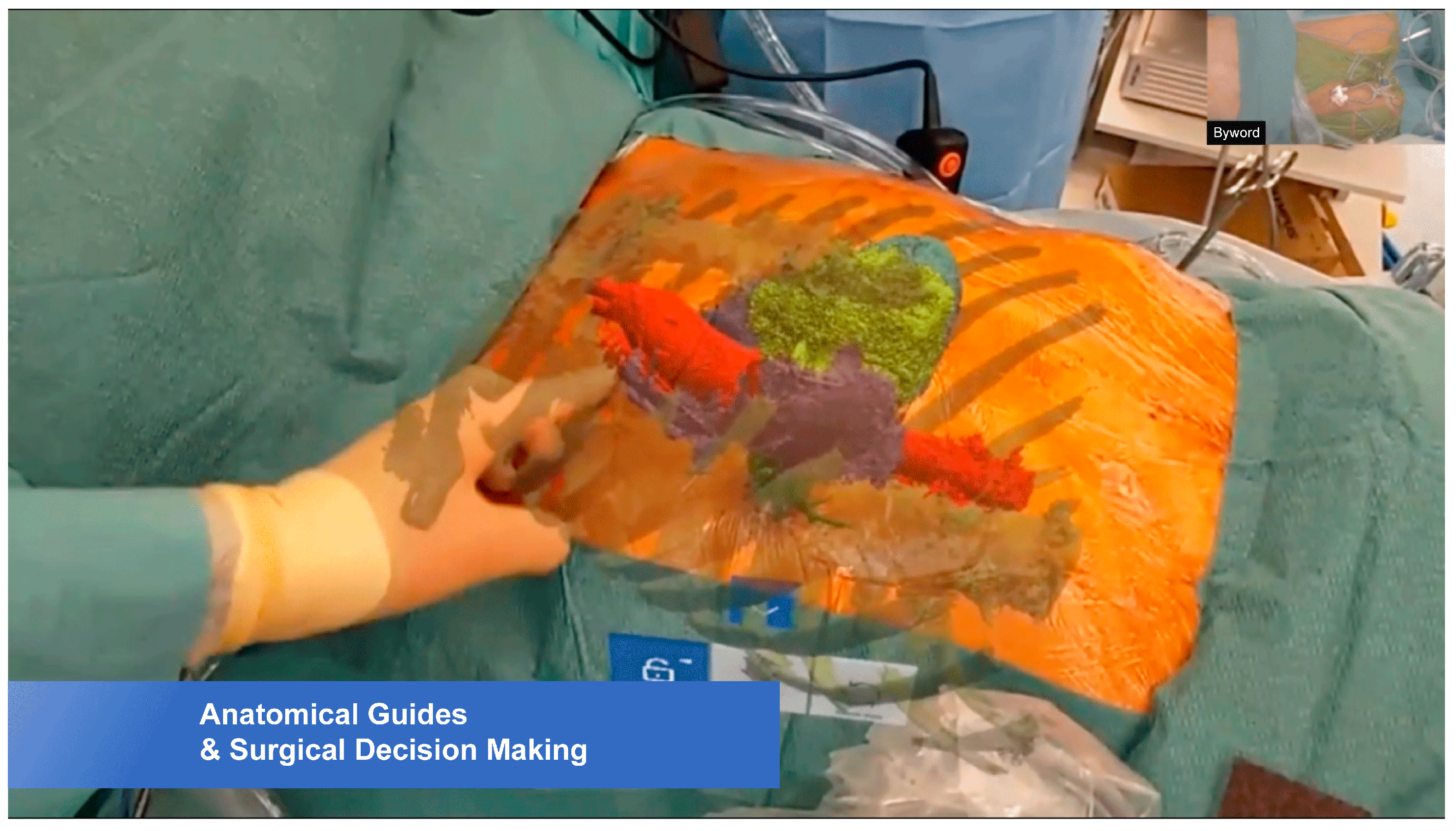
This cognitive problem can be overcome by using HoloLens® 2 (Microsoft Corporation, Redmond, WA, USA), a head-mounted mixed-reality (MR) holographic device [6,7,8] (Figure 1a,b). It provides an immersive experience by superimposing a holographic 3D-rendered model onto the patient’s anatomy [3,9] (Figure 2). Furthermore, the surgeon’s line-in-sight is maintained throughout the procedure so that the operator is not distracted by another screen [9,10].
In virtual reality (VR), the real environment is replaced by a virtual one, whereas augmented reality (AR) overlays virtual objects in the real, physical environment without the user being able to interact with them. Pokémon Go is a prime example of AR [4]. Mixed reality (MR) combines VR and AR [3,4,10,11], allowing surgeons to have simultaneous interactions with the virtual and physical world by superimposing holographic 3D images onto the operative field; thus, MR increases the user’s experience of depth and perception [4,10].
Using HoloLens® 2, the patient’s own virtual holographic model of the heart is anchored by selected landmarks on the thoracic wall, and the model is projected inside the chest cavity [4]. The main advantage of holograms placed inside the operative field is that surgeons have a better appreciation of the vector and magnitude of movement required [9]. HoloLens® 2 guides the surgeon to locate the most appropriate site of incision and auxiliary ports so that optimal visibility of and access to the operative field can be achieved [3].
To date, there are no reports in the MICS literature describing the use of HoloLens® 2 for improving surgical access. We present—to the best of our knowledge—a novel comparative study of three MICS procedures using HoloLens® 2 for preoperative planning and for real-time intraoperative guidance [9].

2. Materials and Methods

2.1. Computed Tomography Angiography (CTA)

A third-generation dual-source 2 × 192-slice scanner (SOMATOM Force, Siemens Healthcare, Forchheim, Germany), and the images are obtained using a prospectively ECG-triggered high-pitch spiral mode (Turbo Flash, 2 × 192 × 0.6 mm detector collimation, Siemens Healthcare) [9]. The slice thickness is based on detector collimation, which is 0.6 mm. The CT protocol was designed by radiologists and radiographers in consultation with the equipment vendor.
The images were then uploaded onto the HoloLens® 2 system to be rendered into a 3D holographic model [6].

2.2. Hologram Creation

Consent for photography or videography was obtained from each patient in accordance with institutional guidelines. CTA DICOM data were extracted from CT scans. Segmentation and alignment of the images were performed. These images were exported as a DICOM standard dataset, which was then uploaded onto HoloLens® 2 via Virtual Surgery Intelligence software version 1.5 (apoQlar GmbH, Hamburg, Germany) that generated the reference cardiothoracic hologram [9]. Additionally, segmentation of individual structures within the raw scan images was performed using 3D Slicer (The Slicer Community, www.slicer.org, v4.11.20210226) via manual segmentation to identify the individual cardiac chambers, valves, coronary arteries, and internal mammary arteries. The segmented images were exported as standard tessellation language (STL) files onto virtual surgery intelligence (VSI) and used for image superimposition and visualization [9]. The entire process of hologram creation took approximately 3 h. It was reviewed by the surgical team preoperatively.

2.3. Surgical Procedures

The patient was positioned on the operating table, and the surgeon marked the preferred site of entry employing conventional anatomical landmarks and their mental model. Then, they superimposed the reconstructed patient’s cardiac hologram onto the chest using the xiphoid and the clavicle as reference landmarks for a real-time anatomical assessment. This enabled the surgeon to look “into” the chest organ topography in their line-in-sight [10]. The best incision site was again marked. In addition, insertion points of surgical instruments and accessories were chosen in reference to the intended incision site marked using HoloLens® 2. Assessment with the hologram also enabled anticipation of difficulties and to foresee any possible spatial confinement.
The technicalities of superimposing the 3D-rendered image on the operative field were initially challenging. They required the surgeon to virtually hold the holographic image in mid-air and place it onto the patient’s chest using the landmarks as accurately as possible [3,10]. Surgeons had preoperative training sessions of approximately 20 min and quickly familiarized themselves with the movements of the holographic imagery [10].
The first procedure was an MIS aortic valve replacement (AVR) via right anterior small thoracotomy using a totally endoscopic technique (Figure 2). Using HoloLens® 2, a holographic image was superimposed onto the actual patient’s chest. The position of the aorta and aortic valve annulus were visualized. With this guidance, the incision was made, and the chest was entered via the second intercostal space. The AVR was performed in the usual manner.
The second procedure was an MIS off-pump coronary artery bypass graft surgery (CABG) via left anterior small thoracotomy (LAST). A similar process was performed. The fixed holographic 3D image guided the best way for the surgeon to access the most appropriate intercostal space and to plan the entry into the chest cavity. It also facilitated decision-making over the placement of bypass grafts and projected the degree of difficulties of exposure.
The 3rd case was an MIS pulmonary valve replacement (PVR) (Figure 3). PVR following repair of Tetralogy of Fallot in childhood is conventionally carried out via redo-median sternotomy. In order to avoid redo-median sternotomy, PVR was planned via LAST. Three-dimensional superimposed holographic visualization adequately projected the position of the main pulmonary artery (MPA) and pulmonary valve annulus (PVA) and guided the most appropriate intercostal space to enter the chest.

3. Results

Approximately 3 hours of preoperative worktime was required to create a 3D holographic image to be viewed on the device. Production was performed by a different in-house team.
The xiphoid process and the clavicle provided consistent reference points to lock the hologram on the patient’s chest, ensuring accurate placement. Hologram superimposition and fine adjustments were straightforward as surgeons had had prior training and were familiar with the technicalities. The OR time taken for the process was less than 10 min from mounting the device on the surgeon’s head to marking the incision site. Dictated by the structures of interest, the most accurate and smallest, incision site could be selected.
In all three cases, the incision site marked with and without the HoloLens® 2 differed by one intercostal space. HoloLens® 2 provided more appropriate access to the operating field: one intercostal space cranial in MIS AVR and MIS PVR and one intercostal space caudal in MIS CABG. The endoscopic tool distribution (camera insertion, left ventricular vent, and their relation to the structures of interest/chest wall) could be better appreciated and/or planned. HoloLens® 2 references were found to be more suitable than the markings based on the surgeon’s interpretation of the structures of interest from preoperative imaging. The transparent visor of the HoloLens® 2 allowed unobstructed views of the operating field. The operative time in 3 MIS cardiac surgical procedures described, with the use of HoloLens 2, was reduced by a mean duration of 34.3 ± 3.2 min. This could be attributed to the entry site being the most accurate without requiring change during the procedure, which can be time-consuming. A survey was also conducted to determine the level of user acceptability for MR devices in surgery. This survey was conducted in 2022, with 30 surgeons who had trialed the solution. Survey feedback was collected using a validated User Evaluation Questionnaire (UEQ), that uses a 26 point matrix to compare the new solution with existing devices. The survey results showed overwhelming positive response to the use of MR in surgery, as shown in Suppelementary File S2.
In this work, the study presented in [12] is expanded upon.

4. Discussion

The concept of holographic visualization and the necessary technology have existed for many decades [13,14]. The use of this technology in the medical field has been delayed by the bulky nature of the devices [3,13] and the inability to view high-quality images [3]. Microsoft HoloLens® 2, a head-mounted MR holographic device, was first made commercially available in 2016 [3] and is currently widely used in various medical and surgical subspecialties. MR technology, interconnected with both VR and AR, allows the user to interact with virtual reality in reality. MR has been applied in numerous medical [15,16,17,18,19,20] and surgical specialties, e.g., orthopedic surgery [21], neurosurgery [22], urology [23], general surgery [24], etc. Advantages have been demonstrated in both educational and clinical settings to help medical professionals better understand complex anatomical structures, so the modality is available for preoperative surgical planning and intraoperative guidance [25,26,27]. VR and 3D-printed heart models were also found to be superior for diagnostic assessment, medical education, and preoperative planning in comparison with conventional visualization techniques [28].

4.1. Benefits

MR devices offer multiple presumed benefits in MICS intraoperative visualization [10,29,30]. The patient’s own 3D holographic model superimposed on the actual anatomy ascertains spatial anatomy. The hologram can be adjusted and rendered to anatomical landmarks in real time [9]. All this contributes to incisional precision and accuracy of access to the target structures [31,32]. In fact, in each of our presented cases, HoloLens® 2’s superimposed model recommended a different intercostal space for chest entry from the one assumed by the surgeon’s mental model. Modification of chest entry by one intercostal space can achieve a significantly better visualization of the structures of interest in a confined operating space in minimally invasive cardiac surgery. The superiority of HoloLens® 2 can be attributed to the fact that holograms not only revealed the appropriate depth and relationships but also the angulation and vector orientation of optimal access.
Furthermore, holographic models propose an optimal distribution and orientation of auxiliary ports for manipulation and visualization. Intrathoracic spatial anatomical findings proved the holographic recommendations and no need for interspace and/or port adjustment occurred. We propose that intraoperative MR technology is the ultimate step in the meticulous preoperative planning process that is required for MICS.
HoloLens® 2’s superimposed model, in line with the surgeon’s vision, optimizes ergonomics, i.e., the surgeon can focus on the progress of the procedure without any interruption to consult with an external 2D flat screen [10] and/or disturbance of the visual–motor axis or hand–eye coordination [7,33]. Assumed benefits include improved surgical performance, reduced procedural length, more accurate visualization, and identification of anatomical structures leading to decreased risk of iatrogenic injuries [5,10,30]. The operator’s integrated perception of virtual and real structures and their spatial relationship contributes to cognitive coherence [11], reduced operator stress and fatigue, and less risk of complications; all this potentially translates into improved patient safety. Intraoperative holography, not involving physical contact with any material structure, is a non-invasive, aseptic modality [29].
MR offers a competitive advantage in avoiding median resternotomy in selected cases. Redo-sternotomy carries inherent risks of potentially fatal injury of the mediastinal structures upon chest entry [34,35]. CT angiography has been advocated as part of the preoperative work-up [36]. CT datasets converted into holographic models open an avenue for an alternative approach, e.g., LAST for PVR [37]. Holographic incisional guidance provides direct access to the target structures and significantly reduces operative risks while increasing precision [38]. The general acceptance and initial user experience were positive throughout our study.

4.2. Limitations

The alignment of 3D holographic images onto a patient’s chest was carried out manually using only two anatomic landmarks, namely the xiphoid and clavicles. More alignment points on exposed landmarks might be required for better precision [4]. Manual alignment needs to be mastered and is time-consuming [10]. Similarly, accommodation to HoloLens® 2 headgear requires some habituation. Co-application of regular head-mounted surgical equipment, such as surgical loupes and headlights typically worn by cardiac surgeons during surgery might be difficult or even impossible with HoloLens® 2 [4]. The holographic images are static [4], and no magnification has yet been integrated into the models [10]. HoloLens® 2 was only applied in the initial phases of the operations, i.e., during chest entry and intrathoracic orientation; the surgical procedures did not require additional spatial guidance. Our study of three surgical procedures only represents our initial experience with MR and HoloLens® 2. The modality requires more exposure with a wider spectrum of anatomies and complexities in order to identify clinical applicability, accuracy, user comfort, and the device’s place in the MICS armamentarium.

5. Conclusions

The use of an MR device in MICS could improve our understanding of anatomical spatial relationships. As an intraoperative form of real-time image guidance, it may be regarded as an important last step in pre- and intraoperative planning that offers improved precision of access and better intrathoracic orientation. In-line 3D visibility conjoining the patient’s intrathoracic anatomy with the superimposed in-line reconstructed holographic model optimizes the surgeon’s ergonomics and potentially enhances patient safety. The real value and place of the modality need further evaluation in an extended cohort of patients with a wider surgical spectrum and more degrees of complexity.

Supplementary Materials

The following supporting information can be downloaded at https://www.mdpi.com/article/10.3390/jcdd12020049/s1, File S1: HoloLens 2 Video; (https://youtu.be/rlC2IilA4i8, accessed on 11 September 2024); File S2: Survey for use of HoloLens 2 in MIS Cardiac Surgery; File S3: Manual Segmentation Protocol.

Author Contributions

Conceptualization, T.K. and L.K.; methodology, T.K.; writing—original draft preparation, W.M.M.A., S.S.K., A.K. and L.K.; writing—review and editing, L.K.; visualization, Y.G.; supervision: L.K. and T.K. All authors have read and agreed to the published version of the manuscript.

Funding

This study received no external funding.

Institutional Review Board Statement

This study was conducted in accordance with the Declaration of Helsinki and approved by the NHG Domain Specific Review Board (DSRB) Approval of Exemption (NHG DSRB Reference Number: 2020/01044) on 8 October 2020.

Informed Consent Statement

Informed consent was obtained from all subjects involved in the study in accordance with institutional guidelines.

Data Availability Statement

The original contributions presented in this study are included in the article/Supplementary Materials. Further inquiries can be directed to the corresponding author.

Acknowledgments

The authors would like thank Ilias Skaltsiotis, Jai Sule, and Guohao Chang for surgical assistance and providing the patients to be involved in this manuscript; Yujia Gao, Ng Kian Wai and Ngiam Kee Yuan for technical assistance and guidance using HoloLens® 2 during the surgeries.

Conflicts of Interest

The authors declare no competing interests. The authors have no financial interest in any of the products or devices that are described in this article.

References

  1. Kofidis, T. Minimally Invasive Cardiac Surgery. A Practical Guide; CRC Press: Boca Raton, FL, USA, 2021. [Google Scholar]
  2. Kiraly, L.; Aye, W.M.; Subbian, S.K.; Kofidis, T.; Al Hakami, A. Minimal incision and less invasive techniques in congenital cardiac surgery: A narrative review. Pediatr. Med. 2024, 7, 14. [Google Scholar] [CrossRef]
  3. Ye, W.; Zhang, X.; Li, T.; Luo, C.; Yang, L. Mixed-reality hologram for diagnosis and surgical planning of double outlet right ventricle: A pilot study. Clin. Radiol. 2021, 76, 237. [Google Scholar] [CrossRef] [PubMed]
  4. Brun, H.; Bugge, R.A.; Suther, L.K.; Birkeland, S.; Kumar, R.; Pelanis, E.; Elle, O.J. Mixed reality holograms for heart surgery planning: First user experience in congenital heart disease. Eur. Heart J. Cardiovasc. Imaging 2019, 20, 883–888. [Google Scholar] [CrossRef]
  5. Liu, S.; Xie, M.; Zhang, Z.; Wu, X.; Gao, F.; Lu, L.; Zhang, J.; Xie, Y.; Yang, F.; Ye, Z. A 3 D Hologram with Mixed Reality Technique to improve Understanding of Pulmonary Lesions Caused by COVID-19; Randomised Control Trial. Med. Internet Res. 2021, 23, e24081. [Google Scholar] [CrossRef]
  6. Karmonik, C.; Boone, T.B.; Khavari, R. Workflow for visualization of neuroimaging data with an augmented reality device. J. Digit. Imaging 2018, 31, 26–31. [Google Scholar] [CrossRef] [PubMed]
  7. Tepper, O.M.; Rudy, H.L.; Lefkowitz, A.; Weimer, K.A.; Marks, S.M.; Stern, C.S.; Garfein, E.S. Mixed reality with HoloLens: Where virtual reality meets augmented reality in the operating room. Plast. Reconstr. Surg. 2017, 140, 1066–1070. [Google Scholar] [CrossRef] [PubMed]
  8. Taylor, A.G. What Is the Microsoft HoloLens? Develop Microsoft HoloLens Apps Now; Springer: Berlin/Heidelberg, Germany, 2016; pp. 3–7. [Google Scholar]
  9. Cai, E.Z.; Gao, Y.; Ngiam, K.Y.; Lim, T.C. Mixed Reality Intraoperative Navigation in Craniomaxillofacial Surgery. Plast. Reconstr. Surg. 2021, 148, 686e–688e. [Google Scholar] [CrossRef] [PubMed]
  10. Scherl, C.; Stratemeier, J.; Rotter, N.; Hesser, J.; Schönberg, S.O.; Servais, J.J.; Männle, D.; Lammert, A. Augmented Reality with HoloLens in Parotid Tumour Surgery: A Prospective Feasibility Study. ORL J. Otorhinolaryngol. Relat. Spec. 2021, 83, 439–448. [Google Scholar] [CrossRef]
  11. Deib, G.; Johnson, A.; Unberath, M.; Yu, K.; Andress, S.; Qian, L.; Osgood, G.; Navab, N.; Hui, F.; Gailloud, P. Image guided percutaneous spine procedures using an optical see-through head mounted display: Proof of concept and rational. J. Neurointerv. Surg. 2018, 10, 1187–1191. [Google Scholar] [CrossRef] [PubMed]
  12. Aye, W.M.M.; Kiraly, L.; Kumar, S.S.; Kasivishvanaath, A.; Gao, Y.; Kofidis, T. Mixed Reality (Holography)-Guided Minimally Invasive Cardiac Surgery—A First-in-Man Comparative Feasibility Study. In Proceedings of the 30th Annual Meeting of the Asian Society for Cardiovascular and Thoracic Surgery (ASCVTS 2022)—Plenary 2, C-PL2-7, Nara, Japan, 25 March 2022. [Google Scholar]
  13. Redman, J.D.; Wolton, W.P.; Shuttleworth, E. Use of holography to make truly three-dimensional X-ray images. Nature 1968, 220, 58–60. [Google Scholar] [CrossRef]
  14. Carbone, M.; Piazza, R.; Condino, S. Commercially Available Head- Mounted Displays Are Unsuitable for Augmented Reality Surgical Guidance: A Call for Focused Research for Surgical Applications. Surg. Innov. 2020, 27, 254–255. [Google Scholar] [CrossRef] [PubMed]
  15. Huber, T.; Hadzijusufovic, E.; Hansen, C.; Paschold, M.; Lang, H.; Kneist, W. Head-mounted mixed-reality technology during robotic-assisted transanal total mesorectal excision. Dis. Colon. Rectum. 2019, 62, 258–261. [Google Scholar] [CrossRef] [PubMed]
  16. Ameri, G.; Baxter, J.S.; Bainbridge, D.; Peters, T.M.; Chen, E.C. Mixed reality ultrasound guidance system: A case study in system development and cautionary tale. Int. J. Comput. Assist. Radiol. Surg. 2018, 13, 495–505. [Google Scholar] [CrossRef] [PubMed]
  17. Coelho, G.; Figueiredo, E.G.; Rabelo, N.N.; Teixeira, M.J.; Zanon, N. Development and evaluation of a new pediatric mixed-reality model for neurosurgical training. J. Neurosurg. Pediatr. 2019, 2, 423–432. [Google Scholar] [CrossRef]
  18. Li, Y.; Chen, X.; Wang, N.; Zhang, W.; Li, D.; Zhang, L.; Qu, X.; Cheng, W.; Xu, Y.; Chen, W.; et al. A wearable mixed-reality holographic computer for guiding external ventricular draining insertion at the bedside. J. Neurosurg. 2018, 10, 1599–1606. [Google Scholar]
  19. Sauer, I.M.; Queisner, M.; Tang, P.; Moosburner, S.; Hoepfner, O.; Horner, R.; Lohmann, R.; Pratschke, J. Mixed reality in visceral surgery development of a suitable workflow and evaluation of intraoperative use-cases. Ann. Surg. 2017, 266, 706–712. [Google Scholar] [CrossRef]
  20. Pratt, P.; Ives, M.; Lawton, G. Through the HoloLens looking glass: Augmented reality for extremity reconstruction surgery using 3D vascular models with perforating vessels. Eur. Radiol. Exp. 2018, 2, 2. [Google Scholar] [CrossRef]
  21. Elmi-Terander, A.; Nachabe, R.; Skulason, H.; Pedersen, K.; Söderman, M.; Racadio, J.; Babic, D.; Gerdhem, P.; Edström, E. Feasibility and accuracy of thoracolumbar minimally invasive pedicle screw placement with augmented reality navigation technology. Spine 2018, 43, 1018–1023. [Google Scholar] [CrossRef] [PubMed]
  22. Li, Z.; Butler, E.; Li, K.; Lu, A.; Ji, S.; Zhang, S. Large-scale exploration of neuronal morphologies using deep learning and augmented reality. Neuroinformatics 2018, 16, 339–349. [Google Scholar] [CrossRef] [PubMed]
  23. Porpiglia, F.; Fiori, C.; Checcucci, E.; Amparore, D.; Bertolo, R. Augmented reality robot-assist- ed radical prostatectomy: Preliminary experience. Urology 2018, 115, 184. [Google Scholar] [CrossRef]
  24. Bong, J.H.; Song, H.J.; Oh, Y.; Park, N.; Kim, H.; Park, S. Endoscopic navigation system with extended field of view using augmented reality technology. Int. J. Med. Robot. 2018, 14, e1886. [Google Scholar] [CrossRef] [PubMed]
  25. Hu, H.Z.; Feng, X.B.; Shao, Z.W.; Xie, M.; Xu, S.; Wu, X.H.; Ye, Z.W. Application and Prospect of Mixed Reality Technology in Medical Field. Curr. Med. Sci. 2019, 39, 1–6. [Google Scholar] [CrossRef] [PubMed]
  26. Wainman, B.; Pukas, G.; Wolak, L.; Mohanraj, S.; Lamb, J.; Norman, G.R. The Critical Role of Stereopsis in Virtual and Mixed Reality Learning Environments. Anat. Sci. Educ. 2020, 13, 401–412. [Google Scholar] [CrossRef]
  27. Hilt, A.D.; Kapllani, K.M.; Hierck, B.P.; Kemp, A.C.; Albayrak, A.; Melles, M.; Schalij, M.J.; Scherptong, R.W. Perspectives of Patients and Professionals on Information and Education After Myocardial Infarction With Insight for Mixed Reality Implementation: Cross-Sectional Interview Study. JMIR Hum. Factors 2020, 7, e17147. [Google Scholar] [CrossRef] [PubMed]
  28. Lau, I.; Gupta, A.; Sun, Z. Clinical Valve of Virtual Reality versus 3D Printing in Congenital Heart Disease. Biomolecules 2021, 11, 884. [Google Scholar] [CrossRef]
  29. Jang, J.; Tschabrunn, C.M.; Barkagan, M.; Anter, E.; Menze, B.; Nezafat, R. Three-dimensional holographic visualization of high-resolution myocardial scar on HoloLens. PLoS ONE 2018, 13, e0205188. [Google Scholar] [CrossRef] [PubMed]
  30. Incekara, F.; Smits, M.; Dirven, C.; Vincent, A. Clinical feasibility of a wearable mixed-reality device in neurosurgery. World Neurosurg. 2018, 118, e422–e427. [Google Scholar] [CrossRef] [PubMed]
  31. Kersten-Oertel, M.; Jannin, P.; Collins, D.L. The State of the Art of Visualization in Mixed Reality Image Guided Surgery. Comput. Med. Imaging Graph. 2013, 37, 98–112. [Google Scholar] [CrossRef] [PubMed]
  32. Wu, X.; Liu, R.; Yu, J.; Xu, S.; Yang, C.; Yang, S.; Shao, Z.; Ye, Z. Mixed Reality Technology Launches in Orthopedic Surgery for Comprehensive Preoperative Management of Complicated Cervical Fractures. Surg. Innov. 2018, 25, 421–422. [Google Scholar] [CrossRef]
  33. El Shallaly, G.; Cuschieri, A. Optimum view distance for laparoscopic surgery. Surg. Endosc. 2006, 20, 1879–1882. [Google Scholar] [CrossRef]
  34. Kirshbom, P.M.; Myung, R.J.; Simsic, J.M.; Kramer, Z.B.; Leong, T.; Kogon, B.E.; Kanter, K.R. One Thousand repeat sternotomies for congenital cardiac surgery: Risk factors for re-entry injury. Ann. Thorac. Surg. 2009, 88, 158–161. [Google Scholar] [CrossRef] [PubMed]
  35. Pahwa, S.; Stephens, E.H.; Dearani, J.A. High-Risk Reoperative Sternotomy-How We Do It, How We Teach It. World J. Pediatr. Congenit. Heart Surg. 2020, 11, 459–465. [Google Scholar] [CrossRef] [PubMed]
  36. Valente, T.; Bocchini, G.; Rossi, G.; Sica, G.; Davison, H.; Scaglione, M. MDCT prior to median re-sternotomy in cardiovascular surgery: Our experiences, infrequent findings and the crucial role of radiological report. Br. J. Radiol. 2019, 92, 20170980. [Google Scholar] [CrossRef] [PubMed]
  37. Henaine, R.; Yoshimura, N.; Di Filippo, S.; Ninet, J. Pulmonary valve replacement in repaired tetralogy of Fallot by left thoracotomy avoid ascending aorta injury. J. Thorac. Cardiovasc. Surg. 2011, 141, 590–592. [Google Scholar] [CrossRef] [PubMed]
  38. Kumar, R.P.; Pelanis, E.; Bugge, R.; Brun, H.; Palomar, R.; Aghayan, D.L.; Fretland, Å.A.; Edwin, B.; Elle, O.J. Use of Mixed Reality for Surgery Planning: Assessment and Development Workflow. J. Biomed. Inform. 2020, 112, 100077. [Google Scholar] [CrossRef]
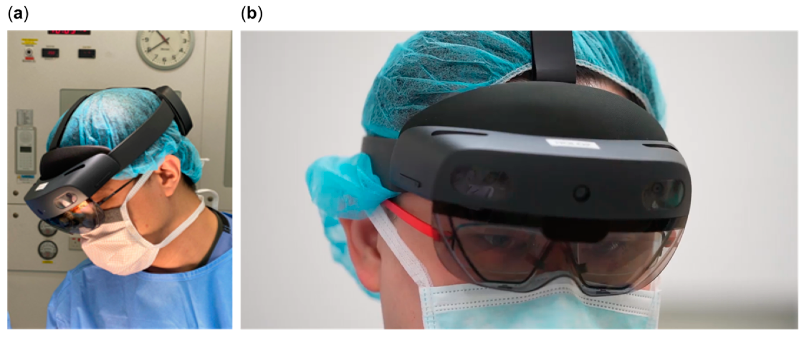
Figure 1. Surgeon wearing HoloLens® 2: (a) Looking at the patient on operating table; (b) looking through the clear visor onto the hologram.
Jcdd 12 00049 g001
Figure 2. HoloLens® 2-guided minimally invasive surgery for aortic valve replacement.
Jcdd 12 00049 g002
Figure 3. HoloLens® 2 guided minimally invasive surgery for pulmonary valve replacement.
Jcdd 12 00049 g003
Disclaimer/Publisher’s Note: The statements, opinions and data contained in all publications are solely those of the individual author(s) and contributor(s) and not of MDPI and/or the editor(s). MDPI and/or the editor(s) disclaim responsibility for any injury to people or property resulting from any ideas, methods, instructions or products referred to in the content.