Article

Sentinel Lymph Node Mapping with Indirect Lymphangiography for Canine Mast Cell Tumour

1
Faculty of Veterinary Medicine, University of Teramo, 64100 Teramo, Italy
2
Clinica Veterinaria Pet Care, 40133 Bologna, Italy
3
Clinica Veterinaria Val Musone, 60027 Osimo, AN, Italy
4
Clinica Veterinaria Pescara Nord, 65124 Pescara, Italy
5
Policlinico Veterinario Roma Sud, 00173 Roma, Italy
6
Clinica Veterinaria Etruria 24H, 53035 Siena, Italy
*
Authors to whom correspondence should be addressed.
Vet. Sci. 2022, 9(9), 484; https://doi.org/10.3390/vetsci9090484
Submission received: 27 July 2022 / Revised: 21 August 2022 / Accepted: 5 September 2022 / Published: 8 September 2022

Abstract

:

Simple Summary

Mast cell tumour (MCT) is a common cutaneous and subcutaneous neoplasia in dogs. Recent studies describe that sentinel lymph node (SLN) assessment is more specific to stage MCT, while regional lymph node (RLN) evaluation is not as specific. SNL is the first site of drainage of a tumour and the first metastatic site in several tumours. The study aims to evaluate the SLN drainage mapping of MCT with indirect lymphography in dogs. The second objective of the study is to compare the SLN to the RLN. Survey radiographs followed by an indirect lymphography were obtained for SLN mapping. Twenty-six dogs with 29 MCTs were included. SLNs were detectable in 26 MCTs and radiographic indirect lymphangiography with Lipiodol was able to detect at least one SLN in 90% of MCTs in dogs. In conclusion, radiographic indirect lymphangiography with Lipiodol is a feasible technique to map SLNs and its draining system in MCTs. The lymph drainage pattern of the MCTs may be different for each MCT and more than one SLN can be involved.

Abstract

Mast cell tumour (MCT) is a common cutaneous and subcutaneous neoplasia in dogs. It can metastasise to lymph nodes (LNs), and this adversely affects the prognosis and treatment. The study aims to evaluate the SLN mapping of MCTs with radiographic indirect lymphography. Dogs that underwent clinical staging were prospectively enrolled. Lipiodol was injected around the MCT or the surgical scar. After 24 h, LNs that picked up contrast were radiographically assessed. Twenty-six dogs with 29 MCTs were included. MCTs were confirmed histologically, while SLNs were evaluated either by cytology and/or histology. SLNs were detectable in 23 dogs with 26 MCTs. Lymphatic vessels were visible in 19 MCTs. In nine MCTs, at least two SLNs picked up contrast. In particular, seven MCTs involved two SLNs, and two MCTs involved three different SLNs. In nine MCTs, at least a SLN was metastatic. This study indicates that the lymph drainage pattern of the MCTs may be different for each MCT, and more than one SLN can be involved. Indirect lymphangiography with Lipiodol allowed the detection of the SLN in 90% of MCTs. This provided clinically relevant information to remove the LN and stage the patient.

1. Introduction

Mast cell tumour (MCT) is a common malignant cutaneous and subcutaneous neoplasia in dogs [1,2,3,4]. It is reported to be 16–21% of all canine integumentary tumours [1,2,3,4]. MCT can metastasise to lymph nodes (LNs), and this adversely affects the prognosis and treatment [1]. The World Health Organisation (WHO) reported a classification to differentiate the staging of MCTs, to better evaluate the prognosis and treatment of MCTs in veterinary medicine [5]. MCTs with metastatic LNs were ranked as stage II, leading to a worse prognosis and requiring different treatment compared to stage 0 or stage I [5]. Initially, regional lymph node (RLN) assessment was considered the standard care for stage MCT [6]. More recent studies describe that sentinel lymph node (SLN) assessment is more accurate to stage MCT, while RLN evaluation is not as accurate [6,7,8,9]. SNL is the first site of drainage of a tumour and in several tumours the first metastatic site [7]. Several studies report a different percentage between SLN and RLN, in particular in Laspley et al. in 27% of dogs, in Worley et al. in 42% of dogs, and in Ferrari et al. in 63% of dogs. [6,7,8,9].
In both the human and veterinary literature, different techniques are reported to help detection of SLN [9,10,11,12,13,14]. SLN mapping for MCT in veterinary medicine has been evaluated with several techniques such as contrast-enhanced ultrasound [10], lymphoscintigraphy [7,8], and computed tomography (CT) indirect lymphangiography [15]. In human medicine, lymphoscintigraphy [16] is the gold standard for mapping SLN, however, in veterinary medicine, only a few centres have the instrumentations to perform it [10]. CT indirect lymphangiography has recently been widely used for different tumour staging [10,13]. There are several disadvantages related to the techniques mentioned above, such as the requirement of general anaesthesia, high cost, and, finally, multiple scans must be performed to correctly detect the lymphatic vessels and SLN [15].
There are only few studies about indirect lymphangiography performed with radiographs. A recent publication mentions the utility of this technique for SLN draining in mammary tumours [12]. Advantages are the wide availability of radiology, the safety of the contrast media injected at the periphery of the tumour, and the possibility to perform the procedure with the dog awake or mildly sedated [12].
The study aimed to evaluate the SLN drainage mapping of MCTs with indirect lymphography in dogs. The second object of the study was to correlate the SLN to the RLN.
We hypothesised that indirect lymphangiography with the radiographic study would be a feasible technique to detect the SLN of canine MCTs. Our second hypothesis was that SLN would not always coincide with the RLN of the MCTs.

2. Materials and Methods

This study protocol N. 10/2020 was approved by Committee on Animal Research and Ethics of the Universities of Chieti-Pescara, Teramo, L’Aquila and of the Experimental Zoo prophylactic Institute of Abruzzo-Molise (CEISA), according to the Italian law decree N. 26/2014 and to EU directive 2010/63. The owner signed an informed consent form before the procedure.
This prospective multicentric observational study was conducted from January 2021 to March 2022 at the Veterinary Teaching Hospital of the University of Teramo, veterinary clinic Pet Care, veterinary clinic Val Musone, veterinary clinic Pescara Nord, veterinary clinic Roma Sud, and veterinary clinic Etruria H24.
Dogs were eligible for the study if they were undergoing staging with complete blood work consistent with CBC and chemistry of a histologically diagnosed MCT. The histology was sent to external laboratories, where board-certified pathologists or experienced operators classified the MCT according to both Kiupel’s 2011 [17] and Patnaik’s 1984 [18] grading system [2], while subcutaneous tumours were measured according to the Thompson grading system [19]. Dogs with scars of already excised MCTs were also eligible if there was an incomplete resection of the margins. Dogs were excluded from the study if the RLN was previously removed.
Survey radiographs followed by an indirect lymphography were obtained for SLN mapping. If needed, premedication was always performed intramuscularly (IM) with methadone (semfortan®; Eurovet Animal Health, Bladel, The Netherlands) ranging from 0.2–0.3 mg kg−1 and dexmedetomidine (Dextroquillan®; Fatro, Bologna, Italy) 0.003–0.006 mg kg−1, according to anaesthetist clinical evaluation. At the end of the procedures, atipamezole (Antisedan®; Vetoquinol, Magny-Vernois, France) was administrated IM at ½ of dexmedetomidine dose.
The radiographic study pre- and post-contrast administration and the injection of contrast medium were performed with the dog awake or mildly sedated according to dog collaboration. If needed, sedation was always performed intramuscularly (IM) with methadone (semfortan®; Eurovet Animal Health, Bladel, The Netherlands) ranging from 0.2–0.3 mg kg−1 and dexmedetomidine (Dextroquillan®; Fatro, Bologna, Italy) 0.003–0.006 mg kg−1, according to anaesthetist clinical evaluation. At the end of the procedures, atipamezole (Antisedan®; Vetoquinol, Magny-Vernois, France) was administrated IM at ½ of the dexmedetomidine dose. Every radiographic study was performed with at least two orthogonal views, according to the location of the primary tumour and the RLNs. If no SLNs were detected in the expected site, an additional region with the radiographic study was included. Immediately after the radiographic examination, Lipiodol Ultra-Fluid TM (iodized ethyl esters of the fatty acids of poppy seed oil, Guerbet, Aulnay-sous-Bois, France; 480 mg iodine mL−1) was injected around the MCT (Figure 1) or the surgical scar. The procedure was performed with a 25 G needle into the subcutis of four quadrants surrounding the tumour, as previously described [12,20]. A slow rate infiltration, 0.2–0.4 mL for each quadrant for a total of 0.8–1.6 mL of contrast medium over 1–2 min, was injected depending on the tumour size [12]. Care was taken to avoid any contrast leakage, or contrast entering the blood vessels or injecting directly into the primary tumour. There are only a few collateral effects reported in human medicine, such as embolism with a 0.12% frequency following lymphangiography, or hypersensitivity reaction that can happen within 60 min of the injection [21,22].
Dogs who were not sedated were monitored to see any possible collateral effects and discharged one hour after lipiodol injection. Otherwise, if the dog underwent sedation, they were carefully monitored and discharged one hour after the end of the sedation and a standing position was obtained. After 24 h, dogs were admitted again and the same radiographic study was repeated; if no SLNs were detected in the expected site additional regions were included in the radiographic study, and the site and number of the SLNs that picked up contrast were radiographically assessed. If visible, the presence of lymphatic vessels was also reported. Each radiographic study was evaluated by a second-year resident ECVDI (A.D.B.) and a board-certified radiologist (M.V.). The two observers independently reviewed the location of SLNs, visibility of lymphatic vessels, and interobserver agreement was assessed. In order to help identification of the location of the LNs, anatomical landmarks were mentioned to better locate the position of the SLNs. In case of disagreement between operators’ assessments, radiologists evaluated images together to find an agreement. The evaluation of if SLN correlated to RLN was divided into 3 groups. Group 1 was “total agreement”, when RLN was the same as the SLN. Group 2 was “partial agreement”, when one SLN coincided with the RLN, but a second SLN did not correspond with the RLN. Group 3 was “no agreement”, when the RLN did not correspond to any SLN. SLNs that were identified through indirect lymphangiography were investigated by either cytology following the Krick et al. classification [23], or were surgically removed and assessed by histological exam according to the Weishaar et al. system, where HN2 and HN3 are considered as metastatic [24] by a board-certified pathologist or an experienced operator.

3. Results

3.1. MCTs

A total of 27 dogs and 31 MCTs were initially included in the study. A single dog who underwent lipiodol staging at the same time with two different MCTs showed three different SLNs. It was not possible to differentiate if the SLNs were draining one, the other, or both the MCTs, so it was removed from the total count, leaving a total of 26 dogs and 29 MCTs. The population sample consisted, for the majority, of mixed breeds and Labrador, two Maltese and English Setter, while the remaining breeds were each represented by a single dog. The population sample is summarised in Table 1.
The masses were located at the level of head and neck (nine), thorax (six), hindlimbs (six), abdomen (three), forelimbs (two), scrotum (two), and prepuce (one). The location of the MTCs is summarised in Table 2.
Twenty-three dogs included in the study have a single MCT. A single dog included in the study has a surgical scar of a single MCT surgically removed with positive margins. One dog presented with two different MCTs, and one subject presented with three different MCTs during the inclusion.

MCTs Histology

The MCTs included in the study were: 8 subcutaneous (scMCT), and 23 cutaneous. Of the cutaneous MCT (cMCT), there were 3 Patnaik grade I–low-grade Kiupel (P1-LK), 16 Patnaik grade II–low-grade Kiupel (P2-LK), and 4 Patnaik grade II–high-grade Kiupel (P2-HK). No grade III Patnaik are presented in the study. The histopathological classification is summarised in Table 3.

3.2. SLNs Mapping

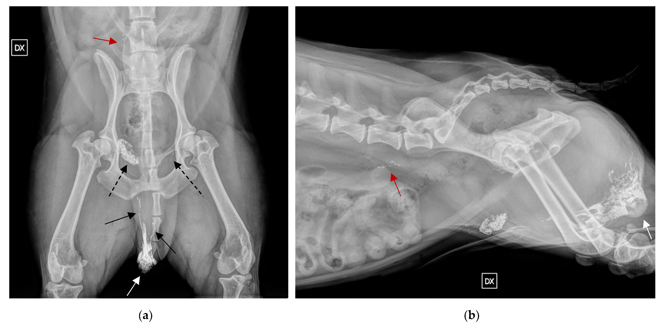
A total of 26 MCTs (90%) and 37 SLNs are correctly identified with indirect lymphangiography (Figure 2). Two cMCTs of different histotypes (2PLK, 2PHK) do not show any SLN with indirect lymphangiography. One scMCT shows just the lymphatic vessels without any SLN. Out of the 26 MCTs that indirect lymphangiography correctly identified the SLN, 17 MCTs show a single SLN, and 9 MCTs present more than one SLN. Among the latter category, seven MCTs present two SLNs and two MCTs present three SLNs with indirect lymphangiography (data summarised in Table 3) (Figure 3). Immediate or delayed side effects are not reported secondary to the injection of Lipiodol in any dog of our study. There is a complete agreement between the observers for the evaluation of SLNs, and for visible lymphatic vessels. We classify 10 SLNs (27%) as in agreement with the RLNs, 7 SLNs (19%) in non-agreement with the RLNs, and 20 SLNs (54%) as in partial agreement with the RLNs. Following the indirect lymphangiography, it is possible to evaluate the lymphatic vessel (Figure 3) (Table 3) in 17 MCTs. Among them, the lymphatic vessel associated with a single SLN in 11 MCTs, with two SLNs in 5 MCTs, three SLNs in 2 MCTs, and only one MCT shows a lymphatic vessel with no evidence of SLN. Otherwise, in 10 MCTs, lymphatic vessels are not visible following indirect lymphangiography. Out of these, five MCTs are associated with one SLN, three MCTs with two SLNs, and two MCTs do not present any SLN.

SLNs Cytology/Histology

In Table 3, the MCTs grading is associated with the cytology/histology of the SLNs. A total of 37 SLNs were analysed through one of the two methods mentioned above. Among the 37 SLNs, 12 (32%) are metastatic (10 histologically diagnosed and 2 through cytology). A total of 20/37 SLNs were evaluated through histology after surgery, while the remaining 17/37 SLNs were assessed with cytology [19]. Out of the 20 SLNs who underwent histological, grading is classified as metastatic in 10 SLNs (three HN2 and seven HN3), while 10 SLNs are not metastatic (seven HN0 and three HN1). Based on the cytological exam, only 2 SLNs out of 17 are metastatic, while in the remaining 15 SLNs, no evidence of metastatic disease is identified according to Krick et al. [23].

4. Discussion

Radiographic indirect lymphangiography with Lipiodol is able to detect at least a SLN in 90% of primary tumours (26/29 MCTs). The results obtained in our study are in accord with our hypothesis, wherein the technique is feasible and allows the evaluation of SLNs in dogs with MCTs. The second aim was to evaluate the agreement between the RLN and SLN. In our study, 26% (7/26) of MCTs have one SLN that does not agree with the RLN, and 34% (9/26) of MCTs have a partial agreement between SLN and the RLN. These findings agree with recent studies where a different SLN from the RLN was detected, ranging from 20 to 60% of cases, through different SLN mapping techniques in dogs with MCTs [6,7,8,9]. Among the MCTs, 9/26 (34%) present at least two different SLNs and, out of those, seven MCTs present two different SLNs, and two MCTs exhibit three different SLNs. This result shows how multiple SLNs, from different centres, can drain the same MCT. Multiple SLNs are reported for MCTs with other mapping methods. Multiple SLNs always represent at least a different SLN from the RLN. Furthermore, in our study, it is important to note that 34% of MCTs have SLNs that only partially agree with RLN, added to the 26% of MCTs with a SLN that does not agree with a RLN, which leads to a total number of 60% of MCTs that, without this mapping technique, would have been improperly evaluated for the correct SLNs and staging [7,15]. There is a complete interobserver agreement for SLNs location and lymphatic vessels visibility. The results represent the relatively easy assessment of the SLNs thanks to the contrast medium within it, leading even a less experienced observer to properly locate the SLNs, giving rise to greater repeatability of the technique.
SLNs mapping gives fundamental information to better evaluate the prognosis of the patient, because, as reported in the literature, SLN usually is the first site of metastasis in MCTs [7]. Studies mention how the presence of metastatic LNs is considered an important prognostic factor, influencing the treatment and the survival time in MCTs [25]. Furthermore, Horta et al. report a higher risk of death (62%) in patients with tumour recurrence and metastatic LNs in MCTs [3].
The results of this study highlight, even more, the necessity of this method for staging dogs instead of evaluating just the RLN. Without indirect lymphangiography, it would not be possible to correctly detect SLN in some cases.
Lipiodol Ultra-Fluid TM in indirect lymphography was used to map the lymphatic drainage of different tumours such as mammary carcinoma [12,26]. To the best of our knowledge, radiographic indirect lymphangiography with Lipiodol for MCTs was never evaluated.
Lymphatic vessels are visible in 65% (19/29) of MCTs. Lymphatic vessels could be useful to help detect the SLNs if the contrast uptake was weak or not homogenous in SLNs. Lymphatic vessels are currently reported through other methods such as CT indirect lymphography and lymphoscintigraphy [7,15,20], although their presence is not correlated with the histologic grade of the MCTs. In our study, no association is found between the classification of the tumour and the presence of the lymphatic vessels or the histologic/cytologic grade of the SLNs. An interesting finding is that 75% (6/8) of scMCTs are associated with visible lymphatic vessels after contrast administration. An explanation for this finding cannot be found, although four of the six subcutaneous MCTs have metastatic SLN, so it may be possible that more aggressive behaviour of the primary tumour leads to the detection of lymphatic vessels with radiographic indirect lymphangiography. On the other hand, out of the other two subcutaneous MCTs that do not show any lymphatic vessel, one of those also has a metastatic SLN. More studies are needed to evaluate this finding.
In three MCTs, the indirect lymphangiography with Lipiodol does not highlight any SLNs; out of these, a single MCT shows lymphatic vessels without any SLNs. A possible explanation would be the failure of the method, such as the contrast being injected too rapidly leading to rupture of the lymphatic vessels. Moreover, the multicentric nature of the study could lead an untrained operator to perform the procedure improperly, such as a fast rate injection of the contrast medium leading to a rupture of the lymphatic vessels, increasing the chance of failure of the procedure. Another potential cause to consider is macro metastasis in the SLNs and lymphatic system. These conditions, as reported in the literature, compromise the visibility of the lymphatic vessels and SLNs [27]. As reported in the literature, the surgical asportation of the MCT could lead, theoretically, to disruption of the normal lymphatic vasculature, and the consequent failure of the indirect lymphangiography on the surgical scar [7]. In our study, the three MCTs in which indirect lymphangiography is not successful are performed at the periphery of the MCTs. On the contrary, in the only dog included with a surgical scar, the method highlights a SLN that is also metastatic, confirmed with histology.
The advantages of radiographic indirect lymphangiography are the wide availability of the technique, and the possibility to perform the study with the dog awake or mildly sedated [12]. Meanwhile, computed tomography SLN mapping is associated with increased costs, more limited availability, and the need to perform the study under general anaesthesia [28,29]. Computed tomography SLN mapping in MCTs correctly identifies SLN in 60% of different tumour types in a study [27], while our indirect lymphangiography with radiographs has a higher success rate (90%). Another difference between the two techniques is the need to repeat the study for every patient in CT at 1, 3, 6, 9, and 12 min, in order to find the time where the contrast enhancement is more homogenous and a proper assessment of the SLN is possible; on the other hand, we only had to repeat the radiographic study once 24 h after the injection to properly evaluate the SLN. The study mentioned in veterinary medicine with lymphoscintigraphy is able to detect SLNs in 91% of MCTs [7], similar to our result of 90% of SLNs detected with indirect lymphangiography in MCTs. The interesting finding that differs from the paper mentioned above is the type of cases in which the lymphoscintigraphy is not successful. The three MCTs in which the method fails were all performed on a surgical scar in the study by Ferrari et al. [7], while, in our study, the indirect lymphangiography performed around the only surgical scar case included successfully identifies the SLN. In fact, the three MCTs wherein the method fails to detect the SLN reported in our study are performed at the periphery of the primary tumour. Unfortunately, in veterinary medicine, not many facilities have the instrumentation for lymphoscintigraphy. Furthermore, care must be considered while manipulating this radiopharmaceutical to guarantee the safety of the operator [30]. Moreover, radiographic indirect lymphangiography is safe to use, and immediate or delayed side effects are not reported secondary to the injection in any dog in our study.
This study has some limitations. The small size of the sample included in the study and a small number of high-grade Kiupel MCTs. This reflects the incidence of the tumour in the literature, because of the lower prevalence of high-grade Kiupel MCTs in dogs compared to the low-grade Kiupel [25,31,32]. Another limitation is the SLNs evaluation with cytology and histology. The gold standard to properly evaluate the malignancy of the LN is histopathology [24]. No intraoperative procedure, such as blue methylene injection, was performed to help detect the SLN identified through the radiographic study, so it cannot be confirmed that the SLNs sampled with FNAs or surgically removed coincided to the one highlighted through the indirect radiograph. This study aimed to evaluate the mapping SLNs with radiographic indirect lymphangiography in MCTs, and not to compare metastasised SLNs, which is why SLNs evaluated through cytology were included, even though including SLNs through cytology could lead to a false negative reading of non-metastatic SLNs. It is possible that if histopathology was performed in every single SLN, the metastatic rate reported in this study could have been even higher. In the study, 12/37 (32%) SLNs are found to be metastatic. Out of those ten, through histology it is confirmed that seven are HN3 and three are HN2. Considering the MCTs in dogs, there are 9/26 (34%) MCTs with SLNs metastasised either by cytology or histology. This relatively high metastatic rate is reported in the literature [25,33,34,35]. In the cMCT group, there are a total of four MCTs with metastatic SLNs. Out of these, two are P2-HK, one P2-LK, and one P1-LK. If we consider the total number of P2-HK included in the study with a successful indirect lymphangiography, we have 2/3 (66%) of these MCTs with metastatic SLNs, while only 2/16 (12%) MCTs include the P1-LK and P2-LK group with metastatic SLNs. These findings agree with what is reported in the literature, where P2-HK has a more aggressive behaviour compared to P1-LK and P2-LK [1]. In the scMCTs group, 5/7 scMCTs are identified with metastatic SLNs. This is controversial to what is reported in the literature, where a recent study mentions a low grade of metastasis of subcutaneous tumours [25]. Is it possible that scMCTs could have more aggressive behaviour than what is reported in the literature, but further studies are needed to confirm this result.

5. Conclusions

In conclusion, radiographic indirect lymphangiography with Lipiodol is a feasible technique to map SLN in MCT. The lymph drainage pattern of the MCTs may be different for each MCT, and more than one SLN can be involved. Few SLNs have a different location from the RLNs of the MCTs. SLNs mapping techniques should become the standard protocol for staging the dog, instead of evaluating RLNs. Detection of SLNs would provide clinically relevant information to remove the LN and stage the dog.

Author Contributions

Conceptualization, M.V.; methodology, M.V. and A.D.B.; validation, V.R., F.C. and R.T. (Roberto Tamburro); formal analysis, A.P.; investigation, J.G., P.F., G.D., E.P. and I.F.; resources, R.T. (Rossella Terragni) and A.B.; data curation, G.D. and E.P.; writing—original draft preparation, A.D.B.; writing—review and editing, A.D.B., A.B., M.V. and V.R.; visualization, V.R., R.B.; supervision, M.V.; project administration, A.D.B. All authors have read and agreed to the published version of the manuscript.

Funding

This research received no external funding.

Institutional Review Board Statement

This study was approved by the Committee on Animal Research and Ethics of the Universities of Chieti-Pescara, Teramo, L’Aquila and of the Experimental Zoo prophylactic Institute of Abruzzo-Molise (CEISA), according to the Italian law decree N. 26/2014 and to EU directive 2010/63.

Informed Consent Statement

Informed consent was obtained from all subjects involved in the study.

Data Availability Statement

Not applicable.

Conflicts of Interest

The authors declare no conflict of interest.

References

  1. Bellamy, E.; Berlato, D. Canine cutaneous and subcutaneous mast cell tumours: A review. J. Small Anim. Pract. 2021, 63, 497–511. [Google Scholar] [CrossRef]
  2. Berlato, D.; Bulman-Fleming, J.; Clifford, C.A.; Garrett, L.; Intile, J.; Jones, P.; Kamstock, D.A.; Liptak, J.M.; Pavuk, A.; Powell, R.; et al. Value, Limitations, and Recommendations for Grading of Canine Cutaneous Mast Cell Tumors: A Consensus of the Oncology-Pathology Working Group. Vet. Pathol. 2021, 58, 858–863. [Google Scholar] [CrossRef]
  3. Horta, R.S.; Lavalle, G.E.; Monteiro, L.N.; Souza, M.C.C.; Cassali, G.D.; Araújo, R.B. Assessment of Canine Mast Cell Tumor Mortality Risk Based on Clinical, Histologic, Immunohistochemical, and Molecular Features. Vet. Pathol. 2018, 55, 212–223. [Google Scholar] [CrossRef]
  4. Kiupel, M.; Camus, M. Diagnosis and Prognosis of Canine Cutaneous Mast Cell Tumors. Vet. Clin. N. Am. Small Anim. Pract. 2019, 49, 819–836. [Google Scholar] [CrossRef]
  5. Owen, L.N.; World Health Organization. TNM Classification of Tumours in Domestic Animals; VPH/CMO/80.20; World Health Organization: Geneva, Switzerland, 1980. [Google Scholar]
  6. Ferrari, R.; Boracchi, P.; Chiti, L.E.; Manfredi, M.; Giudice, C.; De Zani, D.; Spediacci, C.; Recordati, C.; Grieco, V.; Gariboldi, E.M.; et al. Assessing the Risk of Nodal Metastases in Canine Integumentary Mast Cell Tumors: Is Sentinel Lymph Node Biopsy Always Necessary? Animals 2021, 11, 2373. [Google Scholar] [CrossRef]
  7. Ferrari, R.; Chiti, L.E.; Manfredi, M.; Ravasio, G.; De Zani, D.; Zani, D.D.; Giudice, C.; Gambini, M.; Stefanello, D. Biopsy of sentinel lymph nodes after injection of methylene blue and lymphoscintigraphic guidance in 30 dogs with mast cell tumors. Vet. Surg. 2020, 49, 1099–1108. [Google Scholar] [CrossRef]
  8. Worley, D.R. Incorporation of sentinel lymph node mapping in dogs with mast cell tumours: 20 consecutive procedures. Vet. Comp. Oncol. 2014, 12, 215–226. [Google Scholar] [CrossRef]
  9. Lapsley, J.; Hayes, G.M.; Janvier, V.; Newman, A.W.; Peters-Kennedy, J.; Balkman, C.; Sumner, J.P.; Johnson, P. Influence of locoregional lymph node aspiration cytology vs sentinel lymph node mapping and biopsy on disease stage assignment in dogs with integumentary mast cell tumors. Vet. Surg. 2021, 50, 133–141. [Google Scholar] [CrossRef]
  10. Beer, P.; Pozzi, A.; Rohrer Bley, C.; Bacon, N.; Pfammatter, N.S.; Venzin, C. The role of sentinel lymph node mapping in small animal veterinary medicine: A comparison with current approaches in human medicine. Vet. Comp. Oncol. 2018, 16, 178–187. [Google Scholar] [CrossRef]
  11. Fournier, Q.; Thierry, F.; Longo, M.; Malbon, A.; Cazzini, P.; Bisson, J.; Woods, S.; Liuti, T.; Bavcar, S. Contrast-enhanced ultrasound for sentinel lymph node mapping in the routine staging of canine mast cell tumours: A feasibility study. Vet. Comp. Oncol. 2021, 19, 451–462. [Google Scholar] [CrossRef]
  12. Collivignarelli, F.; Tamburro, R.; Aste, G.; Falerno, I.; Del Signore, F.; Simeoni, F.; Patsikas, M.; Gianfelici, J.; Terragni, R.; Attorri, V.; et al. Lymphatic Drainage Mapping with Indirect Lymphography for Canine Mammary Tumors. Animals 2021, 11, 1115. [Google Scholar] [CrossRef] [PubMed]
  13. Soultani, C.; Patsikas, M.N.; Mayer, M.; Kazakos, G.M.; Theodoridis, T.D.; Vignoli, M.; Ilia, T.S.M.; Karagiannopoulou, M.; Ilia, G.M.; Tragoulia, I.; et al. Contrast enhanced computed tomography assessment of superficial inguinal lymph node metastasis in canine mammary gland tumors. Vet. Radiol. Ultrasound 2021, 62, 557–567. [Google Scholar] [CrossRef] [PubMed]
  14. Randall, E.K.; Jones, M.D.; Kraft, S.L.; Worley, D.R. The development of an indirect computed tomography lymphography protocol for sentinel lymph node detection in head and neck cancer and comparison to other sentinel lymph node mapping techniques. Vet. Comp. Oncol. 2020, 18, 634–644. [Google Scholar] [CrossRef] [PubMed]
  15. Grimes, J.A.; Secrest, S.A.; Wallace, M.L.; Laver, T.; Schmiedt, C.W. Use of indirect computed tomography lymphangiography to determine metastatic status of sentinel lymph nodes in dogs with a pre-operative diagnosis of melanoma or mast cell tumour. Vet. Comp. Oncol. 2020, 18, 818–824. [Google Scholar] [CrossRef]
  16. Moncayo, V.M.; Alazraki, A.L.; Alazraki, N.P.; Aarsvold, J.N. Sentinel Lymph Node Biopsy Procedures. Semin. Nucl. Med. 2017, 47, 595–617. [Google Scholar] [CrossRef]
  17. Kiupel, M.; Webster, J.D.; Bailey, K.L.; Best, S.; DeLay, J.; Detrisac, C.J.; Fitzgerald, S.D.; Gamble, D.; Ginn, P.E.; Goldschmidt, M.H.; et al. Proposal of a 2-tier histologic grading system for canine cutaneous mast cell tumors to more accurately predict biological behavior. Vet. Pathol. 2011, 48, 147–155. [Google Scholar] [CrossRef]
  18. Patnaik, A.K.; Ehler, W.J.; MacEwen, E.G. Canine cutaneous mast cell tumor: Morphologic grading and survival time in 83 dogs. Vet. Pathol. 1984, 21, 469–474. [Google Scholar] [CrossRef]
  19. Thompson, J.J.; Yager, J.A.; Best, S.J.; Pearl, D.L.; Coomber, B.L.; Torres, R.N.; Kiupel, M.; Foster, R.A. Canine subcutaneous mast cell tumors: Cellular proliferation and KIT expression as prognostic indices. Vet. Pathol. 2011, 48, 169–181. [Google Scholar] [CrossRef]
  20. Brissot, H.N.; Edery, E.G. Use of indirect lymphography to identify sentinel lymph node in dogs: A pilot study in 30 tumours. Vet. Comp. Oncol. 2017, 15, 740–753. [Google Scholar] [CrossRef]
  21. Pieper, C.C.; Hur, S.; Sommer, C.M.; Nadolski, G.; Maleux, G.; Kim, J.; Itkin, M. Back to the Future: Lipiodol in Lymphography-From Diagnostics to Theranostics. Investig. Radiol. 2019, 54, 600–615. [Google Scholar] [CrossRef]
  22. Kwon, W.J.; Kim, H.J.; Jeong, Y.J.; Lee, C.H.; Kim, K.I.; Kim, Y.D.; Lee, J.H. Direct lipiodol injection used for a radio-opaque lung marker: Stability and histopathologic effects. Exp. Lung Res. 2011, 37, 310–317. [Google Scholar] [CrossRef] [PubMed]
  23. Krick, E.L.; Billings, A.P.; Shofer, F.S.; Watanabe, S.; Sorenmo, K.U. Cytological lymph node evaluation in dogs with mast cell tumours: Association with grade and survival. Vet. Comp. Oncol. 2009, 7, 130–138. [Google Scholar] [CrossRef] [PubMed]
  24. Weishaar, K.M.; Thamm, D.H.; Worley, D.R.; Kamstock, D.A. Correlation of nodal mast cells with clinical outcome in dogs with mast cell tumour and a proposed classification system for the evaluation of node metastasis. J. Comp. Pathol. 2014, 151, 329–338. [Google Scholar] [CrossRef]
  25. De Nardi, A.B.; Dos Santos Horta, R.; Fonseca-Alves, C.E.; de Paiva, F.N.; Linhares, L.C.M.; Firmo, B.F.; Ruiz Sueiro, F.A.; de Oliveira, K.D.; Lourenço, S.V.; De Francisco Strefezzi, R.; et al. Diagnosis, Prognosis and Treatment of Canine Cutaneous and Subcutaneous Mast Cell Tumors. Cells 2022, 11, 618. [Google Scholar] [CrossRef] [PubMed]
  26. Patsikas, M.N.; Karayannopoulou, M.; Kaldrymidoy, E.; Papazoglou, L.G.; Papadopoulou, P.L.; Tzegas, S.I.; Tziris, N.E.; Kaitzis, D.G.; Dimitriadis, A.S.; Dessiris, A.K. The lymph drainage of the neoplastic mammary glands in the bitch: A lymphographic study. Anat. Histol. Embryol. 2006, 35, 228–234. [Google Scholar] [CrossRef] [PubMed]
  27. Rossi, F.; Körner, M.; Suárez, J.; Carozzi, G.; Meier, V.S.; Roos, M.; Rohrer Bley, C. Computed tomographic-lymphography as a complementary technique for lymph node staging in dogs with malignant tumors of various sites. Vet. Radiol. Ultrasound 2018, 59, 155–162. [Google Scholar] [CrossRef]
  28. Hlusko, K.C.; Cole, R.; Tillson, D.M.; Boothe, H.W.; Almond, G.; Coggeshall, W.S.; Matz, B.M. Sentinel lymph node detection differs when comparing lymphoscintigraphy to lymphography using water soluble iodinated contrast medium and digital radiography in dogs. Vet. Radiol. Ultrasound 2020, 61, 659–666. [Google Scholar] [CrossRef]
  29. Majeski, S.A.; Steffey, M.A.; Fuller, M.; Hunt, G.B.; Mayhew, P.D.; Pollard, R.E. Indirect Computed Tomographic Lymphography for iliosacral lymphatic mapping in a cohort of dogs with anal sac gland adenocarcinoma: Technique description. Vet. Radiol. Ultrasound 2017, 58, 295–303. [Google Scholar] [CrossRef]
  30. Manfredi, M.; De Zani, D.; Chiti, L.E.; Ferrari, R.; Stefanello, D.; Giudice, C.; Pettinato, V.; Longo, M.; Di Giancamillo, M.; Zani, D.D. Preoperative planar lymphoscintigraphy allows for sentinel lymph node detection in 51 dogs improving staging accuracy: Feasibility and pitfalls. Vet. Radiol. Ultrasound 2021, 62, 602–609. [Google Scholar] [CrossRef]
  31. Vozdova, M.; Kubickova, S.; Fictum, P.; Fröhlich, J.; Jelinek, F.; Rubes, J. Prevalence and prognostic value of c-kit and TP53 mutations in canine mast cell tumours. Vet. J. 2019, 247, 71–74. [Google Scholar] [CrossRef]
  32. Camus, M.S.; Priest, H.L.; Koehler, J.W.; Driskell, E.A.; Rakich, P.M.; Ilha, M.R.; Krimer, P.M. Cytologic Criteria for Mast Cell Tumor Grading in Dogs with Evaluation of Clinical Outcome. Vet. Pathol. 2016, 53, 1117–1123. [Google Scholar] [CrossRef]
  33. Willmann, M.; Yuzbasiyan-Gurkan, V.; Marconato, L.; Dacasto, M.; Hadzijusufovic, E.; Hermine, O.; Sadovnik, I.; Gamperl, S.; Schneeweiss-Gleixner, M.; Gleixner, K.V.; et al. Proposed Diagnostic Criteria and Classification of Canine Mast Cell Neoplasms: A Consensus Proposal. Front. Vet. Sci. 2021, 8, 755258. [Google Scholar] [CrossRef] [PubMed]
  34. Marconato, L.; Stefanello, D.; Kiupel, M.; Finotello, R.; Polton, G.; Massari, F.; Ferrari, R.; Agnoli, C.; Capitani, O.; Giudice, C.; et al. Adjuvant medical therapy provides no therapeutic benefit in the treatment of dogs with low-grade mast cell tumours and early nodal metastasis undergoing surgery. Vet. Comp. Oncol. 2020, 18, 409–415. [Google Scholar] [CrossRef] [PubMed]
  35. Sabattini, S.; Faroni, E.; Renzi, A.; Ghisoni, G.; Rigillo, A.; Bettini, G.; Pasquini, A.; Zanardi, S.; Guerra, D.; Marconato, L. Longitudinal lymph node step-sectioning for the identification of metastatic disease in canine mast cell tumor. Vet. Pathol. 2022, 59, 768–772. [Google Scholar] [CrossRef] [PubMed]
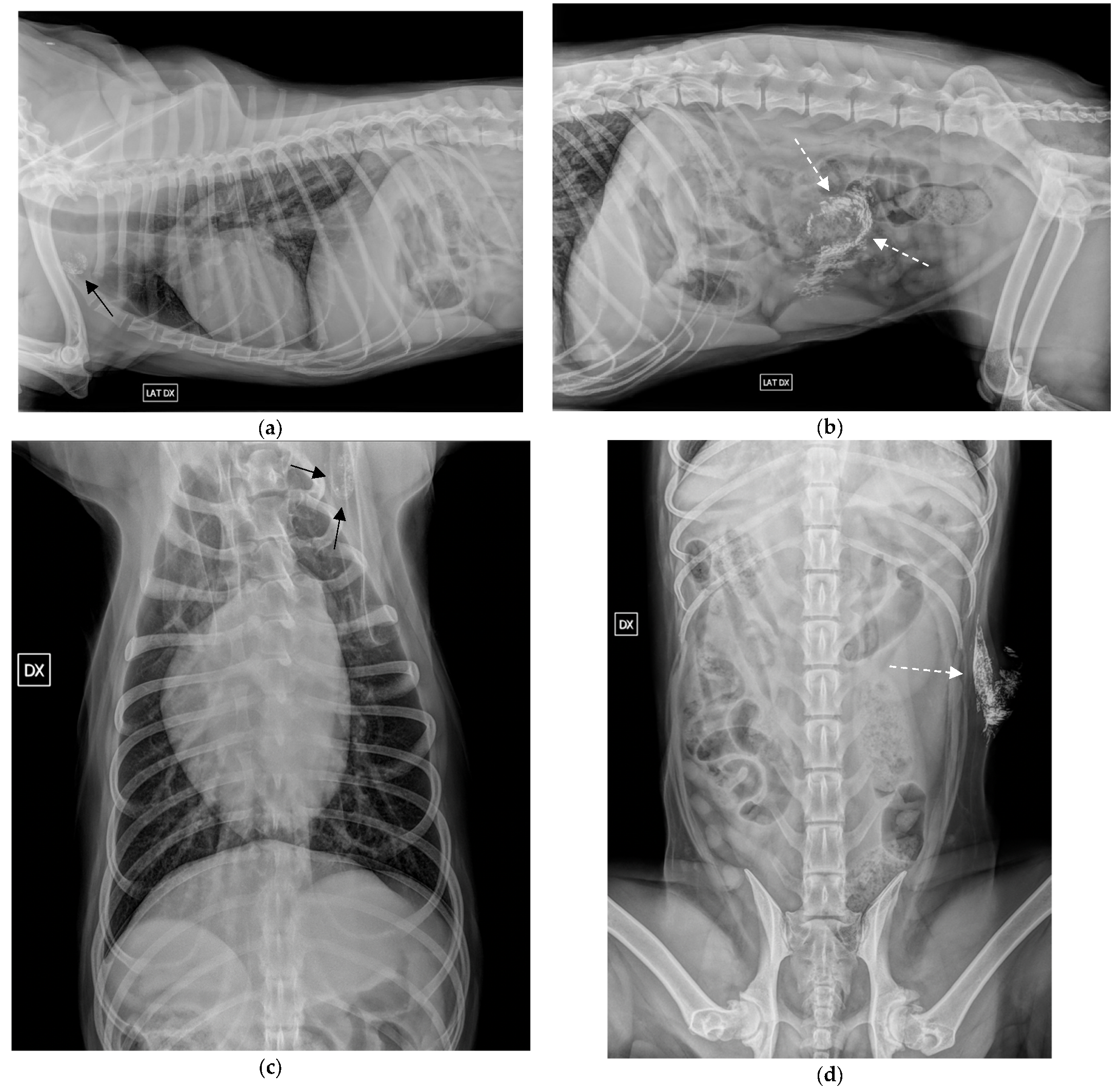
Figure 1. Radiographic indirect lymphangiography 24 h post-Lipiodol of a MCT in the left abdominal wall. (a,c) right lateral and VD view of the thorax. The left axillary SLN is highlighted by the black arrow; (b,d) Right lateral and VD view of the abdomen. The Lipiodol was injected at the periphery of the tumour in the left abdominal wall (white dashed arrow).
Vetsci 09 00484 g001
Figure 2. Number of SLNs detected with radiographic indirect lymphangiography. Abbreviations: SLN, sentinel lymph node; MCT, mast cell tumour.
Vetsci 09 00484 g002
Figure 3. Radiographic indirect lymphangiography 24 h post Lipiodol in two orthogonal views of a MCT in the scrotum with three SLNs: (a) VD view of the pelvic region. The Lipiodol was injected at the periphery of the tumour in the scrotum. Lymphatic vessels are visible on both views extending cranially (black arrows), inguinal lymph nodes are highlighted by the (black dashed arrow); (b) fight lateral view of the pelvic region. The right medial iliac SLN can be assessed ventrally to L6, pointed by the black arrow, and to the right of L6 in the VD highlighted by the red arrow. The location of the primary MCT in both VD and right lateral is highlighted by the white arrow.
Vetsci 09 00484 g003
Table 1. Population data. Breed and sex.
BreedMixLabrador RetrieverEnglish SetterMalteseOther breeds:
(n = 11) 42%
(n = 7) 27%(n = 4) 15%(n = 2) 8%(n = 2) 8%
SexMaleFemaleNeutered MaleSpayed Female
(n = 9) 34.5%(n = 9) 34.5%(n = 3) 11.5%(n = 5) 19.5%
Abbreviation: mix, mixed breed; n, number.
Table 2. Location of MTCs, RLNs, and SLNs assessed with indirect lymphangiography.
MCTLocationRLNSLN Mapping
Case 1Left stifleLeft poplitealLeft inguinal
Case 2Upper left lipLeft mandibularLeft mandibular
Case 3Left neckLeft cervical superficialLeft medial retropharyngeal
Case 4Left prescapularLeft cervical superficialLeft cervical superficial
Case 5Right caudal thighRight inguinalRight inguinal
Case 6Right lateral thighRight inguinalRight inguinal
Case 7 *Left sternumLeft axillaryLeft axillary
Case 8Right sternumRight axillaryRight axillary
Left axillary
Case 9Right prepuceRight inguinalRight inguinal
Case 10Right abdominal wallRight inguinalRight medial iliac
Case 11Right stifleRight poplitealRight popliteal
Right medial iliac
Case 12Left thorax 11th ribRight Axillary/
Case 13Right ventral neckRight cervical superficialRight cervical superficial
Case 14Right thorax 8th ribRight axillaryRight axillary
Right accessory axillary
Case 15Left scrotumLeft inguinalLeft inguinal
Case 16Right pre-scrotumRight inguinalRight inguinal
Left inguinal
Right medial iliac
Case 17Left abdominal wallLeft inguinalLeft axillary
Case 18Left ear baseLeft cervical superficialLeft cervical superficial
Left deep cervical
Case 19Left thighLeft inguinalLeft inguinal
Left medial iliac
Case 20Right abdominal wallRight inguinalRight accessory axillary
Case 21Left ear pinnaLeft cervical superficialLeft cervical superficial
Left mandibular
Case 22Left thighLeft inguinalLeft inguinal
Left popliteal
Left medial iliac
Case 23Left shoulderLeft axillaryLeft axillary
Case 24Right ear pinnaRight cervical superficialRight cervical superficial
Right deep cervical
Case 25Right dorsal neckRight cervical superficialRight mandibular
Case 26Left gluteusLeft inguinal/
Case 27Right neckRight cervical superficialRight mandibular
Case 28Right neckRight cervical superficial/
Case 29Left axillaRight axillaryRight axillary
* MCTs of the dogs who underwent indirect lymphangiography at the surgical scar. Abbreviations: MCT, mast cell tumour; RLN, regional lymph node; SLN, sentinel lymph node. In bold are the SLNs that do not agree with RLN, while in italic are the SLNs who partially agree with RLN.
Table 3. Histological diagnosis of MCTs, cytology/histology of SLNs compared with the location of SLNs, presence or absence of lymphatic vessels.
MCTSLNsCytology/Histology SLNLymphatic VesselHistology MCTs
Case 1Left inguinalHN2 (Histo)YSubcutaneous
Case 2Left mandibularHN0 (Histo)NP2-LK
Case 3Left medial retropharyngealPositive (Cyto)YP2-HK
Case 4Left cervical superficialHN0 (Histo)YP2-HK
Case 5Right inguinalNegative (Cyto)YP2-LK
Case 6Right inguinalNegative (Cyto)YP2-LK
Case 7 *Left axillaryHN3 (Histo)YSubcutaneous
Case 8Left axillary
Right axillary
HN2 (Histo)NSubcutaneous
Case 9Right inguinalHN0 (Histo)YP2-LK
Case 10Right medial iliacNegative (Cyto)YP2-LK
Case 11Right popliteal
Right medial iliac
Negative (Cyto)YP2-LK
Case 12//NP1-LK
Case 13Right cervical superficialNegative (Cyto)YP2-LK
Case 14Right axillary
Right accessory axillary
Negative (Cyto)YP2-LK
Case 15Left inguinalNegative (Cyto)NP2-LK
Case 16Right inguinal
Left inguinal
Right medial iliac
Negative (Cyto)YP2-LK
Case 17Left axillaryHN1 (Histo)NSubcutaneous
Case 18Left cervical superficial
Left deep cervical
Negative (Cyto)NP2-LK
Case 19Left inguinal
Left medial iliac
HN3 (Histo)YP2-LK
Case 20Right accessory axillaryNegative (Cyto)NP2-LK
Case 21Left cervical superficial
Left mandibular
HN1 (Histo)YP2-LK
Case 22Left inguinal
Left popliteal
Left medial iliac
HN0 (Histo)YSubcutaneous
Case 23Left axillaryHN0 (Histo)YP1-LK
Case 24Right cervical superficial
Right deep cervical
HN3 (Histo)NP2-HK
Case 25Right mandibularPositive (Cyto)NP1-LK
Case 26//NP2-HK
Case 27Right mandibularHN3 (Histo)YSubcutaneous
Case 28//YSubcutaneous
Case 29Right axillaryHN3 (Histo)YSubcutaneous
* Single dog with indirect lymphangiography performed on the surgical scar. / MCTs with no SLNs highlighted. Abbreviations: MCT, mast cell tumour; SLN, sentinel lymph node; HN, histological grade; P, Patnaik; Histo, histopathology; Cyto, cytology; Y, yes; N, no; P1-LK, Patnaik I–low-grade Kiupel; P2-LK, Patnaik II–low-grade Kiupel; P2-HK, Patnaik II–high-grade Kiupel.
Publisher’s Note: MDPI stays neutral with regard to jurisdictional claims in published maps and institutional affiliations.