Article

Immune Responses Induced by Recombinant Bacillus subtilis Expressing the PEDV Spike Protein Targeted at Microfold Cells

College of Veterinary Medicine, Nanjing Agricultural University, Weigang 1, Nanjing 210095, China
*
Author to whom correspondence should be addressed.
These authors contributed equally to this work.
Vet. Sci. 2022, 9(5), 211; https://doi.org/10.3390/vetsci9050211
Submission received: 26 March 2022 / Revised: 16 April 2022 / Accepted: 17 April 2022 / Published: 25 April 2022

Abstract

:
Bacillus subtilis (B. subtilis), a probiotic bacterium and feeding additive, is widely used for heterologous antigen expression and protective immunisation. Porcine epidemic diarrhoea virus (PEDV) invades swine via mucosal tissue. To enhance the mucosal immune response to PEDV, we modified B. subtilis to express a PEDV antigen and used it as a mucosal vaccine delivery system. Initially, we constructed a recombinant B. subtilis strain (B.s-RCL) that expressed the PEDV spike protein and L-Lectin-β-GF, with the goal of inducing mucosal secretory immunoglobulin A (sIgA) and anti-PEDV serum immunoglobulin G (IgG) production, as well as to increase the number of microfold cells (M cells). Following the oral administration of B.s-RCL to mice, the small intestinal PEDV-specific sIgA expression levels significantly increased, as well as the increased number of B.s-RCL adhered to M cells. Moreover, we found that mice administered B.s-RCL exhibited markedly higher percentages of CD4+ and CD8+ T cells in the mesenteric lymph nodes and spleen compared to the control mice. Furthermore, we found that intestinal mucosa sIgA and serum anti-PEDV IgG levels were higher in mice orally immunised with B.s-RCL, suggesting that the mice could be more resistant to PEDV. In this study, we developed a novel oral vaccine to prevent porcine diarrhoea epidemics.

1. Introduction

Bacillus subtilis (B. subtilis) is a non-pathogenic Gram-positive bacterium that is widely used as a probiotic. B. subtilis is used as a feed additive to improve the intestinal microbiome [1] and as a mucosal adjuvant to improve mucosal immunity [2,3,4]. Furthermore, B. subtilis has a stable heterologous protein expression system and is commonly used as a vehicle for antigen expression [5,6]. In our previous study, we developed a recombinant B. subtilis strain that could induce an immune response to the transmissible gastroenteritis virus via dendritic cell uptake [7]. However, due to the limited number of dendritic cells in the intestinal mucosa, B. subtilis exhibited poor adhesion to intestinal epithelial cells [8]. This limited the uptake of B. subtilis by intestinal dendritic cells, leading to poor activation of the mucosal immune response. Therefore, a novel antigen delivery vector that can trigger an immune response against mucosal pathogens is needed [9].
Normally in gastrointestinal mucosa, the immunologically active sites are located in Peyer’s patches [10]. Besides dendritic cells, microfold cells (M cells) are another important intestine cell located on the surfaces of Peyer’s patches, where they are involved in antigen uptake, transport and processing [9]. M cells transfer antigens to various immune cells, such as dendritic cells, T cells, B cells and macrophages, thus inducing an immune response [11,12]. In a previous study, recombinant yeast expressing M cells effectively invaded the mucosa [9]. The adhesion protein L-lectin was first discovered in leguminous seeds, where it binds to extracellular sugar chains [13]. Various types of L-lectin proteins can recognise different carbohydrates, despite having similar folding structures [14]. The surface protein L-Lectin-β-GF mediates the adhesion of Staphylococcus aureus to M cells [15]. β-GF binds to secretory immunoglobulin A (sIgA), which is then taken up by M cells in the intestinal mucosa [16]. Therefore, we hypothesised that the M cell-targeting L-Lectin-β-GF protein may improve the adherence of B. subtilis to M cells.
Porcine epidemic diarrhoea virus (PEDV) causes severe enteric disease in pigs, which primarily enters the body via mucosal surfaces and causes disease in the small intestines [17,18,19]. To enhance the mucosal immune response to PEDV, we selected a spike (S) of PEDV as a target to express [20,21]. The S protein contains major antigenic determinants and the core neutralising epitope (COE), which might be useful in preventing PEDV infection [22,23]. To effectively deliver the antigen into the small intestine, we engineered B. subtilis to express the recombinant PEDV COE region of the S protein and L-Lectin-β-GF to enhance the mucosal immunity of mice by increasing their ability to bind to M cells. This modified B. subtilis to express a PEDV antigen can also be used as a mucosal vaccine delivery system to effectively defend PEDV intestines infection.

2. Materials and Methods

2.1. B. subtilis, Plasmids and Animals

The pHT43 plasmids and B. subtilis WB800N were given to Prof. Xuewen Gao, while PEDV SD-M was kindly presented by the Jiangsu Academy of Agricultural Sciences to our lab. The L-Lectin-β-GF sequence was synthesised according to the DNA sequences (GenBank, the accession number of YP_501439.1) and cloned into pET28a. Four to six-week-old SPF C57BL/6 and six to eight-week-old SPF Balb/C mice were bought from Comparative Medical Center of Yangzhou University. Vero cells were cultured in DMEM supplemented with 10% fetal bovine serum, 16 mM HEPES and 100 μg/mL penicillin/streptomycin (Life Technologies, Shanghai, China), which were then incubated in an atmosphere of 5% CO2 at 37 °C.

2.2. The Construction of Recombinant B.S.-RC and B.S.-RCL

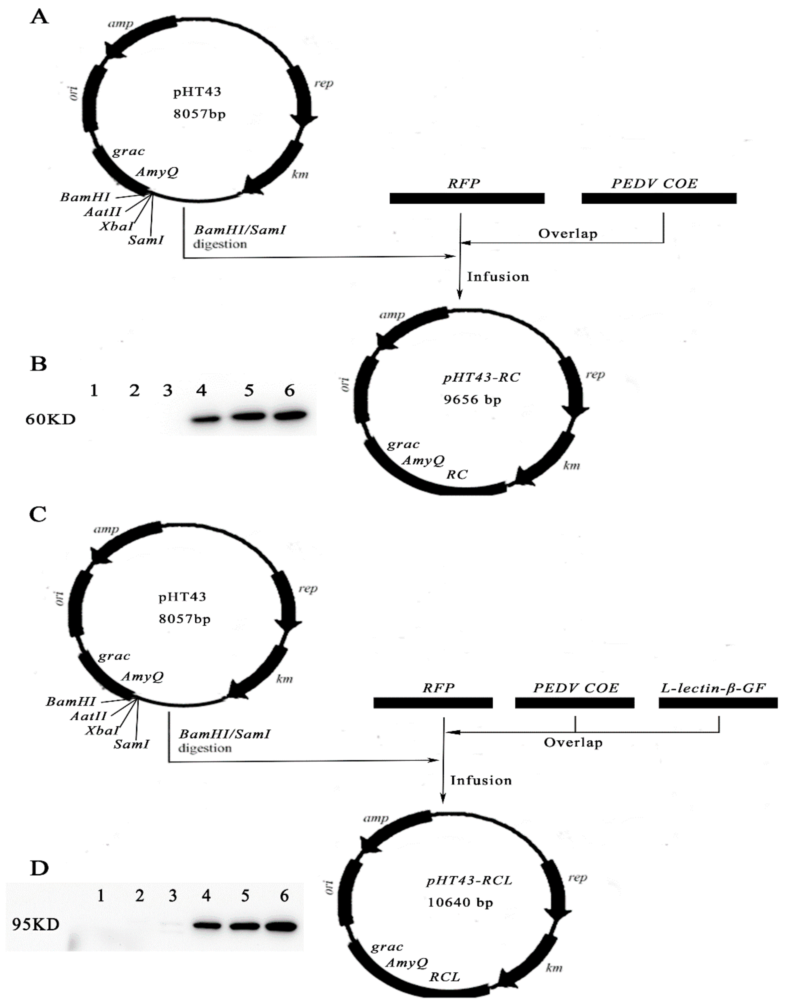
In order to obtain recombinant B. subtilis WB800N, we firstly amplified three fragments. The RFP and COE fragments were cloned from pDsRed-monomer-N1 and PEDV, while the L-Lectin-β-GF fragment was cloned from pET28a-L-Lectin-β-GF. Secondly, the above three fragments were overlapped to generate fusion fragments RFP-COE (RC) and RFP-COE-L-Lectin-β-GF (RCL). Thirdly, the fragments were insert into pHT43 to generate pHT43-RC and pHT43-RCL by infusion clone, respectively. Finally, we transformed plasmids pHT43-RC and pHT43-RCL into WB800N, which were then named B.S.-RC and B.S.-RCL, respectively [24].

2.3. Analysis of Fusion Protein

LB medium with 5 μg/mL chloramphenicol were used to culture B.S.-RC and B.S.-RCL. We then added IPTG (0.1M) when the growth phase of recombinant B. subtilis reached an OD600 = 0.5 and continued the culture for another 3 h. After collecting, B.S.-RC and B.S.-RCL were washed three times with PBS and split by ultrasonication. Moreover, the rabbit anti-PEDV were selected to evaluate their expression by Western blot as previously described [25]. Finally, we used the Super ECL Plus system to visualise the protein bands.

2.4. Ligated Loop Experiments

SPF mice were treated with broad-spectrum antibiotics for 5 days before the experiments. Then, the mice were anesthetised with chloral hydrate according to their weight (350 mg per kg weight, intraperitoneal). After anesthetisation, terminal ileal or jejunal containing a Peyer’s patch ligated loop was injected into DyLight 488-labeled recombinant B.S.-RC and B.S.-RCL (200 mL) or the same volume of PBS (0.01 M) for 15 min or 1 h, as shown in Figure 1. The intestines were removed and fixed with 4% paraformaldehyde at 4 °C for 6 h and subsequently embedded in OCT (Sakura, Torrance, CA, USA) to cut into 8 μm for immunofluorescence [25]. The division can be seen in Table 1 for grouping.
Mice intestinal ligation and oral immunisation experiments were performed. The treatment and sample collections were conduct similar to the above scheme.

2.5. Immunisation Schedule and Samples Collection

C57BL/6 mice were immunised orally (n = 24 per group) at 0 and 7 days with B.S.-RC and B.S.-RCL at the concentration 1010 CFU/kg. Nonimmunised control mice were orally administrated 150 μL of 0.01 M PBS. The mice were sacrificed 14, 21, 28 and 35 days after first immunisation. The division can be seen in Table 1 for the grouping. Firstly, blood samples were taken and allowed to clot overnight at room temperature before collecting the serum for PEDV-specific IgG detection. The small intestine samples were washed with 0.5 mL of 0.01 M PBS, and the suspension was centrifuged and collected for PEDV-specific SIgA detection [2]. Secondly, small intestine containing Peyer’s patches tissue samples were fixed with 4% paraformaldehyde for immunofluorescence after 14 days of immunisation. In the meantime, intestine suspensions were collected for the total SIgA detection, while mesenteric lymph nodes (MLN) and spleen lymphocytes were isolated to assess the CD4+ and CD8+ T-cell populations.

2.6. Immunofluorescence Staining

For M cell staining, the slides were first washed with PBS three times to eliminate the residual OC. Then, the slides were blocked with PBS at room temperature for 30 min, which contained 5% FBS. Moreover, UEA-1-FITC was added and incubated at room temperature for 2 h to dye the M cells, while DAPI was used to stain the cell nuclei. Lastly, we used a Zeiss LSM710 confocal microscope to visualise the staining, and the images were analysed with the guidance of the regional Ethics Committee for Animal Testing (CREEA) (Permit Number No. 69387487) [5].

2.7. Enzyme-Linked Immunosorbent Assay

Nonspecific SIgA and PEDV-specific IgG and SIgA were measured by ELISA according to the instructions as previously described [26]. Briefly, the ELISA plates were first coated with PEDV COE protein at 4 °C overnight and saturated with PBS (1% BSA) at 37 °C for 2 h. Second, the mucosal suspension and serum were added and incubated for another 1 h at 37 °C. Subsequently, HRP-conjugated goat anti-mouse IgG and goat anti-mouse IgA were added and incubated at 37 °C for 1 h. Third, a substrate solution containing H2O2 and o-phenylenediamine was added at room temperature for 15 min before termination. Finally, we used an automated ELISA reader (Molecular Devices, Shanghai, China) to detect the absorbance at OD450 nm.

2.8. Statistical Analysis

All the results were analysed with GraphPad Prism 8.0 and expressed as the mean ± SD. Then, one-way analysis of variance was employed to evaluate the significant differences, followed by Dunnett’s t-test to evaluate the variations between the different groups. Differences were considered to be statistically significant when p < 0.05. Data were collected and combined from at least three independent experiments.

3. Results

3.1. Construction of Recombinant Plasmids and Fusion Protein Expression

The DNA fragments of the fusion proteins red fluorescent protein (RFP)-COE (RC) and RFP-COE-L-lectin-β-GF (RCL) were inserted into the pHT43 plasmid to generate the pHT43-RC and pHT43-RCL shuttle vectors (Figure 2A,C). Then, the pHT43-RC and pHT43-RCL plasmids were transformed into B. subtilis WB800N by electroporation to generate strains B. subtilis-RC (B.s-RC) and B. subtilis-RCL (B.s-RCL). The protein expression was evaluated in B.s-RC and B.s-RCL via Western blotting using anti-PEDV antibodies (Figure 2B,D). Clear, positive bands were present at 60 and 95 kDa in recombinant strains B.s-RC and B.s-RCL, respectively (Supplementary Materials Figures S1 and S2). Our results showed that the RC and RCL fusion proteins were successfully expressed in B. subtilis WB800N.

3.2. Animal Experiments Design and sIgA Levels and M Cell Numbers in Mouse Intestines

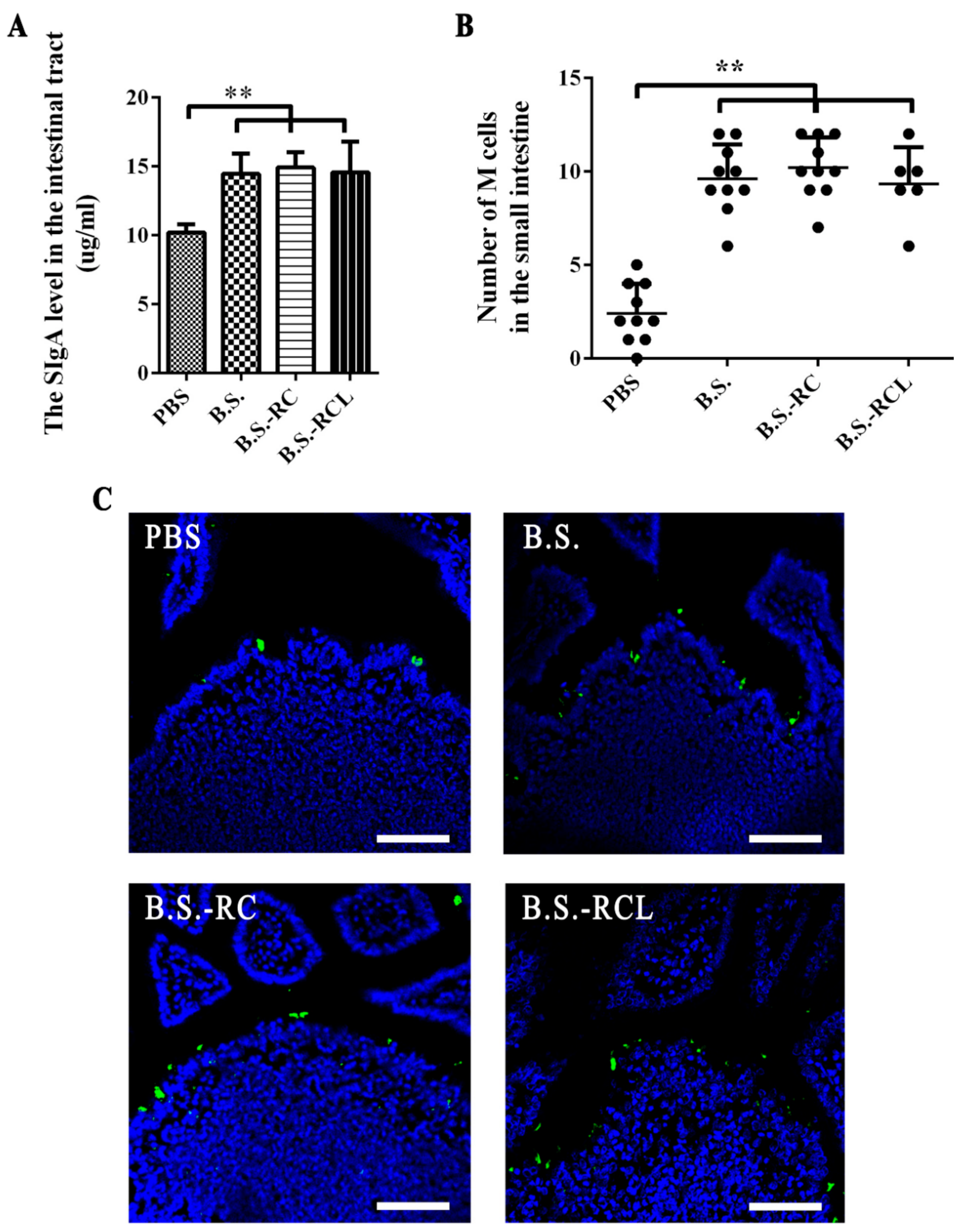
Our animal experiment was divided into two parts, which were the ligated loop experiments and animal immune experiment. The immunisation and sample collection scheme are clearly shown in Figure 2. Since sIgA can interact specifically with mucosal M cells in gut-associated lymphoid tissues [16], we therefore examined the sIgA levels and M cell numbers in mouse intestines following the oral treatment with the recombinant B. subtilis. sIgA levels increased in the intestinal tracts of the mice at 14 days after feeding with B. subtilis (Figure 3A). Moreover, we stained the mouse intestinal M cells 14 days after the first immunisation; the number of M cells in the mouse intestines increased dramatically after feeding with recombinant B. subtilis (Figure 3B,C).

3.3. Co-Localisation of M Cells and Recombinant B. subtilis

M cells are derived from stem cells located within intestinal crypts [27], which can be stimulated by the activator of nuclear factor kappa-Β ligand (RANKL) secreted by subepithelial stromal cells [28]. The L-lectin-β-GF protein binds to sIgA and M cells [15]. To investigate the binding of recombinant B. subtilis and M cells in vivo, we performed ligated loop experiments in mice. We found that B.s-RCL localised to M cells 15 min after the oral administration. Moreover, the number of B. subtilis cells in the follicular-associated epithelium (FAE) was higher in mice fed with recombinant B.s-RCL than in those fed with control B. subtilis at 1 h after administration. Our results suggested that recombinant B.s-RCL effectively adhered to the M cells (Figure 4A,B) and increased the amount of recombinant B. subtilis in the FAE (Figure 4C,D).

3.4. Systemic and Local Immune Responses after Oral Immunisation

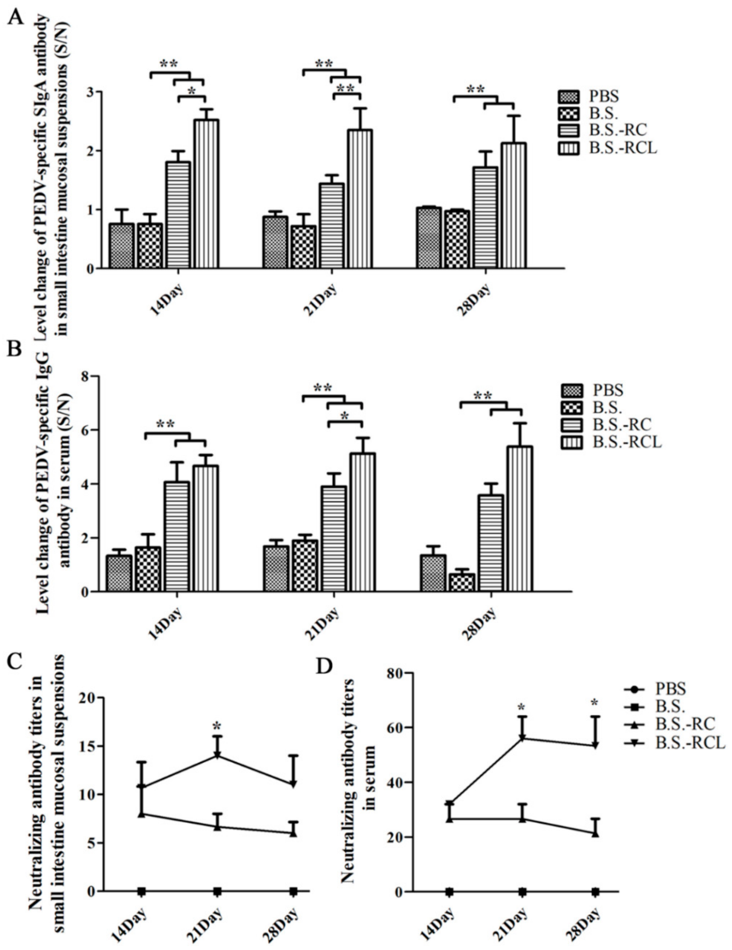
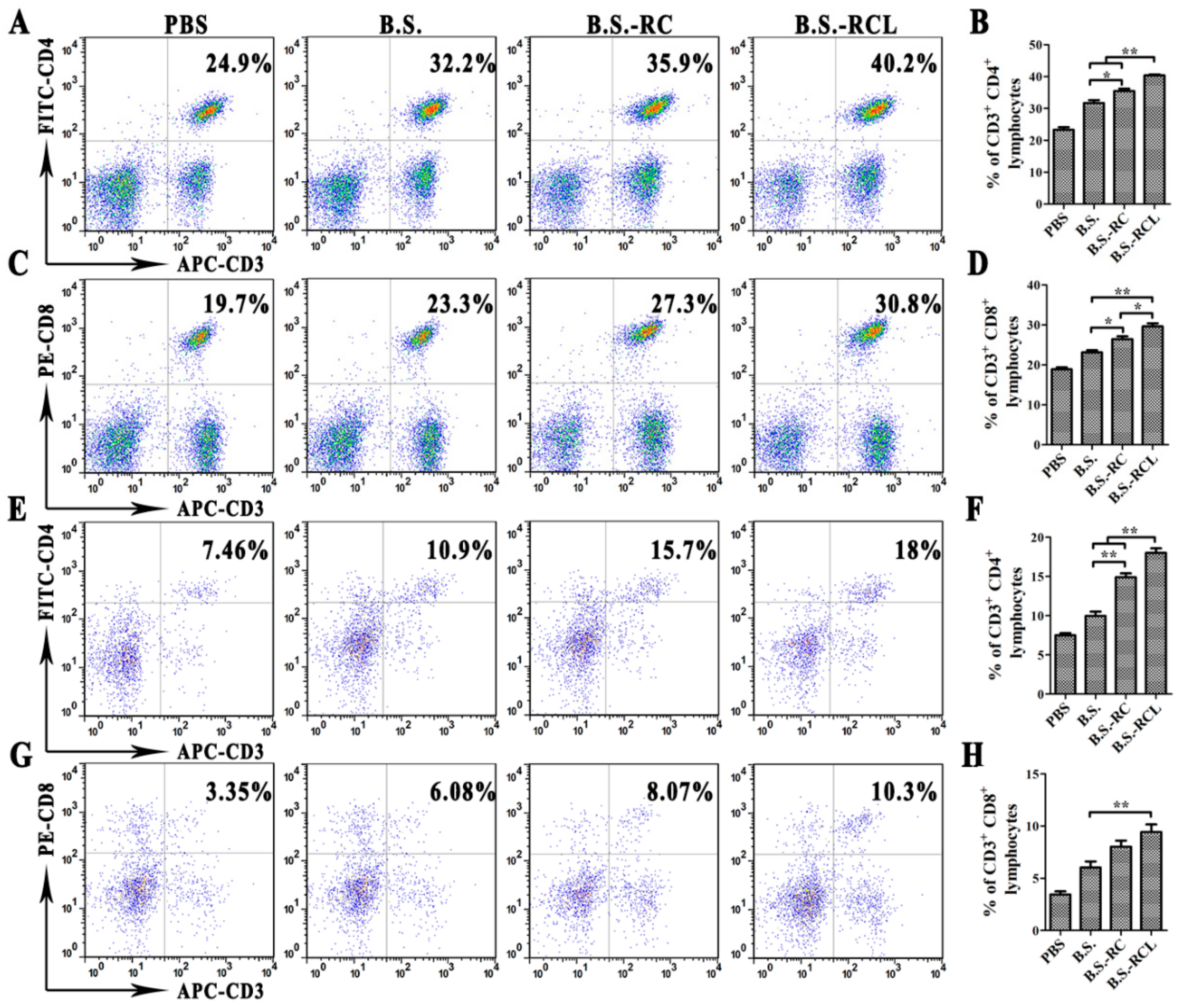
Antigen-specific sIgA is a key player in mucosal immunity. Therefore, we hypothesised that the sIgA-binding L-lectin-β-GF protein could enhance the carrier function and adjuvant properties of B. subtilis. We detected PEDV-specific sIgA (Figure 5A) at 14–28 days after recombinant B. subtilis oral administration. Furthermore, we found that the levels of PEDV-specific sIgA were higher in mice orally administered B.s-RCL compared to those administered B.s-RC at 14–21 days after administration. Mice administered recombinant B. subtilis exhibited higher PEDV-specific immunoglobulin G (IgG) antibody titres compared to those administered controls of B. subtilis at 14–28 days after administration (Figure 5B,C). Similarly result of serum antibody titres were also found enhanced by recombinant B. subtilis stimulation (Figure 5D). In addition, we found that the percentages of CD3+/CD4+ and CD3+/CD8+ T cells in mesenteric lymph nodes (MLNs) and spleen lymphocytes were markedly higher after B.s-RCL stimulation (Figure 6). These results indicate that oral immunisation with recombinant B.s-RCL effectively induced systemic and local immune responses in mice.

4. Discussion

B. subtilis is commonly used to express heterologous proteins and as an antigen delivery vector [5,29,30,31]. The spores formed by B. subtilis have unique resistance properties, enabling their survival under extreme conditions. B. subtilis is also used as a feed additive to improve growth and stimulate the immune responses of livestock [32]. Orally administrated B. subtilis exerts beneficial effects on intestinal lymphoid tissue in chickens [1]. In this study, orally administrated B. subtilis increased intestinal M cells in Peyer’s patches in mice. Although the underlying mechanisms are still unclear, B. subtilis have probably adjuvant effects due to its ability to produce amylase, protease, lipase and amino acids [33]. M cells are primarily derived from stem cells located within dome-associated crypts [27] but also originate from stimulated enterocytes. The increase in M cells likely increased the number of recombinant B. subtilis-binding events while also enhancing the uptake and delivery of antigens.
Recently, several oral vaccines have been developed. Oral vaccines are particularly effective for building defences against mucosal pathogens. Various studies have focused on enhancing antigen deliveries to M cells due to their role in antigen capturing. Some specialised proteins, such as invasion, bind to the surface receptors of M cells to enter the intestine [34,35]. Besides the uptake and transfer of antigens, M cells are also involved in sIgA responses [36]. Hence, we expressed the M cell-targeting protein L-lectin-β-GF to mediate interactions with M cells and trigger sIgA secretion. Our intestinal ligated loop mouse experiments demonstrated that B.s-RCL associated with M cells after 15 min. Furthermore, they revealed that L-lectin-β-GF can enhance the delivery of B. subtilis, demonstrating a novel mucosal immune delivery system. Our findings were similar to those of Rochereau et al., who found that the p24 protein could be employed as a vehicle to induce humoral and cellular immune responses against human immune deficiency virus [37].
PEDV is a serious swine intestinal disease; infections occur at the mucosal surfaces, especially those of the intestinal mucosal epithelium. PEDV entry could be prevented by a well-established mucosal sIgA immune response. Here, we found that B. subtilis treatment increased the sIgA levels in the intestinal tracts of mice, which enhanced the binding of L-lectin-β-GF to M cells, thus further improving the mucosal immunity. Specific sIgAs effectively neutralise viruses. Following the oral administration of B.s-RC, the PEDV-specific sIgA levels increased dramatically. Mice administered B.s-RCL exhibited higher levels of PEDV-specific sIgA at 14 days after the first immunisation.
In this study, we employed B. subtilis as a vehicle to express the PEDV S protein COE and L-lectin-β-GF protein. Our work suggests that recombinant B. subtilis may promote growth and regulate immune responses in piglets while also targeting and binding to M cells to enhance antigen presentation and augment defences against PEDV invasion. However, we did not perform the in vitro M cell experiment to demonstrate that L-lectin-β-GF could enhance the M cell-mediated phagocytic for the lack of a M cell model. This study provided important insights that could facilitate the development of a PEDV vaccine. We demonstrated that immunising mice with B.s-RCL significantly increased faecal PEDV-specific sIgA titres and serum IgG levels.

5. Conclusions

Bacillus subtilis, as a feeding additive, has been widely used for heterologous antigen expression in immune mucosal. Hence, our study tried to defend PEDV by expressing a PEDV antigen with B. subtilis. Initially, we constructed the recombinant B. subtilis strain (B.s-RCL) to induce the mucosal secretory immunoglobulin A (sIgA) and anti-PEDV serum immunoglobulin G (IgG) production, as well as to increase the number of microfold cells (M cells). Then, oral immunisation mice with B.s-RCL not only increased PEDV-specific sIgA in the small intestinal but also upregulated the percentages of CD4+ and CD8+ T cells in mesenteric lymph nodes and the spleen. All in all, our study developed a novel oral vaccine to prevent porcine diarrhoea epidemic.

Supplementary Materials

The following supporting information can be downloaded at: https://www.mdpi.com/article/10.3390/vetsci9050211/s1, Figure S1: Western blot result of the fusion protein B. subtilis RC.; Figure S2: Western blot result of the fusion protein B. subtilis RCL.

Author Contributions

The work presented here was carried out in a collaboration among all the authors. J.L. and C.M. designed and carried out all experiments and analysed and interpreted the results, as well as the writing of the paper, who all contribute to the figure. S.Z. provided excellent technical assistance and contributions to Figure 2, Figure 3 and Figure 4. L.Z. and Y.L. codesigned the experiments, who contributed to Figure 5 and Figure 6. Q.Y. defined the research theme and provided the funding. All authors have read and agreed to the published version of the manuscript.

Funding

This work was supported by the National Key Research and Development Program of China (2021YFD1801105) and the National Natural Science Foundation of China (31930109 and 31772777) to Q.Y. Moreover, this work was also supported by the Jiangsu Excellent Youth Natural Science Foundation (BK20190077), the National Natural Science Foundation of China (32072835) and A Project Funded by the Priority Academic Program Development of Jiangsu Higher Education Institutions (PAPD) to J.L.

Institutional Review Board Statement

All animal experiment of this study was approved by the Institutional Animal Care and Use Committee of Nanjing Agricultural University (SYXK-2017-0007), and followed the National Institutes of Health guidelines for the performance of animal experiments. Also, all our research in this study was approved by the Ethics Committee of Animal Experiments center of Nanjing Agricultural University and accordanced with ARRIVE guidelines (https://arriveguidelines.org, accessed on 22 March 2022).

Informed Consent Statement

Informed consent was obtained from all subjects involved in the study.

Data Availability Statement

All data generated or analysed during this study are included in this published.

Conflicts of Interest

The authors declare no conflict of interest.

References

  1. Lei, X.; Piao, X.; Ru, Y.; Zhang, H.; Peron, A.; Zhang, H. Effect of Bacillus amyloliquefaciens-based Direct-fed Microbial on Performance, Nutrient Utilization, Intestinal Morphology and Cecal Microflora in Broiler Chickens. Asian-Australas. J. Anim. Sci. 2015, 28, 239–246. [Google Scholar] [CrossRef] [PubMed] [Green Version]
  2. Huang, L.; Qin, T.; Yin, Y.; Gao, X.; Lin, J.; Yang, Q.; Yu, Q. Bacillus amyloliquefaciens SQR9 induces dendritic cell maturation and enhances the immune response against inactivated avian influenza virus. Sci. Rep. 2016, 6, 21363. [Google Scholar] [CrossRef] [PubMed] [Green Version]
  3. Lin, J.; Huang, L.L.; Li, Y.C.; Zhang, P.H.; Yu, Q.H.; Yang, Q. Bacillus subtilis Spore-Trained Dendritic Cells Enhance the Generation of Memory T Cells via ICAM1. Cells 2021, 10, 2267. [Google Scholar] [CrossRef]
  4. Zhang, G.Z.; Wang, H.; Zhang, J.W.; Tang, X.M.; Raheem, A.; Wang, M.Y.; Lin, W.D.; Liang, L.; Qi, Y.Z.; Zhu, Y.L.; et al. Modulatory Effects of Bacillus subtilis on the Performance, Morphology, Cecal Microbiota and Gut Barrier Function of Laying Hens. Animals 2021, 11, 1523. [Google Scholar] [CrossRef]
  5. Ferreira, L.C.; Ferreira, R.C.; Schumann, W. Bacillus subtilis as a tool for vaccine development: From antigen factories to delivery vectors. An. Acad. Bras. Cienc. 2005, 77, 113–124. [Google Scholar] [CrossRef] [PubMed] [Green Version]
  6. Hoess, R.H. Bacteriophage lambda as a vehicle for peptide and protein display. Curr. Pharm. Biotechnol. 2002, 3, 23–28. [Google Scholar] [CrossRef] [PubMed]
  7. Mou, C.; Zhu, L.; Xing, X.; Lin, J.; Yang, Q. Immune responses induced by recombinant Bacillus subtilis expressing the spike protein of transmissible gastroenteritis virus in pigs. Antivir. Res. 2016, 131, 74–84. [Google Scholar] [CrossRef]
  8. Spivey, M.A.; Dunn-Horrocks, S.L.; Duong, T. Epithelial cell adhesion and gastrointestinal colonization of Lactobacillus in poultry. Poult. Sci. 2014, 93, 2910–2919. [Google Scholar] [CrossRef]
  9. Kenngott, E.E.; Kiefer, R.; Schneider-Daum, N.; Hamann, A.; Schneider, M.; Schmitt, M.J.; Breinig, F. Surface-modified yeast cells: A novel eukaryotic carrier for oral application. J. Control. Release 2016, 224, 1–7. [Google Scholar] [CrossRef]
  10. Neutra, M.R.; Kozlowski, P.A. Mucosal vaccines: The promise and the challenge. Nat. Rev. Immunol. 2006, 6, 148–158. [Google Scholar] [CrossRef]
  11. Woodrow, K.A.; Bennett, K.M.; Lo, D.D. Mucosal vaccine design and delivery. Annu. Rev. Biomed. Eng. 2012, 14, 17–46. [Google Scholar] [CrossRef] [PubMed] [Green Version]
  12. Gebert, A. The role of M cells in the protection of mucosal membranes. Histochem. Cell Biol. 1997, 108, 455–470. [Google Scholar] [CrossRef] [PubMed]
  13. Srinivas, V.R.; Reddy, G.B.; Ahmad, N.; Swaminathan, C.P.; Mitra, N.; Surolia, A. Legume lectin family, the ‘natural mutants of the quaternary state’, provide insights into the relationship between protein stability and oligomerization. Biochim. Biophys. Acta 2001, 1527, 102–111. [Google Scholar] [CrossRef]
  14. Sharon, N.; Lis, H. Legume lectins—A large family of homologous proteins. FASEB J. Off. Publ. Fed. Am. Soc. Exp. Biol. 1990, 4, 3198–3208. [Google Scholar] [CrossRef] [PubMed]
  15. Yang, Y.H.; Jiang, Y.L.; Zhang, J.; Wang, L.; Bai, X.H.; Zhang, S.J.; Ren, Y.M.; Li, N.; Zhang, Y.H.; Zhang, Z.; et al. Structural insights into SraP-mediated Staphylococcus aureus adhesion to host cells. PLoS Pathog. 2014, 10, e1004169. [Google Scholar] [CrossRef] [PubMed]
  16. Rochereau, N.; Drocourt, D.; Perouzel, E.; Pavot, V.; Redelinghuys, P.; Brown, G.D.; Tiraby, G.; Roblin, X.; Verrier, B.; Genin, C.; et al. Dectin-1 is essential for reverse transcytosis of glycosylated SIgA-antigen complexes by intestinal M cells. PLoS Biol. 2013, 11, e1001658. [Google Scholar] [CrossRef]
  17. Stevenson, G.W.; Hoang, H.; Schwartz, K.J.; Burrough, E.R.; Sun, D.; Madson, D.; Cooper, V.L.; Pillatzki, A.; Gauger, P.; Schmitt, B.J.; et al. Emergence of Porcine epidemic diarrhea virus in the United States: Clinical signs, lesions, and viral genomic sequences. J. Vet. Diagn. Investig. 2013, 25, 649–654. [Google Scholar] [CrossRef] [Green Version]
  18. Suda, Y.; Miyazaki, A.; Miyazawa, K.; Shibahara, T.; Ohashi, S. Systemic and intestinal porcine epidemic diarrhea virus-specific antibody response and distribution of antibody-secreting cells in experimentally infected conventional pigs. Vet. Res. 2021, 52, 2. [Google Scholar] [CrossRef]
  19. Madson, D.M.; Magstadt, D.R.; Arruda, P.H.; Hoang, H.; Sun, D.; Bower, L.P.; Bhandari, M.; Burrough, E.R.; Gauger, P.C.; Pillatzki, A.E.; et al. Pathogenesis of porcine epidemic diarrhea virus isolate (US/Iowa/18984/2013) in 3-week-old weaned pigs. Vet. Microbiol. 2014, 174, 60–68. [Google Scholar] [CrossRef]
  20. Tobler, K.; Bridgen, A.; Ackermann, M. Sequence analysis of the nucleocapsid protein gene of porcine epidemic diarrhoea virus. Adv. Exp. Med. Biol. 1993, 342, 49–54. [Google Scholar]
  21. Pensaert, M.B.; de Bouck, P. A new coronavirus-like particle associated with diarrhea in swine. Arch. Virol. 1978, 58, 243–247. [Google Scholar] [CrossRef] [PubMed] [Green Version]
  22. Maj, M.; Fake, G.M.; Walker, J.H.; Saltzman, R.; Howard, J.A. Oral Administration of Coronavirus Spike Protein Provides Protection to Newborn Pigs When Challenged with PEDV. Vaccines 2021, 9, 1416. [Google Scholar] [CrossRef] [PubMed]
  23. Brian, D.A.; Baric, R.S. Coronavirus genome structure and replication. Curr. Top. Microbiol. Immunol. 2005, 287, 1–30. [Google Scholar] [PubMed] [Green Version]
  24. Narita, J.; Okano, K.; Kitao, T.; Ishida, S.; Sewaki, T.; Sung, M.H.; Fukuda, H.; Kondo, A. Display of alpha-amylase on the surface of Lactobacillus casei cells by use of the PgsA anchor protein, and production of lactic acid from starch. Appl. Environ. Microbiol. 2006, 72, 269–275. [Google Scholar] [CrossRef] [Green Version]
  25. Wang, Z.; Yu, Q.; Gao, J.; Yang, Q. Mucosal and systemic immune responses induced by recombinant Lactobacillus spp. expressing the hemagglutinin of the avian influenza virus H5N1. Clin. Vaccine Immunol. CVI 2012, 19, 174–179. [Google Scholar] [CrossRef] [Green Version]
  26. Mou, C.; Zhu, L.; Xing, X.; Yang, Q. Expression of major antigenic sites A and D in S gene of Transmissible gastroenteritis virus of swine (TGEV) in Escherichia coli and development of indirect ELISA for detection of the antibody against TGEV. Chin. Vet. Sci. 2015, 45, 356–360. [Google Scholar]
  27. Morin, M.J.; Warner, A.; Fields, B.N. A pathway for entry of reoviruses into the host through M cells of the respiratory tract. J. Exp. Med. 1994, 180, 1523–1527. [Google Scholar] [CrossRef] [Green Version]
  28. Taylor, R.T.; Patel, S.R.; Lin, E.; Butler, B.R.; Lake, J.G.; Newberry, R.D.; Williams, I.R. Lymphotoxin-independent expression of TNF-related activation-induced cytokine by stromal cells in cryptopatches, isolated lymphoid follicles, and Peyer’s patches. J. Immunol. 2007, 178, 5659–5667. [Google Scholar] [CrossRef]
  29. Gat, O.; Inbar, I.; Aloni-Grinstein, R.; Zahavy, E.; Kronman, C.; Mendelson, I.; Cohen, S.; Velan, B.; Shafferman, A. Use of a promoter trap system in Bacillus anthracis and Bacillus subtilis for the development of recombinant protective antigen-based vaccines. Infect. Immun. 2003, 71, 801–813. [Google Scholar] [CrossRef] [Green Version]
  30. Olmos-Soto, J.; Contreras-Flores, R. Genetic system constructed to overproduce and secrete proinsulin in Bacillus subtilis. Appl. Microbiol. Biotechnol. 2003, 62, 369–373. [Google Scholar] [CrossRef]
  31. Mauriello, E.M.; Duc Le, H.; Isticato, R.; Cangiano, G.; Hong, H.A.; De Felice, M.; Ricca, E.; Cutting, S.M. Display of heterologous antigens on the Bacillus subtilis spore coat using CotC as a fusion partner. Vaccine 2004, 22, 1177–1187. [Google Scholar] [CrossRef] [PubMed]
  32. Li, Y.; Li, P.; Wang, X.; Yu, Q.; Yang, Q. Co-administration of attenuated Mycoplasma hyopneumoniae 168 strain with bacterial DNA enhances the local and systemic immune response after intranasal vaccination in pigs. Vaccine 2012, 30, 2153–2158. [Google Scholar] [CrossRef] [PubMed]
  33. Santoso, U.; Tanaka, K.; Ohtani, S. Effect of dried Bacillus subtilis culture on growth, body composition and hepatic lipogenic enzyme activity in female broiler chicks. Br. J. Nutr. 1995, 74, 523–529. [Google Scholar] [CrossRef] [PubMed] [Green Version]
  34. Leong, J.M.; Fournier, R.S.; Isberg, R.R. Identification of the integrin binding domain of the Yersinia pseudotuberculosis invasin protein. EMBO J. 1990, 9, 1979–1989. [Google Scholar] [CrossRef]
  35. Marra, A.; Isberg, R.R. Invasin-dependent and invasin-independent pathways for translocation of Yersinia pseudotuberculosis across the Peyer’s patch intestinal epithelium. Infect. Immun. 1997, 65, 3412–3421. [Google Scholar] [CrossRef] [Green Version]
  36. Kerneis, S.; Bogdanova, A.; Kraehenbuhl, J.P.; Pringault, E. Conversion by Peyer’s patch lymphocytes of human enterocytes into M cells that transport bacteria. Science 1997, 277, 949–952. [Google Scholar] [CrossRef]
  37. Rochereau, N.; Pavot, V.; Verrier, B.; Ensinas, A.; Genin, C.; Corthesy, B.; Paul, S. Secretory IgA as a vaccine carrier for delivery of HIV antigen to M cells. Eur. J. Immunol. 2015, 45, 773–779. [Google Scholar] [CrossRef]
Figure 1. Immunisation and sample collection scheme.
Vetsci 09 00211 g001
Figure 2. The construction of the recombinant plasmid and its fusion protein expression. (A) The RFP and PEDV S COE gene fragments were overlapped and inserted into pHT43 to generate pHT43-RC. (B) Western blotting detected the recombinant RFP-COE protein (lanes 1, 2 and 3; B. subtilis WB800N; lanes 4, 5 and 6 and B. subtilis RC). The protein bands were approximately 60 kDa, expected at the size of fusion RFP-COE. (C) The RFP, PEDV S COE gene and L-lectin-β-GF fragments were overlapped and inserted into pHT43 to generate pHT43-RCL. (D) Western blotting detected recombinant RFP-COE- L-lectin-β-GF (lanes 1, 2 and 3; B. subtilis WB800N; lanes 4, 5 and 6 and B. subtilis RCL). Protein bands were approximately 95 kDa, expected at the size of the fusion RFP-COE- L-lectin-β-GF, were detected.
Vetsci 09 00211 g002
Figure 3. Analysation of the SIgA level and M cell number in the mice intestine. (A) The SIgA level in the mice intestine were displayed by histogram (Signifcance diference was expressed as, ** p < 0.01). Orally administrated with PBS, B. subtilis WB800N, B. subtilis RC and B. subtilis RCL at 0 and 7 days and the sample were collected at 14, 21 and 28 days. M cells were stained by UEA-1. (B) The number of M cells in the small intestine of each mouse was displayed. (C) Immunofluorescence staining results of M cells in mice small intestines (oral PBS group, oral B. subtilis WB800N group, oral B. subtilis RC group and oral B. subtilis RCL group. Scale bars = 50 μm).
Vetsci 09 00211 g003
Figure 4. The observation of co-located M cells and recombinant B. subtilis. The ligated loops of mice were injected with Dylight 488-labelled B. subtilis RC and Dylight 488-labelled B. subtilis RCL for 15 min and 1 h, respectively. Frozen sections of the ileum were stained with UEA-1 (green) and DAPI (blue). (A,B) The co-located M cells and Dylight 549-labelled B. subtilis RC or B. subtilis RCL (white arrows) 15 min after recombinant B. subtilis injection. (C,D) The co-located M cells and Dylight 549-labelled B. subtilis RC or B. subtilis RCL (white arrows) 1 h after recombinant B. subtilis injection. Scale bars = 10 μm.
Vetsci 09 00211 g004
Figure 5. The detection of specific antibody levels after oral immunisation. After 14, 21 and 28 days of immunisation, the samples were collected to analyse PEDV-specific SIgA (A), IgG (B) and neutralising antibodies in small intestine mucosal and serum (C,D) (the value of S/N was calculated) by indirect ELISA (n = 6). Data are shown as the mean ± SD of three samples. *, p < 0.05; **, p < 0.01.
Vetsci 09 00211 g005
Figure 6. The changes of the local and systemic cellular immunity after oral immunisation. The MLN and spleen collected from mice 14 days after immunisation, and then, the percentages of CD3+ and/or CD4+ in MLN (A,B), CD3+ and/or CD8+ in MLN (C,D), CD3+ and/or CD4+ in the spleen (E,F) and CD3+ and/or CD8+ in spiralising (G,H) T cells were analysed by FACS (Signifcance diference was expressed as, * p < 0.05, ** p < 0.01).
Vetsci 09 00211 g006
Table 1. Experimental groups and administration strategies.
GroupsTreatments
Intestinal ligationPBS-15 minPBS (200 mL for 15 min or 1 h)
PBS-1 h
B.S.-RC-15 minDyLight 488-labeled recombinant B.S.-RC (200 mL for 15 min or 1 h)
B.S.-RC-1 h
B.S.-RCL-15 minDyLight 488-labeled recombinant B.S.-RCL (200 mL for 15 min or 1 h)
B.S.-RCL-1 h
Oral administrationPBSPBS
PBS (0.01M PBS 150 μL, n = 24)PBS (0.01 M PBS 150 μL, n = 24)
B.S.B.S.
B.S. (1010 cfu/kg)B.S. (1010 cfu/kg)
Publisher’s Note: MDPI stays neutral with regard to jurisdictional claims in published maps and institutional affiliations.