

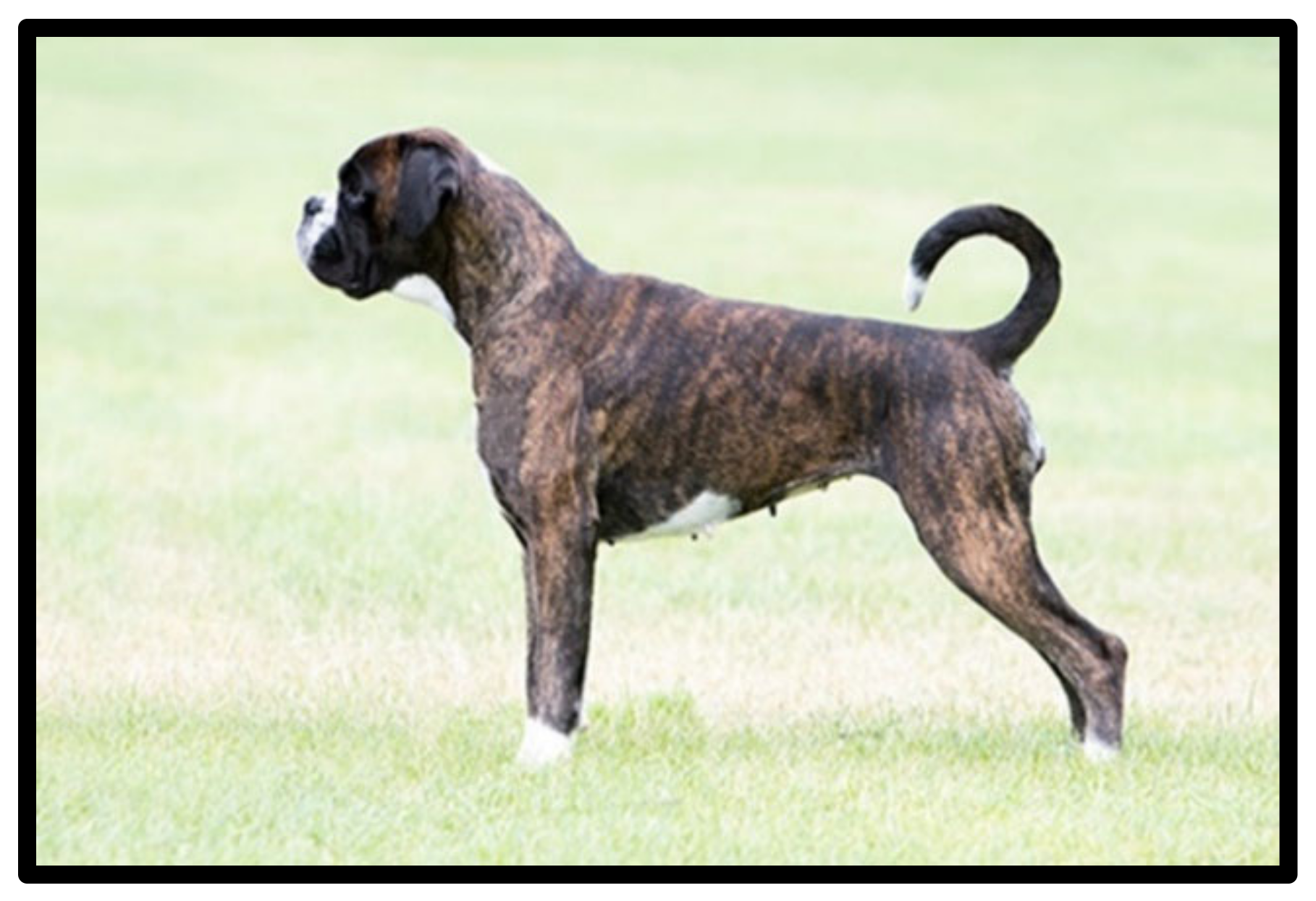
Immune and Genomic Analysis of Boxer Dog Breed and Its Relationship with Leishmania infantum Infection


Abstract
Simple Summary
Abstract
1. Introduction
2. Materials and Methods
2.1. Ethical Approval
2.2. Animals and Epidemiological Data
2.3. Samples Collection and Cytokine Levels
2.4. DNA Extraction and Whole Genome Analysis
2.5. Analysis of Polymorphisms Related to Immune Response
3. Results
4. Discussion
5. Conclusions
Supplementary Materials
Author Contributions
Funding
Institutional Review Board Statement
Informed Consent Statement
Data Availability Statement
Acknowledgments
Conflicts of Interest
References
- The Kennel Club|Welcome to The Kennel Club Website. Available online: https://www.thekennelclub.org.uk/ (accessed on 19 June 2022).
- DEUTSCHER BOXER. Available online: http://www.fci.be/es/nomenclature/BOXER-144.html (accessed on 19 June 2022).
- Boxer|Breeds A to Z|The Kennel Club. Available online: https://www.thekennelclub.org.uk/search/breeds-a-to-z/breeds/working/boxer/ (accessed on 19 June 2022).
- Catchpole, B.; Adams, J.P.; Holder, A.L.; Short, A.D.; Ollier, W.E.R.; Kennedy, L.J. Genetics of Canine Diabetes Mellitus: Are the Diabetes Susceptibility Genes Identified in Humans Involved in Breed Susceptibility to Diabetes Mellitus in Dogs? Vet. J. 2013, 195, 139–147. [Google Scholar] [CrossRef] [PubMed]
- Bussadori, C.; Pradelli, D.; Borgarelli, M.; Chiavegato, D.; D’Agnolo, G.; Menegazzo, L.; Migliorini, F.; Santilli, R.; Zani, A.; Quintavalla, C. Congenital Heart Disease in Boxer Dogs: Results of 6 Years of Breed Screening. Vet. J. 2009, 181, 187–192. [Google Scholar] [CrossRef] [PubMed]
- Meurs, K.M. Genetics of Cardiac Disease in the Small Animal Patient. Vet. Clin. N. Am. Small Anim. Pract. 2010, 40, 701–715. [Google Scholar] [CrossRef] [PubMed]
- Craven, M.; Mansfield, C.S.; Simpson, K.W. Granulomatous Colitis of Boxer Dogs. Vet. Clin. N. Am. Small Anim. Pract. 2011, 41, 433–445. [Google Scholar] [CrossRef] [PubMed]
- Craun, K.; Ekena, J.; Sacco, J.; Jiang, T.; Motsinger-Reif, A.; Trepanier, L.A. Genetic and Environmental Risk for Lymphoma in Boxer Dogs. J. Vet. Intern. Med. 2020, 34, 2068–2077. [Google Scholar] [CrossRef]
- Avery, A.C. The Genetic and Molecular Basis for Canine Models of Human Leukemia and Lymphoma. Front. Oncol. 2020, 10, 23. [Google Scholar] [CrossRef]
- Elvers, I.; Turner-Maier, J.; Swofford, R.; Koltookian, M.; Johnson, J.; Stewart, C.; Zhang, C.-Z.; Schumacher, S.E.; Beroukhim, R.; Rosenberg, M.; et al. Exome Sequencing of Lymphomas from Three Dog Breeds Reveals Somatic Mutation Patterns Reflecting Genetic Background. Genome Res. 2015, 25, 1634–1645. [Google Scholar] [CrossRef]
- Harris, L.J.; Hughes, K.L.; Ehrhart, E.J.; Labadie, J.D.; Yoshimoto, J.; Avery, A.C. Canine CD4+ T-Cell Lymphoma Identified by Flow Cytometry Exhibits a Consistent Histomorphology and Gene Expression Profile. Vet. Comp. Oncol. 2019, 17, 253–264. [Google Scholar] [CrossRef]
- Buckler, J.L.; Liu, X.; Turka, L.A. Regulation of T-Cell Responses by PTEN. Immunol. Rev. 2008, 224, 239–248. [Google Scholar] [CrossRef]
- Chen, L.; Guo, D. The Functions of Tumor Suppressor PTEN in Innate and Adaptive Immunity. Cell Mol. Immunol. 2017, 14, 581–589. [Google Scholar] [CrossRef]
- Alvarez, J.D.; Yasui, D.H.; Niida, H.; Joh, T.; Loh, D.Y.; Kohwi-Shigematsu, T. The MAR-Binding Protein SATB1 Orchestrates Temporal and Spatial Expression of Multiple Genes during T-Cell Development. Genes Dev. 2000, 14, 521–535. [Google Scholar] [CrossRef] [PubMed]
- Cai, S.; Lee, C.C.; Kohwi-Shigematsu, T. SATB1 Packages Densely Looped, Transcriptionally Active Chromatin for Coordinated Expression of Cytokine Genes. Nat. Genet. 2006, 38, 1278–1288. [Google Scholar] [CrossRef] [PubMed]
- Chuenkova, M.V.; Furnari, F.B.; Cavenee, W.K.; Pereira, M.A. Trypanosoma cruzi Trans-Sialidase: A Potent and Specific Survival Factor for Human Schwann Cells by Means of Phosphatidylinositol 3-Kinase/Akt Signaling. Proc. Natl. Acad. Sci. USA 2001, 98, 9936–9941. [Google Scholar] [CrossRef] [PubMed]
- Giri, B.R.; Cheng, G. Host MiR-148 Regulates a Macrophage-Mediated Immune Response during Schistosoma japonicum Infection. Int. J. Parasitol. 2019, 49, 993–997. [Google Scholar] [CrossRef]
- Gómez-Zafra, M.J.; Navas, A.; Jojoa, J.; Murillo, J.; González, C.; Gómez, M.A. Immune Profile of the Nasal Mucosa in Patients with Cutaneous Leishmaniasis. Infect. Immun. 2020, 88, e00881-19. [Google Scholar] [CrossRef]
- Kuroda, S.; Nishio, M.; Sasaki, T.; Horie, Y.; Kawahara, K.; Sasaki, M.; Natsui, M.; Matozaki, T.; Tezuka, H.; Ohteki, T.; et al. Effective Clearance of Intracellular Leishmania major in vivo Requires Pten in Macrophages. Eur. J. Immunol. 2008, 38, 1331–1340. [Google Scholar] [CrossRef]
- Monteiro, C.J.; Mota, S.L.A.; Diniz, L.d.F.; Bahia, M.T.; Moraes, K.C.M. Mir-190b Negatively Contributes to the Trypanosoma cruzi-Infected Cell Survival by Repressing PTEN Protein Expression. Mem. Inst. Oswaldo Cruz. 2015, 110, 996–1002. [Google Scholar] [CrossRef]
- Pan, W.; Xu, H.-W.; Hao, W.-T.; Sun, F.-F.; Qin, Y.-F.; Hao, S.-S.; Liu, H.; Cao, J.-P.; Shen, Y.-J.; Zheng, K.-Y. The Excretory-Secretory Products of Echinococcus granulosus Protoscoleces Stimulated IL-10 Production in B Cells via TLR-2 Signaling. BMC Immunol. 2018, 19, 29. [Google Scholar] [CrossRef]
- Sudarshan, M.; Singh, T.; Singh, B.; Chakravarty, J.; Sundar, S. Suppression of Host PTEN Gene Expression for Leishmania donovani Survival in Indian Visceral Leishmaniasis. Microbes. Infect. 2016, 18, 369–372. [Google Scholar] [CrossRef]
- Edo, M.; Marín-García, P.J.; Llobat, L. Is the Prevalence of Leishmania Infantum Linked to Breeds in Dogs? Characterization of Seropositive Dogs in Ibiza. Animals 2021, 11, 2579. [Google Scholar] [CrossRef]
- França-Silva, J.C.; da Costa, R.T.; Siqueira, A.M.; Machado-Coelho, G.L.L.; da Costa, C.A.; Mayrink, W.; Vieira, E.P.; Costa, J.S.; Genaro, O.; Nascimento, E. Epidemiology of Canine Visceral Leishmaniosis in the Endemic Area of Montes Claros Municipality, Minas Gerais State, Brazil. Vet. Parasitol. 2003, 111, 161–173. [Google Scholar] [CrossRef]
- Solano-Gallego, L.; Miró, G.; Koutinas, A.; Cardoso, L.; Pennisi, M.G.; Ferrer, L.; Bourdeau, P.; Oliva, G.; Baneth, G. LeishVet Guidelines for the Practical Management of Canine Leishmaniosis. Parasites Vectors 2011, 4, 86. [Google Scholar] [CrossRef]
- Leontides, L.S.; Saridomichelakis, M.N.; Billinis, C.; Kontos, V.; Koutinas, A.F.; Galatos, A.D.; Mylonakis, M.E. A Cross-Sectional Study of Leishmania spp. Infection in Clinically Healthy Dogs with Polymerase Chain Reaction and Serology in Greece. Vet. Parasitol. 2002, 109, 19–27. [Google Scholar] [CrossRef]
- Mohebali, M.; Malmasi, A.; Khodabakhsh, M.; Zarei, Z.; Akhoundi, B.; Hajjaran, H.; Azarm, A. Feline Leishmaniosis Due to Leishmania infantum in Northwest Iran: The Role of Cats in Endemic Areas of Visceral Leishmaniosis. Vet. Parasitol. Reg. Stud. Rep. 2017, 9, 13–16. [Google Scholar] [CrossRef]
- Surveillance, Prevention and Control of Leishmaniases in the European Union and Its Neighbouring Countries. Available online: https://www.ecdc.europa.eu/en/publications-data/surveillance-prevention-control-leishmaniases-European-Union-and-neighbouring-countries (accessed on 19 June 2022).
- Gálvez, R.; Montoya, A.; Cruz, I.; Fernández, C.; Martín, O.; Checa, R.; Chicharro, C.; Migueláñez, S.; Marino, V.; Miró, G. Latest Trends in Leishmania infantum Infection in Dogs in Spain, Part I: Mapped Seroprevalence and Sand Fly Distributions. Parasites Vectors 2020, 13, 204. [Google Scholar] [CrossRef]
- Da Silva, L.G.; Costa-Júnior, C.R.L.; Figueiredo-Júnior, C.A.S.; Leal-Balbino, T.C.; Crovella, S.; Otranto, D.; de Queiroz Balbino, V.; Dantas-Torres, F. Canine β-Defensin-1 (CBD1) Gene as a Possible Marker for Leishmania infantum Infection in Dogs. Parasit. Vectors 2017, 10, 199. [Google Scholar] [CrossRef]
- Sanchez-Robert, E.; Altet, L.; Utzet-Sadurni, M.; Giger, U.; Sanchez, A.; Francino, O. Slc11a1 (Formerly Nramp1) and Susceptibility to Canine Visceral Leishmaniasis. Vet. Res. 2008, 39, 36. [Google Scholar] [CrossRef]
- Archer, N.S.; Nassif, N.T.; O’Brien, B.A. Genetic Variants of SLC11A1 Are Associated with Both Autoimmune and Infectious Diseases: Systematic Review and Meta-Analysis. Genes Immun. 2015, 16, 275–283. [Google Scholar] [CrossRef] [PubMed]
- Braliou, G.G.; Kontou, P.I.; Boleti, H.; Bagos, P.G. Susceptibility to Leishmaniasis Is Affected by Host SLC11A1 Gene Polymorphisms: A Systematic Review and Meta-Analysis. Parasitol. Res. 2019, 118, 2329–2342. [Google Scholar] [CrossRef]
- Sanchez-Robert, E.; Altet, L.; Sanchez, A.; Francino, O. Polymorphism of Slc11a1 (Nramp1) Gene and Canine Leishmaniasis in a Case-Control Study. J. Hered. 2005, 96, 755–758. [Google Scholar] [CrossRef]
- Olías-Molero, A.I.; Corral, M.J.; Jiménez-Antón, M.D.; Alunda, J.M. Early Antibody Response and Clinical Outcome in Experimental Canine Leishmaniasis. Sci. Rep. 2019, 9, 18606. [Google Scholar] [CrossRef] [PubMed]
- Chang, C.C.; Chow, C.C.; Tellier, L.C.; Vattikuti, S.; Purcell, S.M.; Lee, J.J. Second-Generation PLINK: Rising to the Challenge of Larger and Richer Datasets. Gigascience 2015, 4, 7. [Google Scholar] [CrossRef] [PubMed]
- Sabeti, P.C.; Reich, D.E.; Higgins, J.M.; Levine, H.Z.P.; Richter, D.J.; Schaffner, S.F.; Gabriel, S.B.; Platko, J.V.; Patterson, N.J.; McDonald, G.J.; et al. Detecting Recent Positive Selection in the Human Genome from Haplotype Structure. Nature 2002, 419, 832–837. [Google Scholar] [CrossRef]
- Danecek, P.; Auton, A.; Abecasis, G.; Albers, C.A.; Banks, E.; DePristo, M.A.; Handsaker, R.E.; Lunter, G.; Marth, G.T.; Sherry, S.T.; et al. The Variant Call Format and VCFtools. Bioinformatics 2011, 27, 2156–2158. [Google Scholar] [CrossRef] [PubMed]
- Voight, B.F.; Kudaravalli, S.; Wen, X.; Pritchard, J.K. A Map of Recent Positive Selection in the Human Genome. PLoS Biol. 2006, 4, e72. [Google Scholar] [CrossRef]
- Batista, L.F.S.; Utsunomiya, Y.T.; Silva, T.B.F.; Dias, R.A.; Tomokane, T.Y.; Pacheco, A.D.; da Matta, V.L.R.; Silveira, F.T.; Marcondes, M.; Nunes, C.M.; et al. Genome-Wide Association Study of Cell-Mediated Response in Dogs Naturally Infected by Leishmania infantum. Infect. Immun. 2016, 84, 3629–3637. [Google Scholar] [CrossRef]
- Batista, L.F.S.; Torrecilha, R.B.P.; Silva, R.B.; Utsunomiya, Y.T.; Silva, T.B.F.; Tomokane, T.Y.; Pacheco, A.D.; Bosco, A.M.; Paulan, S.C.; Rossi, C.N.; et al. Chromosomal Segments May Explain the Antibody Response Cooperation for Canine Leishmaniasis Pathogenesis. Vet. Parasitol. 2020, 288, 109276. [Google Scholar] [CrossRef]
- Batista, L.F.S.; Utsunomiya, Y.T.; Silva, T.B.F.; Carneiro, M.M.; Paiva, J.S.F.; Silva, R.B.; Tomokane, T.Y.; Rossi, C.N.; Pacheco, A.D.; Torrecilha, R.B.P.; et al. Canine Leishmaniasis: Genome-Wide Analysis and Antibody Response to Lutzomyia longipalpis Saliva. PLoS ONE 2018, 13, e0197215. [Google Scholar] [CrossRef]
- Soutter, F.; Solano-Gallego, L.; Attipa, C.; Gradoni, L.; Fiorentino, E.; Foglia Manzillo, V.; Oliva, G.; Tasker, S.; Helps, C.; Catchpole, B. An Investigation of Polymorphisms in Innate and Adaptive Immune Response Genes in Canine Leishmaniosis. Vet. Parasitol. 2019, 269, 34–41. [Google Scholar] [CrossRef]
- Quinnell, R.J.; Kennedy, L.J.; Barnes, A.; Courtenay, O.; Dye, C.; Garcez, L.M.; Shaw, M.-A.; Carter, S.D.; Thomson, W.; Ollier, W.E.R. Susceptibility to Visceral Leishmaniasis in the Domestic Dog Is Associated with MHC Class II Polymorphism. Immunogenetics 2003, 55, 23–28. [Google Scholar] [CrossRef]
- Utsunomiya, Y.T.; Ribeiro, É.S.; Quintal, A.P.N.; Sangalli, J.R.; Gazola, V.R.; Paula, H.B.; Trinconi, C.M.; Lima, V.M.F.; Perri, S.H.V.; Taylor, J.F.; et al. Genome-Wide Scan for Visceral Leishmaniasis in Mixed-Breed Dogs Identifies Candidate Genes Involved in T Helper Cells and Macrophage Signaling. PLoS ONE 2015, 10, e0136749. [Google Scholar] [CrossRef]
- Quilez, J.; Martínez, V.; Woolliams, J.A.; Sanchez, A.; Pong-Wong, R.; Kennedy, L.J.; Quinnell, R.J.; Ollier, W.E.R.; Roura, X.; Ferrer, L.; et al. Genetic Control of Canine Leishmaniasis: Genome-Wide Association Study and Genomic Selection Analysis. PLoS ONE 2012, 7, e35349. [Google Scholar] [CrossRef] [PubMed]
- Altet, L.; Francino, O.; Solano-Gallego, L.; Renier, C.; Sánchez, A. Mapping and Sequencing of the Canine NRAMP1 Gene and Identification of Mutations in Leishmaniasis-Susceptible Dogs. Infect. Immun. 2002, 70, 2763–2771. [Google Scholar] [CrossRef] [PubMed]
- Marín-García, P.J.; Llobat, L. Canine Cytokines Profile in an Endemic Region of L. infantum: Related Factors. Vet. Sci. 2022, 9, 305. [Google Scholar] [CrossRef] [PubMed]
- Paim, F.C.; Da Silva, A.S.; Paim, C.B.V.; França, R.T.; Costa, M.M.; Duarte, M.M.M.F.; Sangoi, M.B.; Moresco, R.N.; Monteiro, S.G.; Lopes, S.T.A. Increased Cytokine and Nitric Oxide Levels in Serum of Dogs Experimentally Infected with Rangelia vitalii. Korean J. Parasitol. 2013, 51, 133–137. [Google Scholar] [CrossRef]
- Galán, A.; Mayer, I.; Rafaj, R.B.; Bendelja, K.; Sušić, V.; Cerón, J.J.; Mrljak, V. MCP-1, KC-like and IL-8 as Critical Mediators of Pathogenesis Caused by Babesia canis. PLoS ONE 2018, 13, e0190474. [Google Scholar] [CrossRef]
- Miranda, S.; Roura, X.; Picado, A.; Ferrer, L.; Ramis, A. Characterization of Sex, Age, and Breed for a Population of Canine Leishmaniosis Diseased Dogs. Res. Vet. Sci. 2008, 85, 35–38. [Google Scholar] [CrossRef]
- Maia, C.; Campino, L. Biomarkers Associated With Leishmania infantum Exposure, Infection, and Disease in Dogs. Front. Cell. Infect. Microbiol. 2018, 8, 302. [Google Scholar] [CrossRef]
- Maia, C.; Campino, L. Cytokine and Phenotypic Cell Profiles of Leishmania infantum Infection in the Dog. J. Trop. Med. 2012, 2012, 541571. [Google Scholar] [CrossRef]
- Ordeix, L.; Montserrat-Sangrà, S.; Martínez-Orellana, P.; Baxarias, M.; Solano-Gallego, L. Toll-like Receptors 2, 4 and 7, Interferon-Gamma and Interleukin 10, and Programmed Death Ligand 1 Transcripts in Skin from Dogs of Different Clinical Stages of Leishmaniosis. Parasites Vectors 2019, 12, 575. [Google Scholar] [CrossRef]
- Ordeix, L.; Montserrat-Sangrà, S.; Martínez-Orellana, P.; Solano-Gallego, L. Toll-Like Receptors 2, 4, and 7, Interferon-Gamma, Interleukin 10, and Programmed Death Ligand 1 Transcripts in Leishmanin Skin Test-Positive Reactions of Ibizan Hound Dogs. J. Immunol. Res. 2020, 2020, 9602576. [Google Scholar] [CrossRef]
- Carrillo, E.; Moreno, J. Cytokine Profiles in Canine Visceral Leishmaniasis. Vet. Immunol. Immunopathol. 2009, 128, 67–70. [Google Scholar] [CrossRef]
- Pedrozo, H.A.; Schwartz, Z.; Mokeyev, T.; Ornoy, A.; Xin-Sheng, W.; Bonewald, L.F.; Dean, D.D.; Boyan, B.D. Vitamin D3 Metabolites Regulate LTBP1 and Latent TGF-Beta1 Expression and Latent TGF-Beta1 Incorporation in the Extracellular Matrix of Chondrocytes. J. Cell. Biochem. 1999, 72, 151–165. [Google Scholar] [CrossRef]
- Park, J.-H.; Yu, Q.; Erman, B.; Appelbaum, J.S.; Montoya-Durango, D.; Grimes, H.L.; Singer, A. Suppression of IL7Ralpha Transcription by IL-7 and Other Prosurvival Cytokines: A Novel Mechanism for Maximizing IL-7-Dependent T Cell Survival. Immunity 2004, 21, 289–302. [Google Scholar] [CrossRef]
- Liu, Y.; Zhang, P.; Li, J.; Kulkarni, A.B.; Perruche, S.; Chen, W. A Critical Function for TGF-Beta Signaling in the Development of Natural CD4+CD25+Foxp3+ Regulatory T Cells. Nat. Immunol. 2008, 9, 632–640. [Google Scholar] [CrossRef]
- Sanjabi, S.; Oh, S.A.; Li, M.O. Regulation of the Immune Response by TGF-β: From Conception to Autoimmunity and Infection. Cold Spring Harb. Perspect. Biol. 2017, 9, a022236. [Google Scholar] [CrossRef]
- Ramia, E.; Chiaravalli, A.M.; Bou Nasser Eddine, F.; Tedeschi, A.; Sessa, F.; Accolla, R.S.; Forlani, G. CIITA-Related Block of HLA Class II Expression, Upregulation of HLA Class I, and Heterogeneous Expression of Immune Checkpoints in Hepatocarcinomas: Implications for New Therapeutic Approaches. Oncoimmunology 2019, 8, 1548243. [Google Scholar] [CrossRef]
- Adhikari, A.; Cobb, B.; Eddington, S.; Becerra, N.; Kohli, P.; Pond, A.; Davie, J. IFN-γ and CIITA Modulate IL-6 Expression in Skeletal Muscle. Cytokine X 2020, 2, 100023. [Google Scholar] [CrossRef]
- Brar, H.K.; Roy, G.; Kanojia, A.; Madan, E.; Madhubala, R.; Muthuswami, R. Chromatin-Remodeling Factor BRG1 Is a Negative Modulator of L. donovani in IFNγ Stimulated and Infected THP-1 Cells. Front. Cell. Infect. Microbiol. 2022, 12, 860058. [Google Scholar] [CrossRef]
- Wojciechowski, W.; DeSanctis, J.; Skamene, E.; Radzioch, D. Attenuation of MHC Class II Expression in Macrophages Infected with Mycobacterium bovis Bacillus Calmette-Guérin Involves Class II Transactivator and Depends on the Nramp1 Gene. J. Immunol. 1999, 163, 2688–2696. [Google Scholar]
- De Vasconcelos, T.C.B.; Furtado, M.C.; Belo, V.S.; Morgado, F.N.; Figueiredo, F.B. Canine Susceptibility to Visceral Leishmaniasis: A Systematic Review upon Genetic Aspects, Considering Breed Factors and Immunological Concepts. Infect. Genet. Evol. 2019, 74, 103293. [Google Scholar] [CrossRef] [PubMed]
- Dou, P.; Zhang, D.; Cheng, Z.; Zhou, G.; Zhang, L. PKIB Promotes Cell Proliferation and the Invasion-Metastasis Cascade through the PI3K/Akt Pathway in NSCLC Cells. Exp. Biol. Med. Maywood 2016, 241, 1911–1918. [Google Scholar] [CrossRef] [PubMed]
- Vergadi, E.; Ieronymaki, E.; Lyroni, K.; Vaporidi, K.; Tsatsanis, C. Akt Signaling Pathway in Macrophage Activation and M1/M2 Polarization. J. Immunol. 2017, 198, 1006–1014. [Google Scholar] [CrossRef] [PubMed]
- Nair, A.; Chakraborty, S.; Banerji, L.A.; Srivastava, A.; Navare, C.; Saha, B. Ras Isoforms: Signaling Specificities in CD40 Pathway. Cell Commun. Signal. 2020, 18, 3. [Google Scholar] [CrossRef] [PubMed]
- Chakraborty, S.; Srivastava, A.; Jha, M.K.; Nair, A.; Pandey, S.P.; Srivastava, N.; Kumari, S.; Singh, S.; Krishnasastry, M.V.; Saha, B. Inhibition of CD40-Induced N-Ras Activation Reduces Leishmania major Infection. J. Immunol. 2015, 194, 3852–3860. [Google Scholar] [CrossRef]
- Montserrat-Sangrà, S.; Alborch, L.; Ordeix, L.; Solano-Gallego, L. TLR-2 and TLR-4 Transcriptions in Unstimulated Blood from Dogs with Leishmaniosis Due to Leishmania infantum at the Time of Diagnosis and during Follow-up Treatment. Vet. Parasitol. 2016, 228, 172–179. [Google Scholar] [CrossRef]
- Shweash, M.; Adrienne McGachy, H.; Schroeder, J.; Neamatallah, T.; Bryant, C.E.; Millington, O.; Mottram, J.C.; Alexander, J.; Plevin, R. Leishmania mexicana Promastigotes Inhibit Macrophage IL-12 Production via TLR-4 Dependent COX-2, INOS and Arginase-1 Expression. Mol. Immunol. 2011, 48, 1800–1808. [Google Scholar] [CrossRef]
- Joutsen, J.; Da Silva, A.J.; Luoto, J.C.; Budzynski, M.A.; Nylund, A.S.; de Thonel, A.; Concordet, J.-P.; Mezger, V.; Sabéran-Djoneidi, D.; Henriksson, E.; et al. Heat Shock Factor 2 Protects against Proteotoxicity by Maintaining Cell-Cell Adhesion. Cell Rep. 2020, 30, 583–597.e6. [Google Scholar] [CrossRef]
- Kanugovi Vijayavittal, A.; Kumar, P.; Sugunan, S.; Joseph, C.; Devaki, B.; Paithankar, K.; Amere Subbarao, S. Heat Shock Transcription Factor HSF2 Modulates the Autophagy Response through the BTG2-SOD2 Axis. Biochem. Biophys. Res. Commun. 2022, 600, 44–50. [Google Scholar] [CrossRef]
- Zanin-Zhorov, A.; Bruck, R.; Tal, G.; Oren, S.; Aeed, H.; Hershkoviz, R.; Cohen, I.R.; Lider, O. Heat Shock Protein 60 Inhibits Th1-Mediated Hepatitis Model via Innate Regulation of Th1/Th2 Transcription Factors and Cytokines. J. Immunol. 2005, 174, 3227–3236. [Google Scholar] [CrossRef]
- Giraldo, E.; Martin-Cordero, L.; Garcia, J.J.; Gehrmann, M.; Gerhmann, M.; Multhoff, G.; Ortega, E. Exercise-Induced Extracellular 72 KDa Heat Shock Protein (Hsp72) Stimulates Neutrophil Phagocytic and Fungicidal Capacities via TLR-2. Eur. J. Appl. Physiol. 2010, 108, 217–225. [Google Scholar] [CrossRef]
- Chen, W.; Zheng, D.; Mou, T.; Pu, J.; Dai, J.; Huang, Z.; Luo, Y.; Zhang, Y.; Wu, Z. Tle1 Attenuates Hepatic Ischemia/Reperfusion Injury by Suppressing NOD2/NF-ΚB Signaling. Biosci. Biotechnol. Biochem. 2020, 84, 1176–1182. [Google Scholar] [CrossRef]
- Dorrington, M.G.; Fraser, I.D.C. NF-ΚB Signaling in Macrophages: Dynamics, Crosstalk, and Signal Integration. Front. Immunol. 2019, 10, 705. [Google Scholar] [CrossRef]
- Sun, S.-C. The Non-Canonical NF-ΚB Pathway in Immunity and Inflammation. Nat. Rev. Immunol. 2017, 17, 545–558. [Google Scholar] [CrossRef]
- Mitchell, S.; Vargas, J.; Hoffmann, A. Signaling via the NFκB System. Wiley Interdiscip. Rev. Syst. Biol. Med. 2016, 8, 227–241. [Google Scholar] [CrossRef]
- Kroviarski, Y.; Debbabi, M.; Bachoual, R.; Périanin, A.; Gougerot-Pocidalo, M.-A.; El-Benna, J.; Dang, P.M.-C. Phosphorylation of NADPH Oxidase Activator 1 (NOXA1) on Serine 282 by MAP Kinases and on Serine 172 by Protein Kinase C and Protein Kinase A Prevents NOX1 Hyperactivation. FASEB J. 2010, 24, 2077–2092. [Google Scholar] [CrossRef]
- Harris, M.L.; Fufa, T.D.; Palmer, J.W.; Joshi, S.S.; Larson, D.M.; Incao, A.; Gildea, D.E.; Trivedi, N.S.; Lee, A.N.; Day, C.-P.; et al. A Direct Link between MITF, Innate Immunity, and Hair Graying. PLoS Biol. 2018, 16, e2003648. [Google Scholar] [CrossRef]
- Zhang, S.; Yue, X.; Yu, J.; Wang, H.; Liu, B. MITF Regulates Downstream Genes in Response to Vibrio Parahaemolyticus Infection in the Clam Meretrix Petechialis. Front. Immunol. 2019, 10, 1547. [Google Scholar] [CrossRef]


| Variable | Categories | No. of Dogs (%) |
|---|---|---|
| Gender | Male | 17 (54.84) |
| Female | 14 (45.16) | |
| Age | Puppy (<1 year) | 4 (12.90) |
| Young (1 to 5 years) | 10 (32.26) | |
| Adult (5 to 10 years) | 14 (45.16) | |
| Elder (>10 years) | 3 (9.68) | |
| Diet | Commercial | 28 (90.32) |
| Home prepared/raw food consumption | 3 (9.68) | |
| Overall | 31 (100.00) |
| Cytokine 1 | n | Range 2 | Mean ± SD 2 | CV (%) |
|---|---|---|---|---|
| IFN-γ | 31 | 0.01–0.78 | 0.22 ± 0.14 | 62.29 |
| IL-2 | 31 | 10.37–721.13 | 68.57 ± 12.09 | 17.63 |
| IL-6 | 31 | 0.40–1.39 | 0.62 ± 0.23 | 37.10 |
| IL-8 | 31 | 58.48–624.40 | 263.75 ± 152.73 | 57.91 |
| IL-18 | 31 | 0–353.45 | 43.08 ± 7.09 | 16.46 |
| Gene 1 | rsID | Chromosome Position | Ref. Alt. 2 | Frequency | Functional Class of Variant |
|---|---|---|---|---|---|
| CIITA | rs24353887 | 6:31796528 | A-G | 0.9792 | Intronic |
| HSF2BP | rs23697150 | 31:37627924 | A-G | 0.7917 | Intronic |
| LTBP1 | rs22564606 | 17:26144284 | C-T | 0.9792 | Intronic |
| rs22598434 | 17:26182161 | A-G | 0.7609 | Intronic | |
| rs22598480 | 17:26192010 | C-T | 0.8478 | Intronic | |
| rs22598531 | 17:26217715 | T-A | 0.7292 | Intronic | |
| rs22598552 | 17:26233826 | G-T | 0.7609 | Intronic | |
| rs22583570 | 17:26237829 | A-G | 0.7292 | Intronic | |
| rs22583641 | 17:26261335 | A-G | 0.7391 | Intronic | |
| rs22583674 | 17:26274101 | C-T | 0.9583 | Intronic | |
| rs22583693 | 17:26283669 | G-A | 0.8750 | Intronic | |
| rs22573465 | 17:26292549 | A-G | 0.8958 | Intronic | |
| rs22583733 | 17:26328978 | T-C | 0.9583 | Intronic | |
| rs22583751 | 17:26345347 | G-A | 0.7292 | Intronic | |
| rs22617468 | 17:26365885 | C-A | 0.8125 | Intronic | |
| rs22617490 | 17:26390401 | C-T | 0.8261 | Intronic | |
| rs22565050 | 17:26412722 | G-A | 0.7292 | Intronic | |
| rs22565078 | 17:26425762 | C-G | 0.7292 | Intronic | |
| rs22565091 | 17:26437220 | A-G | 0.8696 | Intronic | |
| rs22585869 | 17:26451018 | C-G | 0.7391 | Intronic | |
| rs22585928 | 17:26456566 | A-G | 0.7391 | Intronic | |
| rs22600112 | 17:26478030 | G-A | 0.7500 | Intronic | |
| MITF | rs8519356 | 20:21871904 | T-C | 0.7500 | Downstream |
| NOXA1 | rs24534859 | 9:48312935 | G-T | 0.8125 | Intronic |
| PKIB | rs21974900 | 1:62026582 | C(G)-A | 0.9792 | Intronic |
| RAB38 | rs22921195 | 21:12120865 | T-C | 0.7174 | Intronic |
| RASEF | rs21892604 | 1: 76327161 | G-A | 0.7174 | Intergenic |
| rs21885698 | 1: 76366341 | C-T | 0.7174 | Intergenic | |
| rs21894538 | 1: 76416494 | C-T | 0.7174 | Intronic | |
| rs21913661 | 1: 76423140 | A-G | 0.7174 | Intronic | |
| rs21984010 | 1: 76452566 | G-A | 0.7174 | Missense | |
| TLE1 | rs852602083 | 1:77556658 | G-T | 1 | Intronic |
| rs22038874 | 1:77569697 | G-C | 1 | Intronic | |
| rs22038878 | 1:77576847 | T-C | 1 | Intronic | |
| rs21881897 | 1:77607385 | T-A | 0.9783 | Intronic | |
| rs22038945 | 1:77612762 | A-G | 0.9783 | Intronic | |
| rs22038982 | 1:77625675 | C-T | 1 | Intronic | |
| TLR4 | rs22145736 | 11:71364581 | T-C | 0.7500 | 5′UTR |
Publisher’s Note: MDPI stays neutral with regard to jurisdictional claims in published maps and institutional affiliations. |
© 2022 by the authors. Licensee MDPI, Basel, Switzerland. This article is an open access article distributed under the terms and conditions of the Creative Commons Attribution (CC BY) license (https://creativecommons.org/licenses/by/4.0/).
Share and Cite
Álvarez, L.; Marín-García, P.-J.; Rentero-Garrido, P.; Llobat, L. Immune and Genomic Analysis of Boxer Dog Breed and Its Relationship with Leishmania infantum Infection. Vet. Sci. 2022, 9, 608. https://doi.org/10.3390/vetsci9110608
Álvarez L, Marín-García P-J, Rentero-Garrido P, Llobat L. Immune and Genomic Analysis of Boxer Dog Breed and Its Relationship with Leishmania infantum Infection. Veterinary Sciences. 2022; 9(11):608. https://doi.org/10.3390/vetsci9110608
Chicago/Turabian StyleÁlvarez, Luis, Pablo-Jesús Marín-García, Pilar Rentero-Garrido, and Lola Llobat. 2022. "Immune and Genomic Analysis of Boxer Dog Breed and Its Relationship with Leishmania infantum Infection" Veterinary Sciences 9, no. 11: 608. https://doi.org/10.3390/vetsci9110608
APA StyleÁlvarez, L., Marín-García, P.-J., Rentero-Garrido, P., & Llobat, L. (2022). Immune and Genomic Analysis of Boxer Dog Breed and Its Relationship with Leishmania infantum Infection. Veterinary Sciences, 9(11), 608. https://doi.org/10.3390/vetsci9110608