Article

Molecular Detection of Toxigenic Clostridioides difficile among Diarrheic Dogs and Cats: A Mounting Public Health Concern

1
Department of Microbiology, Faculty of Veterinary Medicine, Cairo University, Cairo 12211, Egypt
2
Department of Zoonoses, Faculty of Veterinary Medicine, Cairo University, Cairo 12211, Egypt
*
Author to whom correspondence should be addressed.
Vet. Sci. 2021, 8(6), 88; https://doi.org/10.3390/vetsci8060088
Submission received: 7 April 2021 / Revised: 19 May 2021 / Accepted: 19 May 2021 / Published: 22 May 2021

Abstract

:
Nowadays, pet animals are known to be asymptomatic carriers of Clostridioides difficile. This study was conducted to investigate the burden of toxigenic C. difficile among diarrheic dogs and cats using direct PCR on fecal samples to reveal better insights about the epidemiology of such toxigenic strains referring to its public health significance. For this purpose, fecal samples were obtained from 58 dogs and 42 cats experiencing diarrhea. Following DNA extraction, the extracted DNA was examined for the occurrence of C. difficile as well as toxigenic strains through the detection of C. difficile 16S rRNA and toxin encoding genes (tcdA, tcdB, cdtA and cdtB) using PCR. Moreover, partial DNA sequencing of toxigenic strains retrieved from dog and cat was carried out. Of 100 examined diarrheic animals, 90 (90%) were C. difficile positive, including 93.1% and 85.7% of dogs and cats, respectively. In addition, toxigenic strains were detected in 13 animals, giving an overall prevalence 13% with the following prevalence rates among dogs and cats 12.1% and 14.3%, respectively. Furthermore, the phylogenetic analysis of the obtained sequence revealed high genetic relatedness of tcdA sequence obtained from a cat to strains of human diarrheic cases to point out the public health threat of such sequence. In conclusion, the direct detection of toxigenic C. difficile using PCR among dogs and cats highlights the potential role of household pets as a source for such strains to human contacts.

1. Introduction

Clostridioides difficile (formerly known as Clostridium difficile) is an emerging enteric pathogen in human and veterinary medicine [1]. It is a Gram-positive, strictly anaerobic, spore forming toxin-mediated bacillus [2]. In the last four decades, after admission of broad-spectrum antibiotics, the role of C. difficile in enteric diseases began to flare up to become a remarkable cause of nosocomial associated diarrhea and pseudomembranous colitis among human being [3]. However, nowadays, there is increasing number of C. difficile infection (CDI) cases outside health care settings referring to community acquired CDI, which accounts for one quarter of all reported CDI cases [4,5]. Nonetheless, there is no definitive source of CDI in the community settings [6] and this has urged researchers to investigate the potential role of animals as a vector for transmission of CDI [4]. Notably, this pathogen has been implicated in gastrointestinal diseases among diverse animal species, including food producing animals as well as companion animals [7,8,9,10,11]. Regarding pet animals, there were a lot of reports investigated C. difficile in dogs with gastrointestinal disorders [12,13,14,15] while in cats, little is known concerning association between C. difficile and feline enteric diseases [16,17]. The characteristic diarrhea and gastrointestinal tract inflammation in pet animals and humans are mainly attributed to toxin producing C. difficile [18,19]. Basically, pathogenic C. difficile strains produce two main toxins: toxin A and toxin B which encoded by tcdA and tcdB genes, respectively [20] with some strains producing a binary toxin C. difficile transferase (CDT) [21]. While toxin A is an enterotoxin causing severe gut inflammation, toxin B is a potent cytotoxin that is responsible for cellular death and damage of epithelial tissue [22]. Investigation of C. difficile and its toxins in diarrheic animals relies on conventional methods such as culture may yield underestimated results [13,23]. Recently, direct detection of C. difficile in animal fecal samples using PCR was found to give significantly higher detection rate rather than conventional culture technique [24] whereas, the direct detection of toxin encoding genes is a reliable tool for the detection of toxigenic strains [25]. Accordingly, the current study was carried out to investigate the occurrence of toxigenic C. difficile via direct PCR on feces of pet animals suffering from diarrhea to give insight about the burden of toxigenic C. difficile strains among diarrheic pet animals for better understanding the epidemiology of such strains referring to its public health implication.

2. Materials and Methods

2.1. Ethical Statement

The protocol of this study was approved by ethical committee of Faculty of Veterinary Medicine, Cairo University, Egypt with an ethical approval code: Vet CU28/04/2021/321.

2.2. Sample Collection

Fecal samples were obtained from 100 diarrheic pet animals (58 dogs and 42 cats) from private veterinary clinics where animals of different ages were included in this study. These samples were collected in sterile cups, transported in an icebox to the laboratory and stored at −20 °C for further processing.

2.3. Molecular Investigation of C. difficile and Toxin Encoding Genes

2.3.1. DNA Extraction

DNA was extracted from each fecal sample using FavorPrep™ Stool DNA Isolation Mini Kit (Favorgen, Taiwan, Cat No. FASTI 001-1) according to the manufacturer protocol. Then after, the extracted DNA was stored at −20 °C till further molecular analysis.

2.3.2. Direct Detection of C. difficile

The extracted DNA was screened for the presence of C. difficile via direct detection of C. difficile 16S rRNA using the following primers: B (CCGTCAATTCMTTTRAGTTT) and PG-48 (CTCTTGAAACTGGGAGACTTGA) (Metabion, Steinkirchen, Germany) [26]. The PCR reaction was carried out in a final volume 25 μL where 3 μL of DNA template, 1 μL of each primer, 12.5 μL of Cosmo PCR red master mix (Willowfort, Birmingham, UK, Cat No. WF10203001) and 7.5 μL of nuclease free water were included in each reaction. The thermal profile of PCR reaction was as follows: Initial denaturation at 95 °C for 3 min followed by 40 cycles of denaturation at 95 °C for 30 s, annealing at 44 °C for 30 s, extension at 72 °C for 30 s then final extension at 72 °C for 5 min. Afterwards, amplicons were analyzed with agarose gel electrophoresis (BioRad, Hercules, USA) and photographed to yield specific band at 270 bp (Figure 1).

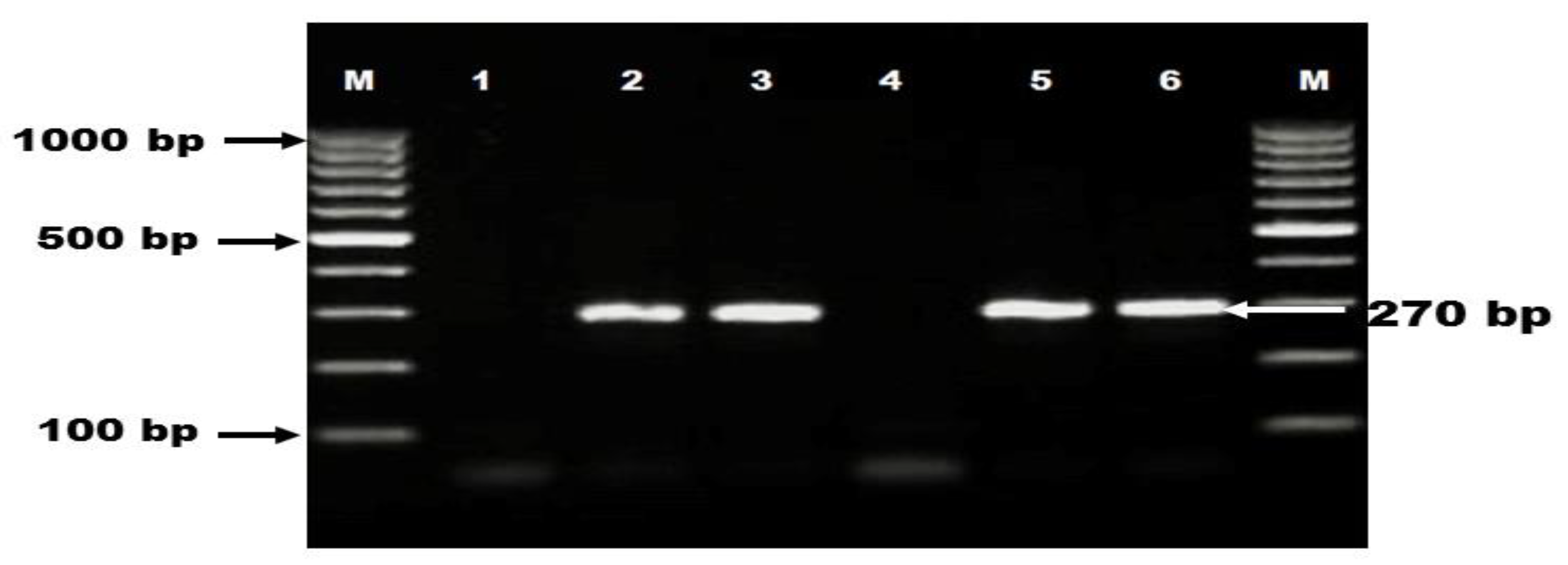
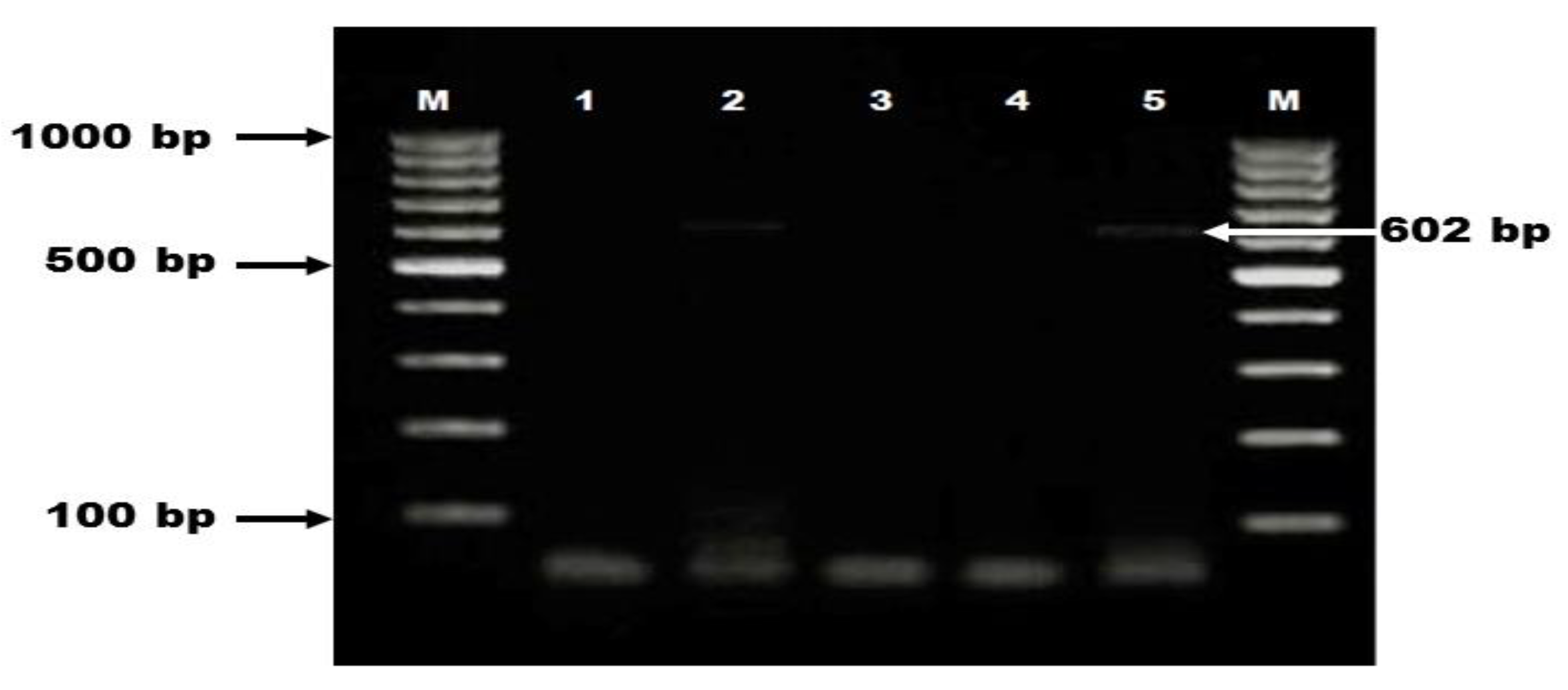
2.3.3. Direct Detection of C. difficile Toxin Genes

tcdA and tcdB Genes

Investigation of C. difficile tcdA and tcdB genes encoding toxin A and toxin B, respectively was carried out in all animals. Primers designed to amplify regions of tcdA and tcdB were as follow: tcdA (YT-28 GCATGATAAGGCAACTTCAGTGG and YT-29 GAGTAAGTTCCTCCTGCTCCATCAA), tcdB (YT-17 GGTGGAGCTGCTTCATTGGAGAG and YT-18 GTGTAACCTACTTTCATAACACCA) (Metabion, Steinkirchen, Germany) [27]. The PCR assay was done at 95 °C for 3 min then 40 cycles of denaturation (95 °C for 20 s), annealing (53 °C, 49 °C for tcdA and tcdB respectively for 25 s), extension (72 °C for 1 min) followed by final extension at 72 °C for 7 min. The PCR products were observed under UV transilluminator (BioRad, Hercules, CA, USA) after electrophoresis step in 1.5% agarose gel (Sigma-Aldrich, Saint Louis, USA, Cat No. A0576) stained with 0.5 μg/mL of ethidium bromide (Sigma-Aldrich, Saint Louis, USA, Cat No. E7637) as specific band of tcdA gene was showed at 602 bp (Figure 2).

cdtA and cdtB Genes

The multiplex PCR amplification for binary toxin genes (cdtA and cdtB) was carried out as follows: after 4 min of initial denaturation at 94 °C, 30 cycles of 94 °C for 45 s, 52 °C for 1 min and 72 °C for 80 s were conducted then followed by 72 °C for 5 min [28].

2.3.4. Partial DNA Sequencing of C. difficile tcdA and tcdB Genes

One PCR product of toxin A obtained from a cat and another one of toxin B retrieved from a dog were purified via a QIAquick purification kit (Qiagen, Hilden, Germany, Cat No. 28104) then they were subjected for sequencing using Big Dye Terminator V3.1 kit (Thermo Fisher, Waltham, MA, USA, Cat No. 4337455) in ABI 3500 Genetic Analyzer (Applied Biosystems, Foster City, CA, USA).

2.4. Nucleotide Sequence Accession Numbers

Partial sequences of C. difficile tcdA and tcdB genes were submitted to GenBank and deposited in GenBank database with the following accession numbers: MW340088 for tcdA and MW357902 for tcdB.

2.5. Sequence Identity BLAST Analysis

The obtained tcdA and tcdB sequences from cat and dog respectively, were compared with C. difficile strains available on GenBank using NCBI website via BLAST analysis to display the identity percentage between our sequences and those of human clinical cases from different countries to clarify the public health significance of such strains.

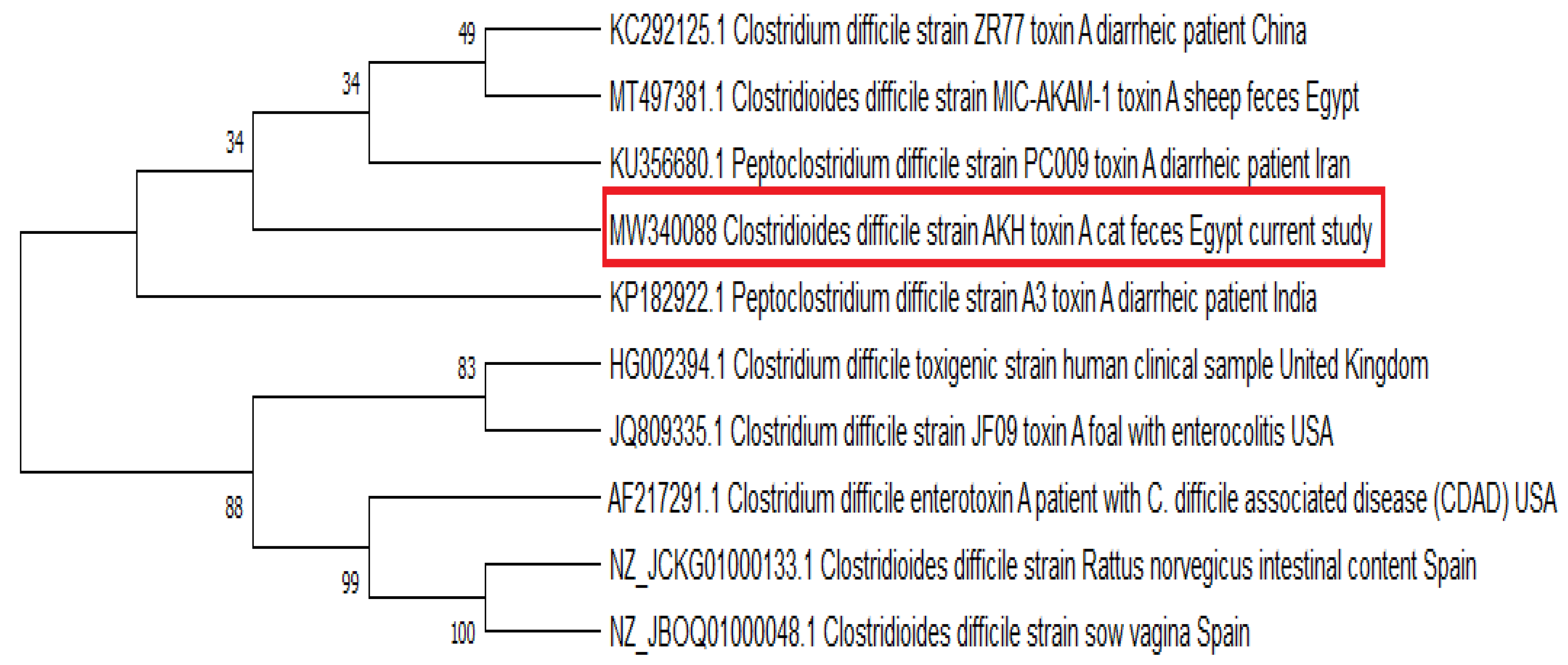
2.6. Phylogenetic Analysis

The recovered tcdA cat strain was aligned against similar C. difficile toxin A sequences retrieved from animals as well as strains obtained from human clinical cases worldwide to confer the genetic relatedness between pets and human strains to understand the public health implications of our findings. Clustal W multiple alignment was conducted using Bioedit software version 7.0.9 while MEGA 7 software was used to construct phylogenetic tree via neighbor-joining approach where bootstrap consensus tree was obtained with 500 replicates (Figure 3).

2.7. Statistical Analysis

The influence of age on prevalence rate of toxigenic C. difficile was analyzed by SPSS software version 18.0 using chi square (χ2) test. The result was considered statistically significant when p-value was less than 0.05.

3. Results

Of 100 examined diarrheic animals, 90 (90%) were C. difficile positive, including 93.1% and 85.7% of dogs and cats, respectively. For toxigenic C. difficile, 13 (13%) out of 100 animals had C. difficile toxins comprising 12.1% (7/58) and 14.3% (6/42) of dogs and cats, respectively. Moreover, according to toxin production type, 4 dogs and 4 cats were positive for tcdA and negative for tcdB while one dog and two cats carried toxin B and negative for toxin A as well as both tcdA and tcdB genes had been detected in two dogs. On the other hand, none of binary toxins (cdtA and cdtB) was found among the examined dogs and cats as shown in Table 1. Regarding animal age, the prevalence of toxigenic C. difficile was as follows: 14% (less than 6 months), 14.8% (6–12 months) and 16.7% (greater than 12 months) (Table 2). Statistically, no significant relationship (p value = 0.94) was observed between toxigenic C. difficile and animal age. The similarity ratios between the obtained C. difficile tcdA and tcdB sequences in this study and those of public health importance according to the BLAST analysis were displayed in Table 3.

4. Discussion

In the last few years, pet dogs and cats were found to be potential reservoirs for some emerging nosocomial pathogens with great public health concern [29,30]. Such previous studies have paved the way for more investigations about the role of pet animals in the epidemiology of other nosocomial pathogens likewise C. difficile and nowadays, the implication of household pets in community acquired CDI is an ongoing public health issue [6]. In the current study, C. difficile was detected in 90% of diarrheic pet animals where 93.1% and 85.7% of dogs and cats were positive, respectively. Our results were higher than those reported in previous studies 6.7% [15], 25% [31] for dogs and 12.9% [17], 15.7% [31] for cats suffering from diarrhea. Such high unexpected results in the current study may be owed to the direct detection of C. difficile by PCR using 16S rRNA primers can detect as little as 10 cells of C. difficile among 1010–1011 total bacterial cells per one gram of stool [32]. On the contrary, other studies recovered C. difficile via conventional culture technique which needs at least 1000 cfu/gram of feces on the selective C. difficile culture media to yield successful cultivation. Therefore, the direct detection of C. difficile by PCR can elucidate the burden of CDI among diarrheic pet animals and consequently triggers a growing public health concern.
Regarding toxigenic C. difficile, 13 out of 100 diarrheic pet animals were positive for toxin encoding genes, whereas 12.1% (7/58) of investigated dogs carried C. difficile toxins. Our finding was lower than that reported by Weese et al. [12] who found 21% (18/87) of examined diarrheic dogs were toxigenic using an ELISA assay. While in cats, the prevalence of toxigenic C. difficile was 14.3% (6/42). Such result was higher than that reported by Silva et al. [17] who detected toxigenic C. difficile in 3 (4.3%) out of 70 diarrheic cats by PCR carried out on recovered C. difficile isolates.
From a public health point of view, C. difficile associated diarrhea in human being is mainly attributed to toxigenic strains [19]. Importantly, in the current study, there were two dogs carried both toxin A and toxin B which may refer to presence of A+/B+ toxinotype. C. difficile A+/B+ is the most predominant toxigenic strain isolated from diarrheic dogs in studies conducted by Wetterwik et al. [13], Andrés-Lasheras et al. [15], Ghavidel et al. [33] and Silva et al. [34]. Likewise, it is the most pathogenic C. difficile toxinotype in human being and is primarily accounted for C. difficile associated disease (CDAD) worldwide [35,36]. Moreover, there were one dog and two cats had tcdB gene but negative for tcdA which may indicate A−/B+ strain. Such toxinotype has attracted the attention of researchers in recent years [37] because it has been incriminated in four nosocomial outbreaks of C. difficile associated diarrhea in Canada [38], Netherlands [39], Japan [40] and Dublin [41] as well as 4 dogs and 4 cats were found to be toxin A positive and toxin B negative; this strain also had been detected among diarrheic patients in intensive care unit [42]. On the other hand, all the examined dogs and cats were negative for binary toxin genes. In agreement with our result, Andrés-Lasheras et al. [15] and Silva et al. [17] who could not find binary toxins in C. difficile isolates recovered from diarrheic dogs and cats, respectively. Pet dogs and cats are in frequent and close contact with their owners and usually share the same places at home like living room and bedroom. Accordingly, diarrheic pet animals may be considered as a potential source for dissemination of toxigenic C. difficile strains within a community. Therefore, our findings indicate that the direct PCR detection of toxigenic C. difficile in feces of diarrheic dogs and cats can give a better insight to understand the epidemiology of toxigenic C. difficile infection among pet animals.
On the other hand, the prevalence of toxin producing C. difficile was found to be increased with age of pet animals as animals of age greater than 12 months had a higher percentage (16.7%) but there was no significant relationship between age and prevalence rate of toxigenic C. difficile. Similarly, Álvarez-Pérez et al. [23] and Diniz et al. [43] reported that shedding of C. difficile and its toxins was increased with pet animals of higher ages.
Interestingly, in this study, we provide C. difficile tcdA and tcdB partial sequences from a cat and a dog, respectively, where cat and dog strains showed high identity percentage of 99.81%–100% and 99.43%–99.72% respectively to C. difficile isolates retrieved from patients with diarrhea and pseudomembranous colitis worldwide to highlight the public health impact of such strains. In the meantime, phylogenetic tree was constructed to encompass C. difficile toxin A sequences from animals as well as human strains including diarrheic patients from different countries (Figure 3). It was obvious that tcdA sequence from a cat was grouped within the same cluster with that reported in sheep in the same country (Egypt) and those of human diarrheic cases originated from Asian countries (China, India, and Iran). Thus, the high genetic relatedness of our sequence to those of humans points out a potential relationship between cats and diarrheal infection in human being rendering pet animals a potential zoonotic source for toxigenic C. difficile human infection.

5. Conclusions

This study provides more knowledge regarding the epidemiology of toxigenic C. difficile infection among diarrheic dogs and cats. Remarkably, the direct detection of toxigenic C. difficile using PCR in animal samples opens a gate for better assessment of toxigenic CDI burden among household pets which subsequently, reflects on human health.

Author Contributions

K.A.A.-M. and A.S.: Idea, study design, supervising the work and writing manuscript. H.M.Z.: Sample collection, molecular techniques and writing manuscript. All authors have read and agreed to the published version of the manuscript.

Funding

This research received no external funding.

Institutional Review Board Statement

The protocol of this study was approved by ethical committee of Faculty of Veterinary Medicine, Cairo University, Egypt with an ethical approval code: Vet CU28/04/2021/321.

Informed Consent Statement

Not applicable.

Data Availability Statement

Not applicable.

Conflicts of Interest

The authors declare no conflict of interest.

References

  1. Keessen, E.C.; Gaastra, W.; Lipman, L.J. Clostridium difficile infection in humans and animals, differences and similarities. Vet. Microbiol. 2011, 153, 205–217. [Google Scholar] [CrossRef]
  2. Zhu, D.; Sorg, J.A.; Sun, X. Clostridioides difficile Biology: Sporulation, Germination, and Corresponding Therapies for C. difficile Infection. Front. Cell Infect. Microbiol. 2018, 8, 29. [Google Scholar] [CrossRef] [PubMed] [Green Version]
  3. Burnham, C.D.; Carroll, K.C. Diagnosis of Clostridium difficile Infection: An Ongoing Conundrum for Clinicians and for Clinical Laboratories. Clin. Microbiol. Rev. 2013, 26, 604–630. [Google Scholar] [CrossRef] [PubMed] [Green Version]
  4. Hensgens, M.P.; Keessen, E.C.; Squire, M.M.; Riley, T.V.; Koene, M.G.; de Boer, E.; Lipman, L.J.; Kuijper, E.J.; European Society of Clinical Microbiology and Infectious Diseases Study Group for Clostridium difficile (ESGCD). Clostridium difficile infection in the community: A zoonotic disease? Clin. Microbiol. Infect. 2012, 18, 635–645. [Google Scholar] [CrossRef] [PubMed] [Green Version]
  5. Gupta, A.; Khanna, S. Community-acquired Clostridium difficile infection: An increasing public health threat. Infect. Drug Resist. 2014, 7, 63–72. [Google Scholar] [PubMed] [Green Version]
  6. Hernandez, B.G.; Vinithakumari, A.A.; Sponseller, B.; Tangudu, C.; Mooyottu, S. Prevalence, Colonization, Epidemiology, and Public Health Significance of Clostridioides difficile in Companion Animals. Front. Vet. Sci. 2020, 7, 512551. [Google Scholar] [CrossRef]
  7. Kachrimanidou, M.; Tzika, E.; Filioussis, G. Clostridioides (Clostridium) Difficile in Food-Producing Animals, Horses and Household Pets: A Comprehensive Review. Microorganisms 2019, 7, 667. [Google Scholar] [CrossRef] [Green Version]
  8. Hammitt, M.C.; Bueschel, D.M.; Keel, M.K.; Glock, R.D.; Cuneo, P.; DeYoung, D.W.; Reggiardo, C.; Trinh, H.T.; Songer, J.G. A possible role for Clostridium difficile in the etiology of calf enteritis. Vet. Microbiol. 2008, 127, 343–352. [Google Scholar] [CrossRef]
  9. Avberšek, J.; Pirš, T.; Pate, M.; Rupnik, M.; Ocepek, M. Clostridium difficile in goats and sheep in Slovenia: Characterization of strains and evidence of age-related shedding. Anaerobe 2014, 28, 163–167. [Google Scholar] [CrossRef] [PubMed]
  10. Weese, J.S.; Staempfli, H.R.; Prescott, J.F. A prospective study of the roles of Clostridium difficile and enterotoxigenic Clostridium perfringens in equine diarrhoea. Equine Vet. J. 2001, 33, 403–409. [Google Scholar] [CrossRef]
  11. Weese, J.S.; Armstrong, J. Outbreak of Clostridium difficile-associated disease in a small animal veterinary teaching hospital. J. Vet. Intern Med. 2003, 17, 813–816. [Google Scholar] [PubMed]
  12. Weese, J.S.; Staempfli, H.R.; Prescott, J.F.; Kruth, S.A.; Greenwood, S.J.; Weese, H.E. The roles of Clostridium difficile and enterotoxigenic Clostridium perfringens in diarrhea in dogs. J. Vet. Intern Med. 2001, 15, 374–378. [Google Scholar] [CrossRef]
  13. Wetterwik, K.J.; Trowald-Wigh, G.; Fernström, L.L.; Krovacek, K. Clostridium difficile in faeces from healthy dogs and dogs with diarrhea. Acta Vet. Scand. 2013, 55, 23. [Google Scholar] [CrossRef] [Green Version]
  14. Orden, C.; Blanco, J.L.; Álvarez-Pérez, S.; Garcia-Sancho, M.; Rodriguez-Franco, F.; Sainz, A.; Villaescusa, A.; Harmanus, C.; Kuijper, E.; Garcia, M.E. Isolation of Clostridium difficile from dogs with digestive disorders, including stable metronidazole-resistant strains. Anaerobe 2017, 43, 78–81. [Google Scholar] [CrossRef] [PubMed]
  15. Andrés-Lasheras, S.; Martín-Burriel, I.; Mainar-Jaime, R.C.; Morales, M.; Kuijper, E.; Blanco, J.L.; Chirino-Trejo, M.; Bolea, R. Preliminary studies on isolates of Clostridium difficile from dogs and exotic pets. BMC Vet. Res. 2018, 14, 77. [Google Scholar] [CrossRef] [PubMed]
  16. Weese, J.S. Clostridium (Clostridioides) difficile in animals. J. Vet. Diagn. Investig. 2020, 32, 213–221. [Google Scholar] [CrossRef] [PubMed]
  17. Silva, R.O.S.; Ribeiro, M.G.; de Paula, C.L.; Pires, I.H.; Oliveira Junior, C.A.; Diniz, A.N.; de Araújo Nunes, T.A.; Lobato, F.C.F. Isolation of Clostridium perfringens and Clostridioides difficile in diarrheic and nondiarrheic cats. Anaerobe 2020, 62, 102164. [Google Scholar] [CrossRef] [PubMed]
  18. Hussain, I.; Sharma, R.K.; Borah, P.; Rajkhowa, S.; Hussain, I.; Barkalita, L.M.; Hasin, D.; Choudhury, M.; Rupnik, M.; Deka, N.K.; et al. Isolation and characterization of Clostridium difficile from pet dogs in Assam, India. Anaerobe 2015, 36, 9–13. [Google Scholar] [CrossRef] [PubMed]
  19. Voth, D.E.; Ballard, J.D. Clostridium difficile toxins: Mechanism of action and role in disease. Clin. Microbiol Rev. 2005, 18, 247–263. [Google Scholar] [CrossRef] [Green Version]
  20. Carter, G.P.; Rood, J.I.; Lyras, D. The role of toxin A and toxin B in Clostridium difficile-associated disease: Past and present perspectives. Gut Microbes 2010, 1, 58–64. [Google Scholar] [CrossRef] [Green Version]
  21. Gülke, I.; Pfeifer, G.; Liese, J.; Fritz, M.; Hofmann, F.; Aktories, K.; Barth, H. Characterization of the enzymatic component of the ADP-ribosyltransferase toxin CDTa from Clostridium difficile. Infect. Immun. 2001, 69, 6004–6011. [Google Scholar] [CrossRef] [PubMed] [Green Version]
  22. Samie, A.; Obi, C.L.; Franasiak, J.; Archbald-Pannone, L.; Bessong, P.O.; Alcantara-Warren, C.; Guerrant, R.L. PCR detection of Clostridium difficile triose phosphate isomerase (tpi), toxin A (tcdA), toxin B (tcdB), binary toxin (cdtA, cdtB), and tcdC genes in Vhembe District, South Africa. Am. J. Trop. Med. Hyg. 2008, 78, 577–585. [Google Scholar] [CrossRef] [PubMed] [Green Version]
  23. Álvarez-Pérez, S.; Blanco, J.L.; Harmanus, C.; Kuijper, E.J.; García, M.E. Prevalence and characteristics of Clostridium perfringens and Clostridium difficile in dogs and cats attended in diverse veterinary clinics from the Madrid region. Anaerobe 2017, 48, 47–55. [Google Scholar] [CrossRef]
  24. Fathy, M.; Abdel-Moein, K.A.; Osman, W.A.; Erfan, M.A.; Prince, A.; Hafez, A.A.; Mahmoud, H.E.; Mosallam, T.E.; Samir, A. Performance of different laboratory methods for detection of clostridium difficile in animal samples. Adv. Anim. Vet. Sci. 2021, 9, 132–136. [Google Scholar]
  25. Wongwanich, S.; Rugdeekha, S.; Pongpech, P.; Dhiraputra, C. Detection of Clostridium difficile toxin A and B genes from stool samples of Thai diarrheal patients by polymerase chain reaction technique. J. Med. Assoc. Thai. 2003, 86, 970–975. [Google Scholar] [PubMed]
  26. Zhang, T.; Lin, Q.Y.; Fei, J.X.; Zhang, Y.; Lin, M.Y.; Jiang, S.H.; Wang, P.; Chen, Y. Clostridium Difficile Infection Worsen Outcome of Hospitalized Patients with Inflammatory Bowel Disease. Sci. Rep. 2016, 6, 29791. [Google Scholar] [CrossRef] [Green Version]
  27. Cohen, S.H.; Tang, Y.J.; Silva, J., Jr. Analysis of the pathogenicity locus in Clostridium difficile strains. J. Infect. Dis. 2000, 181, 659–663. [Google Scholar] [CrossRef] [Green Version]
  28. Stubbs, S.; Rupnik, M.; Gibert, M.; Brazier, J.; Duerden, B.; Popoff, M. Production of actin-specific ADP-ribosyltransferase (binary toxin) by strains of Clostridium difficile. FEMS Microbiol. Lett. 2000, 186, 307–312. [Google Scholar] [CrossRef]
  29. Abdel-Moein, K.A.; El-Hariri, M.; Samir, A. Methicillin-resistant Staphylococcus aureus: An emerging pathogen of pets in Egypt with a public health burden. Transbound. Emerg. Dis. 2012, 59, 331–335. [Google Scholar] [CrossRef]
  30. Abdel-Moein, K.A.; Samir, A. Occurrence of extended spectrum β-lactamase-producing Enterobacteriaceae among pet dogs and cats: An emerging public health threat outside health care facilities. Am. J. Infect. Control 2014, 42, 796–798. [Google Scholar] [CrossRef] [PubMed]
  31. Koene, M.G.; Mevius, D.; Wagenaar, J.A.; Harmanus, C.; Hensgens, M.P.; Meetsma, A.M.; Putirulan, F.F.; van Bergen, M.A.; Kuijper, E.J. Clostridium difficile in Dutch animals: Their presence, characteristics and similarities with human isolates. Clin. Microbiol. Infect. 2012, 18, 778–784. [Google Scholar] [CrossRef] [PubMed] [Green Version]
  32. Gumerlock, P.H.; Tang, Y.J.; Meyers, F.J.; Silva, J., Jr. Use of the polymerase chain reaction for the specific and direct detection of Clostridium difficile in human feces. Rev. Infect. Dis. 1991, 13, 1053–1060. [Google Scholar] [CrossRef]
  33. Ghavidel, M.; Salari Sedigh, H.; Razmyar, J. Isolation of Clostridium difficile and molecular detection of binary and A/B toxins in faeces of dogs. Iran J. Vet. Res. 2016, 17, 273–276. [Google Scholar]
  34. Silva, R.O.; Santos, R.L.; Pires, P.S.; Pereira, L.C.; Pereira, S.T.; Duarte, M.C.; de Assis, R.A.; Lobato, F.C. Detection of toxins A/B and isolation of Clostridium difficile and Clostridium perfringens from dogs in Minas Gerais, Brazil. Braz. J. Microbiol. 2013, 44, 133–137. [Google Scholar] [CrossRef] [PubMed] [Green Version]
  35. Drudy, D.; Fanning, S.; Kyne, L. Toxin A-negative, toxin B-positive Clostridium difficile. Int. J. Infect. Dis. 2007, 11, 5–10. [Google Scholar] [CrossRef] [Green Version]
  36. Jalali, M.; Khorvash, F.; Warriner, K.; Weese, J.S. Clostridium difficile infection in an Iranian hospital. BMC Res. Notes 2012, 5, 159. [Google Scholar] [CrossRef] [PubMed] [Green Version]
  37. Janezic, S.; Marín, M.; Martín, A.; Rupnik, M. A new type of toxin A-negative, toxin B-positive Clostridium difficile strain lacking a complete tcdA gene. J. Clin. Microbiol. 2015, 53, 692–695. [Google Scholar] [CrossRef] [PubMed] [Green Version]
  38. al-Barrak, A.; Embil, J.; Dyck, B.; Olekson, K.; Nicoll, D.; Alfa, M.; Kabani, A. An outbreak of toxin A negative, toxin B positive Clostridium difficile-associated diarrhea in a Canadian tertiary-care hospital. Can. Commun. Dis. Rep. 1999, 25, 65–69. [Google Scholar] [PubMed]
  39. Kuijper, E.J.; de Weerdt, J.; Kato, H.; Kato, N.; van Dam, A.P.; van der Vorm, E.R.; Weel, J.; van Rheenen, C.; Dankert, J. Nosocomial outbreak of Clostridium difficile-associated diarrhoea due to a clindamycin-resistant enterotoxin A-negative strain. Eur. J. Clin. Microbiol. Infect. Dis. 2001, 20, 528–534. [Google Scholar] [CrossRef]
  40. Sato, H.; Kato, H.; Koiwai, K.; Sakai, C. [A nosocomial outbreak of diarrhea caused by toxin A-negative, toxin B-positive Clostridium difficile in a cancer center hospital]. Kansenshogaku Zasshi 2004, 78, 312–319. [Google Scholar] [CrossRef] [Green Version]
  41. Drudy, D.; Harnedy, N.; Fanning, S.; O’Mahony, R.; Kyne, L. Isolation and characterization of toxin A-negative, toxin B-positive Clostridium difficile in Dublin, Ireland. Clin. Microbiol. Infect. 2007, 13, 298–304. [Google Scholar] [CrossRef] [PubMed] [Green Version]
  42. Rezazadeh Zarandi, E.; Mansouri, S.; Nakhaee, N.; Sarafzadeh, F.; Iranmanesh, Z.; Moradi, M. Frequency of antibiotic associated diarrhea caused by Clostridium difficile among hospitalized patients in intensive care unit, Kerman, Iran. Gastroenterol. Hepatol. Bed. Bench. 2017, 10, 229–234. [Google Scholar] [PubMed]
  43. Diniz, A.N.; Coura, F.M.; Rupnik, M.; Adams, V.; Stent, T.L.; Rood, J.I.; de Oliveira, C.A., Jr.; Lobato, F.C.F.; Silva, R.O.S. The incidence of Clostridioides difficile and Clostridium perfringens netF-positive strains in diarrheic dogs. Anaerobe 2018, 49, 58–62. [Google Scholar] [CrossRef] [PubMed]
Figure 1. Occurrence of C. difficile 16S rRNA among diarrheic dogs and cats. Lane M: DNA ladder (100 bp); lane 1: negative control; lanes 2, 3, 5, 6: positive samples with specific band at 270 bp; lane 4: negative sample.
Vetsci 08 00088 g001
Figure 2. Molecular detection of C. difficile tcdA gene among diarrheic dogs and cats. Lane M: DNA ladder (100 bp); lane 1: negative control; lanes 2,5: positive samples showed specific band at 602 bp; lanes 3,4: negative samples.
Vetsci 08 00088 g002
Figure 3. Phylogenetic consensus tree was constructed using neighbor-joining approach via Mega 7 software to display the genetic relatedness between tcdA sequence obtained from a cat and those retrieved from Genbank.
Vetsci 08 00088 g003
Table 1. Occurrence of C. difficile 16S rRNA and toxin encoding genes among diarrheic dogs and cats.
Animal SpeciesNo. of Examined AnimalsNo. of Positive Animals (%)
C. difficile 16S rRNA Toxigenic C. difficile
tcdA+tcdB-tcdA-tcdB+tcdA+tcdB+Binary Toxins (CDT)Total
Dogs5854 (93.1)4(6.9)1 (1.7)2 (3.4)0 (0)7 (12.1)
Cats4236 (85.7)4 (9.5)2 (4.8)0 (0)0 (0)6 (14.3)
Total10090 (90)8 (8)3 (3)2 (2)0 (0)13 (13)
Table 2. Occurrence of toxigenic C. difficile among pet animals of different ages.
Age of AnimalsNo. of Examined AnimalsPositive Animals
No.%
<6 M43614
6–12 M27414.8
>12 M30516.7
Total1001515
Table 3. The identity percentage of obtained C. difficile tcdA and tcdB partial sequences in this study with C. difficile strains deposited in Genbank of public health significance.
SequenceGenbank IDIsolation SourceCountry% Identity
MW340088
(tcdA cat sequence)
KP182922.1Diarrheic patientIndia100
CP022524.1Hospitalized pediatric patient with diarrheaUSA99.81
CP010905.2Patient with severe pseudomembranous colitisSwitzerland99.81
KC292061.1Diarrheic patientChina99.81
MW357902
(tcdB dog sequence)
DQ117266.1Patient with antibiotic associated diarrheaFrance99.72
KC292138.1Diarrheic patientChina99.48
CP010905.2Patient with severe pseudomembranous colitisSwitzerland99.48
DQ117268.1Patient with pseudomembranous colitisFrance99.43
Publisher’s Note: MDPI stays neutral with regard to jurisdictional claims in published maps and institutional affiliations.

