Article

Molecular Diversity of Hard Tick Species from Selected Areas of a Wildlife-Livestock Interface Ecosystem at Mikumi National Park, Morogoro Region, Tanzania

1
Department of Molecular Biology and Biotechnology, University of Dar es Salaam, 35091 Dar es Salaam, Tanzania
2
Department of Biomedical Sciences and Veterinary Public Health, Swedish University of Agricultural Sciences, Box 7036, SE-750 07 Uppsala, Sweden
3
Department of Clinical Sciences, Swedish University of Agricultural Sciences, Box 7054, SE-750 07 Uppsala, Sweden
*
Author to whom correspondence should be addressed.
Vet. Sci. 2021, 8(3), 36; https://doi.org/10.3390/vetsci8030036
Submission received: 27 December 2020 / Revised: 7 February 2021 / Accepted: 9 February 2021 / Published: 24 February 2021

Abstract

:
Ticks are one of the most important arthropod vectors and reservoirs as they harbor a wide variety of viruses, bacteria, fungi, protozoa, and nematodes, which can cause diseases in human and livestock. Due to their impact on human, livestock, and wild animal health, increased knowledge of ticks is needed. So far, the published data on the molecular diversity between hard ticks species collected in Tanzania is scarce. The objective of this study was to determine the genetic diversity between hard tick species collected in the wildlife-livestock interface ecosystem at Mikumi National Park, Tanzania using the mitochondrion 16S rRNA gene sequences. Adult ticks were collected from cattle (632 ticks), goats (187 ticks), and environment (28 ticks) in the wards which lie at the border of Mikumi National Park. Morphological identification of ticks was performed to genus level. To identify ticks to species level, molecular analysis based on mitochondrion 16S rRNA gene was performed. Ticks representing the two genera (Hyalomma and Rhipicephalus) were identified using morphological characters. Six species were confirmed based on mitochondrion 16S rRNA gene, including Rhipicephalus microplus, Rhipicephalus evertsi, Hyalomma rufipes, Hyalomma truncatum, Hyalomma marginatum, and Hhyalomma turanicum. The presence of different clusters of tick species reflects the possible biological diversity of the hard ticks present in the study region. Further studies are however required to quantify species of hard ticks present in the study region and the country in general over a larger scale.

1. Introduction

Ticks are responsible for great economic, social, and conservation losses because of their negative impacts on human, livestock, and wild animal health [1]. Ticks can cause reduction in animal body weight, limit animal production and induce anemia especially when animal is heavily infested [2]. Tick bites may cause irritation resulting in reduced quality of the hides [2]. Certain tick species, Rhipicephalus evertsi, Ixodes rubicundus, and Hyalomma truncatum inject toxin to animals, which causes paralysis [1]. Ticks are also reservoirs and vectors of viruses, bacteria, fungi, protozoa, and nematodes, which can cause diseases in human, livestock, and wild animals [3].
Most of the tick species belong to the two main families, Ixodidae (hard ticks) and Argasidae (soft ticks) [4,5]. Hard ticks of the genera Rhipicephalus, Hyalomma, and Amblyomma are the most important and widely distributed species found in many parts of Africa, including Tanzania [6,7,8].
More than 95% of the cattle and goat population in Tanzania are reared under traditional pastoral systems [9]. Nomadic and pastoralist lifestyles, especially those at the wildlife-livestock interface, can allow direct and indirect contact with wild animals that can facilitate exposure and sharing of tick spp., [1].
In areas which lie at the border of Mikumi National Park, people practice nomadic pastoralism, keeping large number of indigenous cattle and goats. During dry seasons there is the movement of people and livestock to areas very close and sometimes entering beyond the National Park boundary, where water and pasture are abundant long after the rains have gone. Likewise, there is migration of wild animals and birds outside of the National Park boundaries. These migration patterns facilitate the potential transfer of ticks, presenting the opportunity for exchange of diverse tick species between the domestic, wild animal, and even human populations [1]. Therefore, the area is considered to be one of the hotspots for tick species, although the diversity of the tick species inhabiting the local area is scarce [1,10].
Studies aiming at quantifying and identifying tick species in Tanzania are still limited. The published studies related to tick species composition in Tanzania were conducted in Ngorongoro district [9], Iringa region, Maswa district [7], Mara region [8], Singida region, Mbeya region [6], where Hyalomma rufipes, Rhipicephalus microplus, and Rhipicephalus evertsi were reported. However, their information was restricted on the morphological characters only. The current study provides molecular diversity information on tick species and generates the mitochondrion 16S rRNA gene sequence data bases for the hard tick species infecting cattle, goats, and those collected from the environment in wildlife-livestock interface ecosystem of Mikumi National park, Tanzania. Molecular identification of Ixodidae ticks will provide valuable information to farmers and other stakeholders about different tick species present in the area [11]. Therefore, data from this field study can be useful for rational control strategies of ticks and tick-borne diseases in Tanzania [11,12].

2. Methods

2.1. Study Site

This study was conducted in the wards which lie at the border of Mikumi National Park, Morogoro region of Tanzania, namely, Doma, Melela, Kisaki, Tindiga, Kilangali, Ulaya, Mikumi, Ruhembe, Kidodi, and Kidatu (Figure 1). Mikumi National Park is located in the Morogoro region of Tanzania and lies between latitudes 7 °C and 10 °C south of the equator and between longitudes 36 °C and 37 °C East of Greenwich (Figure 1).The geography of the study area consists of plain land in most areas while some parts are covered by hills.
The area has typical tropical climate, with annual rainfall ranging from 600 to 1500 mm and average annual temperatures ranging from 20–30 °C. Rainfall distribution in the area is bimodal, with wet season from March to May. The dry seasons are experienced for six months (June, July, August, September, January, and February).
The area is the home to a pastoralist community for whom keeping livestock is their way of life. Poor animal husbandry and grazing practices put great pressure on land resources, which results in the need to continuously move large numbers of animals, especially cattle and goats, in search of pasture. This often brings livestock to share the pasture with wild animals in the wildlife-livestock interface ecosystem bordering the conserved area of Mikumi National Park. Nevertheless, significant human-animal conflicts are common as the wild animals straying from the reserve boundaries to the homesteads. This migration pattern facilitates the movement of potentially ticks across great distances, presenting the opportunity for the exchange of tick species between the domestic and wild animals.
Interaction between wild animals and livestock in the study area increase the likelihood of vectors and their pathogens parasitizing different vertebrate groups, resulting in pathogen spillover. In addition, ticks, among other vectors, may be facilitating the transmission of infectious pathogens among these groups of potential hosts.

2.2. Collection of Ticks

Ticks were collected from the body of domestic animals (632 ticks in cattle and 187 ticks in goats) as well as from the environment (28 free living, also termed questing ticks). A total of436 domestic animals were examined, including 260 cattle and 176 goats. All adult ticks from the animals were removed using forceps and placed in sterile plastic vials [13]. Questing (free living) ticks were collected from host resting areas and burrows, host routes and areas surrounding watering hole [4]. Collection methods for questing ticks included dragging of a flag, hand picking from vegetation and Carbon dioxide trapping using an improvised Carbon dioxide trap [4].
Following collection, the ticks were transported live to the laboratory in tubes plugged with cotton swabs. In the laboratory, the sampled ticks were washed with sterile water to remove excess environmental particulate contamination and then rinsed with 70% ethanol [11]. The washed ticks were then transferred to sterile vials and stored at −20 °C until processing for identification [4].

2.3. Tick Identification

2.3.1. Morphological Identification of Ticks

Identification of ticks was done using morphological characteristics based on modified procedures as described by Walker et al., 2003 [5]. Sampled ticks were thawed at room temperature and rinsed once again with 70% ethanol. They were then mounted on slides and examined using a stereo microscope. Identification of the ticks was by genus using appropriate identification keys [5]. The morphologically identified ticks from all surveyed wards were pooled together into their respective genus.

2.3.2. Molecular Identification of Ticks

For each genus of tick identified morphologically (Rhipicephalus and Hyalomma), 10 representative individuals were randomly selected for molecular analysis to assess the species diversity of the ticks in the wildlife-livestock interface of the ecosystem that lie at the border of Mikumi National Park and not for each specific ward.

DNA Extraction

The ticks were mechanically crushed in liquid nitrogen using a mortar and pestle followed by addition of 1 mL lysis buffer (NaCl 0.1 M, Tris-HCl 0.21 M, pH8 EDTA 0.05 M, SDS 0.5%). Enzymatic digestion of the hard tick protein cuticle was performed using the proteinase K. DNA extraction was carried out using phenol-chloroform extraction method [14]. The DNA was then precipitated with absolute ethanol before re-suspended in 200 µL of 1 × TE buffer (Tris 10 mM, EDTA 1 mM, pH8).

DNA Amplification

Specific primer setof 16S+1 (5′-CTGCTCAATGATTTTTTAAATTGCTGTGG-3′) and 16S-1 (5′-CCGGTCTGAACTCAGATCAAGT-3′) were used to target the mitochondrial 16S rRNA gene of ticks [15]. The PCR reactions were conducted in a final volume of 20μL; 10μLof PCR Master Mix, 6μLof nuclease free water, 1μLof 10 μmol/L of each primer, and 2μLof DNA template. The PCR condition for the 16S rRNA gene amplification was: initial denaturation 95 °C for 5 min; followed by 10 cycles of 92 °C for 1 min, 48 °C for 1 min and 72 °C for 30 s; 32 cycles of 92 °C for 1 min, 54 °C for 35 s, 72 °C for 30 s, followed by final extension of 72 °C for 7 min [16].

Agarose Gel Analysis and Sequencing of the 16S rRNA Amplicons

PCR products were visualized in 1.4% agarose gel(CSL-AG500, Cleaver Scientific Ltd., Rugby, UK) stained with EZ-vision®Bluelight DNA Dye (Bio-Rad Laboratories, Irvine, CA, USA). A100bp DNA ladder was used asa standard marker. PCR products were then purified using Qiagen PCR Purification Kit (Qiagen, Hilden, Germany) according to manufacturer’s protocol. Fragments were sequenced at Inqaba South Africa using the same forward and reverse primers as used to generate the PCR products. The labeled products were then cleaned with the ZR-96 DNA Sequencing Clean-up Kit (Zymoresearch, Irvine, CA, USA) (http://www.zymoresearch (accessed on 18 November 2019)). The cleaned products were injected on the Applied Biosystems ABI 3500XL Genetic Analyzer with a 50cm array using POP7 (Applied Biosystem, Foster city, CA, USA) (https://www.thermofisher.com (accessed on 18 November 2019)). Sequence chromatogram analysis was performed using Finch TV analysis software (Applied Biosystem, Foster city, CA, USA) (https://www.softpedia.com/get/Science-CAD/FinchTV.shtml (accessed on 18 November 2019)).

Sequences Editing, Blast Analysis, and Alignment

Sequences of the 16S rRNA gene from the present study were compared with the available data on GenBank using Blast on the NCBI website (https://blast.ncbi.nlm.nih.gov/Blast.cgi (accessed on 18 November 2019)) after trimming low-quality sequences at both ends and edited using BioEdit software in MEGA X (version 5.1). Representative mitochondrion 16S rRNA gene sequences of Rhipicephalusmicroplus and R.evertsi were downloaded from GenBank for analysis of Rhipicephalus species [17]. Similarly, mitochondrion 16S rRNA gene sequences for Hyalommarufipes, H.marginatum, H.truncutum, and H. turanicum for analysis of Hyalomma species were downloaded from GenBank [18] Multiple sequence alignments were then conducted using Clustal W in MEGA X software [19] The sequences acquired in this study have been deposited in the GenBank database with accession numbers MN961110 to MN961129.

Evolutionary Relationships of Taxa

The evolutionary history was inferred using the Unweighted Pair GroupMethod with Arithmetic mean (UPGMA) method [19]. The optimal tree with the sum of branch length = 0.54688856 was used. The percentage of replicate trees in which the associated taxa clustered together in the bootstrap test (1000 replicates) was used [19]. The tree was drawn to scale, with branch lengths in the same units as those of the evolutionary distances used to infer the phylogenetic tree. The evolutionary distances were computed using the Kimura 2-parameter method and were in the units of the number of base substitutions per site [19]. The analysis involved 26 nucleotide sequences. All ambiguous positions were removed for each sequence pair (pairwise deletion option) [19]. Evolutionary analyses were conducted in MEGA X [19].

3. Results

3.1. Data Acquisition and Morphological Identification of Ticks

The prevalence of tick-infestation in both cattle and goats were high (Table 1). From the animal’s body surface, the high tick infestation was found in udder, anal and ear in both cattle and goats. As shown in Table 2, there was variation in the mean tick intensity between cattle and goats.
Using the morphological characters as described by Walker et al., 2003 [5], ticks which belong into two genera namely; Hyalomma and Rhipicephalus were recorded in the present study (Table 3).
Both Rhipicephalus and Hyalomma tick spp. were collected in large amounts in cattle (Table 3). On the other hand, larger amounts of Hyalomma than Rhipicephalus tick spp. were collected in goats (Table 3). Very few amounts of adult ticks in both genera Hyalomma and Rhipicephalus ticks were collected from the environment in the present study (Table 3).
As indicated in Table 4, the mean abundance of ticks was 2.4 in cattle and 1.1 in goats, whereas the overall mean tick abundance was 1.8.

3.2. Molecular Identification and Classification of Ticks

DNA was isolated from 20 tick specimens selected randomly,10 from each genus of ticks initially identified by morphological characters. After partial amplification of the PCR products, all 20 samples were positive, and all were sequenced. The sequences of mitochondrion 16SrRNA fragments of 20 tick specimens used in this study were aligned and compared with the downloaded sequences from the GenBank (Table 5).
The lengths of the aligned sequences varied from 401 to 455 base pairs and the nucleotide components indicate that mitochondrion16S rRNA of these ticks is highly A-T rich with average nucleotide frequencies of Thymine (36.77%), Cytosine (9.51%), Adenine (39.71%), and Guanine (13.99%). Morphological identifications for ticks of the Rhipicephalus and Hyalomma genera were consistent with GenBank BLAST using sequences of the mitochondrion16S rRNA gene, with sequence identity (Table 5).
For the 16S rRNA gene sequences of R. microplus from the present study (Accession number MN961110, MN961111, MN961118, MN961122 and MN961125) the closest sequence was from an R. microplus isolate collected in Mozambique (GenBank: EU918187.1) and Thailand (GenBank:KT428016.1 and KC17O742.1) (Table 5).
For the 16S rRNA gene sequences of R. evertsi from the present study (Accession number MN961124), the closest sequence was from an R. evertsi isolate collected in South Africa (GenBank: KJ613642.1) (Table 5).
For the 16S rRNA gene sequences of H. marginatum (Accession number MN961114, MN961117, MN961123, MN961127), the closest sequences were from H. marginatum isolate collected from Algeria and Israel (GenBank: KP776645.1and KT391060.1), whereas H. turanicum (Accession number MN961115 and MN961126) from this study were closest to the sequence of H. turanicum isolate from Israel (GenBank:KT391063.1) (Table 5).
The sequences of H. rufipes (Accession number MN961111, MN961116, 961119, MN961120 and MN961129) from the present study were closest to the sequence of H. rufipes isolate from Egypt (GenBank: MK737649.1) and Hungary (GenBank: KU170517.1) (Table 5).
For the 16S rRNA gene sequences of H. truncatum (Accession number MN961113, MN961121, MN961128), the closest sequence was from a H. truncatum isolate collected from South Africa (GenBank: KU130478.1) (Table 5).

3.3. EvolutionaryRelationships of Taxa(Phylogenetic Analysis)

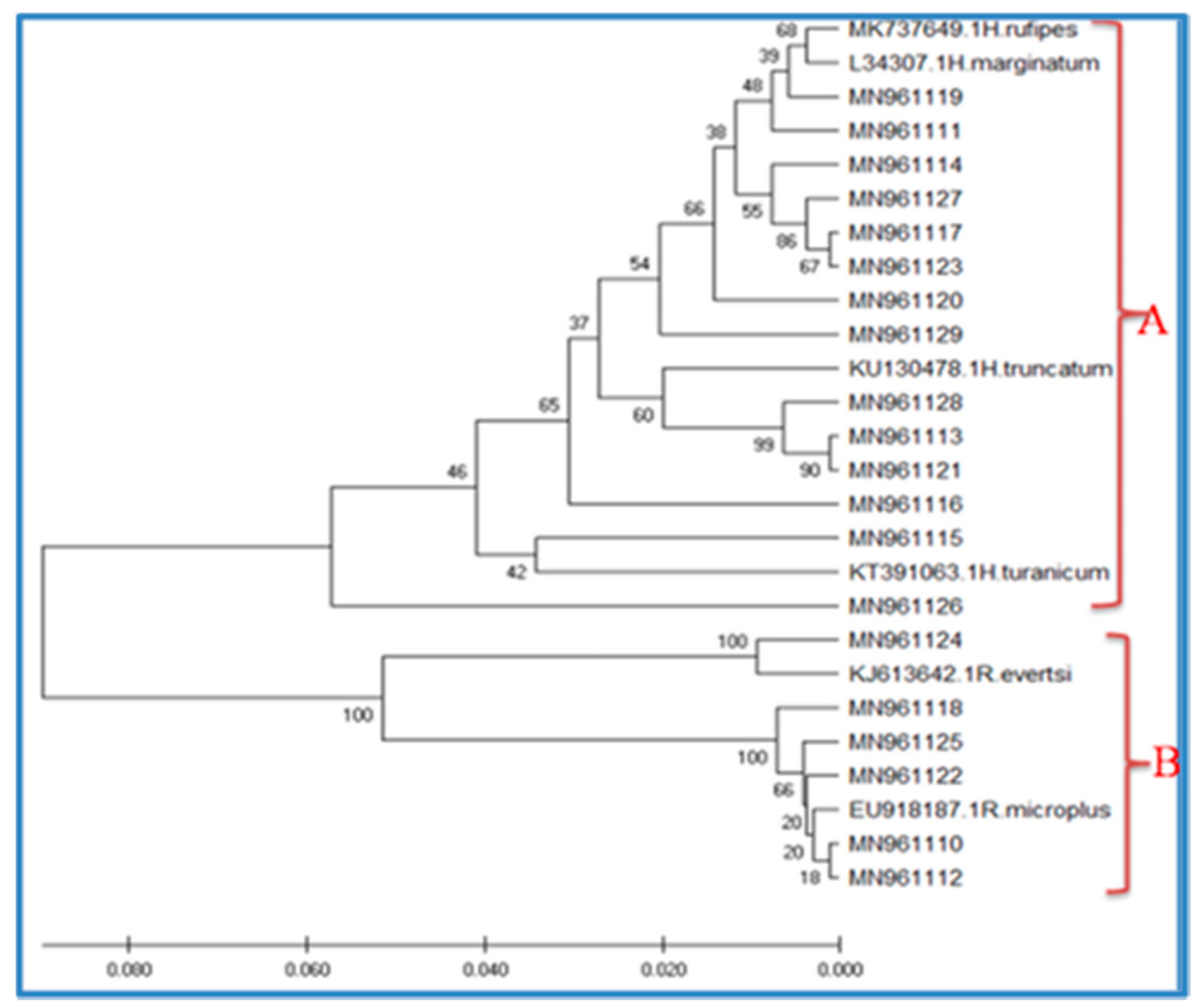
Based on the phylogenetic analysis of themitochondrion16S rRNA gene, two species have been reported in the Rhipicephalus genus, namely R. microplus and R. evertsi (Figure 2). All R. microplus and R. evertsi sequences generated in the present study clustered into one clade (B), together with other R. mmicroplus and R. evertsi isolates from other areas (Figure 2). For Hyalomma spp., sequences from the present study were selected for phylogenetic analysis and four species of H. rufipes, H. truncatum, H. marginatum, and H. mturanicum were recognized. Likewise, all the sequences of the Hyalomma spp. from the present study and GenBank formed another single clade (A) (Figure 2). However, H. turanicum (MN961115 and MN961126) formed a separate group from the H. rufipes, H. truncatum, and H. marginatum within the same clades. H. rufipes and H. marginatum collected during the present study clustered together and formed a complex group (Figure 2).

4. Discussion

Most of the previous studies from Tanzania were only based on morphological identification and most of them identified tick samples at the genus level [7,8]. There is therefore a need for molecular identification in order to generate genetic data bases for tick species and develop better control measures for ticks and tick borne pathogens [20,21].Two tick genera (Rhipicephalus and Hyalomma) were identified based on morphological characters. The two genera of hard ticks identified in this study, have also been reported by other researchers in some parts of Tanzania [6,7,8]. The presence of the tick spp. in the present study, similar to studies conducted in other sites in Tanzania, may be associated with unrestricted cattle movement from one area to another and cattle trade [22,23], which is a common phenomenon in Tanzania.
It is evident from the results in the present study that the mean tick intensity and the mean tick abundance differed between the two animal hosts (cattle and goats), which concur with previous studies [20,21]. The variations in infestation intensity and abundance could be due to a matter of feeding behavior differences between cattle and goats, as goats are browsers [21]. The observed high tick intensity in cattle as compared to goats may also be linked with the body surface area, host genetics, and small number of goats in our study [20]. However, limited information is available about ticks’ prevalence in small ruminants in Tanzania.
Most of the ticks in this study infested the sites with shorter hair and thinner skin [22]. The high tick infestations on these sites could be ascribed to the fact that ticks prefer warm, moist, and hidden sites with a good vascular supply and thin skin [23].
Six species have been confirmed based on mitochondrion16S rRNA analysis, including R. microplus, R. evertsi, H. rufipes, H. truncatum, H. marginatum, and H. turanicum. The evolutionary tree generated in the present study recorded several clusters of mitochondrial16S rRNA gene sequences, indicating divergence of gene sequences of hard ticks present in this wildlife-livestock interface ecosystem. Clusters of similar sequences represent species/subspecies clearly separated from other clusters (species/subspecies).
R. microplus was one among the tick species recorded in the present study. The finding of the R. microplus in the study region is in accordance with those of a local study conducted on Singida and Mbeya region of Tanzania [6], where the authors recorded high prevalence of R. microplus. The rapid expansion of R. microplus Tanzania is likely attributable to the shorter life-cycle and higher egg production capacity [24]. This phenomenon has recently been reported in several other African countries, including South Africa and Ivory Coast [25,26,27]. Moreover, the ability of R. microplus to develop resistance to most available acaricides might also have favored its expansion at the expense of more susceptible species [28]. According to Walker et al., 2003 [5], this species is widespread in tropical and subtropical regions and is considered to be the most important tick infesting livestock in the world. The higher prevalence of this species in the study area is of great interest because it is known to be a good vector of highly pathogenic Babesia bigemina and Babesia bovis, causing bovine babesiosis [29]. In addition, this species in terms of control management is well-known to be resistant to numerous pyrethroid and organophosphate compounds [30]. This species is also a vector of Anaplasma marginale, which causes anaplasmosis in cattle [31].
Another species belonging to the Rhipicephalus genus found in the present study was Rhipicephalus evertsi. This species has been reported with 33.8% prevalence in Mbeya region, Tanzania [6], to be prevalent in wild animals [7], and 10.9% prevalence in the Somali Regional State, Ethiopia [32]. According to Walker et al., 2003 [5], R. evertsi prefer the tropical geographical region in sub-Saharan Africa. Its distribution includes desert, steppe, savanna, and temperate climatic regions [5]. R. evertsi is of veterinary importance since it transmits the Babesia caballi and Theileria equi to horses, both causing forms of equine piroplasmosis [5]. Furthermore, this tick transmits the bacterium Anaplasma marginale to cattle causing bovine anaplasmosis. The saliva of female ticks contains a toxin that causes paralysis, particularly in lambs, but it may also affect calves and adult sheep [29].
Hyalomma rufipes was also recorded in the present study. This observation is in agreement with a previous report by Kerario et al., 2017 [6], with the highest prevalence (13.5% and 35.6%) in Singida and Mbeya regions, Tanzania respectively. However, this observation does not concur with Kwak et al., 2014 [7],who assessed Ixodidae tick infestation in Iringa and Maswa districts of Tanzania. In their study, this species was not observed. The observation of H. rufipes in the present study area is in agreement with Walker et al., 2003 [5], who assessed ticks of domestic animals in Africa. According to Walker et al., 2003 [5], H. rufipes is widely distributed in much of Africa and has been recorded from every climatic region from desert to rain forest. The presence of this species in the study area of Tanzania is of great importance, as it is known to be the most important vector in southern Africa of the virus causing Crimean-Congo hemorrhagic fever in human [29]. Furthermore, H. rufipes transmits A. marginale, a causative agent of anaplasmosis in cattle [33,34]. The feeding of adults on cattle causes large lesions at the attachment sites, leading to the formation of severe abscesses [3].
Hyalomma marginatum is another tick species that was recorded in the present study. According to Walker et al., 2003 [5] and Perveen et al., 2021 [35], H. marginatum occurs in areas with the humid Mediterranean climate of northern Africa and southern Europe and of steppe climate. The findings of Walker et al., 2003 [5] are not in agreement with our observation in the present study conducted in Morogoro region, which has hot and a moderate wet climate. The results of the current study could probably be due to changes in the tick populations since that time. There have been many changes since 2003 in the conditions required by the various tick species, including decrease of wild hosts, increase in the human population, increase in livestock and other domestic animals, the role of migratory birds in ticks transfer, changes in vegetation, changes in climate, changes in agricultural activities, increase in tick control regimes and its effectiveness, which together may well have had a great influence on the tick population [3]. The observation of this species in the study area is of great interest because it transmits the protozoa Babesia caballi causing babesiosis in horses, and it is known to transmit Theileria annulata causing theilerosis [3]. It is also responsible for transmission to humans of the virus causing Crimean-Congo haemorrhagic fever [29,35]. Furthermore, it can cause serious damage to cattle hides because of its long mouthparts. Notably, these ticks preferentially feed on the udder and teats of cattle and may cause serious problems in the suckling of calves [34].
Hyalomma truncatum is another tick species that was recorded in the present study. This observation is agreeable with Walker et al., 2003 [5] who assessed ticks of domestic animals in Africa. In their study, this species was observed to be endemic to the tropical region and thus is generally restricted to areas south of the Sahara, although it has been recorded from northern Sudan and from Egypt [35]. The observation of this species in the present study area is of great veterinary importance as certain strains of Hyalomm truncatum have a toxin in their saliva that causes the skin diseases known as sweating sickness in cattle. The long mouth-parts cause tissue damage and secondary bacterial infections may lead to infected abscesses [5].
The last and most interesting tick species found in the present study, Hyalommat ranicum, had not been reported from the study region before. According to Walker et al., 2003 [5], H. turanicum is distributed in areas with steppe and desert climate, and in Africa it occurs mainly in the central and western arid regions of southern Africa. The results of the current study could probably be due to a change in the tick populations since that time, as there have been enormous changes since 2003 in the conditions required by the various tick species, same conditions as discussed previously for H. marginatum apply to both species [3]. The presence of this species in the study area of Tanzania is of great importance as it is considered to be vector of the virus causing Crimean-Congo hemorrhagic fever in humans [29,35], although H. turanicum is not known to be a main vector of pathogens causing disease in domestic animals.

5. Conclusions

Present study provides genetic characterization of the mitochondrion16SrRNAgenes diversity of the hard ticks collected from the wildlife-livestock interface at Mikumi National Park, Morogoro region, Tanzania. The tick species infesting domestic animals (cattle and goats) in the study area were Rhipicephalus microplus, Rhipicephalus evertsi, Hyalomma rufipes, and Hyalomma truncatum. Hyalomma marginatum and Hyalomma turanicum were also found, whereby these two species had not been reported from the study region before. The presence of different groups of tick species and the observation of the species which had not been reported from the study region before, reflect the possible biological diversity of hard ticks present in this wildlife-livestock interface ecosystem. Therefore, further work is required to investigate species of hard tick present in the study region over a larger scale.

Author Contributions

Conceptualization, D.D., M.D., J.J.W., and M.B.; methodology, D.D., M.D., J.J.W. and M.B.; software D.D.; validation, D.D., M.D., J.J.W., and M.B.; formal analysis D.D., J.J.W., and MB.; investigation, D.D., M.D., J.J.W., and M.B.; resources, D.D., M.D., J.J.W., and M.B.; data curation, D.D.; writing- original draft preparation, D.D.; writing review and editing, D.D., M.D., J.J.W., and M.B.; visualization, D.D., M.D., J.J.W., and M.B.; supervision, M.D., J.J.W., and M.B.; project administration, M.D., J.J.W., and M.B.; funding acquisition, M.D., J.J.W., and M.B. All authors have read and agreed to the published version of the manuscript.

Funding

This research was funded by the Swedish International Development Cooperation Agency (UDSM-SIDA), grant number 2221.

Informed Consent Statement

Not applicable.

Data Availability Statement

The data supporting the conclusions of this article are included within the article. The sequences generated in the present study were submitted to GenBank under the accession numbers MN961110 to MN961129.

Acknowledgments

In particular, we thank the Swedish Government and Swedish International Development Cooperation Agency (SIDA) for contributions and financial support. We also extend special thanks to the University of Dar es Salaam, Tanzania, and Swedish University of Agricultural Sciences, Sweden for supporting this research work, as well as our colleagues and researchers at the Swedish University of Agricultural Sciences Global Bioinformatics Centre who provided valuable knowledge and skills concerning Bioinformatics. We thank the livestock keepers, village and ward leaders from each location for their help with tick collection, as well as entomologists from Sokoine University of Agriculture who assisted in tick morphological identification.

Conflicts of Interest

The authors declare that they have no competing interests. The funders had no role in the design of the study; in the collection, analyses, or interpretation of data; in the writing of the manuscript, or in the decision to publish the results.

Abbreviations

BLAST: Basic Local Alignment Search Tool, H: Hyalomma, K2P: Kimura’s 2-parameter, R: Rhipicephalus, UPGMA: Unweighted Pair Group Method with Arithmetic mean.

References

  1. Yu, Z.; Wang, H.; Wang, T.; Sun, W.; Yang, X.; Liu, J.-Z. Tick-borne pathogens and the vector potential of ticks in China. Parasites Vectors 2015, 8, 24–28. [Google Scholar] [CrossRef] [Green Version]
  2. Mansfield, K.L.; Jizhou, L.; Phipps, L.P.; Johnson, N. Emerging Tick-Borne Viruses in the Twenty-First Century. Front. Cell. Infect. Microbiol. 2017, 7, 298. [Google Scholar] [CrossRef] [Green Version]
  3. Jongejan, F.; Uilenberg, G. The global importance of ticks. Parasitology 2004, 129, S3–S14. [Google Scholar] [CrossRef] [PubMed]
  4. Estrada-Peña, A.; Gray, J.S.; Kahl, O.; Lane, R.S.; Nijhof, A.M. Research on the ecology of ticks and tick-borne pathogens-methodological principles and caveats. Front. Cell. Infect. Microbiol. 2013, 3, 29. [Google Scholar] [CrossRef] [Green Version]
  5. Walker, A.R.; Bouattour, A.; Camicas, J.J.; Estrada-Pena, A.; Horak, I.G.; Latif, A.A.; Pegram, R.G.; Preston, P.M. Ticks of Domestic Animals in Africa: A Guide to Identification of Species; Bioscience Reports: Edinburgh, UK, 2003; pp. 1–1221. [Google Scholar]
  6. Kerario, I.; Muleya, W.; Chenyambuga, S.; Koski, M.; Seong-Gu Hwang, S.; Simuunza, M. Abundance and distribution of Ixodid tick speciesinfesting cattle reared under traditional farmingsystems in Tanzania. Afr. J. Agric. Sci. 2017, 12, 286–299. [Google Scholar]
  7. Kwak, Y.S.; Kim, T.Y.; Man, S.H.; Lee, I.Y.; Kim, H.P.; Mduma, S.; Keyyu, J.; Fyumagwa, R.; Yong, T.S. Ixodid tick infestation in cattle and w ild animals in Maswa and Iringa, Tanzania. Korean J. Parasitol. 2014, 5, 565–568. [Google Scholar] [CrossRef]
  8. Laisser, E.L.K.; Kipanyula, M.J.; Msalya, G.; Mdegela, R.H.; Karimuribo, E.D.; Mwilawa, A.J.; Mwega, E.D.; Kusiluka, L.J.M.; Chenyambuga, S.W. Tick burden and prevalence of Theileriaparvainfection in Tarime zebu cattle in the Lake zone of Tanzania. Trop. Anim. Health Prod. 2014, 46, 1391–1396. [Google Scholar] [CrossRef]
  9. Swai, E.S.; Mbise, A.N.; Kessy, V.; Kaaya, E.; Sanka, P.; Loomu, P.M. Farm Constraints, Cattle Disease Perception and Tick Managementpractices in Pastoral Maasai Community, Ngorongoro, Tanzania. 2005. Available online: http://www.lrrd.org/17/2/cont1702 (accessed on 18 October 2019).
  10. Labuda, M.; Nuttall, P.A. Tick-borneviruses. Parasitology 2004, 129, S221–S245. [Google Scholar] [CrossRef]
  11. Muruthi, C.W.; Lwande, O.W.; Makumi, J.N.; Runo, S.; Otiende, M.; Makori, W.A. Phenotypic and genotypicidentification of ticks sampled from wildlife species in selected conservation sites of Kenya. Vet. Sci. Technol. 2016, 7, 1–8. [Google Scholar]
  12. Man, S.Q.; Qiao, K.; Cui, J.; Feng, M.; Fu, Y.F.; Cheng, X.J. A case of human infection with a novel Babesia species in China. Infect. Dis. Poverty 2016, 5, 28. [Google Scholar] [CrossRef] [Green Version]
  13. Apanaskevich, M.A.; Apanaskevich, D.A. Description of new Dermacentor(Acari: Ixodidae) speciesfrom Malaysia and Vietnam. J. Med. Entomol. 2015, 52, 156–162. [Google Scholar] [CrossRef] [Green Version]
  14. Wallace, D.M. Large and Small Scale Phenol Extractions Guide to Molecular Cloning Techniques; Academic Press: Orlando, FL, USA, 1987; pp. 31–41. [Google Scholar]
  15. Hill, C.A.; Gutierrez, J.A. Method for extraction and analysis of high quality genomicDNA from ixodid ticks. Med. Vet. Entomol. 2003, 12, 224–227. [Google Scholar] [CrossRef] [PubMed]
  16. Mauel, M.J.; Carlton, S.J.; Mather, T.N. Polymerase chain reaction detection efficiency of thehuman granulocytic ehrlichiosis agent (Rickettsiaceae: Ehrlichieae) in ticks (Acari: Ixodidae) is dependent on the DNA extraction method. J. Med. Entomol. 1999, 36, 649–652. [Google Scholar] [CrossRef]
  17. Zemtsova, G.E.; Apanaskevich, D.A.; Reeves, W.K.; Hahn, M.; Snellgrove, A.; Levin, M.L. Phylogeography of Rhipicephalus sanguineussensulato and its relationships with climatic factors. Exp. Appl. Acarol. 2016, 69, 191–203. [Google Scholar] [CrossRef] [PubMed] [Green Version]
  18. Takano, A.; Fujita, H.; Kadosaka, T.; Takahashi, M.; Yamauchi, T.; Ishiguro, F. Construction of a DNA database for ticks collected in Japan: Application of molecular identification based on the mitochondrial 16S rDNA gene. Med. Entomol. Zool. 2014, 65, 13–21. [Google Scholar] [CrossRef] [Green Version]
  19. Kumar, S.; Tamura, K.; Nei, M. MEGA: Molecular Evolutionary Genetics Analysis software for microcomputers. Comput. Appl. Biosci. 1994, 10, 189–191. [Google Scholar] [CrossRef] [Green Version]
  20. Zeryehun, T.; Atomsa, M. Ectoparasiotes infestations of sheep and goats. Eurasian J. Vet. Sci. 2012, 28, 185–189. [Google Scholar]
  21. Kaiser, M.N.; Surtherst, R.W.; Bourne, A.S. Relationshipbetweenticks and Zebu in southern Uganda. Trop. Anim. Health Prod. 1982, 14, 63–74. [Google Scholar] [CrossRef]
  22. Moges, N.; Bogale, B.; Fentahun, T. Hard Ticks (Ixodidae) Species Composition, Seasonal Dynamics and Body Site Distribution on cattle in Chilga District, Northwest Ethiopia. Asian J. Agric. Sci. 2012, 4, 341–345. [Google Scholar]
  23. Awaa, D.N.; Adakald, H.; Luogboua, N.D.D.; Wachonga, K.H.; Leinyuya, I.; Achkwiaa, M.D. Is Ripicephalus (Boophilus) microplus absentin Cameroon and the central African region? Ticks Tick-Borne Dis. 2015, 6, 117–122. [Google Scholar] [CrossRef]
  24. Tønnesen, M.; Penzhorn, B.; Bryson, N.; Stoltsz, W.; Masibigiri, T. Displacement of Boophilusdecoloratus by Boophilus microplus in theSoutpansberg region, Limpopo Province, South Africa. Exp. Appl. Acarol. 2004, 32, 199–208. [Google Scholar] [CrossRef] [PubMed]
  25. Lynen, G.; Zeman, P.; Bakuname, C.; Di Giulio, G.; Mtui, P.; Sanka, P. Shifts in the distributional ranges of Boophilusticks in Tanzania: Evidence that a parapatric boundary between Boophilus microplus and B. decoloratus follows climate gradients. Exp. Appl. Acarol. 2008, 44, 147–164. [Google Scholar] [CrossRef]
  26. Nyangiwe, N.; Harrison, A.; Horak, I.G. Displacement of Rhipicephalus decoloratus by Rhipicephalusmicroplus(Acari: Ixodidae) in the Eastern Cape Province, South Africa. Exp. Appl. Acarol. 2013, 61, 371–382. [Google Scholar] [CrossRef] [Green Version]
  27. Madder, M.; Thys, E.; Achi, L.; Touré, A.; De Deken, R. Rhipicephalus (Boophilus) microplus: A most successful invasive tick species in West-Africa. Exp. Appl. Acarol. 2011, 53, 139–145. [Google Scholar] [CrossRef]
  28. Busch, J.D.; Stone, N.E.; Nottingham, R.; Araya-Anchetta, A.; Lewis, J.; Hochhalter, C. Widespread movement of invasive cattle fever ticks (Rhipicephalus microplus) in southern Texas leads to shared local infestations on cattle and deer. Parasites Vectors 2014, 7, 188. [Google Scholar] [CrossRef] [Green Version]
  29. Bock, R.; Jackson, L.; de Vos, A.; Jorgensen, W. Babesiosis ofcattle. Parasitology 2004, 129, S247–S269. [Google Scholar] [CrossRef]
  30. Baffi, M.A.; de Sauza, G.R.; de Sauza, G.S.; Ceron, C.R.; Bonetti, A.M. Esterase enzymes involved in pyrethroid and organophosphateresistance in a Brazilian population of Rhipicephalus (Boophilus) microplus(Acari, Ixodidae). Mol. Biochem. Parasitol. 2008, 160, 70–73. [Google Scholar] [CrossRef]
  31. Kocan, K.M.; de La Fuente, J.; Blouin, E.F.; Garcia, J.C. Anaplasmamarginale (Rickettsiales: Anaplasmataceae): Resent advances in defining host-pathogen adaptation of a tick-borne rickettisia. Parasitology 2004, 129, S285–S300. [Google Scholar] [CrossRef] [PubMed]
  32. Abebe, R.; Fantahun, T.; Abera, M.; Bekele, J. Survey ofticks (Acari:Ixodidae) infesting cattle in two districts of SomaliRegional State. Ethiopia. Vet. World 2010, 3, 539–543. [Google Scholar]
  33. Potgieter, F.T. Epidemiology and control of Anaplasmosis in South Africa. J. S. Afr. Vet. Assoc. 1979, 504, 367–372. [Google Scholar]
  34. Tadesse, F.; Abadfaji, G.; Girma, S.; Kumsa, B.; Jibat, T. Identification of tick species and their preferred site on cattle’s body in and around MizanTeferi, Southwestern Ethiopia. J. Vet. Med. Anim. Health 2012, 4, 1–5. [Google Scholar]
  35. Perveen, N.; Muzaffar, S.; Al-Deeb, M.A. Ticks and Tick-borne Diseases of Livestock in the Middle East and North Africa. Insects 2021, 12, 83. [Google Scholar] [CrossRef]
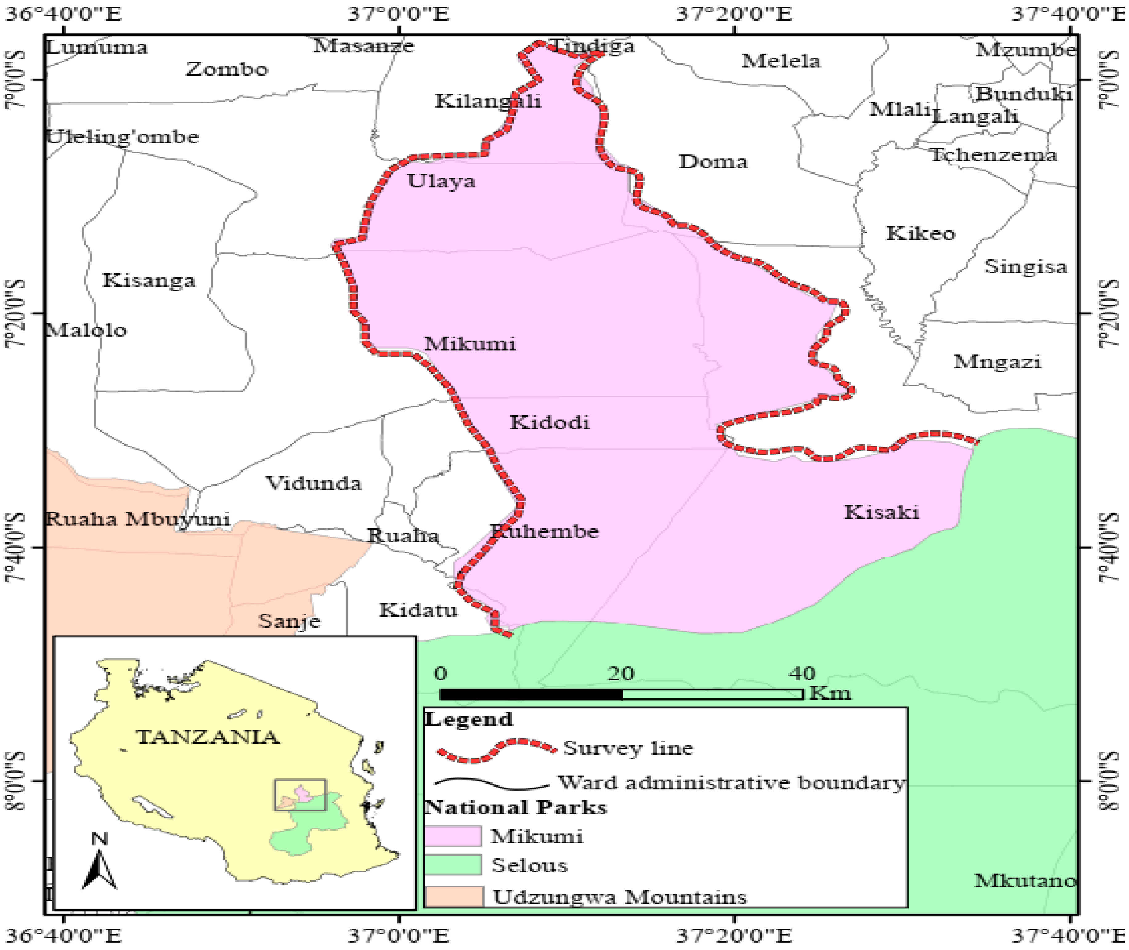
Figure 1. Map showing wildlife-livestock interface ecosystem of Mikumi National Park. Data source; (National Bureau of Statistics (Geograhic Information System) data base; https://www.nbs.go.tz/index.php/en/) (accessed on 18 November 2019).
Vetsci 08 00036 g001
Figure 2. Overall phylogenetic tree (inferred using the Unweighted Pair Group Method with Arithmetic mean (UPGMA)) for tick species based on the 16S rRNA gene, including sequences obtained in the present study and representative sequences of the known species from GenBank (indicated with a species name). A and B in the phylogenetic tree are the clades.
Vetsci 08 00036 g002
Table 1. Prevalence of ticks in cattle and goats a.
AnimalsTypeTotal
Cattle N (%)Goats N (%)
Examined260176436
Infested with ticks134 (51.5)78 (44.3)212 (48.6)
a Number of infested animals/Number of examined animal× 100.
Table 2. Mean ticks intensity in cattle and goats b.
CategoryCattleGoatsTotal
Animals infested 134 78 212
Ticks collected 632 187 819
Mean tick intensity4.72.43.9
b Number of collected ticks/Number of infested animals.
Table 3. Number (N) of ticks collected from cattle, goats, and environment (free) c.
Tick GenusSourceTotal
Cattle N (%)Goats N (%)Free N (%)
Rhipicephalus378 (59.8)36 (19.3)16 (57.1)430 (51)
Hyalomma254 (40.2)151 (80.7)12 (42.9)417 (49)
Total632 (100)187 (100)28 (100)847 (100)
c Number of tick spp. from (cattle, goats, environment)/Total tick spp. from (cattle, goats, environment) × 100.
Table 4. Mean ticks abundance in cattle and goats d.
CategoryCattleGoatsTotal
Animal analyzed260176436
Ticks collected632187819
Mean abundance2.41.11.8
d Nunber of ticks collected/Number of analyzed animals.
Table 5. Identity of tick species and percentage similarity value with the reference sequences.
Sample AccessionSourceSite/WardGeneBank ReferenceIdentity %Species
MN961110CattleDomaEU918187.1100Rhipicephalusmicroplus
MN961111EnvironmentMikumiKU170517.198.76Hyalomma refipes
MN961112CattleMangaeKT428016.1100Rhipicephalusmicroplus
MN961113CattleKidodiKU13478.197.15Hyalomma truncatun
MN961114CattleMkataKP776645.198.99Hyalomma marginatum
MN961115CattleMikumiKT391063.193.98Hyalomma turanicum
MN961116CattleDomaMK737649.194.70Hyalomma rufipes
MN961117CattleUlayaKP776645.198.74Hyalomma marginatum
MN961118CattleMelelaKT428016.199.75Rhipicephalusmicroplus
MN961119EnvironmentKilangaliKU170517.198.99Hyalomma rufipes
MN961120GoatDomaKU17051799.97Hyalomma rufipes
MN961121CattleRuhembeKU130478.196.63Hyalomma truncatum
MN961122EnvironmentMangaeKT428016.199.26Rhipicephalusmicroplus
MN961123GoatMikumiKT391060.198.50Hyalomma marginatum
MN961124CattleMkataKJ613642.198.42Rhipicephalus evertsi
MN961125GoatTindigaKC170742.199.50Rhipicephalusmicroplus
MN961126CattleMelelaKP77664591.54Hyalomma turanicum
MN961127CattleKisakiKP776645.198.50Hyalomma marginatum
MN961128GoatMangaeKU130478.196.37Hyalomma truncatum
MN961129CattleKidatuMK737650.197.47Hyalomma rufipes
Publisher’s Note: MDPI stays neutral with regard to jurisdictional claims in published maps and institutional affiliations.