Article

Evaluation of the Dual Antiviral and Immunomodulatory Effects of Phallus indusiatus in a Feline Infectious Peritonitis Model Using PBMCs

by
Chularat Hlaoperm
1,2,
Wassamon Moyadee
1,2,
Emwalee Wongsaengnoi
3,
Wiwat Klankaew
4,
Amonpun Rattanasrisomporn
5,
Atchara Paemanee
6,7,
Kiattawee Choowongkomon
4,
Oumaporn Rungsuriyawiboon
3 and
Jatuporn Rattanasrisomporn
1,2,*
1
Graduate Program in Animal Health and Biomedical Sciences, Faculty of Veterinary Medicine, Kasetsart University, Bangkok 10900, Thailand
2
Department of Companion Animal Clinical Sciences, Faculty of Veterinary Medicine, Kasetsart University, Bangkok 10900, Thailand
3
Department of Veterinary Technology, Faculty of Veterinary Technology, Kasetsart University, Bangkok 10900, Thailand
4
Department of Biochemistry, Faculty of Science, Kasetsart University, Bangkok 10900, Thailand
5
Interdisciplinary of Genetic Engineering and Bioinformatics, Graduate School, Kasetsart University, Bangkok 10900, Thailand
6
National Omics Center, National Center for Genetic Engineering and Biotechnology (BIOTEC), National Science and Technology Development Agency (NSTDA), Khlong Luang, Pathum Thani 12120, Thailand
7
Food Biotechnology Research Team, Functional Ingredients and Food Innovation Research Group, National Center for Genetic Engineering and Biotechnology (BIOTEC), National Science and Technology Development Agency (NSTDA), Khlong Luang, Pathum Thani 12120, Thailand
*
Author to whom correspondence should be addressed.
Vet. Sci. 2025, 12(9), 847; https://doi.org/10.3390/vetsci12090847
Submission received: 29 July 2025 / Revised: 23 August 2025 / Accepted: 29 August 2025 / Published: 1 September 2025
(This article belongs to the Special Issue Advanced Therapy in Companion Animals—2nd Edition)

Simple Summary

This study explores the potential of Phallus indusiatus (P. indusiatus), a medicinal mushroom, as a dual-action treatment for feline infectious peritonitis (FIP), a fatal disease caused by feline coronavirus. P. indusiatus demonstrated the most pronounced inhibitory effect among the 17 mushroom extracts tested (69.2% inhibition of FIPV Mpro), showing a level of activity approaching that of standard antivirals such as lopinavir and ritonavir. The extract also significantly reduced nitric oxide production (a marker of inflammation) in lipopolysaccharide (LPS)-stimulated peripheral blood mononuclear cells (PBMCs) from healthy cats, indicating anti-inflammatory effects. However, immune cells from cats already suffering from FIP, which were in a chronically inflamed state, did not respond to the extract or to dexamethasone. This highlights the complexity of FIP-related inflammation. This study also successfully cultured PBMCs from FIP fluid for the first time, providing a novel in vitro model for future research. Overall, the results suggest that P. indusiatus has promising antiviral and anti-inflammatory properties, though further research is needed to isolate active compounds and test in live infection models.

Abstract

Feline infectious peritonitis (FIP) is a progressive and often fatal disease caused by a virulent biotype of feline coronavirus (FCoV). Although antiviral treatments are now available, relapse and resistance remain ongoing concerns. This study investigates the therapeutic potential of P. indusiatus, a medicinal mushroom, for its antiviral and anti-inflammatory activities against FIP. The main protease (FIPV Mpro) of feline infectious peritonitis virus (FIPV) was recombinantly expressed and purified to facilitate enzyme inhibition screening. P. indusiatus exhibited the strongest FIPV Mpro inhibitory activity among the 17 mushroom extracts tested (69.2%), showing a notable level of inhibition relative to standard antiviral agents such as lopinavir and ritonavir. To assess its anti-inflammatory potential, PBMCs derived from healthy cats and FIP-associated effusions (FIP fluid) were cultured and stimulated with LPS to induce inflammation. In healthy PBMCs, P. indusiatus significantly reduced nitrite levels, with effects similar to dexamethasone. However, PBMCs from FIP fluid, already in an activated state, showed no additional response. Notably, this study is the first to successfully isolate and culture PBMCs from FIP fluid, providing a new platform for future immunological research. These findings suggest that P. indusiatus possesses both antiviral and anti-inflammatory properties, positioning it as a potential dual-action therapeutic candidate for FIP. Further investigation into cytokine signaling pathways is warranted to clarify its mechanisms of action and advance future therapeutic development.

1. Introduction

Feline coronavirus (FCoV) is an enveloped, positive-sense single-stranded RNA virus in the Coronaviridae family commonly found in domestic cats [1]. It exists in two biotypes: feline enteric coronavirus (FECV) [2], which typically causes mild gastrointestinal signs and is often self-limiting in immunocompetent cats, and feline infectious peritonitis virus (FIPV), the pathogenic variant responsible for feline infectious peritonitis (FIP) [3]. First reported in 1966, FIP is fatal, immune-mediated disease that primarily affects kittens, adolescents, and young adult cats, and remains a significant cause of mortality in this population [4]. Approximately 12% of FCoV-infected cats progress to FIP, with environmental stress and immune dysfunction as a key contributing factors [5]. Although antiviral drugs have recently become available, delayed diagnosis still results in high mortality, emphasizing the need for timely and accurate detection [6]. Pathogenesis involves efficient replication of FCoV in activated monocytes and macrophages, leading to systemic inflammation, pyogranulomatous lesions, and T-cell depletion [6,7]. Altered cytokine profiles, including elevated interleukin (IL)-1β, IL-6, IL-10, IL-12p40, and tumor necrosis factor-alpha (TNF-α), have been identified in affected cats, indicating that immune dysregulation plays a key role in the advancement of FIP [8].
Edible mushrooms have been reported to possess multiple biomedical properties, including antiviral, anti-inflammatory activities, and immunomodulatory effects through cytokine stimulation [7,8]. These effects are largely attributed to bioactive compounds such as polysaccharides, terpenoids, and alkaloids. These compounds enhance phagocytic activity, increase IL-1β and TNF-α expression, and activate key signaling pathways such as ERK1/2, JNK, and p38 MAPKs. They also promote NF-κB p65 phosphorylation and nuclear translocation and can bind to macrophages via Toll-like receptor 4 (TLR4) [8]. Certain mushrooms may also help alleviate clinical signs associated with cytokine storms. While some compounds have shown antiviral activity, scientific evaluation in feline models remains limited.
In the preliminary inflammation model of this study, both lipopolysaccharide (LPS) and FIPV (or viruses in general) are potent activators of the NF-κB and AP-1 signaling pathways [9,10,11]. These pathways function as central molecular “switches” that regulate the transcription of various inflammatory mediators, including NO, inducible nitric oxide synthase (iNOS), cyclooxygenase-2 (COX-2), and prostaglandin E2 (PGE2), and the pro-inflammatory cytokines (e.g., TNF-α, IL-6, and IL-1β) [12,13]. Therefore, using LPS as an inflammatory stimulant in PBMCs to investigate the anti-inflammatory effects of mushroom extracts is well justified as a preliminary inflammation model, as it activates shared pathways involved in cytokine and iNOS expression mechanisms that are also triggered by FIPV (Figure 1).
Coronaviruses are RNA viruses that hijack the host’s translational machinery to generate viral proteins. The resulting polyproteins must be cleaved to release individual functional proteins necessary for viral replication and transcription. The virus encodes proteases, including the main protease (Mpro), also known as 3CLpro, which has conserved sequences that facilitate the assembly of the viral replicase complex [14]. The Mpro protein of FCoV is essential for the replication cycle, as observed in other members of the Coronaviridae family [15]. Therefore, FIPV Mpro protein is considered a prominent drug target for antiviral therapy against FIP (e.g., GC376) [14,16].
The immune system plays a crucial role in controlling viral infections, including the suppression of FCoV replication and prevention of progression to FIP. In this study, 17 mushroom extracts were screened as potential candidates for alternative immunomodulatory strategies in cats. Screening was conducted to assess antiviral activity by them against FIPV Mpro to identify active compounds. The extract with the strongest inhibitory effect was selected for further assessment of its anti-inflammatory potential. PBMCs were isolated from both healthy cats and cats with FIP-associated effusions, including pleural, peritoneal, or a combination of both, and were cultured under optimized conditions. These PBMCs were then stimulated with LPS and treated with the selected extract to assess nitrite-scavenging activity. The resulting data will serve as a foundation for future investigations into cytokine modulation, particularly in the context of FIP-related inflammation and antiviral response.

2. Materials and Methods

2.1. Ethics Statement

This study was approved by the Institutional Animal Care and Use Committee of Kasetsart University, Bangkok, Thailand, under protocol number ACKU67-VET-124. Additionally, all sample collections were performed with the informed consent of the cat owners.

2.2. Protein Expression and Purification

The expression and purification of FIPV’s main protease in this study, called “FIPV Mpro”, have been described previously [17]. In this study, the FIPV Mpro was derived from the FIPV WSU-79-1146 strain. The coding sequence for Mpro included an N-terminal 10xHis tag and a TEV cleavage site, resulting in a recombinant protein with an approximate molecular weight of 37.2 kDa. The gene was synthesized into the pGEX-4T-1 vector (GenScript, USA) and transformed into Escherichia coli strain BL21(DE3) for protein expression. Cultures were grown in LB medium incubated at 37 °C for 3 to 4 h, the absorbance was measured at OD 600 to 0.8, and then the sample was induced by 0.4 mM isopropyl-β-d-thiogalactopyranoside at 37 °C for 18–20 h. The FIPV Mpro was purified by Ni2+ affinity column (Cytiva AB, Sweden). Western blotting was performed to confirm the protein expression, using an anti-His tag primary antibody (Thermo Fisher Scientific, USA) and an HRP-conjugated Goat Anti-Mouse IgG secondary antibody (Thermo Fisher Scientific, USA) for signal detection.

2.3. Inhibition of FIPV Mpro Protease

The FIPV Mpro modified with Dabcyl-KTSAVLQSGFRKM-E (Edans) (Genscript USA, Piscataway, NJ, USA) was prepared in a buffer containing 20 mM Tris (pH 7.5), 100 mM NaCl, 2 mM DTT, and 0.05 mM EDTA. The enzyme activity was subsequently examined in a 384-well plate containing 1 µM FIPV Mpro in buffer solution. Subsequently, enzyme activity inhibitors, including lopinavir and itonavir as positive controls, were added for comparison with 10 µg/mL mushroom extracts. The final inhibitor concentration in the reaction was set to 10 µg/mL. The reaction was initiated by adding the substrate at a final concentration of 40 µM. The increase in fluorescence intensity of the substrate at 340 nm was measured to determine the relationship between Relative Fluorescence Units (RFUs) per second, using a microplate reader (Infinite 200, Tecan Group Ltd., Männedorf, Switzerland) and GraphPad version 8.0 software, as described in Equation (1).
%   Relative inhibition = V 0   e n z V 0   I n h i b i t o r × 100 V 0   e n z
V0 represents the initial reaction velocity of the enzyme. It indicates how fast the enzyme converts substrate to product at the start of the reaction. V0 enz is the initial reaction velocity of the enzyme without an inhibitor. V0 Inhibitor is the initial reaction velocity of the enzyme in the presence of an inhibitor.
Given the constraints associated with cell culture systems and the limited availability of animal-derived samples, particularly those obtained from experimental models, this phase of the study was designed to reduce animal usage by utilizing an in vitro screening approach. This model was employed to identify mushroom extracts exhibiting the highest inhibitory activity against the FIPV Mpro. The extract demonstrating the most potent inhibitory effect will be selected for further investigation of its anti-inflammatory properties.

2.4. Animals and Experimental Design

This research was approved for sample collection from the Kasetsart University Veterinary Teaching Hospital in Bangkok, Thailand. PBMCs were isolated from healthy donor cats (n = 20). In parallel, pleural and/or ascitic fluid samples were collected from cats clinically diagnosed with effusive FIP, based on a combination of consistent clinical, laboratory, and imaging findings. These included serum biochemical and hematological abnormalities, clinical signs such as anorexia, depression, intermittent fever, abdominal distension, or dyspnea due to pleural effusion. Radiographic or ultrasonographic imaging confirmed the presence of fluid accumulation in body cavities and involvement of internal organs. Fluid analysis and cytology revealed pyogranulomatous inflammation, with a proteinaceous, granular background, and a cell population consisting of macrophages, non-degenerate neutrophils, and few lymphocytes. The effusions were classified as modified transudates or non-septic exudates. The diagnosis was further supported by additional findings, including a serum albumin-to-globulin (A:G) ratio of less than 0.8, a positive Rivalta’s test, and/or a positive PCR result for FCoV, in accordance with previously published criteria [18,19]. The pleural and/or ascitic fluid samples were subsequently used for mononuclear cell isolation. These samples (n = 20) were subsequently used for mononuclear cell isolation. For simplicity, mononuclear cells isolated from effusions are referred to as “PBMCs from FIP fluid” throughout this study. The sample size was derived from data in other similar studies. All analyses were performed using GraphPad Prism version 9 (GraphPad Software, LLC, Boston, MA, USA) [20].

2.5. The Preparation of the Mushroom Extraction Procedure

A total of 17 types of medical mushrooms were selected, including Pleurotus ostreatus, Flammulina filiformis, Agrocybe cylindracea, Lentinula edodes, Tuber Uncinatum, Lignosus rhinocerotis, Volvariella volvacea, Ganoderma oregonense, Ganoderma formosanum, Ganoderma lucidum, Inonotus obliquus, Hericium erinaceus, Psilocybe cubensis, Phellinus igniarius, Dasoclema simensis, Pleurotus ostreatus, and Phallus indusiatus. A dry sample of 100 g was air-dried and grinded by mixer grinder, immersed in 95% ethyl alcohol (ethanol) 200 mL at a temperature of 37 °C, and shaken for 12 h. Afterward, the liquid part was separated and the ethanol was evaporated using a rotary evaporator under vacuum. The crude extract was stored at a temperature of −20 °C until analysis. The extracts from the 17 types of mushrooms will be further tested for their FIPV Mpro inhibitory activity. In this study, crude extracts were prepared using the same stock and extraction protocol described by Theeraraksakul, K. et al., 2023 [21].

2.6. Preparation of PBMCs

Whole blood from healthy donor cats and effusion samples (pleural, ascitic, or both) from FIP cats were processed to isolate PBMCs by separating the buffy coats using centrifugation with Lymphoprep™ (Corning®, USA) at 600 g for 30 min to isolate PBMCs. Subsequently, complete RPMI-1640 medium (Corning®, USA) containing 10% fetal bovine serum (FBS; Gibco®, USA) and 1% penicillin–streptomycin (Gibco®, USA) was added, and the cell concentration was adjusted to 1 × 10⁶ cells/mL. The cells were counted, and their viability was assessed by mixing the separated cells with a 0.4% Trypan Blue solution (Gibco®, USA) in a 1:1 ratio. A hemocytometer was used to count the cells under a microscope.
In the case of isolating PBMCs from effusion samples, referred to in this study as “PBMCs from FIP fluid” the sample characteristics of the FIP fluid were carefully considered. If the sample exhibited high viscosity, it was diluted with 1× phosphate-buffered saline (PBS; Cytiva HyClone™, USA) to facilitate efficient cell pelleting. The diluted sample was then centrifuged at 600× g for 10 min. The supernatant was discarded, and the cell pellet was retained. The pellet was subsequently washed three times with RPMI medium at a 1:1 ratio, each wash involving centrifugation at 600× g for 10 min. Once the cells were isolated, cell counting was performed, and the cells were cultured under the same conditions as previously described.

2.7. In Vitro Optimize Toxicity of P. indusiatus, LPS, and DMSO

The toxicity of P. indusiatus extract at concentrations of 10,000, 5000, 2500, 625, 156, 78, 39, and 19 µg/mL was tested on PBMCs isolated from the whole blood of donor cats. PBMCs (1 × 10⁶ cells/mL) were seeded into a 96-well plate and pre-incubated for 1–2 h in a humidified atmosphere with 5% CO2 at 37 °C. Subsequently, the cells were treated with various concentrations of P. indusiatus extract dissolved in 0.5% DMSO. The PBMCs were incubated for 24 h. Following treatment, cell viability was assessed using the Cell Counting Kit-8 (CCK-8) colorimetric assay (Dojindo, Kumamoto, Japan). Briefly, 10 µL of CCK-8 solution was added to each well and incubated for 2–4 h. The absorbance of each well was then measured at 450 nm using a microplate reader (EnSight® Multimode Plate Reader, PerkinElmer, Waltham, MA, USA). Cell viability was determined by calculating the percentage of absorbance relative to the control group (non-treated PBMCs).
The testing involved determining optimal concentrations and exposure times in generating PBMCs after stimulation with LPS (Sigma-Aldrich®, Israel) at various concentrations (0.001, 0.01, 0.1, and 1 µg/mL). Additionally, testing was conducted to determine suitable concentrations for cell viability using dimethyl sulfoxide (DMSO, Uvasol, Japan) at concentrations of 0.5%, 1%, 1.5%, and 2%, compared to a control group without DMSO. Cell viability was assessed using the CCK-8 colorimetric assay (Dojindo, Kumamoto, Japan) under the same conditions as described previously.

2.8. Anti-Inflammatory Assay Using Nitrite Scavenging Activity

To determine the effect of P. indusiatus extract on nitrite production, a primary breakdown product of nitric oxide, cells were suspended in a 96-well plate containing complete RPMI-1640 medium and pre-incubated for 1–2 h in a humidified atmosphere with 5% CO2 at 37 °C. Subsequently, the cells were treated with P. indusiatus at a concentration of 19 µg/mL and dexamethasone at 5 µg/mL as a positive drug control in the presence of LPS (1 µg/mL) and incubated for 24 h. Nitrite production was determined by calculating the percentage of absorbance relative to the control group (non-treated PBMCs) using the Griess Reagent System (Promega, USA). A parallel test was conducted on PBMCs isolated from FIP fluid under the same conditions, but without LPS stimulation. This study investigates the potential effects of P. indusiatus extract on inflammation in PBMCs isolated from whole blood and fluid samples of cats diagnosed with FIP. The primary aim was to evaluate whether the extract can modulate inflammation by measuring nitrite concentrations, which is a marker of inflammatory response.

2.9. Statistical Analysis

Data are presented as mean ± Standard Error of the Mean (SEM), with each experiment replicated at least twice to ensure reliability. PBMC viability from whole blood and FIP fluid was compared using two-way ANOVA, with comparisons made between cell means within each row (p < 0.05, n = 5). The effect of varying LPS concentrations (0.001, 0.01, 0.1, and 1 µg/mL) on nitrite levels was assessed after 24 h using one-way ANOVA, compared to a no-LPS control (p < 0.05). The impact of 19 µg/mL P. indusiatus and 5 µg/mL dexamethasone on nitrite production in PBMCs from donor cat whole blood was evaluated after 24 h, with data shown as mean ± SEM (n = 8), comparing column means. Inflammation modulation in PBMCs from FIP fluid treated with P. indusiatus extract (n = 5) was analyzed using one-way ANOVA (p < 0.05). Differences in treatment responses between PBMCs from whole blood and FIP fluid were assessed by two-way ANOVA (p < 0.05, n = 4).

3. Results

3.1. Expression and Purification of FIPV Mpro

Recombinant His-tagged FIPV 3CLpro, expressed in E. coli BL21 (DE3), was purified using a Ni2+ affinity column. The expression of the FIPV Mpro or FIPV-3C-like Protease, (3CLpro), was induced with 0.4 mM isopropyl β-d-1-thiogalactopyranoside (IPTG), followed by incubation for 18–24 h. Quantitative analysis using SDS-PAGE confirmed the expression of the FIPV Mpro protein with a molecular weight of 37.20 kDa (Figure 2A).
Qualitative analysis was performed using the Western blot technique, which utilized the specificity of the primary antibody (Anti-His Tag (HRP)) for proteins labeled with a 6x His-Tag. A secondary antibody (Goat Anti-Mouse IgG (HRP)) was employed to bind with HRP substrates. This revealed a band corresponding to the FIPV Mpro protein at a size of 37.20 kDa (Figure 2B). The results of determining the optimal concentration of FIPV Mpro enzymatic activity, using Dabcyl-KTSAVLQSGFRKM-E (Edans) as the substrate at a final concentration of 40 µM, showed that the reaction rate (or fluorescence intensity) of the substrate increased as the enzyme concentration was raised to 0.5 and 1 µM, separately (Figure 2C).

3.2. Inhibition of FIPV Mpro Activity by Antiviral Drugs and Crude Mushroom Extract

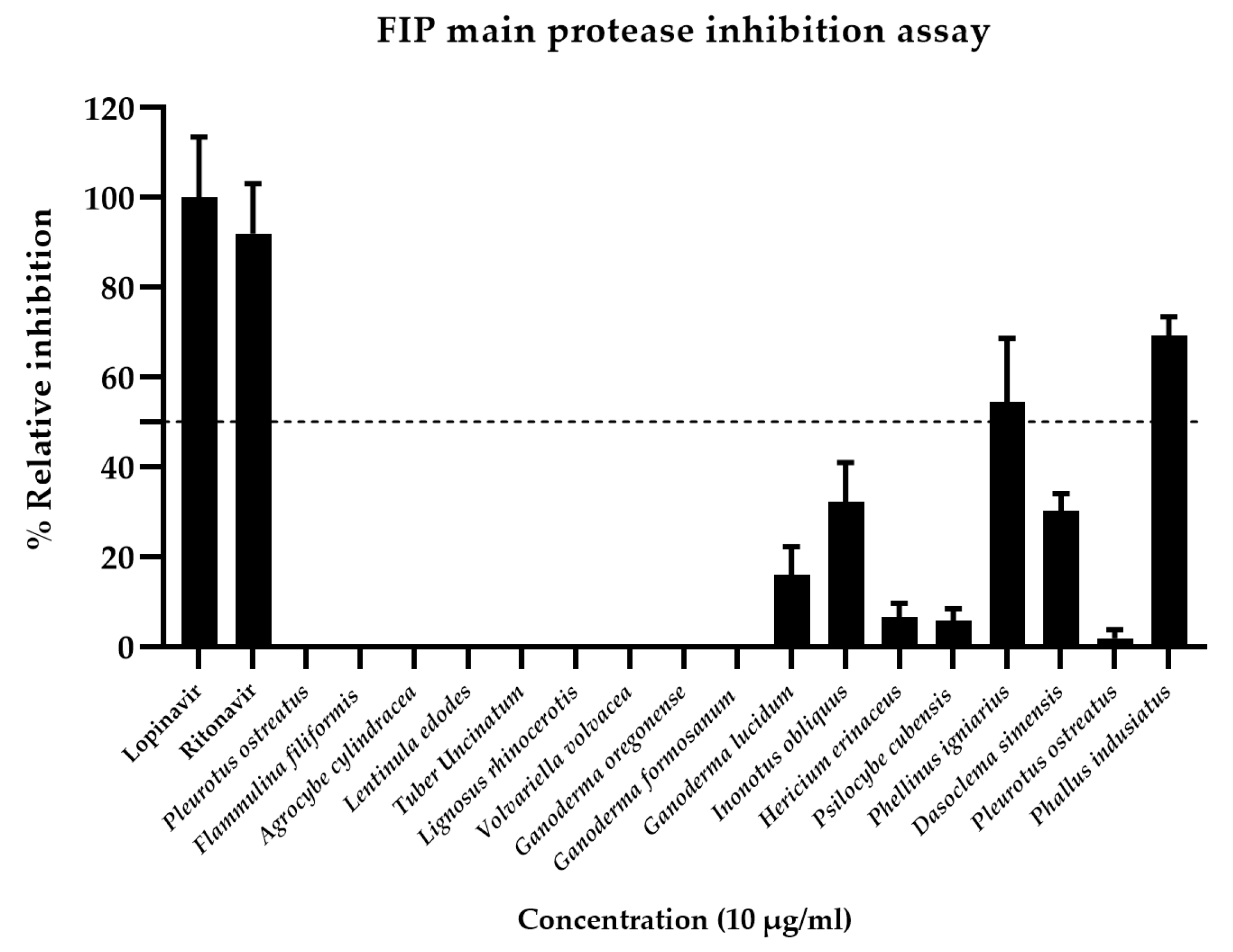
The effect of various inhibitors on FIPV Mpro activity at a concentration of 1 µM was evaluated using protease inhibitors or antiviral drugs, with lopinavir and ritonavir (National Cancer Institute (NCI, USA)) serving as positive controls for comparison with mushroom extracts. Lopinavir and ritonavir were used at final concentrations of 10 and 100 µg/mL to assess enzyme inhibition by monitoring the reaction rate. This was performed by measuring the fluorescence intensity over 30 min. The initial reaction rate (V0) was then used to calculate the percentage of enzyme activity inhibition. At a concentration of 10 µg/mL, lopinavir inhibited enzyme activity by 84.13%, while ritonavir inhibited it by 71.88% (Figure 3).
Relative inhibition of the FIPV Mpro assay was evaluated using crude mushroom extracts. The experimental results showed that P. indusiatus exhibited the highest inhibition of FIPV Mpro activity at 69.2%. Therefore, the extract from P. indusiatus was selected for further testing on its anti-inflammatory effects (Figure 3).

3.3. Optimization of PBMC Culture Conditions Using Whole Blood Samples and FIP Fluid

The characteristics of PBMCs isolated from both whole blood of donor cat and FIP fluid revealed that PBMCs from whole blood contained a small number of platelets and red blood cell contamination (Figure 4A). In contrast, PBMCs isolated from FIP fluid were less pure in terms of white blood cells compared to those from whole blood. The PBMCs sometimes exhibited platelet clumping, which was attributed to the diseased state of the animals. Occasionally, proteins were also present, contributing to the platelet clumping (Figure 4B). Therefore, for the PBMCs isolated from FIP fluid, samples without platelet clumping will be used (Figure 4C).
The experimental results show that at a DMSO concentration of 0.5%, the viability of PBMCs (1 × 10⁶ cells/mL) isolated from whole blood and FIP fluid was 93.75 ± 2.92% and 90.23 ± 2.1%, respectively. This concentration is suitable for further use as a solvent for extracts as it is non-toxic to the cells. However, concentrations of 1%, 1.5%, and 2% were found to be unsuitable as solvents since the viability of PBMCs was below 90%. Therefore, in this study, DMSO at a concentration not exceeding 0.5% was chosen as the solvent for the crude mushroom extract, because PBMCs isolated from both whole blood and FIP fluid had a viability percentage greater than 90% (shown in Figure S1)
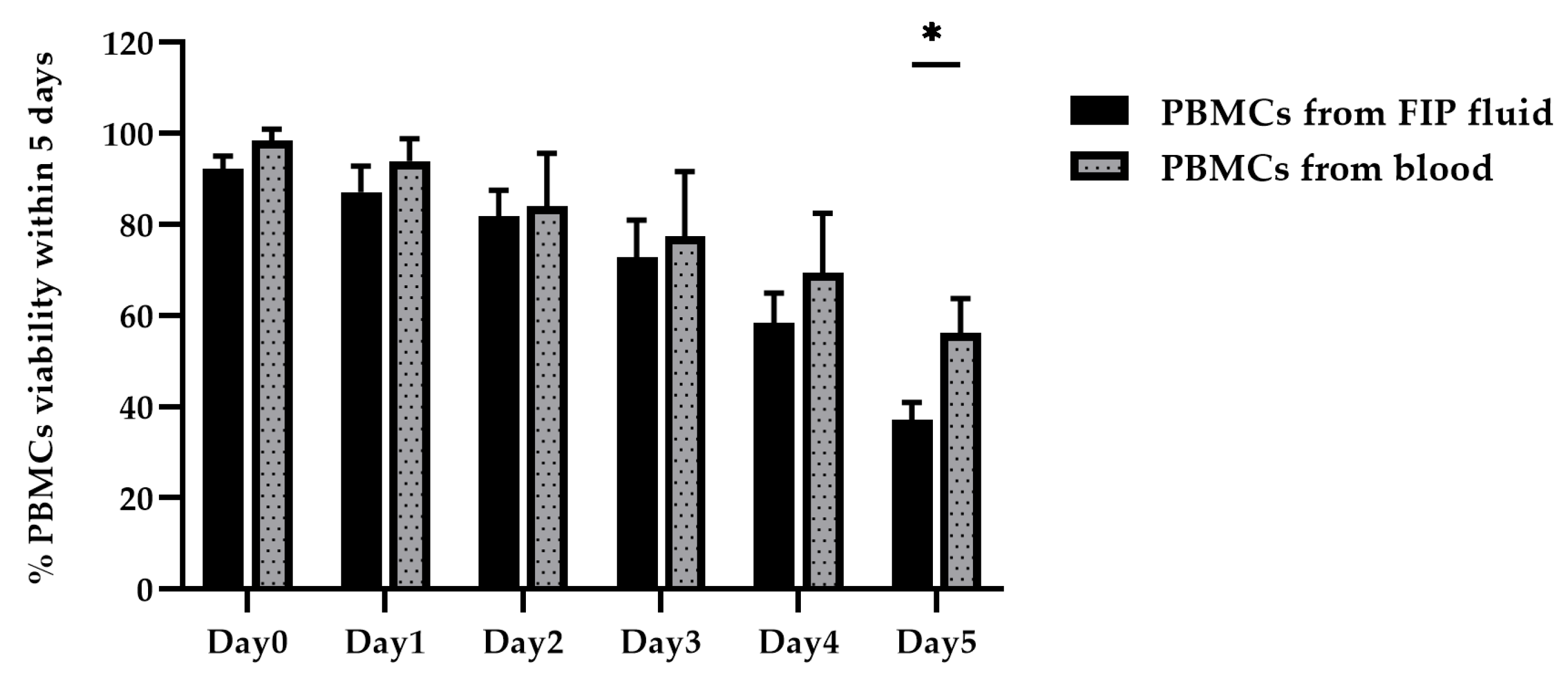
The viability assessment of PBMCs (1 × 10⁶ cells/mL) isolated from whole blood and FIP fluid, conducted using the Trypan Blue Exclusion Assay, revealed that on Day 0, cell viability exceeded 90% for both sources, making them suitable for experiments. By Day 1, PBMCs from whole blood remained suitable for experimentation, while PBMCs from FIP fluid were not, as their viability had dropped below 90%. When comparing each day to Day 0, no statistically significant differences were observed from Day 1 to Day 3, whereas statistically significant differences were observed from Day 4 to Day 5.
A comparison of PBMC viability isolated from both whole blood and FIP fluid was performed using a two-way ANOVA (p < 0.05). The results indicated no statistically significant differences in PBMC viability between the two sample types from Day 0 to Day 4. However, on Day 5, a statistically significant difference in PBMC viability was observed between PBMCs isolated from whole blood and FIP fluid (Figure 5).
However, in this study, PBMCs isolated from samples will be used for testing within 24 h, as they exhibit the highest cell viability, exceeding 90% [22]. This summary confirms that PBMCs isolated from both donor and fluid of FIP retain high viability and are appropriate for subsequent experimental applications.

3.4. LPS-Stimulated PBMCs

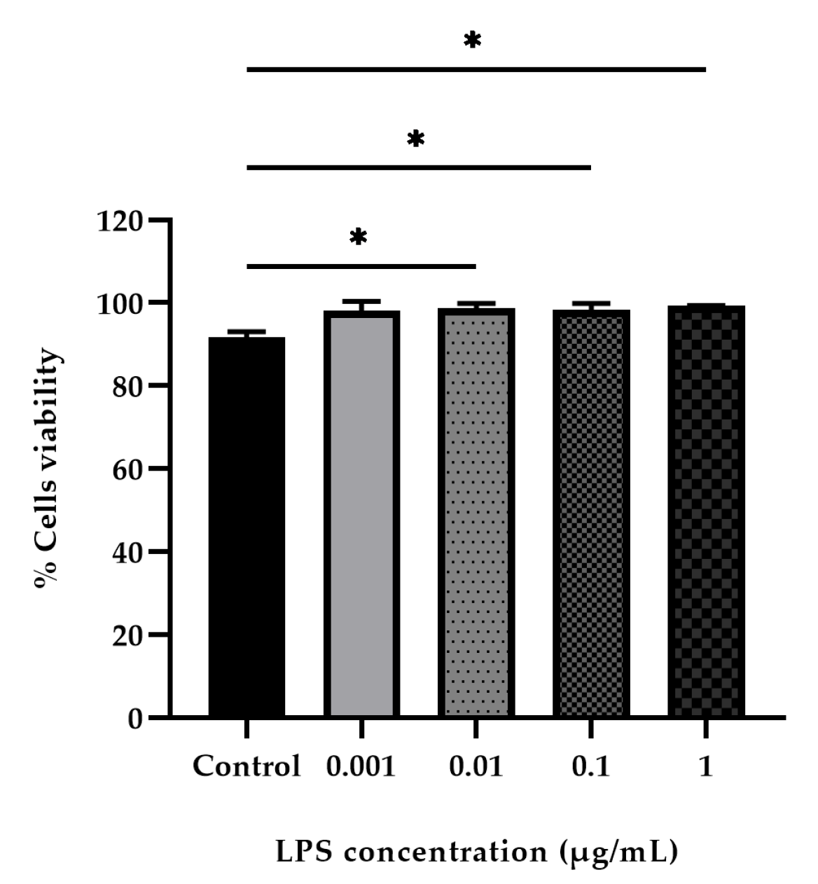
PBMCs isolated from donor cat blood samples were stimulated with LPS at concentrations ranging from 1 to 0.001 µg/mL for 24 h. Cell viability and proliferation were then assessed using the CCK-8 colorimetric assay. The results showed that increasing LPS concentrations significantly enhanced cell viability. In the control group without LPS stimulation, cell viability was 91.60 ± 0.85%, whereas at the highest LPS concentration (1 µg/mL), cell viability increased to 99.21 ± 0.10% (Figure 6).
Subsequently, a comparison of PBMCs viability at each concentration with the control group without LPS addition revealed that at a concentration of 0.001 µg/mL, there was no statistically significant difference compared to the control group. However, at concentrations of 0.01, 0.1, and 1 µg/mL, there were statistically significant differences from the control group (Figure 6). The concentrations of LPS used did not exhibit cytotoxicity, as cell viability exceeded 90% in all cases. Therefore, an LPS concentration of 1 µg/mL was selected to stimulate its effect on inducing cellular inflammation.

3.5. Effect of P. indusiatus on the Cell Viability of PBMCs

The toxicity test of P. indusiatus extract at various concentrations on PBMCs derived from the whole blood of donor cats over 24 h was assessed using the CCK-8 colorimetric assay. The results revealed that at concentrations of 10,000, 5000, 2500, 625, 156, 78, and 39 µg/mL, the extract remained toxic to the cells, as cell viability was below 90%. The PBMCs exhibited morphological changes, such as reduced size and loss of sheen, indicating cell death, which was distinct from the control group. However, at a concentration of 19 µg/mL, the cells retained their normal morphology, showing no changes compared to the control group, which is consistent with the 90% viability results. Consequently, a concentration of 19 µg/mL is suitable for further testing. The IC50 value was calculated to be 1890 µg/mL (as shown in Table S1).

3.6. Evaluation of Nitrite Scavenging Activity of P. indusiatus Extract in LPS-Stimulated PBMCs

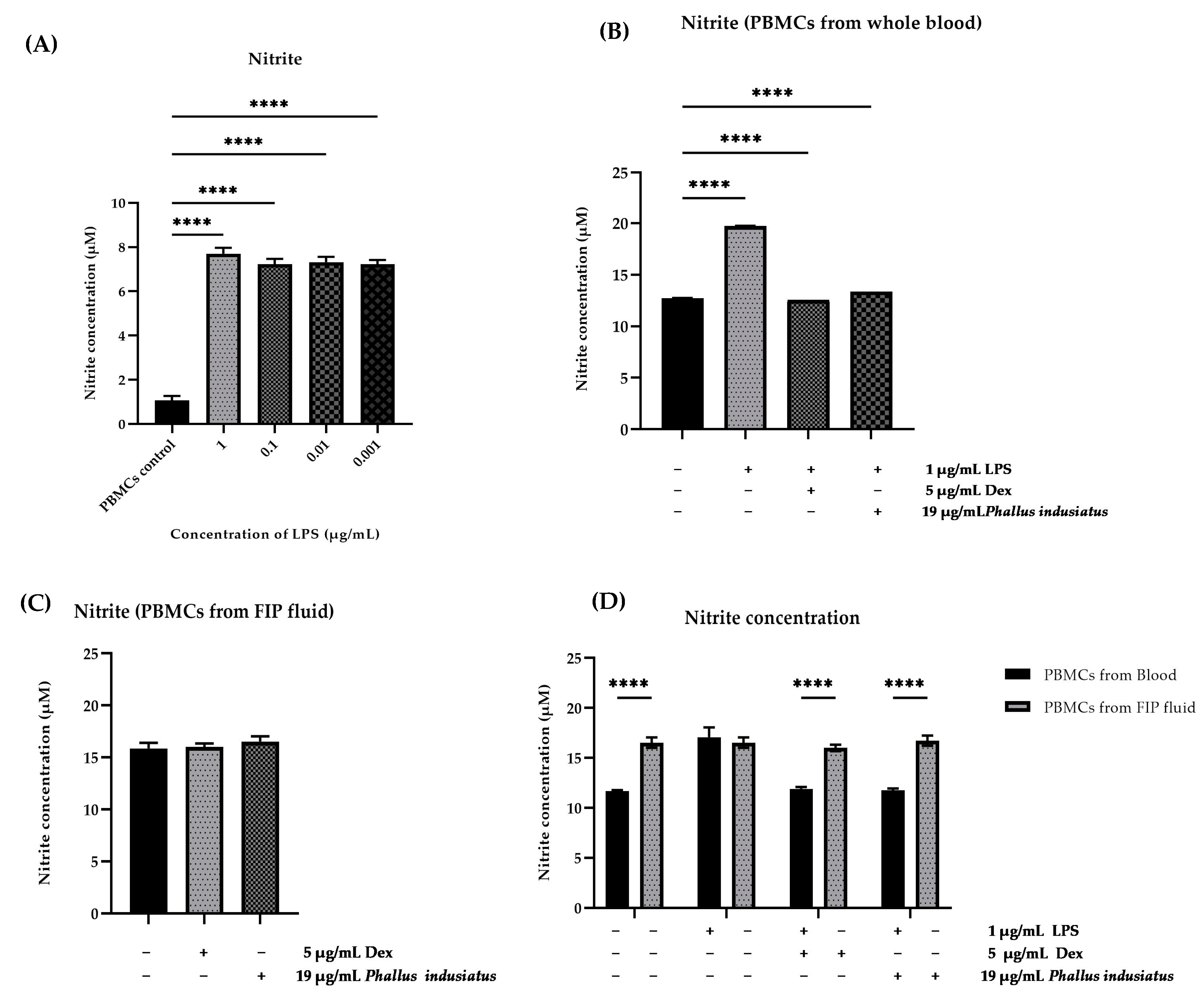
The evaluation of nitrite (NO2) concentration, resulting from the oxidation of nitric oxide (NO), which serves as an indicator for assessing inflammation in LPS-stimulated cells, revealed statistically significant differences compared to the control group at LPS concentrations of 1, 0.1, 0.01, and 0.001 µg/mL (Figure 7A). The nitrite production measured showed a concentration of 7.7 µM, indicating the induction of inflammation in PBMC stimulated with LPS at the respective concentrations. However, considering the viability of PBMCs, it was found that at a concentration of 1 µg/mL, the cells exhibited the highest viability, consistent with the cell viability results assessed using the CCK-8 assay, which showed 99.21 ± 0.01% (Figure 7A). Therefore, an LPS concentration of 1 µg/mL was selected for inducing inflammation in PBMCs.
The test results showed that PBMCs stimulated with 1 µg/mL LPS (negative control) released a nitrite concentration of 19.73 µM. In contrast, the group treated with the anti-inflammatory drug dexamethasone at 5 µg/mL (positive control) exhibited a reduced nitrite concentration of 12.56 µM. Moreover, the group treated with 19 µg/mL of P. indusiatus mushroom extract demonstrated a further reduction in nitrite concentration to 13.36 µM. These results suggest that the P. indusiatus mushroom extract may have potential anti-inflammatory properties (Figure 7B).
The results also showed that PBMCs isolated from FIP fluid exhibited inflammation, as assessed by the nitrite concentration shown in Figure 7C, which measured 15.87 µM without stimulation by LPS. This indicates that the cat with FIP already had inflammation in its PBMCs. In the group treated with the anti-inflammatory drug dexamethasone (positive control), the nitrite concentration was 16.13 µM, which was not statistically different from the control group. Similarly, the test group treated with P. indusiatus extract had a nitrite concentration of 16.52 µM, which also showed no significant difference compared to the control group.
Figure 7D shows a statistically significant difference between PBMCs isolated from blood and those from FIP fluid. PBMCs from FIP fluid exhibited a higher level of cellular inflammation compared to those from blood. However, when comparing PBMCs isolated from blood and stimulated with LPS to PBMCs from FIP fluid, no significant difference was found (Figure 7D). This statistical analysis confirms that PBMCs isolated from FIP fluid are true inflammatory responses. These results indicate that, since there is no significant difference between PBMCs isolated from blood stimulated with LPS and PBMCs from FIP fluid, the latter are naturally inflamed [23]. In the experimental groups treated with dexamethasone (positive control) and the extract of P. indusiatus, a statistically significant difference was observed. PBMCs isolated from blood showed a reduction in nitrite production in both the positive control group and the group treated with P. indusiatus extract. However, PBMCs isolated from FIP fluid did not exhibit a reduction in nitrite production.

3.7. Phytochemical Composition and Literature Support for Anti-Inflammatory Activity
In this study, a crude extract of P. indusiatus was prepared following the extraction protocol described by Theeraraksakul, K. et al., 2023 [21]. Phytochemical analysis confirmed the presence of key anti-inflammatory constituents, including phenolic compounds, flavonoids, and tannins. The extract demonstrated inhibitory effects on the production of nitric oxide (NO), interleukin-1 (IL-1), interleukin-6 (IL-6), and tumor necrosis factor-alpha (TNF-α). Furthermore, their study reported that the crude extract of P. indusiatus also contained various bioactive compounds, such as catechins, flavonoids, polysaccharides, monosaccharides, mucopolysaccharides, allantoin, alkaloids, epicatechin, gallic acid, caffeic acid, and other polyphenolic compounds are also important contributors to the anti-inflammatory activity of the extract [24,25,26]. Although the crude extract of P. indusiatus has not been previously evaluated for its anti-inflammatory effects in cat PBMCs, previous studies have identified bioactive compounds within the extract that possess anti-inflammatory properties. In this study, the observed reduction in NO production in LPS-stimulated PBMCs isolated from healthy cats serves as an important in vitro model (Figure 7B). These findings provide a basis for the future development of anti-inflammatory strategies targeting monocytes and macrophages infected with the FIP virus (Table 1).
Figure 7. The effect of LPS on nitrite production in stimulated PBMCs was quantified after 24 h of exposure to LPS at concentrations of 0.001, 0.01, 0.1, and 1 µg/mL (A). The effect of 19 µg/mL P. indusiatus and 5 µg/mL dexamethasone (Dex) on nitrite production in stimulated PBMCs isolated from whole blood of donor cats was quantified after 24 h. The data are presented as mean ± SEM, comparing the mean of each column with the means of every other column (n = 8) (B). The stimulation and suppression of inflammation in PBMCs isolated from FIP fluid using P. indusiatus extract (n = 5) (C). There was analysis by one-way ANOVA; p < 0.05 reveals statistically significant differences. The difference in response to treatments between PBMCs isolated from whole blood and those from FIP fluid was analyzed using two-way ANOVA (p < 0.05) (n = 4) (D). **** p < 0.0001.
Vetsci 12 00847 g007
Table 1. Pharmacological results of P. indusiatus efficacy from in vitro studies.
PreparationBioactive CompoundExperimental ModelKey FindingsReferences
Crude extract, hot water, and ethanol.Phenolics, flavonoids, and tannins.Murine macrophages used to study the anti-inflammatory effects of LPS stimulation.The anti-inflammatory activity was demonstrated by a reduction in the level of released nitric oxide (NO).Theeraraksakul, K. et al., 2023
[21]
Crude extract, peel and green mixture, core, and whole mushroom.Catechins, flavonoids, polysaccharides, monosaccharides, mucopolysaccharides, allantoin, alkaloids, polyphenolic compounds, epicatechin, gallic acid, and caffeic acid.The experiment was conducted using LPS-induced RAW 264.7 macrophages.The anti-inflammatory activity was demonstrated by the inhibition of NO, IL-1β, IL-6, and TNF-α secretion.Ruksiriwanich, W. et al., 2022.,
Nazir, Y., et al. 2021.,
Sedtananun, S. et al., 2025
[24,25,26]
Hot water extract of crude polysaccharide from mushroom fruiting body.Polysaccharides.Colitis was induced in mice using dextran sulfate sodium (DSS).Reduced the expression of inducible iNOSKanwal, S. et al., 2020
[27]
This study utilized an ethanolic extract derived from the whole mushroom.Phenolics, flavonoids, and tannins. [21]PBMCs from healthy cats were stimulated with LPS and PBMCs from FIP fluid The anti-inflammatory activity was demonstrated by the inhibition of NO in PBMCs isolated from healthy cats.Hlaoperm, C. et al.
Bioactive compounds from P. indusiatus demonstrate biochemical and cellular mechanisms of action, ranging from general antioxidant and anti-inflammatory effects to the modulation of specific signaling pathways [7]. Therefore, based on the findings of this study and prior research, there is compelling evidence that extracts from P. indusiatus possess anti-inflammatory properties and may modulate the immune response, potentially contributing to the inhibition of FIPV in cats.

4. Discussion

In this study, the FIPV Mpro had a size of 34 kDa, which increased to 37.2 kDa after the addition of a 10 × His tag and a TEV cleavage site at the N-terminus. The FIPV Mpro protein was induced by IPTG and analyzed both quantitatively and qualitatively (Figure 2A). Notably, the FIPV Mpro comparable in size to the SARS-CoV-2 Mpro, which has a molecular weight of approximately 33 kDa [28]. These findings indicate that the research was successful at this stage.
The present study revealed a significant finding: the mushroom extract demonstrated FIPV protease inhibition at 69.2%, indicating its potential as a natural source of antiviral compounds. This activity is slightly lower than both ritonavir (71.88%) and lopinavir (84.13%), both of which are established broad-spectrum protease inhibitors clinically used in Human Immunodeficiency Virus (HIV) therapy [29] (Figure 3). These agents have also been shown to inhibit SARS-CoV by strongly binding to its Mpro [30,31]. In our study, they served as general protease inhibitory benchmarks, since they are not used for treating FIP. It is important to note that this protease inhibition mechanism differs from that of currently used FIP treatments such as molnupiravir or GS-441524, which primarily target the viral RNA-dependent RNA polymerase (RdRp), and are therefore not appropriate controls for a protease inhibition assay [32,33]. The absence of GC376, a highly specific FIPV Mpro inhibitor [34,35], was due to its unavailability in our laboratory at the time of the study.
The experimental results revealed that P. indusiatus at a concentration of 10 µg/mL achieved the highest inhibition of FIPV Mpro activity among the tested mushroom extracts, with an inhibition rate of 69.2% (Figure 3). Despite the mushroom extract’s inhibition rate being marginally lower than ritonavir and lopinavir, its efficacy as a natural extract warrants further investigation, suggesting the presence of bioactive molecules with FIPV Mpro inhibitory properties. This finding is particularly relevant because, while current gold standard FIP treatments focus on RdRp, viral proteases (specifically Mpro or 3CLpro) represent an equally critical and well-validated drug target for coronaviruses due to their essential role in processing polyproteins translated from viral RNA [35,36,37]. This enzyme is indispensable for viral polyprotein processing, which is a fundamental step for replication [37,38]. The success of protease inhibitors in other viral infections (e.g., Human HIV and Hepatitis C virus [HCV]), along with the promising results of FIPV-specific protease inhibitors such as GC376, underscores the druggability of this enzyme [39]. Also, due to the high conservation of Mpro during viral mutagenesis and its critical role in the coronavirus life cycle, Mpro is considered as a pivotal target for antivirus drug development [16]
Mushroom extracts are widely recognized for their antiviral and anti-inflammatory properties [40,41]. Previous studies have shown that polysaccharides and adenosine from crude mushroom extract inhibited SARS-CoV protein expression, genomic RNA synthesis, and spike protein activity [42,43,44]. El-Mekkawy, Sahar et al. (1998) have reported that triterpenes extracted from the fruiting bodies of Ganoderma lucidum can inhibit the essential protease enzyme of HIV-1 at a concentration of 7.8 µg/mL [45]. The findings of this study are consistent with previous reports. The crude extract from P. indusiatus is particularly noteworthy for further investigation of its anti-inflammatory properties, as it demonstrated the highest enzyme inhibition among the tested mushroom species and also showed potential for inhibiting the FIPV Mpro. Exploring protease inhibitors from diverse natural sources, such as mushroom extracts, represents a vital strategy—offering novel compounds, alternative mechanisms to combat resistance, and a foundation for future combination therapies. Therefore, in this study, the crude extract from P. indusiatus was selected for testing its anti-inflammatory effects on PBMCs of donor cats and FIP fluid.
The PBMCs isolated from both the whole blood of donor cats and FIP fluid, characterized by monocytes and macrophages, are essential components of the innate immune system [46,47,48]. The selection of samples should prioritize PBMCs that are already isolated, do not clump, and exhibit a round, glossy appearance (Figure 4). The HIV/AIDS Network Coordination Standard Operating Procedure (HANC-SOP) recommends limiting the processing time to a maximum of 8 h [49]. Delays exceeding 24 h have been associated with decreased cell viability [22]. However, in this study, PBMCs were successfully separated from both whole blood and FIP fluid and tested within 24 h (Figure 5).
In the present study, we employed LPS to activate PBMCs as a model of inflammation. LPS, commonly referred to as endotoxin, is a potent pathogen-associated molecular pattern (PAMP) derived from the outer membrane of Gram-negative bacteria. It is recognized primarily by Toll-like receptor 4 (TLR4) expressed on the surface of immune cells, including monocytes/macrophages [50,51]. Engagement of TLR4 by LPS initiates downstream signaling cascades, notably the myeloid differentiation primary response 88 (MyD88)-dependent pathway, culminating in the activation of key transcription factors such as NF-κB and activator protein 1 (AP-1) [9,50,52,53]. Although FIPV is not a bacterial pathogen, viruses similarly trigger innate immune responses by expressing PAMPs, such as double-stranded RNA (dsRNA) and specific viral proteins [54]. These are recognized by pattern recognition receptors (PRRs), including endosomal Toll-like receptors (TLRs), such as TLR3, TLR7, TLR8, and TLR9, as well as cytosolic sensors like retinoic acid-inducible gene I (RIG-I) and melanoma differentiation-associated protein 5 (MDA5) [55,56]. Activation of these receptors initiates similar signaling pathways that converge on the activation of transcription factors NF-κB and AP-1 [10,11] (Figure 1). Although LPS was used as an inflammatory stimulant in this study, mimicking viral infection in feline PBMCs remains challenging due to the lack of a suitable viral culture system in our lab. Nevertheless, both LPS (via TLR4) and viral components (via other TLRs or cytosolic receptors) activate key pro-inflammatory pathways, particularly NF-κB and AP-1. This results in the upregulation of mediators such as TNF-α, IL-1β, IL-6, and iNOS, supporting the use of LPS as a relevant initial model for evaluating general anti-inflammatory effects.
The innate immune system’s ability to initiate responses to LPS was also examined [57]. In this report, 0.001, 0.01, 0.1, and 1 µg/mL of LPS were used to stimulate PBMCs isolated from whole blood of donor cats, compared with the absence of LPS (Figure 6). The data presented in this publication indicate that LPS increased cell viability by approximately 98–99% within 24 h, correlating with the report by Jansky et al. (2003) [58]. A study reported that LPS stimulation increased PBMCs proliferation by activating monocytes and T lymphocytes, thereby promoting their proliferation [59,60,61]. Based on the study results, LPS may have the potential to increase the number of PBMCs [59,60,61]. However, PBMCs isolated from FIP fluid do not exhibit LPS-induced inflammatory responses, likely because virus-infected cats already present with systemic leukocyte activation [23]. This diminished responsiveness may be attributed to the fact that FIPV specifically targets monocytes and macrophages, leading to their functional dysregulation [62,63].
The effect of P. indusiatus on nitrite scavenging activity in LPS-stimulated PBMCs was assessed. During the inflammatory response, nitric oxide production, measured as nitrite levels in the PBMCs’ supernatant, can be upregulated in freshly isolated PBMCs by in vitro stimulation with LPS [64,65,66,67,68]. LPS triggers the TLR4 pathway, leading to the activation of NF-κB and the upregulation of iNOS [50,51]. In LPS-induced samples, monocytes/macrophages are the primary NO producers (Figure 7A) [50,52]. In this study, a group of PBMCs, isolated from donor cat whole blood and stimulated with 1 µg/mL LPS, was evaluated for its anti-inflammatory effects, with a reduction in nitrite serving as an indicator [68]. The crude extract of P. indusiatus may possess potential anti-inflammatory properties (Figure 7B).
The test on the reduction of nitrite production by PBMCs isolated from FIP fluid (Figure 7D) clearly demonstrates that cats suffering from this disease naturally exhibit PBMC inflammation, even in the absence of LPS stimulation. The PRRs, such as TLR3, TLR7, RIG-I, and MDA5, lead to IFN signaling [69]. This may trigger IFN responses (IFN-α, IFN-β, IFN-γ), which can either promote NO production or have other immune effects [54]. The host’s recognition of viral DNA initiates multiple signal transduction pathways that converge on iNOS upregulation, leading to increased endogenous NO production [70]. The oxidative degradation of NO results in the formation of its breakdown products, nitrate and nitrite. This process, primarily identified in macrophages, involves the expression of the inducible iNOS enzyme [71,72]. Consequently, nitrite levels are significantly higher in PBMCs isolated from FIP fluid compared to those from the whole blood of healthy cats, with a statistically significant difference. This aligns with the research conducted by Özbek et al. (2022), which investigated serum nitric oxide levels in cats affected by FIP [73]. This elevation in nitrite serves as a reliable indicator of inflammation (Figure 7D) [74,75].
The results of this study show a statistically significant difference in the response to treatments between PBMCs isolated from whole blood and those from FIP fluid. PBMCs from whole blood responded to both treatments (dexamethasone and P. indusiatus extract) by reducing nitrite production, indicating potential anti-inflammatory effects. However, PBMCs from FIP fluid (possibly from an inflamed environment) did not respond in the same way (Figure 7C,D). The lack of anti-inflammatory effects from both the mushroom extract and dexamethasone in PBMCs from FIP-affected cats underscores the complex and severe nature of FIP-associated inflammation [23,54,62,63,69]. Unlike the acute response induced by LPS, FIP involves chronic, systemic inflammation driven by persistent viral replication in macrophages and a dysregulated immune response, characterized by persistently elevated levels of various inflammatory mediators (a systemic cytokine storm) [52,57,76,77]. In this hyper-inflammatory state, NO production may be maximally upregulated and “locked in”, reducing responsiveness to conventional treatments, including dexamethasone [54,55,56]. This suggests that FIP-related inflammation may involve distinct or more resistant pathways, as well as nitric oxide production through multiple mechanisms, compared to LPS models [10,11,54,56]. Additionally, the direct presence PBMCs from FIP fluid may alter cellular signaling compared to LPS-stimulated healthy PBMCs, potentially affecting drug responsiveness. The previous study suggests that dexamethasone has direct anti-inflammatory effects, acting by altering gene transcription—upregulating anti-inflammatory genes (e.g., IL-10), downregulating pro-inflammatory ones (e.g., IL-6, TNF-α, COX-2, iNOS), and inhibiting the NF-κB pathway [78,79]. In contrast, P. indusiatus modulates immunity and oxidative stress through compounds like β-glucans, which stimulate TNF-α, IL-6, and IL-10 to restore immune balance [7,80]. While dexamethasone suppresses NF-κB, P. indusiatus fine-tunes it to regulate immune responses [81,82].
Limitations of this study include, firstly, the use of LPS as a model of a general inflammatory stimulus instead of direct FIPV infection. Although both can activate similar inflammatory pathways (e.g., NF-κB leading to TNF-α, IL-1β, IL-6, and iNOS expression). FIPV infection is more complex, involving intricate virus–host interactions that LPS cannot fully mimic. Secondly, attempts to develop an in vitro FIPV infection model were unsuccessful. Although efforts were made to establish an FIPV infection model by infecting CRFK (Crandell-Rees Feline Kidney) cells in vitro, these attempts failed due to challenges in isolating the virus from FIP fluid samples, difficulties in quantifying viral load, and the inconsistency of infection. As a result of these limitations, it was not possible to use the actual virus in the study, making it necessary to rely on the LPS-based simulation system, which may not fully reflect the true nature of FIPV infection.
Future investigations may consider employing alternative approaches to better mimic the natural course of FIPV infection. For example, pseudotyped viral systems expressing the FIPV spike protein could provide a safe and controllable platform for examining viral entry and replication, while FIPV-infected cell lines may offer a more physiologically relevant model for studying virus–host interactions [83,84]. Although these models are not without limitations, they could complement the LPS-based stimulation system used in this study and thereby provide a more comprehensive evaluation of the antiviral and anti-inflammatory potential of the tested extracts.
Despite this, this study represents the first preliminary investigation to evaluate extracts from the mushroom P. indusiatus for both anti-FIPV Mpro activity and anti-inflammatory effects. It also represents the first assessment of the extract’s effects on PBMCs isolated from FIP fluid compared to PBMCs from healthy donors. The extract demonstrated strong inhibitory effects on FIPV Mpro. Based on these results, this study is considered successful and has provided important and useful information, opening opportunities for developing new compounds, alternative mechanisms to overcome resistance, and serving as a foundation for future combination therapy research.
Future research should focus on elucidating the roles of pro-inflammatory cytokines (e.g., IL-6, TNF-α) and other relevant inflammatory mediators involved in the pathogenesis of FIP. Bioactive constituents from P. indusiatus should be investigated, and their chemical structures elucidated using advanced analytical techniques (e.g., liquid chromatography–mass spectrometry; LC-MS/MS). Subsequent evaluations should include both in vitro and in vivo studies to determine the pharmacological properties of the isolated compounds. Initial in vitro assays should assess antiviral efficacy and anti-inflammatory activity in relevant cell-based models. Promising compounds should then undergo in vivo validation using appropriate animal models. Additionally, comparative analyses with currently available FIPV-specific therapeutics are recommended to benchmark their relative efficacy and therapeutic potential.

5. Conclusions

This study provides compelling preliminary evidence for the dual antiviral and anti-inflammatory potential of P. indusiatus extract in the context of FIP. The extract showed a strong inhibitory effect (69.2%) on the FIPV Mpro, comparable to established protease inhibitors such as ritonavir and lopinavir. It also significantly reduced NO production in LPS-stimulated PBMCs from healthy cats, indicating potent anti-inflammatory activity. Importantly, the extract was also tested on PBMCs isolated from FIP fluid, which are characterized by chronic inflammation and immune dysregulation. However, these cells showed limited responsiveness (nonresponse) to both dexamethasone and P. indusiatus extract, underscoring the complexity of immune activation in FIP. Although LPS was used as a surrogate inflammatory stimulus and a direct FIPV infection model was not available, this is the first study to evaluate P. indusiatus for both antiviral and anti-inflammatory effects in the context of FIP. These findings highlight the potential of P. indusiatus as a natural source of immunomodulatory agents. Future research should focus on isolating its active compounds, elucidating their mechanisms in relevant infection models, and evaluating their therapeutic potential in vivo, including direct comparisons with FIPV-specific inhibitors.

Supplementary Materials

The following supporting information can be downloaded at https://www.mdpi.com/article/10.3390/vetsci12090847/s1, Figure S1: The appropriate concentration of DMSO for the viability of PBMCs., and Table S1: Optimization of P. indusiatus PBMC viability.

Author Contributions

Conceptualization, C.H., A.P., K.C., O.R., and J.R.; methodology, C.H., W.M., E.W., and W.K.; software, C.H., E.W., and W.K.; validation, C.H., A.P., O.R., and J.R.; formal analysis, C.H., E.W., and W.K.; investigation, C.H., E.W., and W.K.; resources, A.R., A.P., K.C., O.R., and J.R.; data curation, C.H.; writing—original draft preparation, C.H.; writing—review and editing, C.H., W.M., E.W., A.P., K.C., O.R., and J.R.; visualization, C.H., E.W., and W.K.; supervision, A.P., K.C., O.R., and J.R.; project administration, J.R.; funding acquisition, A.P., K.C., and J.R. All authors have read and agreed to the published version of the manuscript.

Funding

This research is funded by the High-Quality Research Graduate Development Cooperation Project between Kasetsart University and the National Science and Technology Development Agency (NSTDA), Kasetsart University Research and Development Institute (KURDI), grant number FF(KU) 7.68 and The Faculty of Veterinary Medicine, Kasetsart University, Bangkok, Thailand.

Institutional Review Board Statement

This study was approved by the Ethics Committee of Kasetsart University under protocol number ACKU67-VET-124 and all cat owners provided written consent.

Informed Consent Statement

Not applicable.

Data Availability Statement

The raw data supporting the conclusions of this article will be made available by the authors on request. Further inquiries can be directed to the corresponding author.

Acknowledgments

I would like to thank Somruedee Sommart, a veterinary nurse at the Kasetsart University Veterinary Teaching Hospital, for collecting whole blood from donors. I am also grateful to the staff the feline clinic, blood bank unit, emergency room, outpatient department (OPD), and Central Lab at the Kasetsart University Veterinary Teaching Hospital, Faculty of Veterinary Medicine, Kasetsart University, Bangkhen Campus for granting access to the data. Additionally, I extend my gratitude to Nuttanit Jirapanth, a veterinary nurse at the Faculty of Veterinary Medicine, Kasetsart University, for her collaboration with The Nine Animal Hospital and M.J. Animal Hospital in screening and selecting FIP fluid samples.

Conflicts of Interest

The authors confirm that they have no competing interests.

References

  1. Hartmann, K. Feline infectious peritonitis. Vet. Clin. N. Am. Small Anim. Pract. 2005, 35, 39–79. [Google Scholar] [CrossRef] [PubMed]
  2. Decaro, N.; Mari, V.; Lanave, G.; Lorusso, E.; Lucente, M.S.; Desario, C.; Colaianni, M.L.; Elia, G.; Ferringo, F.; Alfano, F.; et al. Mutation analysis of the spike protein in Italian feline infectious peritonitis virus and feline enteric coronavirus sequences. Res. Vet. Sci. 2021, 135, 15–19. [Google Scholar] [CrossRef]
  3. Gao, Y.Y.; Wang, Q.; Liang, X.Y.; Zhang, S.; Bao, D.; Zhao, H.; Li, S.B.; Wang, K.; Hu, G.X.; Gao, F.S. An updated review of feline coronavirus: Mind the two biotypes. Virus Res. 2023, 326, 199059. [Google Scholar] [CrossRef]
  4. Xing, L.; Remick, D.G. Relative cytokine and cytokine inhibitor production by mononuclear cells and neutrophils. Shock 2003, 20, 10–16. [Google Scholar] [CrossRef]
  5. Addie, D.; Belák, S.; Boucraut, B.C.; Egberink, H.; Frymus, T.; Gruffydd, J.T.; Hartmann, K.; Hosie, M.J.; Lloret, A.; Lutz, H.; et al. Feline infectious peritonitis: ABCD guidelines on prevention and management. J. Feline Med. Surg. 2009, 11, 594–604. [Google Scholar] [CrossRef]
  6. Saylor, S.S.; Coggins, S.; Barker, E.N.; Gunn-Moore, D.; Jeevaratnam, K.; Norris, J.M.; Hughes, D.; Stacey, E.; MacFarlane, L.; O’Brien, C.; et al. Retrospective study and outcome of 307 cats with feline infectious peritonitis treated with legally sourced veterinary compounded preparations of remdesivir and GS-441524 (2020–2022). J. Feline Med. Surg. 2023, 25, 1098612X231194460. [Google Scholar] [CrossRef]
  7. Habtemariam, S. The chemistry, pharmacology and therapeutic potential of the edible mushroom Dictyophora indusiata (Vent ex. Pers.) Fischer (syn. Phallus indusiatus). Biomedicines 2019, 7, 98. [Google Scholar] [CrossRef]
  8. Suleiman, W.B.; Shehata, R.M.; Younis, A.M. In vitro assessment of multipotential therapeutic importance of Hericium erinaceus mushroom extracts using different solvents. Bioresour. Bioprocess. 2022, 9, 1–13. [Google Scholar] [CrossRef]
  9. Luo, R.; Zhang, Z.; Li, Y. An examination of the LPS–TLR4 immune response through the analysis of molecular structures and protein–protein interactions. Cell Commun. Signal. 2025, 23, 142. [Google Scholar] [CrossRef]
  10. Hennessy, C.; McKernan, D.P. Anti-viral pattern recognition receptors as therapeutic targets. Cells 2021, 10, 2258. [Google Scholar] [CrossRef]
  11. Yu, L.; Gao, F.; Li, Y.; Su, D.; Han, L.; Li, Y.; Zhang, X.; Feng, Z. Role of pattern recognition receptors in the development of MASLD and potential therapeutic applications. Biomed. Pharmacother. 2024, 175, 116724. [Google Scholar] [CrossRef]
  12. Lee, M.S.; Kim, Y.J. Signaling pathways downstream of pattern-recognition receptors and their cross talk. Annu. Rev. Biochem. 2007, 76, 447–480. [Google Scholar] [CrossRef]
  13. Subedi, L.; Lee, J.H.; Yumnam, S.; Ji, E.; Kim, S.Y. Anti-inflammatory effect of sulforaphane on LPS-activated microglia potentially through JNK/AP-1/NF-κB inhibition and Nrf2/HO-1 activation. Cells 2019, 8, 194. [Google Scholar] [CrossRef] [PubMed]
  14. Vuong, W.; Khan, M.B.; Fischer, C.; Arutyunova, E.; Lamer, T.; Shields, J.; Saffran, H.A.; McKay, R.T.; van Belkum, M.J.; Joyce, M.A.; et al. Feline coronavirus drug inhibits the main protease of SARS-CoV-2 and blocks virus replication. Nat. Commun. 2020, 11, 4282. [Google Scholar] [CrossRef] [PubMed]
  15. Citarella, A.; Scala, A.; Piperno, A.; Micale, N. SARS-CoV-2 Mpro: A potential target for peptidomimetics and small-molecule inhibitors. Biomolecules 2021, 11, 607. [Google Scholar] [CrossRef] [PubMed]
  16. Jiang, Z.; Piao, L.; Ren, C.; Zhang, W.; Zhu, Y.; Kong, R. Identifying natural products as feline coronavirus Mpro inhibitors by structure-based virtual screening and enzyme-based assays. ACS Omega 2025, 10, 2092–2101. [Google Scholar] [CrossRef]
  17. Wang, J.; Wang, F.; Tan, Y.; Chen, X.; Zhao, Q.; Fu, S.; Li, S.; Chen, C.; Yang, H. Crystallization and preliminary crystallographic study of feline infectious peritonitis virus main protease in complex with an inhibitor. Acta Crystallogr. Sect. F Struct. Biol. Cryst. Commun. 2014, 70, 1612–1615. [Google Scholar] [CrossRef]
  18. Moyadee, W.; Roytrakul, S.; Jaresitthikunchai, J.; Phaonakrop, N.; Choowongkomon, K.; Ploypetch, S.; Tansakul, N.; Rattanasrisomporn, A.; Rattanasrisomporn, J. Serum proteomic approach to identifying differentially expressed proteins in effusive feline infectious peritonitis. Sci. Rep. 2025, 15, 18899. [Google Scholar] [CrossRef]
  19. Khumtong, K.; Rapichai, W.; Saejung, W.; Khamsingnok, P.; Meecharoen, N.; Ratanabunyong, S.; Dong, H.V.; Tuanthap, S.; Rattanasrisomporn, A.; Choowongkomon, K.; et al. Colorimetric Reverse Transcription Loop-Mediated Isothermal Amplification with Xylenol Orange Targeting Nucleocapsid Gene for Detection of Feline Coronavirus Infection. Viruses 2025, 17, 418. [Google Scholar] [CrossRef]
  20. Kosukwatthana, P.; Rungsuriyawiboon, O.; Rattanasrisomporn, J.; Kimram, K.; Tansakul, N. Cytotoxicity and Immunomodulatory Effects of Cannabidiol on Canine PBMCs: A Study in LPS-Stimulated and Epileptic Dogs. Animals 2024, 14, 3683. [Google Scholar] [CrossRef]
  21. Theeraraksakul, K.; Jaengwang, K.; Choowongkomon, K.; Tabtimmai, L. Exploring the biological functions and anti-melanogenesis of Phallus indusiatus for mushroom-based cosmetic applications. Cosmetics 2023, 10, 121. [Google Scholar] [CrossRef]
  22. Bull, M.; Lee, D.; Stucky, J.; Chiu, Y.L.; Rubin, A.; Horton, H.; McElrath, M.J. Defining blood processing parameters for optimal detection of cryopreserved antigen-specific responses for HIV vaccine trials. J. Immunol. Methods 2007, 322, 57–69. [Google Scholar] [CrossRef]
  23. Bull, M.; Lee, D.; Stucky, J.; Chiu, Y.L.; Rubin, A.; Horton, H.; McElrath, M.J. Study of Macrophage Activity in Cats with FIP and Naturally FCoV-Shedding Healthy Cats. Pathogens 2024, 13, 437. [Google Scholar]
  24. Nazir, Y.; Linsaenkart, P.; Khantham, C.; Chaitep, T.; Jantrawut, P.; Chittasupho, C.; Rachtanapun, P.; Jantanasakulwong, K.; Phimolsiripol, Y.; Sommano, S.R.; et al. High efficiency in vitro wound healing of Dictyophora indusiata extracts via anti-Inflammatory and collagen stimulating (MMP-2 inhibition) mechanisms. J. Fungi. 2021, 7, 1100. [Google Scholar] [CrossRef] [PubMed]
  25. Ruksiriwanich, W.; Khantham, C.; Linsaenkart, P.; Chaitep, T.; Rachtanapun, P.; Jantanasakulwong, K.; Phimolsiripol, Y.; Režek Jambrak, A.; Nazir, Y.; Yooin, W.; et al. Anti-inflammation of bioactive compounds from ethanolic extracts of edible bamboo mushroom (Dictyophora indusiata) as functional health promoting food ingredients. Int. J. Food Sci. Technol. 2022, 57, 110–122. [Google Scholar] [CrossRef]
  26. Sedtananun, S.; Sujiwattanarat, P.; Hoang Tra, M.H.; Suphakun, P.; Seetaha, S.; Auetragul, A.; Choowongkomon, K.; Tabtimmai, L. Phallus indusiatus Extracts Promoted MCF-7 Apoptosis Under TNFα-induced Tumor Microenvironment by Attenuating NF-kappaB and Akt Activation. Asian Pac. J. Cancer Prev. 2025, 26, 479. [Google Scholar] [CrossRef]
  27. Kanwal, S.; Joseph, T.P.; Aliya, S.; Song, S.; Saleem, M.Z.; Nisar, M.A.; Wang, Y.; Meyiah, A.; Ma, Y.; Xin, Y. Attenuation of DSS induced colitis by Dictyophora indusiata polysaccharide (DIP) via modulation of gut microbiota and inflammatory related signaling pathways. J. Funct. Foods 2020, 64, 103641. [Google Scholar] [CrossRef]
  28. Rong, Y.; Zhang, C.; Gao, W.C.; Zhao, C. Optimization of the expression of the main protease from SARS-CoV-2. Protein Expr. Purif. 2023, 203, 106208. [Google Scholar] [CrossRef]
  29. Ortiz, R.; Dejesus, E.; Khanlou, H.; Voronin, E.; van Lunzen, J.; Andrade-Villanueva, J.; Fourie, J.; De Meyer, S.; De Pauw, M.; Lefebvre, E.; et al. Efficacy and safety of once-daily darunavir/ritonavir versus lopinavir/ritonavir in treatment-naive HIV-1-infected patients at week 48. AIDS 2008, 22, 1389–1397. [Google Scholar] [CrossRef]
  30. Schoergenhofer, C.; Jilma, B.; Stimpfl, T.; Karolyi, M.; Zoufaly, A. Pharmacokinetics of lopinavir and ritonavir in patients hospitalized with coronavirus disease 2019 (COVID-19). Ann. Intern. Med. 2020, 173, 670–672. [Google Scholar] [CrossRef]
  31. Nutho, B.; Mahalapbutr, P.; Hengphasatporn, K.; Pattaranggoon, N.C.; Simanon, N.; Shigeta, Y.; Hannongbua, S.; Rungrotmongkol, T. Why are lopinavir and ritonavir effective against the newly emerged coronavirus 2019? Atomistic insights into the inhibitory mechanisms. Biochemistry 2020, 59, 1769–1779. [Google Scholar]
  32. Tsika, A.C.; Gallo, A.; Fourkiotis, N.K.; Argyriou, A.I.; Sreeramulu, S.; Löhr, F.; Rogov, V.V.; Richter, C.; Linhard, V.; Gande, S.L.; et al. Binding adaptation of GS-441524 diversifies Macro domains and downregulates SARS-CoV-2 de-MARylation capacity. J. Mol. Biol. 2022, 434, 167720. [Google Scholar] [CrossRef]
  33. Wang, Z.; Yang, L.; Song, X.Q. Oral GS-441524 derivatives: Next-generation inhibitors of SARS-CoV-2 RNA-dependent RNA polymerase. Front. Immunol. 2022, 13, 1015355. [Google Scholar] [CrossRef]
  34. Perera, K.D.; Rathnayake, A.D.; Liu, H.; Pedersen, N.C.; Groutas, W.C.; Chang, K.O.; Kim, Y. Characterization of amino acid substitutions in feline coronavirus 3C-like protease from a cat with feline infectious peritonitis treated with a protease inhibitor. Vet. Microbiol. 2019, 237, 108398. [Google Scholar] [CrossRef] [PubMed]
  35. Pedersen, N.C.; Kim, Y.; Liu, H.; Galasiti Kankanamalage, A.C.; Eckstrand, C.; Groutas, W.C.; Bannasch, M.; Meadows, J.M.; Chang, K.O. Efficacy of a 3C-like protease inhibitor in treating various forms of acquired feline infectious peritonitis. J. Feline Med. Surg. 2018, 20, 378–392. [Google Scholar] [CrossRef] [PubMed]
  36. Xiong, M.; Su, H.; Zhao, W.; Xie, H.; Shao, Q.; Xu, Y. What coronavirus 3C-like protease tells us: From structure, substrate selectivity, to inhibitor design. Med. Res. Rev. 2021, 41, 1965–1998. [Google Scholar] [CrossRef] [PubMed]
  37. Zhu, W.; Shyr, Z.; Lo, D.C.; Zheng, W. Viral proteases as targets for coronavirus disease 2019 drug development. J. Pharmacol. Exp. Ther. 2021, 378, 166–172. [Google Scholar] [CrossRef]
  38. Abian, O.; Ortega-Alarcon, D.; Jimenez-Alesanco, A.; Ceballos-Laita, L.; Vega, S.; Reyburn, H.T.; Rizzuti, B.; Velazquez-Campoy, A. Structural stability of SARS-CoV-2 3CLpro and identification of quercetin as an inhibitor by experimental screening. Int. J. Biol. Macromol. 2020, 164, 1693–1703. [Google Scholar] [CrossRef]
  39. Majerová, T.; Konvalinka, J. Viral proteases as therapeutic targets. Mol. Aspects Med. 2022, 88, 101159. [Google Scholar] [CrossRef]
  40. Varghese, R.; Efferth, T.; Ramamoorthy, S. Exploring the anti-COVID-19 potential of mushroom metabolites: Current status and perspectives. In Traditional Medicines and Natural Products as Preventive and Therapeutic Agents Against COVID-19; Academic Press: Cambridge, MA, USA, 2025; pp. 317–337. [Google Scholar]
  41. Chun, S.; Gopal, J.; Muthu, M. Antioxidant activity of mushroom extracts/polysaccharides—Their antiviral properties and plausible anti-COVID-19 properties. Antioxidants 2021, 10, 1899. [Google Scholar] [CrossRef]
  42. Zhang, Y.; Zhang, G.; Ling, J. Medicinal fungi with antiviral effect. Molecules 2022, 27, 4457. [Google Scholar] [CrossRef] [PubMed]
  43. Verma, A.K. Cordycepin: A bioactive metabolite of Cordyceps militaris and polyadenylation inhibitor with therapeutic potential against Covid-19. J. Biomol. Struct. Dyn. 2020, 40, 3745–3752. [Google Scholar] [CrossRef] [PubMed]
  44. Zhao, C.; Gao, L.; Wang, C.; Liu, B.; Jin, Y.; Xing, Z. Structural characterization and antiviral activity of a novel heteropolysaccharide isolated from Grifola frondosa against enterovirus 71. Carbohydr Polym. 2016, 144, 382–389. [Google Scholar] [CrossRef] [PubMed]
  45. El-Mekkawy, S.; Meselhy, M.R.; Nakamura, N.; Tezuka, Y.; Hattori, M.; Kakiuchi, N.; Shimotohno, K.; Kawahata, T.; Otake, T. Anti-HIV-1 and anti-HIV-1-protease substances from Ganoderma lucidum. Phytochemistry 1998, 49, 1651–1657. [Google Scholar] [CrossRef]
  46. Bienzle, D.; Reggeti, F.; Clark, M.E.; Chow, C. Immunophenotype and functional properties of feline dendritic cells derived from blood and bone marrow. Vet. Immunol. Immunopathol. 2003, 96, 19–30. [Google Scholar] [CrossRef]
  47. Parihar, A.; Eubank, T.D.; Doseff, A.I. Monocytes and macrophages regulate immunity through dynamic networks of survival and cell death. J. Innate Immun. 2010, 2, 204–215. [Google Scholar] [CrossRef]
  48. Lopes, R.; Sampaio, F.; Carvalho, H.L.; Garcês, A.; Fernandes, C.; Neves, C.V.; Brito, A.S.; Marques, T.; Sousa, C.; Silva, A.R.; et al. Feline Infectious Peritonitis Effusion Index: A Novel Diagnostic Method and Validation of Flow Cytometry-Based Delta Total Nucleated Cells Analysis on the Sysmex XN-1000V®. Vet. Sci. 2024, 11, 563. [Google Scholar] [CrossRef]
  49. Browne, D.J.; Miller, C.M.; Doolan, D.L. Technical pitfalls when collecting, cryopreserving, thawing, and stimulating human T-cells. Front. Immunol. 2024, 15, 1382192. [Google Scholar] [CrossRef]
  50. Xu, Y.; Jagannath, C.; Liu, X.D.; Sharafkhaneh, A.; Kolodziejska, K.E.; Eissa, N.T. Toll-like receptor 4 is a sensor for autophagy associated with innate immunity. Immunity 2007, 27, 135–144. [Google Scholar] [CrossRef]
  51. Oshio, T.; Kawashima, R.; Kawamura, Y.I.; Hagiwara, T.; Mizutani, N.; Okada, T.; Otsubo, T.; Inagaki-Ohara, K.; Matsukawa, A.; Haga, T.; et al. Chemokine receptor CCR8 is required for lipopolysaccharide-triggered cytokine production in mouse peritoneal macrophages. PLoS ONE 2014, 9, e94445. [Google Scholar] [CrossRef]
  52. Tucureanu, M.M.; Rebleanu, D.; Constantinescu, C.A.; Deleanu, M.; Voicu, G.; Butoi, E.; Calin, M.; Manduteanu, I. Lipopolysaccharide-induced inflammation in monocytes/macrophages is blocked by liposomal delivery of Gi-protein inhibitor. Int. J. Nanomed. 2018, 13, 63–76. [Google Scholar] [CrossRef]
  53. Chen, H.; Wang, F.; Mao, H.; Yan, X. Degraded λ-carrageenan activates NF-κB and AP-1 pathways in macrophages and enhances LPS-induced TNF-α secretion through AP-1. Biochim. Biophys. Acta 2014, 1840, 2162–2170. [Google Scholar] [CrossRef]
  54. Thompson, M.R.; Kaminski, J.J.; Kurt-Jones, E.A.; Fitzgerald, K.A. Pattern recognition receptors and the innate immune response to viral infection. Viruses 2011, 3, 920. [Google Scholar] [CrossRef]
  55. Kolli, D.; Velayutham, T.S.; Casola, A. Host-viral interactions: Role of pattern recognition receptors (PRRs) in human pneumovirus infections. Pathogens 2013, 2, 232–263. [Google Scholar] [CrossRef] [PubMed]
  56. Okamoto, M.; Tsukamoto, H.; Kouwaki, T.; Seya, T.; Oshiumi, H. Recognition of viral RNA by pattern recognition receptors in the induction of innate immunity and excessive inflammation during respiratory viral infections. Viral Immunol. 2017, 30, 408–420. [Google Scholar] [CrossRef] [PubMed]
  57. Ngkelo, A.; Meja, K.; Yeadon, M.; Adcock, I.; Kirkham, P.A. LPS induced inflammatory responses in human peripheral blood mononuclear cells is mediated through NOX4 and G i α dependent PI-3kinase signalling. J. Inflamm. 2012, 9, 1–7. [Google Scholar] [CrossRef]
  58. Jansky, L.; Reymanova, P.; Kopecky, J. Dynamics of cytokine production in human peripheral blood mononuclear cells stimulated by LPS, or infected by Borrelia. Physiol. Res. 2003, 52, 593–598. [Google Scholar] [CrossRef]
  59. Lin, Z.; Huang, Y.; Jiang, H.; Zhang, D.; Yang, Y.; Geng, X.; Li, Y. Functional differences and similarities in activated peripheral blood mononuclear cells by lipopolysaccharide or phytohemagglutinin stimulation between human and cynomolgus monkeys. Ann. Transl. Med. 2021, 9, 257. [Google Scholar] [CrossRef]
  60. Kubin, M.; Kamoun, M.; Trinchieri, G. Interleukin 12 synergizes with B7/CD28 interaction in inducing efficient proliferation and cytokine production of human T cells. J. Exp. Med. 1994, 180, 211–222. [Google Scholar] [CrossRef]
  61. Ulmer, A.J.; Flad, H.D.; Rietschel, T.; Mattern, T. Induction of proliferation and cytokine production in human T lymphocytes by lipopolysaccharide (LPS). Toxicology 2000, 152, 37–45. [Google Scholar] [CrossRef]
  62. Malbon, A.J.; Michalopoulou, E.; Meli, M.L.; Barker, E.N.; Tasker, S.; Baptiste, K.; Kipar, A. Colony stimulating factors in early feline infectious peritonitis virus infection of monocytes and in end stage feline infectious peritonitis; a combined in vivo and in vitro approach. Pathogens 2020, 9, 893. [Google Scholar] [CrossRef]
  63. Cornelissen, E.; Dewerchin, H.L.; Van Hamme, E.; Nauwynck, H.J. Absence of surface expression of feline infectious peritonitis virus (FIPV) antigens on infected cells isolated from cats with FIP. Vet. Microbiol. 2007, 121, 131–137. [Google Scholar] [CrossRef] [PubMed]
  64. Hirano, T.; Matsuda, T.; Turner, M.; Miyasaka, N.; Buchan, G.; Tang, B.; Sato, K.; Shimi, M.; Maid, R.; Feldmann, M.; et al. Excessive production of interleukin 6/B cell stimulatory factor-2 in rheumatoid arthritis. Eur. J. Immunol. 1988, 18, 1797–1802. [Google Scholar] [CrossRef] [PubMed]
  65. Bergroth, V.; Zvaifler, N.J.; Firestein, G.S. Cytokines in chronic inflammatory arthritis. III. Rheumatoid arthritis monocytes are not unusually sensitive to γ-interferon, but have defective γ-interferon–mediated HLA–DQ and HLA–DR induction. Arthritis Rheum. 1989, 32, 1074–1079. [Google Scholar] [CrossRef] [PubMed]
  66. Firestein, G.S.; Alvaro-Gracia, J.M.; Maki, R. Quantitative analysis of cytokine gene expression in rheumatoid arthritis. J. Immunol. 1990, 144, 3347–3353. [Google Scholar] [CrossRef]
  67. Sakurai, H.; Kohsaka, H.; Liu, M.F.; Higashiyama, H.; Hirata, Y.; Kanno, K.; Saito, I.; Miyasaka, N. Nitric oxide production and inducible nitric oxide synthase expression in inflammatory arthritides. J. Clin. Investig. 1995, 96, 2357–2363. [Google Scholar] [CrossRef]
  68. Davies, C.A.; Rocks, S.A.; O’Shaughnessy, M.C.; Perrett, D.; Winyard, P.G. Analysis of nitrite and nitrate in the study of inflammation. In Inflammation Protocols, Methods in Molecular Biology; Humana Press: Totowa, NJ, USA, 2003; pp. 305–320. [Google Scholar]
  69. Capozza, P.; Pratelli, A.; Camero, M.; Lanave, G.; Greco, G.; Pellegrini, F.; Tempesta, M. Feline coronavirus and alpha-herpesvirus infections: Innate immune response and immune escape mechanisms. Animals 2021, 11, 3548. [Google Scholar] [CrossRef]
  70. Garren, M.R.; Ashcraft, M.; Qian, Y.; Douglass, M.; Brisbois, E.J.; Handa, H. Nitric oxide and viral infection: Recent developments in antiviral therapies and platforms. Appl. Mater. Today 2021, 22, 100887. [Google Scholar] [CrossRef]
  71. Omar, M.; Abdelal, H.O. Nitric oxide in parasitic infections: A friend or foe? J. Parasit. Dis. 2022, 46, 1147–1163. [Google Scholar] [CrossRef]
  72. Olyslaegers, D.A.; Dedeurwaerder, A.; Desmarets, L.M.; Vermeulen, B.L.; Dewerchin, H.L.; Nauwynck, H.J. Altered expression of adhesion molecules on peripheral blood leukocytes in feline infectious peritonitis. Vet. Microbiol. 2013, 166, 438–449. [Google Scholar] [CrossRef]
  73. Özbek, M.; Özkan, C.; Kaya, A.; Yıldırım, S.; Kozat, S.; Akgül, Y. Clinicopathological and biochemical evaluation of Feline Infectious Peritonitis in Turkish Van cats. J. Hell. Vet. Med. Soc. 2022, 73, 4379–4388. [Google Scholar] [CrossRef]
  74. Waltz, P.; Escobar, D.; Botero, A.M.; Zuckerbraun, B.S. Nitrate/nitrite as critical mediators to limit oxidative injury and inflammation. Antioxid. Redox Signal. 2015, 23, 328–339. [Google Scholar] [CrossRef] [PubMed]
  75. Gao, X.Q.; Fei, F.; Huo, H.H.; Huang, B.; Meng, X.S.; Zhang, T.; Liu, B.L. Impact of nitrite exposure on plasma biochemical parameters and immune-related responses in Takifugu rubripes. Aquat. Toxicol. 2020, 218, 105362. [Google Scholar] [CrossRef] [PubMed]
  76. Sherding, R.G. Feline infectious peritonitis (feline coronavirus). J. Small Anim. Pract. 2009, 50, 132–143. [Google Scholar]
  77. Malbon, A.J.; Fonfara, S.; Meli, M.L.; Hahn, S.; Egberink, H.; Kipar, A. Feline infectious peritonitis as a systemic inflammatory disease: Contribution of liver and heart to the pathogenesis. Viruses 2019, 11, 1144. [Google Scholar] [CrossRef]
  78. Zappia, C.D.; Torralba-Agu, V.; Echeverria, E.; Fitzsimons, C.P.; Fernández, N.; Monczor, F. Antihistamines potentiate dexamethasone anti-inflammatory effects. Impact on glucocorticoid receptor-mediated expression of inflammation-related genes. Cells 2021, 10, 3026. [Google Scholar]
  79. Jiang, Z.; Zhu, L. Update on molecular mechanisms of corticosteroid resistance in chronic obstructive pulmonary disease. Pulm. Pharmacol. Ther. 2016, 37, 1–8. [Google Scholar] [CrossRef]
  80. Yadav, D.; Negi, P.S. Role of mushroom polysaccharides in improving gut health and associated diseases. In Microbiome, Immunity, Digestive Health and Nutrition; Academic Press: London, UK; Elsevier: Amsterdam, The Netherlands, 2022; pp. 431–448. [Google Scholar]
  81. Thrikawala, S.U.; Anderson, M.H.; Rosowski, E.E. Glucocorticoids Suppress NF-κB–Mediated Neutrophil Control of Aspergillus fumigatus Hyphal Growth. J. Immunol. 2024, 213, 971–987. [Google Scholar] [CrossRef]
  82. Kumar, Y.; Xu, B. New Insights into Chemical Profiles and Health-Promoting Effects of Edible Mushroom Dictyophora indusiata (Vent ex. Pers.) Fischer: A Review. J. Fungi. 2025, 11, 75. [Google Scholar] [CrossRef]
  83. Liu, S.; Zhang, L.; Fu, W.; Liang, Z.; Yu, Y.; Li, T.; Tong, J.; Liu, F.; Nie, J.; Lu, Q.; et al. Optimization and validation of a virus-like particle pseudotyped virus neutralization assay for SARS-CoV-2. MedComm 2024, 5, e615. [Google Scholar] [CrossRef]
  84. Luo, H.; Lv, L.; Yi, J.; Zhou, Y.; Liu, C. Establishment of replication deficient vesicular stomatitis virus for studies of PEDV spike-mediated cell entry and its inhibition. Microorganisms 2023, 11, 2075. [Google Scholar] [CrossRef]
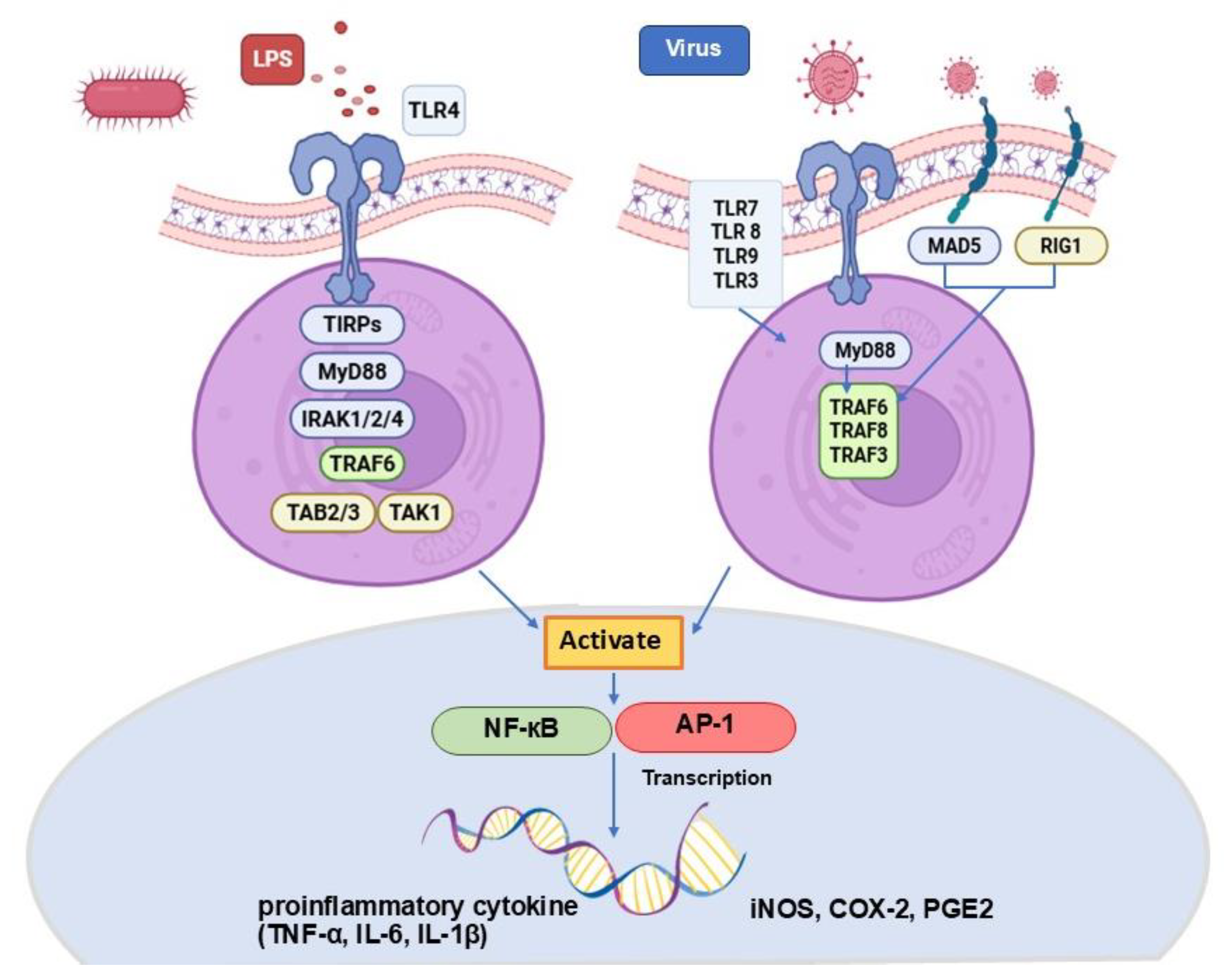
Figure 1. This figure illustrates the activation of TLR–NF-κB and AP-1 signaling pathways in response to LPS or viral stimulation, resulting in the upregulation of inducible iNOS, COX-2, PGE2, and pro-inflammatory cytokines. This schematic was adapted from Luo R. et al., 2025, Yu, L. et al., 2024, and Hennessy & McKernan., 2021 [9,10,11].
Vetsci 12 00847 g001
Figure 2. Protein purification was performed using a Ni2+ affinity column, with protein expression induced by 0.4 mM IPTG. The FIPV Mpro was detected at 37.2 kDa on an SDS-PAGE gel. lane1: Marker, lane2: Pellet FIP, lane3: Supernatant FIP, lane4: Flowthrough, lane5: Wash A, lane6: 20% Buffer B, lane7: 40% Buffer B, lane 8–9: 60% Buffer B, lane10–11: 80% Buffer B, lane12–14: 100% Buffer B (A). Western blot analysis confirmed the presence of the FIPV Mpro protein at 37.20 kDa (B). The reaction rates of the FIPV Mpro enzyme (Relative Fluorescence Units/sec) at enzyme concentrations of 1 and 0.5 µM (C).
Vetsci 12 00847 g002
Figure 3. The relationship between the percentage inhibition of FIPV Mpro (1 µM) and crude mushroom extracts at a concentration of 10 µg/mL was evaluated. The inhibition of FIPV Mpro enzyme activity was evaluated at a concentration of 1 µM. At a concentration of 10 µg/mL, lopinavir inhibited enzyme activity by 84.13%, while ritonavir inhibited it by 71.88%. P. indusiatus exhibited the highest inhibition of FIPV Mpro activity at 69.2%. The dotted line represents 50% relative inhibition.
Vetsci 12 00847 g003
Figure 4. Morphological studies by inverted microscope at actual magnification 50 µm on PBMCs. Normal PBMCs without clumping can be observed when isolated from whole blood (A). PBMCs isolated from the fluid showed signs of platelet clumping (B). PBMCs separated from FIP fluid without clumping are shown in (C).
Vetsci 12 00847 g004
Figure 5. The PBMC viability was analyzed by comparing PBMCs from whole blood collection and FIP fluid samples using a two-way ANOVA. The data are presented as mean ± SEM for PBMCs viability, comparing each cell mean with the other cell means in the same row (n = 5). * p < 0.05, PBMCs from FIP fluid vs. PBMCs from blood.
Vetsci 12 00847 g005
Figure 6. Testing the appropriate concentration of LPS compared to the control group without adding LPS, using concentrations of 0.001, 0.01, 0.1, and 1 µg/mL for 24 h, which were analyzed by one-way ANOVA; * p < 0.05 revealed statistically significant differences (n = 3).
Vetsci 12 00847 g006
Disclaimer/Publisher’s Note: The statements, opinions and data contained in all publications are solely those of the individual author(s) and contributor(s) and not of MDPI and/or the editor(s). MDPI and/or the editor(s) disclaim responsibility for any injury to people or property resulting from any ideas, methods, instructions or products referred to in the content.