Preoperative and Postoperative Ozone Therapy in Cats Presenting Extensive Wounds Treated by Reconstructive Surgery Methods—A Short Case Series
Simple Summary
Abstract
1. Introduction
2. Description of Cases
2.1. Case 1
2.2. Case 2
2.3. Case 3
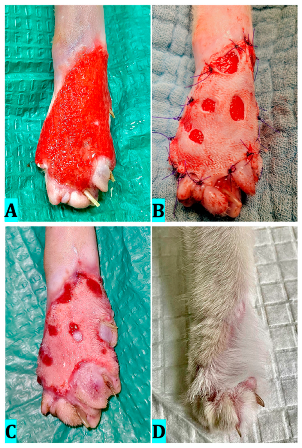
2.4. Case 4
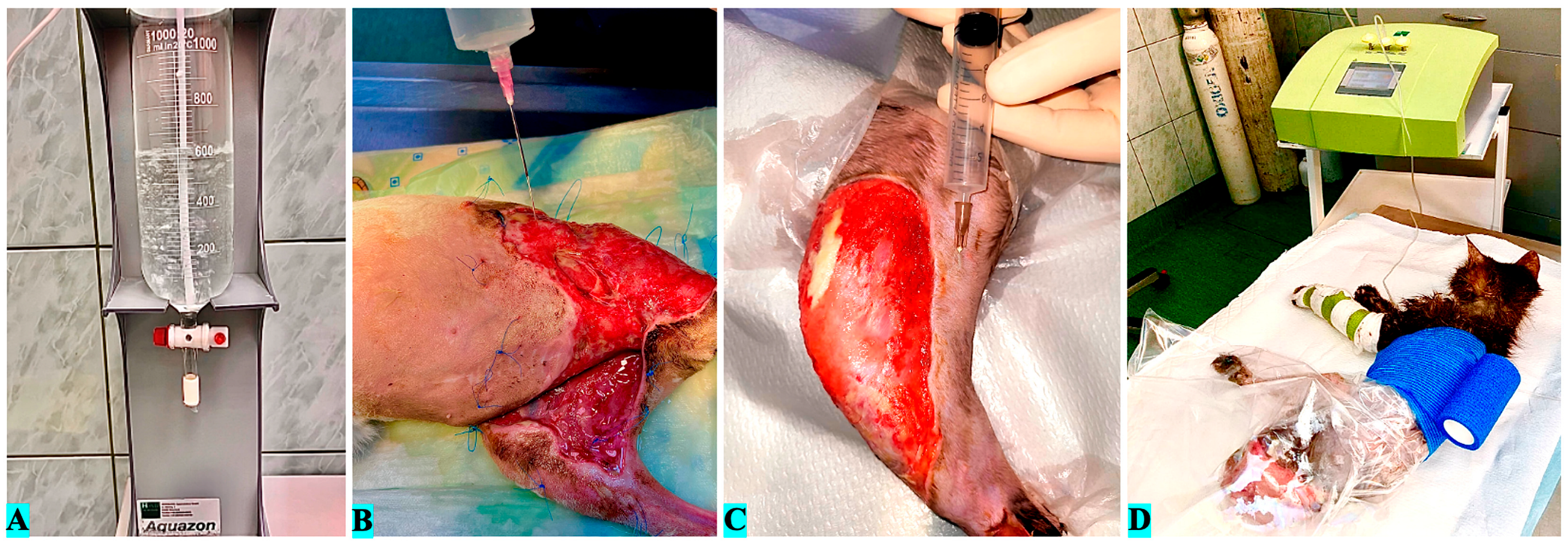
3. Ozone Therapy
4. Surgical Procedure and Results
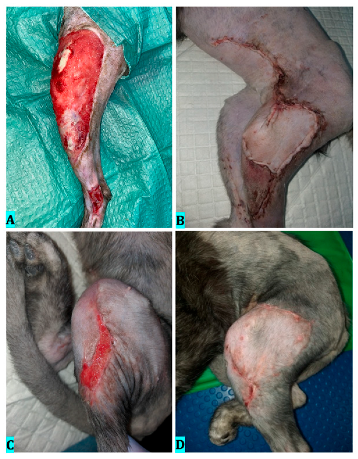
4.1. Case 1
4.2. Case 2
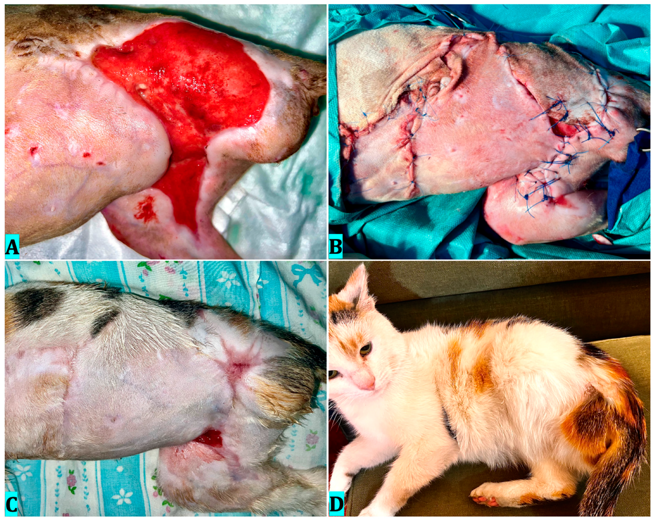
4.3. Case 3
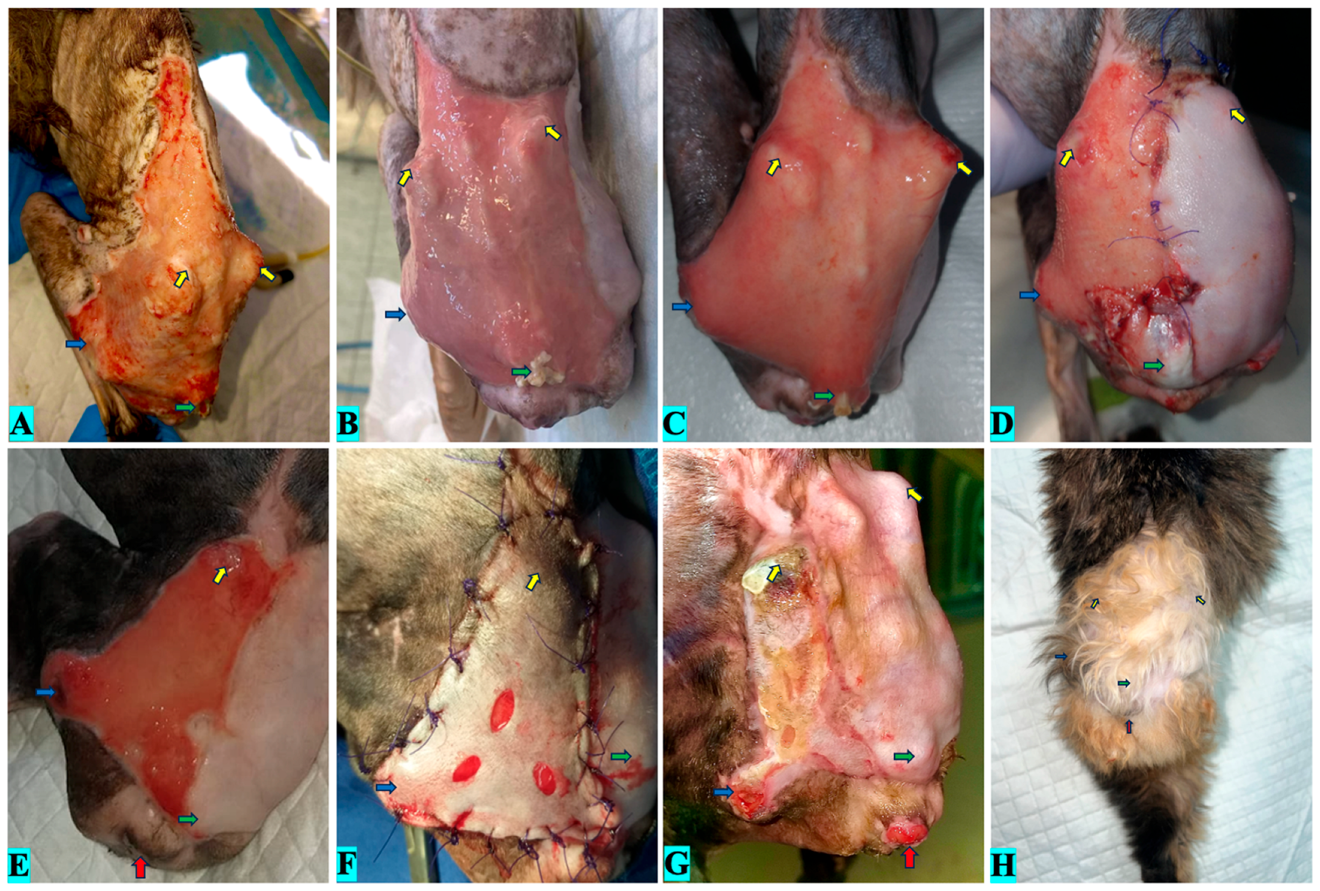
4.4. Case 4
5. Discussion
6. Conclusions
Author Contributions
Funding
Institutional Review Board Statement
Informed Consent Statement
Data Availability Statement
Acknowledgments
Conflicts of Interest
References
- Sorg, H.; Tilkorn, D.J.; Hager, S.; Hauser, J.; Mirastschijski, U. Skin Wound Healing: An Update on the Current Knowledge and Concepts. Eur. Surg. Res. 2016, 58, 81–94. [Google Scholar] [CrossRef] [PubMed]
- Corr, S. Intensive, Extensive, Expensive: Management of Distal Limb Shearing Injuries in Cats. J. Feline Med. Surg. 2009, 11, 747–757. [Google Scholar] [CrossRef]
- Martínez-Sánchez, G.; Definitions of terms in ozone therapy. International Scientific Committee of Ozone Therapy. 2019. Available online: https://isco3.org/wp-content/uploads/2015/09/ISCO3-QAU-00-04-Definitios-and-terms-in-O3-Final.pdf (accessed on 5 February 2024).
- WFOT’s Review on Evidence Based Ozone Therapy. World Federation of Ozone Therapy—WFOT. 2015. Available online: https://www.wfoot.org/libray/WFOT-OZONE-2015-ENG.pdf (accessed on 5 February 2024).
- Bocci, V.; Di Paolo, N. Oxygen-Ozone Therapy in Medicine: An Update. Blood Purif. 2009, 28, 373–376. [Google Scholar] [CrossRef]
- Bocci, V.A. Scientific and Medical Aspects of Ozone Therapy. State of the Art. Arch. Med. Res. 2006, 37, 425–435. [Google Scholar] [CrossRef]
- Kim, H.S.; Noh, U.S.; Inn, Y.W.; Kim, K.M.; Kang, H.; Kim, H.O.; Park, Y.M. Therapeutic Effects of Topical Application of Ozone on Acute Cutaneous Wound Healing. J. Korean Med. Sci. 2009, 24, 368. [Google Scholar] [CrossRef]
- Valacchi, G.; Lim, Y.; Belmonte, G.; Miracco, C.; Zanardi, I.; Bocci, V.; Travagli, V. Ozonated Sesame Oil Enhances Cutaneous Wound Healing in SKH1 Mice. Wound Repair Regen. 2010, 19, 107–115. [Google Scholar] [CrossRef]
- Travagli, V.; Zanardi, I.; Valacchi, G.; Bocci, V. Ozone and Ozonated Oils in Skin Diseases: A Review. Mediat. Inflamm. 2010, 2010, 610418. [Google Scholar] [CrossRef]
- Sagai, M.; Bocci, V. Mechanisms of Action Involved in Ozone Therapy: Is Healing Induced via a Mild Oxidative Stress? Med. Gas Res. 2011, 1, 29. [Google Scholar] [CrossRef]
- Martínez-Sánchez, G.; Al-Dalain, S.M.; Menéndez, S.; Re, L.; Giuliani, A.; Candelario-Jalil, E.; Álvarez, H.; Fernández-Montequín, J.I.; León, O.S. Therapeutic Efficacy of Ozone in Patients with Diabetic Foot. Eur. J. Pharmacol. 2005, 523, 151–161. [Google Scholar] [CrossRef]
- Bohling, M.W.; Henderson, R.A.; Swaim, S.F.; Kincaid, S.A.; Wright, J.C. Cutaneous Wound Healing in the Cat: A Macroscopic Description and Comparison with Cutaneous Wound Healing in the Dog. Vet. Surg. 2004, 33, 579–587. [Google Scholar] [CrossRef] [PubMed]
- Schwartz, A.; Martínez-Sánchez, G.; Sabbah, F.; Hernández Avilés, M. Madrid Declaration on Ozone Therapy, 3rd ed.; International Scientific Committee of Ozone Therapy: Madrid, Spain, 2020; Available online: https://ozonewithoutborders.ngo/wp-content/uploads/2021/04/2020-Madrid-Declaration.pdf (accessed on 5 February 2024).
- Orlandin, J.R.; Machado, L.C.; Ambrósio, C.E.; Travagli, V. Ozone and its derivatives in veterinary medicine: A careful appraisal. Vet. Anim. Sci. 2021, 13, 100191. [Google Scholar] [CrossRef]
- Degli Agosti, I.; Ginelli, E.; Mazzacane, B.; Peroni, G.; Bianco, S.; Guerriero, F.; Ricevuti, G.; Perna, S.; Rondanelli, M. Effectiveness of a Short-Term Treatment of Oxygen-Ozone Therapy into Healing in a Posttraumatic Wound. Case Rep. Med. 2016, 2016, 9528572. [Google Scholar] [CrossRef]
- Gajendrareddy, P.K.; Sen, C.K.; Horan, M.P.; Marucha, P.T. Hyperbaric Oxygen Therapy Ameliorates Stress-Impaired Dermal Wound Healing. Brain Behav. Immun. 2005, 19, 217–222. [Google Scholar] [CrossRef]
- Re, L.; Sanchez, G.M.; Mawsouf, N. Clinical Evidence of Ozone Interaction with Pain Mediators. Saudi Med. J. 2010, 31, 1363–1367. [Google Scholar] [PubMed]
- Fuccio, C.; Luongo, C.; Capodanno, P.; Giordano, C.; Scafuro, M.; Siniscalco, D.; Lettieri, B.; Rossi, F.; Maione, S.; Berrino, L. A Single Subcutaneous Injection of Ozone Prevents Allodynia and Decreases the Over-Expression of Pro-Inflammatory Caspases in the Orbito-Frontal Cortex of Neuropathic Mice. Eur. J. Pharmacol. 2009, 603, 42–49. [Google Scholar] [CrossRef] [PubMed]
- Amsellem, P. Complications of Reconstructive Surgery in Companion Animals. Vet. Clin. N. Am. Small Anim. Pract. 2011, 41, 995–1006. [Google Scholar] [CrossRef]
- Elvis, A.M.; Ekta, J.S. Ozone Therapy: A Clinical Review. J. Nat. Sci. Biol. Med. 2011, 2, 66–70. [Google Scholar] [CrossRef]
- Bocci, V.; Zanardi, I.; Huijberts, M.S.P.; Travagli, V. Diabetes and Chronic Oxidative Stress. A perspective based on the possible use of ozone therapy. Diabetes Metab. Syndr. Clin. Res. Rev. 2011, 5, 45–49. [Google Scholar] [CrossRef]
- Roy, D.; Wong, P.K.; Engelbrecht, R.S.; Chian, E.S. Mechanism of Enteroviral Inactivation by Ozone. Appl. Environ. Microbiol. 1981, 41, 718–723. [Google Scholar] [CrossRef] [PubMed]
- Zeng, J.; Lu, J. Mechanisms of Action Involved in Ozone-Therapy in Skin Diseases. Int. Immunopharmacol. 2018, 56, 235–241. [Google Scholar] [CrossRef]
- Swaim, S.F. Skin Grafts. Vet. Clin. N. Am. Small Anim. Pract. 1990, 20, 147–175. [Google Scholar] [CrossRef]
- Oros, N.-V.; Repciuc, C.; Ober, C.; Mihai, M.; Oana, L.-I. Combined Oxygen-Ozone Therapy for Mesh Skin Graft in a Cat with a Hindlimb Extensive Wound. Animals 2023, 13, 513. [Google Scholar] [CrossRef]
- Sun, H.; Heng, H.; Liu, X.; Geng, H.; Liang, J. Evaluation of the Healing Potential of Short-Term Ozone Therapy for the Treatment of Diabetic Foot Ulcers. Front. Endocrinol. 2023, 14, 1304034. [Google Scholar] [CrossRef] [PubMed]
- Sen, C.K.; Roy, S. Redox Signals in Wound Healing. Biochim. Biophys. Acta-Gen. Subj. 2008, 1780, 1348–1361. [Google Scholar] [CrossRef]
- Kırkıl, C.; Yiğit, M.V.; Özercan, İ.H.; Aygen, E.; Gültürk, B.; Artaş, G. The Effect of Ozonated Olive Oil on Neovascularizatıon in an Experimental Skin Flap Model. Adv. Ski. Wound Care 2016, 29, 322–327. [Google Scholar] [CrossRef] [PubMed]
- Valacchi, G.; Bocci, V. Studies on the Biological Effects of Ozone: 10. Release of Factors from Ozonated Human Platelets. Mediat. Inflamm. 1999, 8, 205–209. [Google Scholar] [CrossRef] [PubMed]
- Oros, N.V.; Repciuc, C.C.; Ober, C.; Peștean, C.; Mircean, M.V.; Oana, L.I. Clinical Evaluation of Medical Ozone Use in Domestic Feline Cutaneous Wounds—A Short Case Series. Animals 2023, 13, 2796. [Google Scholar] [CrossRef]
- Hosgood, G. Wound repair and specific tissue response to injury. In Textbook of Small Animal Surgery, 3rd ed.; WB Saunders: Philadelphia, PA, USA, 2003; pp. 66–86. [Google Scholar]
- Smith, N.L.; Wilson, A.L.; Gandhi, J.; Vatsia, S.; Khan, S.A. Ozone therapy in veterinary medicine: A review. Vet. Sci. 2020, 7, 13. [Google Scholar] [CrossRef]
- Ziegler, G.B.; Ziegler, E.; Witzenhausen, R. [Vital Fluorescent Staining of Microorganisms by 3′,6′-Diacetyl-Fluoresceine for Determination of Their Metabolic Activity (Author’s Transl)]. Zentralblatt Bakteriol. Parasitenkd. Infekt. Hygiene. Erste Abt. Originale. Reihe A Med. Mikrobiol. Parasitol. 1975, 230, 252–264. [Google Scholar]
- Thanomsub, B.; Anupunpisit, V.; Chanphetch, S.; Watcharachaipong, T.; Poonkhum, R.; Srisukonth, C. Effects of Ozone Treatment on Cell Growth and Ultrastructural Changes in Bacteria. J. Gen. Appl. Microbiol. 2002, 48, 193–199. [Google Scholar] [CrossRef]
- Silva, R.A.; Garotti, J.E.R.; Silva, R.S.B.; Navarins, A.; Pacheco, A.M., Jr. Analysis of the bactericidal effect of ozone pneumoperitoneum. Acta Cir. Bras. 2009, 24, 124–127. [Google Scholar] [CrossRef] [PubMed]
- Fontes, B.; Cattani Heimbecker, A.M.; de Souza Brito, G.; Costa, S.F.; van der Heijden, I.M.; Levin, A.S.; Rasslan, S. Effect of Low-Dose Gaseous Ozone on Pathogenic Bacteria. BMC Infect. Dis. 2012, 12, 358. [Google Scholar] [CrossRef]
- Romary, D.J.; Landsberger, S.A.; Bradner, K.N.; Ramirez, M.; Leon, B.R. Liquid Ozone Therapies for the Treatment of Epithelial Wounds: A Systematic Review and Meta-Analysis. Int. Wound J. 2023, 20, 1235–1252. [Google Scholar] [CrossRef]
- Pchepiorka, R.; Moreira, M.S.; Lascane, N.A.d.S.; Catalani, L.H.; Allegrini, S., Jr.; de Lima, N.B.; Gonçalves, F. Effect of Ozone Therapy on Wound Healing in the Buccal Mucosa of Rats. Arch. Oral Biol. 2020, 119, 104889. [Google Scholar] [CrossRef]
- Valacchi, G.; Sticozzi, C.; Zanardi, I.; Belmonte, G.; Cervellati, F.; Bocci, V.; Travagli, V. Ozone Mediators Effect on “in Vitro” Scratch Wound Closure. Free Radic. Res. 2016, 50, 1022–1031. [Google Scholar] [CrossRef] [PubMed]
- Viebahn-Haensler, R.; León Fernández, O.S. Ozone in Medicine. The Low-Dose Ozone Concept and Its Basic Biochemical Mechanisms of Action in Chronic Inflammatory Diseases. Int. J. Mol. Sci. 2021, 22, 7890. [Google Scholar] [CrossRef] [PubMed]

| Case | Etiology | Affected Area | Bacteria Involved | Wound Area | Method of Ozonetherapy Before Recontructive Surgery | Days of Persecundam Management | Type of Recontructive Surgery | Method of Ozonetherapy After Recontructive Surgery | Total Time to Healing (Days) |
|---|---|---|---|---|---|---|---|---|---|
| 1 | Traumatic origin | -left hind limb | Staphylococcus spp. | 96.7 cm2 | - bagging conc. 20–60 μg/mL for 5–20 min - perilesional infiltration with 15 μg/mL - lavage with ozonized saline solution at 60 μg/mL | 15 | Superficial caudal epigastric artery | Bagging | 60 |
| 2 | Post-amputation dehiscent wound | -left flank -sacral region, -groin -right hind limb | Pseudomonas spp. | 163.2 cm2 | - bagging conc. 20–60 μg/mL for 5–20 min - perilesional infiltration with 15 μg/mL - lavage with ozonized saline solution at 60 μg/mL | 20 | Local advancing skin flaps | Bagging | 90 |
| 3 | Unknow | -lumbosacral area | Escherichia coli, Proteus spp., and Pseudomonas aeruginosa | 64.8 cm2 | - bagging conc. 20–60 μg/mL for 5–20 min - perilesional infiltration with 15 μg/mL - lavage with ozonized saline solution at 60 μg/mL | 31 | Local advancing skin flaps and Free skin graft | Bagging | 90 |
| 4 | Car accident | -right forelimb’s dorsal metacarpal-phalangeal | Negative | 34 cm2 | - bagging conc. 20–60 μg/mL for 5–20 min - lavage with ozonized saline solution at 60 μg/mL | 20 | Free skin graft | Bagging | 35 |
| Patient | Stage | Timepoint | Score (0–5) | Approx. Coverage (%) | Wound Clinical State |
|---|---|---|---|---|---|
| 1 | Initial presentation | day 0 | 0 | 0% | Large open wound with mostly necrotic superficial tissue, no epithelial margin. |
| Mid-ozone therapy | day 15 | 1 | ≈15% | Visible epithelial migration from edges; central wound still granulating. | |
| Postoperative | day 29 | 4 | ≈95% | Near-complete coverage, small peripheral granulating areas. | |
| Final follow-up | day 60 | 5 | 100% | Complete epithelial coverage with hair regrowth. | |
| 2 | Initial presentation | day 0 | 0 | 0% | Large open wound with isles of necrotic tissue, discrete epithelial margin and granulation. |
| Mid-ozone therapy | day 20 | 2 | ≈40% | Well-developed granulation tissue with early epithelial advance from periphery. | |
| Postoperative | day 50 | 4 | ≈90% | Majority of surface covered with reduced flap retraction areas remain. | |
| Final follow-up | day 90 | 5 | 100% | Fully epithelialized, mature tissue, visible hair regrowth. | |
| 3 | Initial presentation | day 0 | 0 | 0% | Open wound with isles of necrotic tissue, discrete granulation. |
| Mid-ozone therapy | day 30 | 2 | ≈45% | Epithelial migration from margins more evident, granulation still prominent. | |
| Post 1st graft | day 33 | 3 | ≈65% | Epithelial migration from margins evident, granulation still prominent | |
| Post 2nd graft | day 65 | 4 | ≈95% | Almost complete epithelialization; few epithelializing isles remain | |
| Final follow-up | day 90 | 5 | 100% | Fully epithelialized, mature tissue with hair regrowth | |
| 4 | Initial presentation | day 0 | 0 | 0% | Extensive open wound with necrotic margins, no epithelial tissue. |
| Mid-ozone therapy | day 20 | 1 | ≈25% | Early epithelial growth at wound margins, predominantly granulation. | |
| Post-graft | day 29 | 4 | ≈90% | Majority of wound epithelialized; small residual granulating areas. | |
| Final follow-up | day 60 | 5 | 100% | Full epithelial coverage, hair regrowth present. |
Disclaimer/Publisher’s Note: The statements, opinions and data contained in all publications are solely those of the individual author(s) and contributor(s) and not of MDPI and/or the editor(s). MDPI and/or the editor(s) disclaim responsibility for any injury to people or property resulting from any ideas, methods, instructions or products referred to in the content. |
© 2025 by the authors. Licensee MDPI, Basel, Switzerland. This article is an open access article distributed under the terms and conditions of the Creative Commons Attribution (CC BY) license (https://creativecommons.org/licenses/by/4.0/).
Share and Cite
Oros, N.V.; Repciuc, C.C.; Bel, L.V.; Melega, I.; Pertea, A.N.; Oana, L.I. Preoperative and Postoperative Ozone Therapy in Cats Presenting Extensive Wounds Treated by Reconstructive Surgery Methods—A Short Case Series. Vet. Sci. 2025, 12, 786. https://doi.org/10.3390/vetsci12080786
Oros NV, Repciuc CC, Bel LV, Melega I, Pertea AN, Oana LI. Preoperative and Postoperative Ozone Therapy in Cats Presenting Extensive Wounds Treated by Reconstructive Surgery Methods—A Short Case Series. Veterinary Sciences. 2025; 12(8):786. https://doi.org/10.3390/vetsci12080786
Chicago/Turabian StyleOros, Nicușor Valentin, Călin Cosmin Repciuc, Lucia Victoria Bel, Iulia Melega, Andreea Niculina Pertea, and Liviu Ioan Oana. 2025. "Preoperative and Postoperative Ozone Therapy in Cats Presenting Extensive Wounds Treated by Reconstructive Surgery Methods—A Short Case Series" Veterinary Sciences 12, no. 8: 786. https://doi.org/10.3390/vetsci12080786
APA StyleOros, N. V., Repciuc, C. C., Bel, L. V., Melega, I., Pertea, A. N., & Oana, L. I. (2025). Preoperative and Postoperative Ozone Therapy in Cats Presenting Extensive Wounds Treated by Reconstructive Surgery Methods—A Short Case Series. Veterinary Sciences, 12(8), 786. https://doi.org/10.3390/vetsci12080786


