Article

Molecular Epidemiology of Brucella spp. in Aborted Livestock in the Ningxia Hui Autonomous Region, China

1
College of Veterinary Medicine, Gansu Agricultural University, Lanzhou 730070, China
2
Ningxia Hui Autonomous Region Center for Animal Disease Control and Prevention, Yinchuan 750011, China
3
Ningxia Hui Autonomous Region Center for Disease Control and Prevention, Yinchuan 750001, China
4
School of Agriculture, Ningxia University, Yinchuan 750021, China
*
Authors to whom correspondence should be addressed.
Vet. Sci. 2025, 12(8), 702; https://doi.org/10.3390/vetsci12080702
Submission received: 28 May 2025 / Revised: 9 July 2025 / Accepted: 23 July 2025 / Published: 28 July 2025

Simple Summary

Brucellosis is a bacterial infection caused by the Brucella species that leads to a series of symptoms ranging from fever to abortion both in livestock and humans. It severely impacts the development of the livestock industry and public health. The human Brucellosis epidemic in Ningxia has gradually worsened in recent years. To reduce the impact of this epidemic and the effect of Brucellosis on livestock, the “Ningxia Brucellosis Prevention and Control Special Three-Year Action Implementation Plan (2022–2024)” was implemented by the government of Ningxia. The plan includes information regarding the surveillance, epidemiological investigations, and ongoing compulsory immunization of newborn livestock. As a part of the surveillance and epidemiological investigation, this study comprises the first systematic study on Brucella spp. from the aborted tissues of livestock in Ningxia, China. In this cross-sectional study, which was conducted in Ningxia from 2022 to 2023, we reported that 8.68% of the aborted tissue samples of livestock were identified as being positive for Brucella spp. This indicates a high risk of transmission from the aborted livestock to other animals and humans. Therefore, comprehensive disinfection and safe treatment should be implemented after the abortion of livestock to interrupt the transmission pathway in order to prevent the further infection of livestock and humans.

Abstract

Brucellosis is caused by Brucella spp.; it can result in fetal loss and abortion, resulting in economic losses and negative effects on human health. Herein, a cross-sectional study on the epidemiology of Brucella spp. in aborted livestock in Ningxia from 2022 to 2023 was conducted. A total of 749 aborted tissue samples from 215 cattle and 534 sheep were collected from farmers who reported abortions that were supported by veterinarians trained in biosecurity. The samples were analyzed using qPCR and were cultured for Brucella spp. when a positive result was obtained; the samples were speciated using AMOS-PCR. MLST and MLVA were employed for genotype identification. The results demonstrated that 8.68% of the samples were identified as being positive for Brucella spp. based on qPCR results. In total, 14 field strains of Brucella spp. were subsequently isolated, resulting in 11 B. melitensis, 2 B. abortus, and 1 B. suis. being identified via AMOS-PCR. Four sequence types were identified via MLST—ST7 and ST8 (B. melitensis), ST2 (B. abortus), and ST14 (B. suis)—with ST8 predominating. Five MLVA-8 genotypes and seven MLVA-11 genotypes were identified, with MLVA-11 GT116 predominating in livestock. Thus, at least three Brucella species are circulating in aborted livestock in Ningxia. This suggests a significant risk of transmission to other animals and humans. Therefore, disinfection and safe treatment procedures for aborted livestock and their products should be carried out to interrupt the transmission pathway; aborted livestock should be examined to determine zoonotic causes and targeted surveillance should be strengthened to improve the early detection of infectious causes, which will be of benefit to the breeding industry and public health security.

1. Introduction

Abortion represents a serious issue in pregnant animals, leading to significant financial challenges, especially in developing nations [1]. This phenomenon not only limits production but also severely impacts food production and the agricultural industry. Furthermore, potential zoonotic pathogens related to abortion may pose serious threats to public health. Identifying the causal agent responsible for an abortion event is significant for effective disease control and management in order to address the public health threats that are associated with zoonotic diseases, in addition to preventing further outbreaks [2].
Brucellosis is a highly prevalent zoonotic disease worldwide. Globally, the annual number of infections in humans is approximately 2.1 million [3]. This systemic zoonotic infection is manifested as a series of symptoms ranging from fever to miscarriage. Various species within the Brucella genus are responsible for Brucellosis, which results in substantial economic losses and considerable public health challenges [4]. It is a major contributor to infectious abortion in livestock across various nations [5] and presents a grave occupational risk for individuals such as slaughterhouse workers, farmers, shepherds or traders, veterinarians, and lab technicians [6].
Brucella spp. live within cells, serving as abortive agents in various animal species, particularly affecting cattle, goats, sheep, swine, and dogs [7]. The bacteria can enter hosts through ingestion, the inhalation of air or dust, or through the conjunctiva or damaged skin [8]. The Brucella genus comprises several species, with B. abortus, B. melitensis, and B. suis being found most frequently in domesticated animals [9]. B. melitensis is responsible for the majority of Brucellosis cases in sheep and goats and can cause abortions in infected pregnant animals [10]. Human forms of Brucellosis are most commonly contracted from cattle, sheep, goats, camels, and pigs [11]. Typically, humans contract the infection via direct or indirect exposure to infected animals or contaminated materials, such as tissues from aborted livestock [12]. Numerous zoonotic species of Brucella are obtained from animals that have experienced abortions.
Brucellosis is commonly found in China, particularly in the northern regions, where the local economy relies heavily on ruminant animals [13], and B. melitensis ST8 is the most common species, with it being widespread in 1990–2010 [14]. The Ningxia Hui Autonomous Region (104°17′–107°39′ E, 35°14′–39°23′ N) in northwest China has a dry climate and sufficient daylight. It has a long history in relation to the development of the livestock industry. By the end of 2022, the number of dairy cow, beef cattle, and Tan sheep herds in the region reached 0.837 million, 1.484 million, and 7.105 million, respectively (http://nxccpit.nx.gov.cn/xwzx/zzqxx/202304/t20230426_4047068.html (accessed on 4 May 2025)). Due to the rapid expansion of animal farming and frequent animal movement, the prevalence of bovine Brucellosis in the area was 2.46% and that of sheep was 3.85% in 2022. The human Brucellosis epidemic has gradually worsened, with the incidence increasing from 0.441/100,000 in 2005 to 86.83/100,000 in 2022 in Ningxia [15].
In order to systematically investigate the presence of Brucellosis in aborted livestock in Ningxia, China, and analyze the genetic characteristics of the isolated Brucella spp., bioinformatics techniques such as Multilocus Sequence Typing (MLST) and multiple-locus variable-number tandem repeat analysis (MLVA) genotyping were employed to perform analyses. In this study, we aim to clarify its molecular evolution and improve prevention and control strategies.

2. Materials and Methods

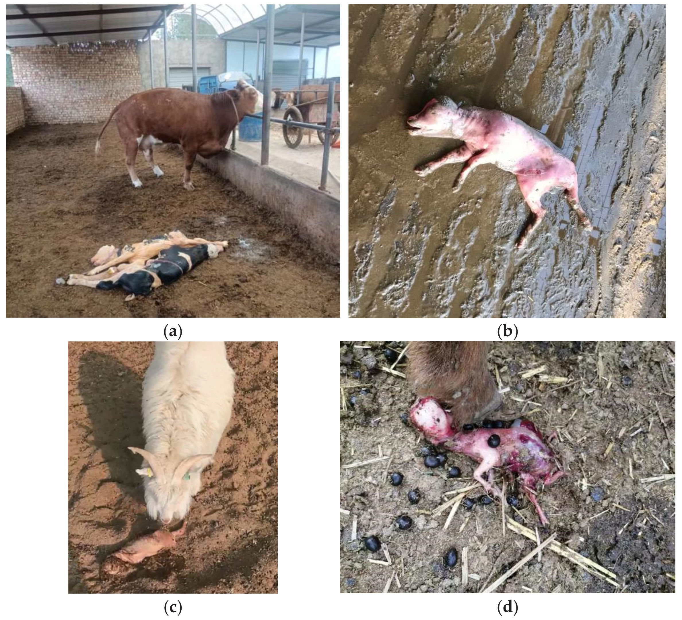
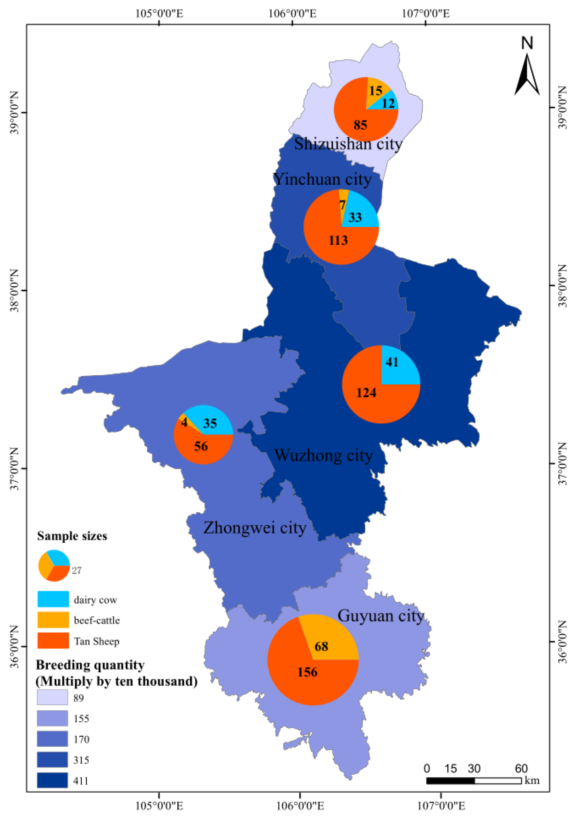
2.1. Sample Collection

The following study was a cross-sectional study performed from January 2022 to December 2023. The study population comprised livestock that had experienced abortions in Ningxia, China. Veterinarians from various districts and areas were informed about the study’s aim and received training on biosafety procedures. Where possible, samples of aborted placenta or fetal spleen were taken from farms reporting livestock that had experienced abortions. The samples were collected into 50 mL sterile conical centrifuge tubes. These tubes were sealed twice in biohazard plastic bags by veterinarians who were equipped with appropriate personal protective equipment [1]. The cold chain was maintained, with samples being transported on ice and stored at −20 °C before analysis. After collecting the samples, thorough disinfection and safe treatment procedures were performed.

2.2. Isolation and Culture of Brucella spp. from Aborted Samples

Brucella spp. were isolated and cultured in a BSL-3 laboratory of the Ningxia Hui Autonomous Region Center for Disease Control and Prevention and subsequently tested following a previously described method [16,17]. Briefly, tissue homogenates were inoculated onto Brucella Selective Medium plates with the following antibiotics: bacitracin (25,000 IU/L), mycostatin (10,000 IU/L), polymyxin B (5000 IU/L), vancomycin (20 mg/L), and actinone (10 mg/L). These plates were incubated in the presence of 5% CO2 at 37 °C and subsequently examined every day for 3 weeks. Suspected colonies were preliminarily identified using biochemical tests, such as urease production and carbon dioxide requirement and Gram and Ziehl-Neelsen staining. Suspected colonies were purified, and agglutination was observed with monospecific antisera A, M, and R for strain typing and biotyping.

2.3. Molecular Analysis

2.3.1. DNA Extraction

Bacteria present in the samples were inactivated through heating at 100 °C for 5 min. A Nucleic Acid Automatic Extraction System (Xi’an Tianlong Technology Co., Ltd., Xi’an, China) was then employed to extract the bacterial genomic DNA from the cultures and tissues according to the supplier’s protocols. The extracted DNA was maintained at −20 °C until use.

2.3.2. Detection of Brucella spp. via qPCR

Primers for bcsp31 were designed using Primer Premier 6 (Premier Biosoft, San Francisco, CA, USA). Forward Primer: 5′-GGCCATCTCGAACGGTATTT-3′, Reverse Primer: 5′-GTTTCCGCCACGTCCTTATAG-3′, probe: 5′-(FAM, 6-carboxyfluorescein) CTTCTTCTCGAAGGATGTGGTTCCTGC-BHQ1 (Black Hole Quencher 1)-3′. Sangon Biotech (Shanghai) Co., Ltd. (Shanghai, China) synthesized the primers and probe.
The qPCR 25 µL reaction comprised 12.5 µL of Premix Ex Taq (Takara Bio Inc., Dalian, China), 0.5 µL of each primer (final concentration = 0.2 μM), 1 µL of each probe (final concentration = 0.2 μM), 2 µL of DNA template, and 8.5 μL of ddH2O. The source of DNA for qPCR was tissues and culture isolation. The qPCR analysis was performed on a CFX96 Real-Time PCR Detection System (Bio-Rad, Hercules, CA, USA) using cycling comprising initial denaturation at 95 °C for 20 s and then 40 cycles of denaturation at 95 °C for 5 s, before annealing/extension at 60 °C for 20 s. The FAM channel was chosen to monitor the fluorescence signal at 60 °C.

2.3.3. Identification of Brucella spp.

The vaccine strains (S2 and A19) of Brucella spp. were identified using an identification kit (Aodong Inspection & Testing Co., Ltd., Shenzhen, China).
B. abortus biovar (bv). 1, 2, and 4; B. melitensis bv. 1, 2, and 3; B. ovis; and B. suis bv. 1 were identified using a multiplex AMOS-PCR assay (named after the species detected: B. abortus, B. melitensis, B. ovis, and B. suis) [18]. The positive controls comprised B. suis, B. melitensis, and B. abortus nucleic acids (Pudao Standard Technology Co., Ltd., Beijing, China). A PCR Instrument (Mastercycler Nexus GX2, Eppendorf, Hamburg, Germany) was used to perform the amplification reactions. The reaction (25 μL) contained 1 × MyRaq Red PCR Mix, four species-specific forward primers and reverse primer IS711 (final concentrations = 0.1 μM and 0.5 μM, respectively), and 2 μL of DNA template. The PCR cycling conditions comprised 95 °C for 3 min; 40 cycles of 95 °C for 1 min, 60 °C for 2 min, and 72 °C for 1 min; and lastly 72 °C for 10 min. UV illumination was used to visualize amplicons after electrophoretic separation on a 1.5% agarose gel stained with GelRed.

2.3.4. MLST Genotyping

Nine separate genomic loci were selected for MLST genotyping, comprising seven housekeeping genes (cobQ, trpE, gyrB, dnaK, glk, aroA, and gap), one intergenic fragment (int-hyp), and one outer membrane protein gene (omp25) [19]. The PCR reactions were carried out in 50 μL volumes, and 5 μL of each reaction for each of the nine loci was subjected to capillary electrophoresis by Sangon Biotech (Shanghai) Co., Ltd. A web-based MLST service (Brucella Base, http://59.99.226.203/brucellabase/mlst.html (accessed on 4 May 2025)) was used to predict each sequence type (ST) over all loci.
To further investigate the molecular epidemiology of Brucella spp. strains at the national and international levels, data from previous studies listed in public databases for molecular typing and microbial genome diversity (https://pubmlst.org/organisms/brucella-spp (accessed on 4 May 2025)) were employed. PHYLOViZ 2.0 software [20] was used to construct a minimum spanning tree (MST) to evaluate the genetic relationships among the 14 isolates.

2.3.5. MLVA Genotyping

MLVA analysis was conducted using a previously described method [21]. Briefly, 16 primer pairs were used, grouped into three panels: Panel 1 (bruce06, bruce08, bruce11, bruce12, bruce42, bruce43, bruce45, and bruce55); Panel 2A (bruce18, bruce19, and bruce21); and Panel 2B (bruce04, bruce07, bruce09, bruce16, and bruce30). The PCR amplifications were performed in a 50 μL reaction volume, 5 μL of which was subjected to analysis using 2% gel electrophoresis (with gel red nucleic acid staining), followed by UV visualization and photography. BioNumerics version 5.1 (Applied Maths NV, Sint-Martens-Latem, Belgium) was employed to estimate the band densities. The allele numbering system [22] was then used to convert the band densities into repeat units. BioNumerics version 5.1 was then used to generate arithmetic averages for unweighted pair group phylogenetic analysis of the 14 strains isolated from aborted livestock in the study and 21 strains isolated by Liu et al. from humans [15].

2.4. Analysis of the Data

We calculated the proportion of positive animals by dividing the total number of animals that tested positive by the total number of animals in the study. Microsoft Excel 2010 spreadsheets (Microsoft Corp., Redmond, WA, USA) were employed to record the data. Proportions were calculated, and positive results were assessed using the χ2 test in Epi-Info 7 version 10 (CDC, Atlanta, GA, USA).

3. Results

3.1. Detecting Brucella spp. in Cattle and Sheep Abortion Tissues

In total, 831 cases of abortion were reported during the study period, and 749 samples of aborted placenta or fetal spleen were collected from the aborted livestock (Figure 1); the remaining 82 cases were disposed of by the farmers themselves and were not sampled. The samples were obtained from 121 (16.15%) dairy cows, 94 (12.55%) beef cattle, and 534 (71.29%) sheep. Among them, 14.95% (112/749) originated from Shizuishan City, 20.43% (153/749) originated from Yinchuan City, 22.03% (165/749) originated Wuzhong City, 12.68% (95/749) originated from Zhongwei City, and 29.91% (224/749) originated from Guyuan City (Figure 2).
In this study, 8.68% (65/749) of the samples were identified as positive for Brucella DNA using qPCR. The PCR-positive rate of aborted tissues from beef cattle was 13.83% (13/94); for dairy cows, the rate was 9.09% (11/121), which was higher than the rate of 7.68% (41/534) for sheep. In total, 14 strains of Brucella spp. were isolated.

3.2. Identification of Brucella spp.

Amplicons of 731 bp (B. melitensis), 498 bp (B. abortus), and 285 bp (B. suis) were detected using AMOS-PCR in Brucella cultures from aborted cattle and sheep tissues. Among the 14 wild strains, 11 were recognized as B. melitensis, 2 were recognized as B. abortus, and 1 was recognized as B. suis.

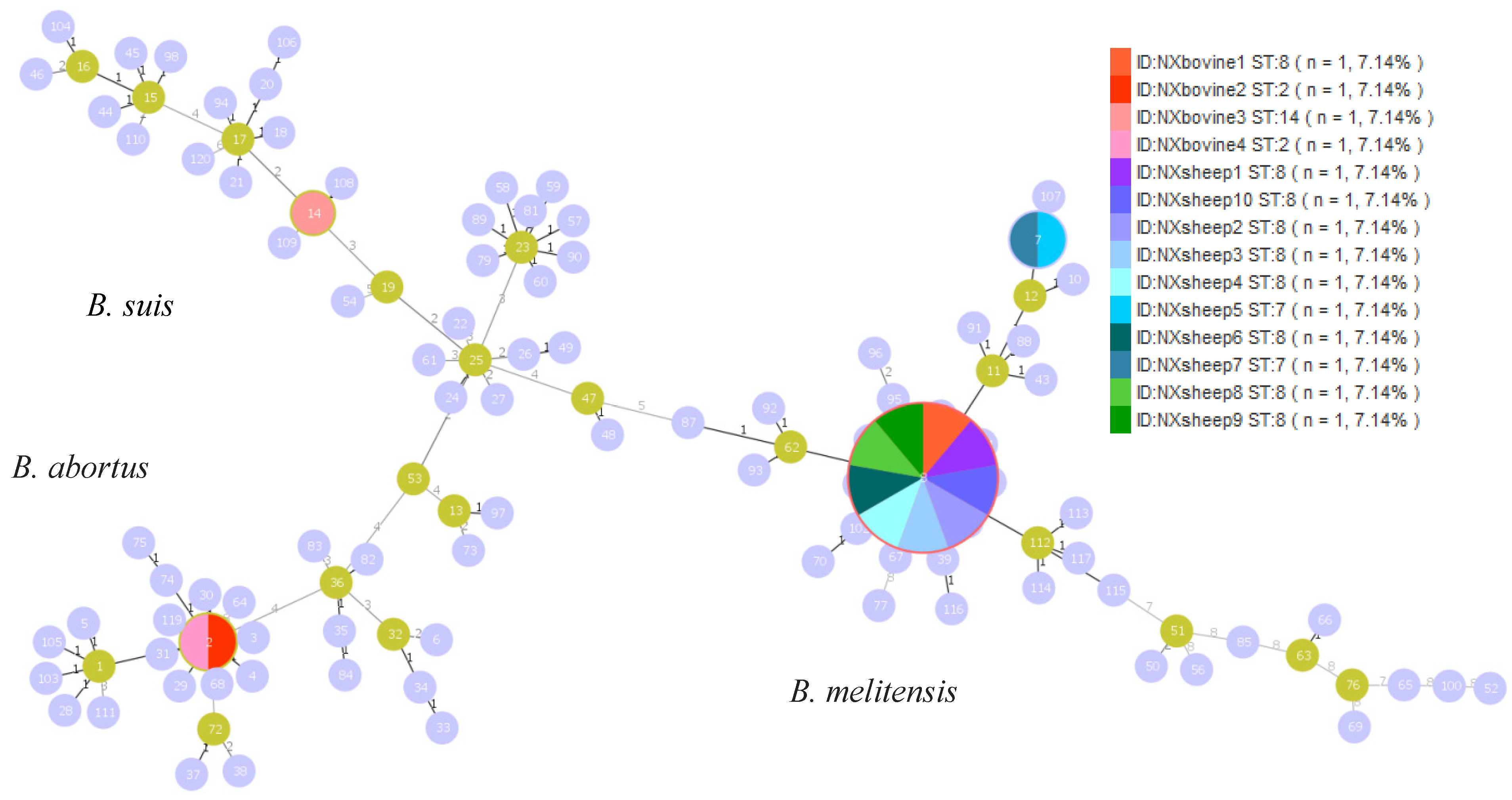
3.3. MLST Genotyping

Using the nine MLST loci, four known STs were identified: ST2 (2-1-2-2-1-3-1-1-1; n = 2), ST7 (3-5-3-2-1-5-3-2-10; n = 2), ST8 (3-2-3-2-1-5-3-2-8; n = 9), and ST14 (1-6-4-1-4-3-5-1-2; n = 1). The predominant ST in the Ningxia region was ST8 (Figure 3).

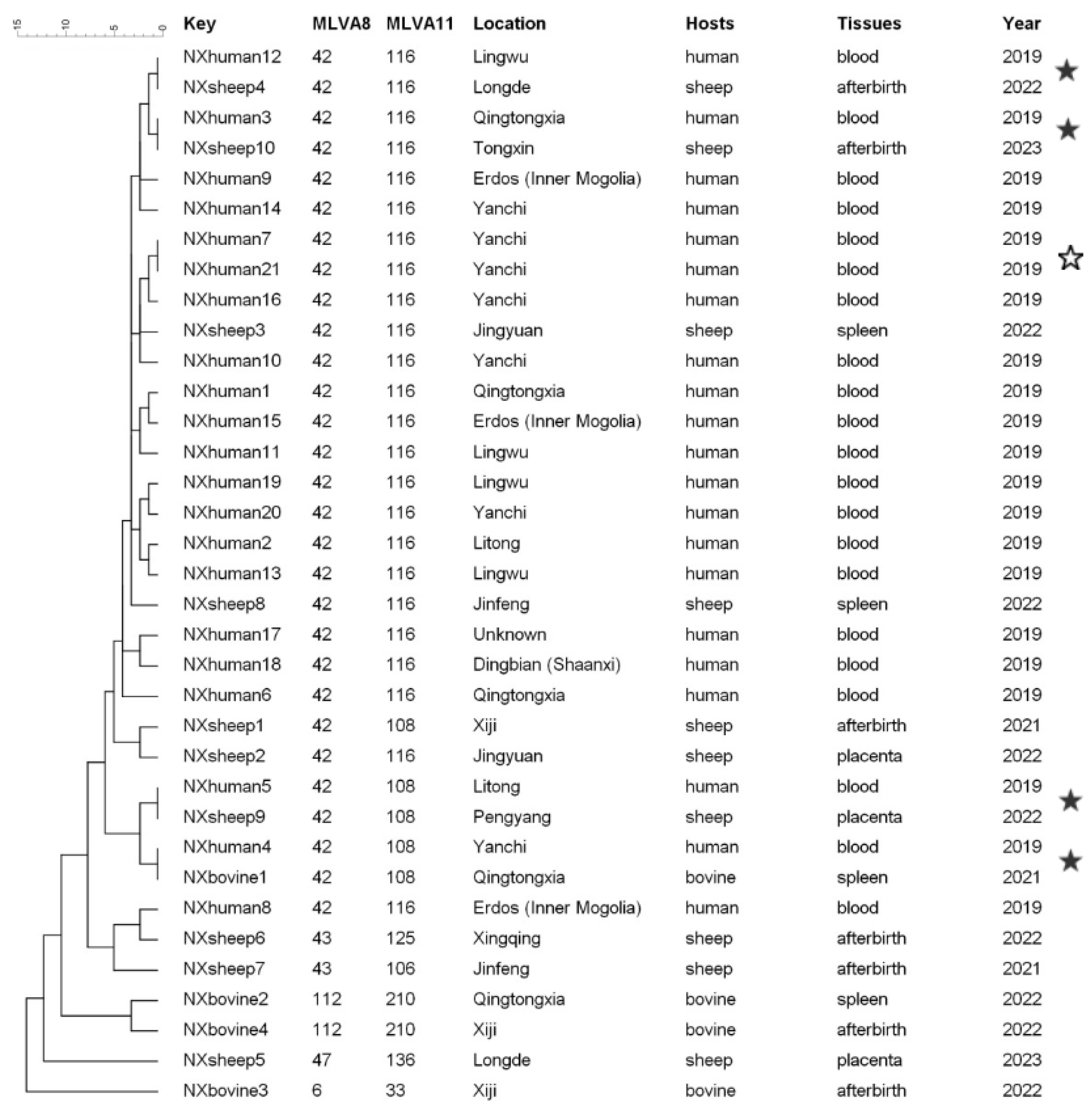
3.4. MLVA Genotyping

MLVA was employed to characterize 14 strains isolated from aborted livestock. The unweighted pair group method with arithmetic averages (UPGMA) was used to cluster the strains based on their genetic similarities. A total of 21 strains isolated from humans, reported in the study by Liu et al., from Ningxia in 2019 were selected and analyzed together [15]. Panel 1 shows that the strains could be clustered into five MLVA-8 genotypes (42 (1-5-3-13-2-2-3-2; n = 29), 43 (1-5-3-13-2-3-3-2; n = 2), 47 (3-4-2-13-4-2-3-3; n = 1), 112 (1-5-3-13-2-3-3-2; n = 2), and 6 (2-3-6-10-4-1-5-2; n = 1)). Seven MLVA-11 genotypes were identified (GT116, GT108, GT125, GT106, GT210, GT136, and GT33), with the MLVA-11 genotype 116 representing the predominant genotype circulating in both humans and livestock, which is widely distributed throughout Ningxia.
Ten samples contained shared genotypes (NXhuman7 and NXhuman21, NXhuman12 and NXsheep4, NXhuman3 and NXsheep10, NXhuman5 and NXsheep9, NXhuman4, and NXbovine1), eight of which shared genotypes in humans and livestock. The other 10 strains isolated from aborted livestock and 15 strains isolated from humans were represented by unique strains (Figure 4).

4. Discussion

Various factors can contribute to pregnancy loss, including issues of a noninfectious nature such as nutritional deficiencies and physical stress [23]. In addition, infectious agents, such as viruses, bacteria, fungi, and protozoa, can also cause abortions. It is estimated that infectious pathogens are responsible for roughly 90 percent of abortion cases, with the main factors responsible being Brucella spp., Chlamydia abortus, Toxoplasma gondii, bovine viral diarrhea virus (BVDV), and so forth [24]. Notably, the genus Brucella spp. is one of the primary bacterial agents that causes abortions in livestock across numerous developing nations [1].
Brucellosis can lead to a series of symptoms, such as miscarriage and orchitis, both in animals and humans, with it having significant global economic impacts related to animal health and financial losses, which also threaten human health [8]. In India, Brucellosis has led to financial losses estimated at USD 3.4 billion, with an average loss of roughly 18.2 dollars for each buffalo, 6.8 dollars for each cow, 0.7 dollars for sheep, 0.6 dollars for pigs, and 0.5 dollars for goats [25]. Furthermore, bovine brucellosis has caused economic damages of up to 600 million dollars in Latin America [26]. There has been a steady rise in human Brucellosis cases from the year 2000, affecting all of mainland China. The number of human cases has grown from 18,416 in 2005 to 75,858 in 2023 (https://www.ndcpa.gov.cn/jbkzzx/c100016/common/list.html (accessed on 4 May 2025)). In Ningxia, the number of human cases increased from 26 in 2005 to 6292 in 2022 [15]. The most effective method to prevent human Brucellosis cases is to control the disease in livestock. Effective strategies, such as regular livestock screening, removal of infected groups, and immunizing healthy animals, have led to the successful management of the disease in numerous developed countries [12].
To reduce the impact of Brucellosis on livestock, promote further development of the livestock industry, and ensure public health security, the “Ningxia Brucellosis Prevention and Control Special Three-Year Action Implementation Plan (2022–2024)” was implemented by the government of Ningxia [27]. The Plan encompasses a series of systematic measures aimed at both humans and livestock. These measures include the ongoing compulsory immunization of newborn lambs with the S2 vaccine and calves with the A19 vaccine and comprehensive disinfection and safe treatment of aborted livestock. In addition, the Plan emphasizes surveillance and epidemiological investigations, enhanced public awareness campaigns, and the training of both professional and reserve personnel. Furthermore, it highlights the implementation of a special “mask and gloves initiative” to ensure comprehensive safety. The initiative provides free gloves and masks to workers in high-risk industries, such as breeding and slaughtering. Vaccination can effectively decrease the incidence of Brucellosis in livestock and correlates with a reduced number of reported cases in humans, despite there currently being no human vaccines available [28]. Importantly, reducing infectious disease spread is supported by appropriate protective measures [27]. Employees involved in breeding, transport, slaughtering, and processing have seen a significant increase in their awareness of zoonotic diseases such as Brucellosis, tuberculosis, and anthrax prevention and control. Furthermore, public awareness of epidemic prevention and the control of zoonotic diseases have been continuously improved through the implementation of systematic measures. The number of reported human Brucellosis cases in the entire region has decreased by 28.5% year-on-year since 2023.
As a part of surveillance and epidemiological investigations, this study is a vital element of the Plan to reduce reported human cases, comprising the first systematic study on Brucella spp. from aborted tissues of livestock in the Ningxia Hui Autonomous Region of China. Our results indicate that in Ningxia, at least three Brucella species (B. suis, B. abortus, and, B. melitensis) are circulating, among which B. melitensis ST8 was the most frequently detected species.
In specific areas of infection, determining effective treatment strategies, tracing transmission, clarifying the reservoir and source, evaluating epidemiological intensity, and classifying foci are crucially informed by the identification of Brucella genotype, biovars, and species [9]. Correlations among Brucella spp. cannot be analyzed using classical bio-typing methods; however, tracking infection spread, genetic diversity, and population structure can be aided by molecular epidemiological studies, thereby facilitating and improving effective measures for disease control and prevention [29]. Moreover, Brucella MLST and MLVA have been suggested as supplemental methodologies to traditional bio-typing techniques [13].
The main Brucella spp. in China include the following: B. melitensis biovar 1 and 3 (such as ST7, ST8, ST34, ST35, and ST37), B. abortus biovar 1 and 3 (such as ST1, ST2, ST5, ST28, ST29, ST30, ST31, ST32, ST33, and ST38), and B. suis biovar 1 and 3 (such as ST14, ST17 and ST36), with B. melitensis as the main epidemic pathogenic species [14]. B. melitensis ST8 also constitutes the main species in Inner Mongolia [30], Xinjiang [24], Qinghai [31], and Guangxi [32], with it being responsible for numerous cases of human Brucellosis. This finding is also supported by the detection of 11 B. melitensis strains, and 9 ST8 among the 14 isolated strains in this study. B. melitensis ST8’s prevalence in Northwest China, and even in southern China more recently, may reflect the regional fitness advantages of the strain. Its prevalence has mainly resulted from the frequent transportation and circulation of live livestock on the Chinese mainland.
Although MLST and MLVA showed similarly consistent discrimination at the Brucella species level, MLVA could discriminate the genotype with higher resolution than MLST [13]. A complete MLVA-16 assay is often employed to traceback infection sources and investigate molecular epidemiological associations among Brucella isolates [30]. In the study presented herein, there eight strains shared the same MLVA-16 genotypes in humans and livestock. This finding demonstrates that the human and livestock cases were caused by a common source. Other strains were represented by unique strains. Brucellosis is primarily transmitted via direct or indirect contact between infected animals or products and humans [12], which indicates that some human cases cannot be traced to animal sources and that the distribution of Brucella species among domestic animals is in fact more diverse.
Bacterial isolation is considered indisputable evidence of disease [12]. However, some samples (aborted fetuses and vaginal swabs) assessed as positive based on PCR results but negative based on culture likely occurred as a result of contamination [24] and also arose from an infection that had run its course, with no live bacteria present but instead residual debris from dead cell’s DNA. In addition, there can also be a gap of several days between the time of reporting abortion and the time of sampling; in particular, at higher temperatures, old and decaying aborted tissues may also decrease the isolation rate of the bacteria.
This study suffers from some inherent limitations. Firstly, the submitted number of abortion cases may not provide an accurate representation of the actual number of cases. The underreporting of livestock abortions represents a global issue, even when reporting is required. This issue impacts the early identification of infectious and zoonotic factors and could result in the transmission of possible pathogens [23]. Evidence from other studies demonstrates that farmers and their veterinarians frequently neglect to report abortion cases despite the existence of mandatory reporting requirements, with such scenarios usually driven by self-interest and concerns about potential consequences, including farm isolation for suspected cases of Brucellosis or other notifiable diseases, in addition to financial and logistical challenges [33,34,35]. In addition, Hui people are largely concentrated in Ningxia and their lifestyle and dietary practices follow Islamic traditions [15]. We also found that some Hui populations do not report abortion cases because they are under religious influence to not profit from dead animals. Efforts have thus been made in attempt to increase the reporting of abortion cases by publicizing information on the transmission of Brucellosis and its main threats; despite these efforts, little progress has been made in some villages.
Secondly, animals that test negative for all Brucella spp. should be tested to determine whether other abortigenic pathogens, including Campylobacter fetus and Leptospira spp., are present in order to control ruminant abortions [35]. However, due to limited funding and laboratory capabilities, another limitation of this study is the fact that the aborted samples were not tested to determine if other pathogens were present.
Despite these limitations, this study provides valuable epidemiological information on Brucellosis in aborted livestock in Ningxia. The results could aid in the analysis of the spread and evolution of Brucella spp. in Ningxia and lead to novel vaccine development. They could provide enhanced insights into strain evolution and transmission pathways employing higher-resolution genomic markers (e.g., Whole-Genome Sequencing) in future studies.

5. Conclusions

In this study, B. melitensis, B. abortus, and B. suis isolated from aborted tissues of livestock from 2022 to 2023 in Ningxia were identified and analyzed using MLVA and MLST. Our research results suggest that comprehensive disinfection and safe treatment should be implemented after the abortion of livestock to interrupt transmission pathways to prevent livestock infection and thus human transmission. Moreover, aborted livestock should be examined to determine possible zoonotic causes, and targeted surveillance should be strengthened to improve the early detection of infectious causes, patricianly Brucellosis, which would be of benefit to the breeding industry and public health security. Such measures can be further developed by proposing prioritized monitoring of sheep flocks (primary hosts for B. melitensis ST8), particularly those experiencing abortions, and evaluating S2 vaccine efficacy in this context.

Author Contributions

Methodology, C.Y. (Cai Yin) and C.Y. (Cong Yang); software, C.Y. (Cai Yin); formal analysis, C.Y. (Cai Yin) and C.Y. (Cong Yang); investigation, Y.W. (Yawen Wu), J.D., T.B., L.L., S.Z., and L.M.; resources, Y.W. (Yumei Wang); data curation, Y.Z.; writing—original draft, C.Y. (Cai Yin); writing—review and editing, Q.Z. and Z.L.; visualization, C.Y. (Cong Yang); supervision, X.W. and Z.L.; project administration, Q.Z.; funding acquisition, Y.Z. and Z.L. All authors have read and agreed to the published version of the manuscript.

Funding

This research was supported by grants from the Autonomous Region Animal Disease Purification Technology Innovation Team Project [grant number 2020CXTDLX05]; the Key Research and Development Project for Characteristic Industries in Ningxia Hui Autonomous Region in 2024 [grant number 2024BBF02021]; the Local Science and Technology Development Projects [grant number 2024FRD05072]; and the Natural Science Foundation of Ningxia Province [grant numbers 2022AAC03712 and 2023AAC02075]. The funder had no role in study design, data collection, analysis, or interpretation, or the decision to publish the results.

Institutional Review Board Statement

Not applicable.

Informed Consent Statement

Informed consent was obtained from all animals owners.

Data Availability Statement

All data are included in the manuscript.

Acknowledgments

The authors gratefully acknowledge the Ningxia youth top talent “Autonomous Region Academic technology leader” reserve candidate talent project, and Xueyi Wang, Ruigang Wang, and Kuiju Zhang for their invaluable assistance with sample collection and photography.

Conflicts of Interest

The authors declare no conflicts of interest.

References

  1. Ntivuguruzwa, J.B.; Kolo, F.B.; Mwikarago, E.I.; van Heerden, H. Characterization of Brucella spp. and other abortigenic pathogens from aborted tissues of cattle and goats in Rwanda. Vet. Med. Sci. 2022, 8, 1655–1663. [Google Scholar] [CrossRef]
  2. Clune, T.; Beetson, S.; Besier, S.; Knowles, G.; Paskin, R.; Rawlin, G.; Suter, R.; Jacobson, C. Ovine abortion and stillbirth investigations in Australia. Aust. Vet. J. 2020, 99, 72–78. [Google Scholar] [CrossRef]
  3. Godfroid, J.; Cloeckaert, A.; Liautard, J.-P.; Kohler, S.; Fretin, D.; Walravens, K.; Garin-Bastuji, B.; Letesson, J.-J. From the discovery of the Malta fevers agent to the discovery of a marine mammal reservoir, brucellosis has continuously been a re-emerging zoonosis. Vet. Res. 2005, 36, 313–326. [Google Scholar] [CrossRef]
  4. Pal, M.; Gizaw, F.; Fekadu, G.; Alemayehu, G.; Kandi, V. Public Health and Economic Importance of Bovine Brucellosis: An Overview. Am. J. Epidemiol. Infect. Dis. 2017, 5, 27–34. [Google Scholar] [CrossRef]
  5. Mustafa, S.; Qureshi, A.S.; Deeba, F.; Rashid, I.; Usman, M. Brucellosis in Cattle and Buffaloes. Int. J. Agric. Biol. 2023, 2, 25–33. [Google Scholar]
  6. Spickler, A.R. Brucellosis. 2018. Available online: http://www.cfsph.iastate.edu/DiseaseInfo/factsheets.php (accessed on 22 July 2025).
  7. Fereig, R.M.; Wareth, G.; Abdelbaky, H.H.; Mazeed, A.M.; El-Diasty, M.; Abdelkhalek, A.; Mahmoud, H.Y.A.H.; Ali, A.O.; El-tayeb, A.; Alsayeqh, A.F.; et al. Seroprevalence of Specific Antibodies to Toxoplasma gondii, Neospora caninum, and Brucella spp. in Sheep and Goats in Egypt. Animals 2022, 12, 3327. [Google Scholar] [CrossRef]
  8. Ma, H.-R.; Xu, H.-J.; Wang, X.; Bu, Z.-Y.; Yao, T.; Zheng, Z.-R.; Sun, Y.; Ji, X.; Liu, J. Molecular characterization and antimicrobial susceptibility of human Brucella in Northeast China. Front. Microbiol. 2023, 14, 1137932. [Google Scholar] [CrossRef]
  9. Daugaliyeva, A.; Daugaliyeva, S.; Kydyr, N.; Peletto, S. Molecular typing methods to characterize Brucella spp. from animals: A review. Vet. World 2024, 17, 1778–1788. [Google Scholar] [CrossRef]
  10. Moreno, E. Retrospective and prospective perspectives on zoonotic brucellosis. Front. Microbiol. 2014, 5, 213. [Google Scholar] [CrossRef]
  11. Mesner, O.; Riesenberg, K.; Biliar, N.; Borstein, E.; Bouhnik, L.; Peled, N.; Yagupsky, P. The Many Faces of Human-to-Human Transmission of Brucellosis: Congenital Infection and Outbreak of Nosocomial Disease Related to an Unrecognized Clinical Case. Clin. Infect. Dis. 2007, 45, e135–e140. [Google Scholar] [CrossRef]
  12. Yagupsky, P.; Morata, P.; Colmenero, J.D. Laboratory Diagnosis of Human Brucellosis. Clin. Microbiol. Rev. 2019, 33, e00073-19. [Google Scholar] [CrossRef]
  13. Tan, Q.; Wang, Y.; Liu, Y.; Tao, Z.; Yu, C.; Huang, Y.; Yang, X.; Ying, X.; Hu, Y.; Li, S. Molecular epidemiological characteristics of Brucella in Guizhou Province, China, from 2009 to 2021. Front. Microbiol. 2023, 14, 1188469. [Google Scholar] [CrossRef]
  14. Piao, D.-R.; Liu, X.; Di, D.-D.; Xiao, P.; Zhao, Z.-Z.; Xu, L.-Q.; Tian, G.-Z.; Zhao, H.-Y.; Fan, W.-X.; Cui, B.-Y.; et al. Genetic polymorphisms identify in species/biovars of Brucella isolated in China between 1953 and 2013 by MLST. BMC Microbiol. 2018, 18, 7. [Google Scholar] [CrossRef]
  15. Liu, G.; Ma, X.; Zhang, R.; Lü, J.; Zhou, P.; Liu, B.; Liu, T.; Ren, H.; Liu, Z.; Li, Z.; et al. Epidemiological changes and molecular characteristics of Brucella strains in Ningxia, China. Front. Microbiol. 2024, 15, 1320845. [Google Scholar] [CrossRef]
  16. Ali, S.; Ali, Q.; Melzer, F.; Khan, I.; Akhter, S.; Neubauer, H.; Jamal, S.M. Isolation and identification of bovine Brucella isolates from Pakistan by biochemical tests and PCR. Trop. Anim. Health Prod. 2013, 46, 73–78. [Google Scholar] [CrossRef]
  17. Yang, C.; Gao, J.; Xian, R.; Liu, X.; Kuai, W.; Yin, C.; Fan, H.; Tian, J.; Ma, X.; Ma, J. Molecular epidemiology of Brucella abortus isolated from the environment in Ningxia Hui autonomous region, China. Infect. Genet. Evol. 2024, 123, 105635. [Google Scholar] [CrossRef]
  18. Bricker, B.J.; Halling, S.M. Differentiation of Brucella abortus bv. 1, 2, and 4, Brucella melitensis, Brucella-ovis, and Brucella suis bv. 1 by PCR. J. Clin. Microbiol. 1994, 32, 2660–2666. [Google Scholar] [CrossRef]
  19. Whatmore, A.M.; Koylass, M.S.; Muchowski, J.; Edwards-Smallbone, J.; Gopaul, K.K.; Perrett, L.L. Extended Multilocus Sequence Analysis to Describe the Global Population Structure of the Genus Brucella: Phylogeography and Relationship to Biovars. Front. Microbiol. 2016, 7, 2049. [Google Scholar] [CrossRef]
  20. Nascimento, M.; Sousa, A.; Ramirez, M.; Francisco, A.P.; Carriço, J.A.; Vaz, C.; Kelso, J. PHYLOViZ 2.0: Providing scalable data integration and visualization for multiple phylogenetic inference methods. Bioinformatics 2017, 33, 128–129. [Google Scholar] [CrossRef]
  21. Al Dahouk, S.; Flèche, P.L.; Nöckler, K.; Jacques, I.; Grayon, M.; Scholz, H.C.; Tomaso, H.; Vergnaud, G.; Neubauer, H. Evaluation of Brucella MLVA typing for human brucellosis. J. Microbiol. Meth. 2007, 69, 137–145. [Google Scholar] [CrossRef]
  22. Jiang, H.; Wang, H.; Xu, L.; Hu, G.; Ma, J.; Xiao, P.; Fan, W.; Di, D.; Tian, G.; Fan, M.; et al. MLVA Genotyping of Brucella melitensis and Brucella abortus Isolates from Different Animal Species and Humans and Identification of Brucella suis Vaccine Strain S2 from Cattle in China. PLoS ONE 2013, 8, e76332. [Google Scholar] [CrossRef]
  23. Van Loo, H.; Bogado Pascottini, O.; Ribbens, S.; Hooyberghs, J.; Opsomer, G.; Pardon, B. Enhancing bovine abortion surveillance: A learning experience. J. Dairy Sci. 2024, 107, 1766–1777. [Google Scholar] [CrossRef]
  24. Clegg, S.; Zhang, H.; Deng, X.; Cui, B.; Shao, Z.; Zhao, X.; Yang, Q.; Song, S.; Wang, Z.; Wang, Y.; et al. Abortion and various associated risk factors in dairy cow and sheep in Ili, China. PLoS ONE 2020, 15, e0232568. [Google Scholar]
  25. Singh, B.B.; Dhand, N.K.; Gill, J.P.S. Economic losses occurring due to brucellosis in Indian livestock populations. Prev. Vet. Med. 2015, 119, 211–215. [Google Scholar] [CrossRef]
  26. Dadar, M.; Tiwari, R.; Sharun, K.; Dhama, K. Importance of brucellosis control programs of livestock on the improvement of one health. Vet. Q. 2021, 41, 137–151. [Google Scholar] [CrossRef]
  27. Gong, W.; Sun, P.; Zhai, C.; Yuan, J.; Chen, Y.; Chen, Q.; Zhao, Y. Accessibility of the three-year comprehensive prevention and control of brucellosis in Ningxia: A mathematical modeling study. BMC Infect. Dis. 2023, 23, 292. [Google Scholar] [CrossRef]
  28. Heidary, M.; Dashtbin, S.; Ghanavati, R.; Mahdizade Ari, M.; Bostanghadiri, N.; Darbandi, A.; Navidifar, T.; Talebi, M. Evaluation of Brucellosis Vaccines: A Comprehensive Review. Front. Vet. Sci. 2022, 9, 925773. [Google Scholar] [CrossRef]
  29. Kydyshov, K.; Usenbaev, N.; Berdiev, S.; Dzhaparova, A.; Abidova, A.; Kebekbaeva, N.; Abdyraev, M.; Wareth, G.; Brangsch, H.; Melzer, F.; et al. First record of the human infection of Brucella melitensis in Kyrgyzstan: Evidence from whole-genome sequencing-based analysis. Infect. Dis. Poverty 2022, 11, 120. [Google Scholar] [CrossRef]
  30. Liu, Z.; Wang, M.; Ta, N.; Fang, M.; Mi, J.; Yu, R.; Luo, Y.; Cao, X.; Li, Z. Seroprevalence of human brucellosis and molecular characteristics of Brucella strains in Inner Mongolia Autonomous region of China, from 2012 to 2016. Emerg. Microbes Infect. 2020, 9, 263–274. [Google Scholar] [CrossRef]
  31. Ma, J.-Y.; Wang, H.; Zhang, X.-F.; Xu, L.-Q.; Hu, G.-Y.; Jiang, H.; Zhao, F.; Zhao, H.-Y.; Piao, D.-R.; Qin, Y.-M.; et al. MLVA and MLST typing of Brucella from Qinghai, China. Infect. Dis. Poverty 2016, 5, 26. [Google Scholar] [CrossRef]
  32. Liu, Z.-g.; Wang, M.; Zhao, H.-y.; Piao, D.-r.; Jiang, H.; Li, Z.-j. Investigation of the molecular characteristics of Brucella isolates from Guangxi Province, China. BMC Microbiol. 2019, 19, 292. [Google Scholar] [CrossRef]
  33. Bronner, A.; Hénaux, V.; Fortané, N.; Hendrikx, P.; Calavas, D. Why do farmers and veterinarians not report all bovine abortions, as requested by the clinical brucellosis surveillance system in France. BMC Vet. Res. 2014, 10, 93. [Google Scholar] [CrossRef]
  34. Elbers, A.R.W.; Gorgievski-Duijvesteijn, M.J.; Zarafshani, K.; Koch, G. To report or not to report: A psychosocial investigation aimed at improving early detection of avian influenza outbreaks. Rev. Sci. Tech. 2010, 29, 435–449. [Google Scholar] [CrossRef]
  35. Jonker, A.; Thompson, P.N.; Michel, A.L. Approaches to increase recovery of bacterial and fungal abortion agents in domestic ruminants. Onderstepoort J. Vet. Res. 2023, 90, e1–e10. [Google Scholar] [CrossRef]
Figure 1. Aborted livestock used in this study. (a) Aborted beef cattle and stillbirths in Longde, Guyuan city, in June 2023. (b) Dairy cow stillbirth in Shapotou, Zhongwei city, in September 2023. (c) Aborted sheep and stillbirths in Yanchi, Wuzhong city, in 2022.12. (d) Sheep stillbirth in Xiji, Guyuan city, in March 2023. (We thank Xueyi Wang, Ruigang Wang, and Kuiju Zhang for their assistance in taking the photographs).
Vetsci 12 00702 g001
Figure 2. Sampling sizes of aborted livestock in this study and the number of breeding livestock in Ningxia.
Vetsci 12 00702 g002
Figure 3. The minimum spanning tree of the 14 strains identified in this study was constructed using PHYLOZIV 2.0 software. Orange, red, light orange, and pink colors indicate strains isolated from cattle; other colors indicate strains isolated from sheep.
Vetsci 12 00702 g003
Figure 4. MLVA dendrogram of 14 strains isolated from aborted livestock in this study and 21 strains isolated from humans in the study by Liu et al. in Ningxia [15]. The hollow pentagram show shared genotypes in humans; the solid pentagram shows the shared genotypes in humans and livestock.
Vetsci 12 00702 g004
Disclaimer/Publisher’s Note: The statements, opinions and data contained in all publications are solely those of the individual author(s) and contributor(s) and not of MDPI and/or the editor(s). MDPI and/or the editor(s) disclaim responsibility for any injury to people or property resulting from any ideas, methods, instructions or products referred to in the content.

