Article

A Multidrug-Resistant Escherichia coli Caused the Death of the Chinese Soft-Shelled Turtle (Pelodiscus sinensis)

by
Mingyang Xue
1,2,†,
Xiaowei Hu
1,†,
Nan Jiang
1,
Wei Liu
1,
Zidong Xiao
1,
Chunjie Zhang
1,
Yeying Wu
3,
Tianwang Liang
3,
Huixuan Zhang
3,
Yuding Fan
1,
Yan Meng
1,* and
Yong Zhou
1,*
1
Yangtze River Fisheries Research Institute, Chinese Academy of Fishery Sciences, Wuhan 430223, China
2
Freshwater Fisheries Research Center, Chinese Academy of Fishery Sciences, Wuxi 214081, China
3
School of Life Science, Wuchang University of Technology, Wuhan 430223, China
*
Authors to whom correspondence should be addressed.
These authors contributed equally to this work.
Vet. Sci. 2025, 12(5), 473; https://doi.org/10.3390/vetsci12050473
Submission received: 18 March 2025 / Revised: 17 April 2025 / Accepted: 13 May 2025 / Published: 14 May 2025

Simple Summary

Disease outbreaks caused by bacterial pathogens, particularly antimicrobial-resistant strains, represent a critical challenge in Chinese soft-shelled turtle (Pelodiscus sinensis) aquaculture. In this investigation, researchers characterized a multidrug-resistant Escherichia coli strain (HD-593) isolated from moribund specimens. Antimicrobial susceptibility profiling revealed resistance to 14 clinically relevant antibiotics and had several resistance genes. Experimental challenge trials demonstrated significant pathogenicity, with a median lethal dose of 6.53 × 10⁵ CFU/g. Infected turtles had damaged organs and changes in serum levels. This study shows that this multidrug-resistant E. coli is a threat to aquaculture. It is important to use antibiotics carefully and find alternatives which can help protect animal and public health.

Abstract

The rapid increase in drug resistance in recent years has become a significant global public health concern. Escherichia coli are ubiquitous bacteria, widely distributed in various environments. This study isolated a bacterial strain (HD-593) from diseased Chinese soft-shelled turtles (Pelodiscus sinensis). The bacterium was identified based on morphology, biochemical tests, and 16S rRNA sequencing, confirming it as E. coli. Drug susceptibility tests revealed that the HD-593 strain was highly resistant to ceftriaxone, enrofloxacin, doxycycline, sulfadiazine, gentamicin, neomycin, florfenicol, carbenicillin, cefradine, erythromycin, penicillin, ampicillin, midecamycin, and streptomycin. Resistance gene analysis confirmed the presence of quinolone resistance genes (oqxA and oqxB), aminoglycoside resistance genes (aac(3)-II and aphA1), a β-lactam resistance gene (blaTEM), and an acylaminol resistance gene (floR) in HD-593. The median lethal dose (LD50) of HD-593 for P. sinensis was 6.53 × 10⁵ CFU/g. Biochemical analysis of serum revealed that HD-593 infection caused a significant reduction in total protein, albumin, and globulin levels, while markedly increasing the levels of aspartate aminotransferase, alanine aminotransferase, and alkaline phosphatase. Histopathological analysis revealed severe intestinal damage characterized by villi detachment and muscle cell necrosis. Additionally, extensive splenocyte necrosis with nuclear marginalization, glomerular swelling, and pronounced hepatic steatosis accompanied by distended sinusoids were observed. This study identified a multidrug-resistant E. coli strain from deceased P. sinensis, suggesting that drug resistance genes may circulate in aquaculture ecosystems, posing potential risks to aquaculture.

1. Introduction

The Chinese soft-shelled turtle (Pelodiscus sinensis), also known as Trionyx sinensis, is a valuable aquaculture species in East Asia, particularly in China, due to its high economic and nutritional value [1]. Production of P. sinensis has steadily increased in recent years, primarily in regions like Zhejiang, Anhui, Guangdong, Hubei, Jiangsu, and Jiangxi [2]. The industry has trended toward industrialization, standardization, and intensified production methods [3,4]. However, the increasing scale of P. sinensis aquaculture has been accompanied by the emergence of various diseases. Disease outbreaks, particularly bacterial infections, during the summer months pose a significant challenge for P. sinensis farmers. Several bacterial pathogens, including Bacillus cereus [5], Aeromonas veronii [6,7], Aeromonas hydrophila [3,8], and Morganella morganii [9], have been identified as causative agents of P. sinensis disease. Among the bacterial pathogens affecting aquaculture species, Escherichia coli is notable due to its dual role as a common gut bacterium and a potential pathogen under certain conditions. Although E. coli is generally non-pathogenic, certain strains have acquired virulence factors that enable them to cause infections in a variety of hosts, including aquatic animals [10].
Multidrug resistance among bacterial pathogens has become a major concern worldwide, driven by the widespread use and misuse of antibiotics in both human and animal medicine [11]. Treating infections caused by antimicrobial-resistant pathogens presents a significant challenge to global healthcare systems and the breeding industry, driving up costs and placing considerable strain on resources [12]. In aquaculture, the indiscriminate application of antibiotics to control bacterial infections has exacerbated this issue, leading to the emergence of antibiotic-resistant bacteria that can survive multiple classes of antibiotics. Such bacteria not only threaten the sustainability of aquaculture but also pose a potential risk for zoonotic transmission, creating a pathway for antibiotic resistance to spread to human populations.
This study documents a case of mortality in P. sinensis caused by a multidrug-resistant E. coli strain, aiming to provide insights into the pathogen’s resistance profile and pathogenicity. A strain, designated E. coli HD-593, was isolated from the diseased P. sinensis. Antibiotic susceptibility testing, combined with resistance gene analysis, revealed that E. coli HD-593 exhibited multidrug resistance. Furthermore, artificial infection experiments and histopathological analysis were performed to evaluate the pathogenicity of E. coli HD-593. This investigation not only identified the pathogen responsible for the disease outbreak in P. sinensis but also offered valuable data and theoretical support for promoting the rational use of antibiotics in aquaculture.

2. Materials and Methods

2.1. Animal

The diseased P. sinensis (n = 11) weighing approximately 1100 ± 30 g were acquired from a breeding facility in Hubei Province, China. These diseased individuals were transported to the laboratory for pathogen isolation. Healthy P. sinensis (n = 200) with no history of the disease were obtained from the same breeding facility. The healthy P. sinensis were temporarily housed in laboratory aquariums measuring 350 cm × 200 cm × 50 cm and maintained at water temperatures of 30 ± 2 °C for 14 days. Healthy P. sinensis were randomly divided into six groups, with 30 P. sinensis in each group. All animal experiments conducted in this investigation were approved by the Animal Experimental Ethical Inspection of the Laboratory Animal Center, Yangtze River Fisheries Research Institute, Chinese Academy of Fishery Sciences (ID Number: YFI 2023-zhouyong-101).

2.2. Pathogen Isolation

Diseased P. sinensis was anesthetized, and then disinfected with 75% ethanol. After dissection, some liver tissue was inoculated onto sterile agar plates containing brain heart infusion (BHI) medium (HopeBio, Qingdao, China). These plates were incubated at 30 °C for 24 h. Next, single colonies were selected and purified by repeated streaking on fresh BHI agar plates. The purified bacterial strain was designated HD-593.

2.3. Morphological Characteristic

The bacteria were preserved by inoculation into BHI liquid medium and cultured at 30 °C with stirring at 200 rpm for 24 h. The cultured bacterial fluids were diluted for Gram staining (Jiancheng, Nanjing, China), and optical micrographs (Olympus, Tokyo, Japan) were used to visualize bacterial morphology [13]. The bacterium was then stabilized in 2.5% glutaraldehyde solution and dehydrated to dryness [14]. Bacterial morphology was examined using scanning electron microscopy (Hitachi, Tokyo, Japan).

2.4. Biochemical Characterization

The biochemical characterization of HD-593 was performed using the Microbial Identification and Phenotype MicroArrays System (MIPAS). The HD-593 strain was introduced into IF-A inoculation liquid (Biolog, Hayward, CA, USA). The inoculum fluid was then dispensed at 100 μL per well onto GEN III recognition plates. The GEN III plate was validated automatically by inserting it into the Biolog Automated Microbiology Recognition System (Biolog, Hayward, CA, USA).

2.5. Molecular Biology Analysis

The HD-593 strain was cultured at 30 °C and 200 rpm for 18 h. Subsequently, the cultured bacteria were centrifuged at 12,000 rpm for 10 min. DNA was extracted from the bacterial pellets using the Bacterial DNA Extraction Kit (Tiangen, Beijing, China) to serve as templates for subsequent PCR reactions. The 16S rRNA gene sequence was amplified using the universal primers 27F and 1492R. The reactions contained 12.5 μL 2× Taq PCR Master Mix (Tiangen, Beijing, China), 1 μL of each primer (Huayu Gene, Wuhan, China), 1 μL of template DNA, and deionized water. The amplification program was as follows: 95 °C for 5 min, followed by 35 cycles of 94 °C for 1 min, 55 °C for 1 min, 72 °C for 1 min, and 72 °C for 10 min. The amplified products were analyzed using 1% agarose gel electrophoresis and visualized with an ultraviolet light transilluminator (Bio-Rad, Hercules, CA, USA). These sequences were compared using BLAST on the National Center for Biotechnology Information (NCBI) GenBank database (https://blast.ncbi.nlm.nih.gov/Blast.cgi, accessed on 5 November 2023). The phylogenetic tree was constructed using MEGA software (Version 11.0.13).

2.6. Drug Susceptibility and Drug Resistance Genes Testing

The antibiotic sensitization of HD-593 was determined using the Kirby–Bauer disc diffusion technique. The concentration of bacteria was adjusted to 1 × 10⁸ CFU/mL using sterile PBS. Subsequently, 100 μL of the bacterial suspension was uniformly coated on the MHA medium, and antibiotic discs (ceftriaxone, enrofloxacin, doxycycline, sulfonamide, gentamycin, neomycin, florfenicol, carbenicillin, cefradine, erythromycin, penicillin, ampicillin, midecamycin, streptomycin, furazolidone, and polymyxin; diameter: 6 mm) were placed on the plate. After overnight incubation at 30 °C, the diameters of the inhibition zones were measured.
The drug resistance genes–quinolone resistance genes (oqxA, oqxB), aminoglycoside resistance genes (aac(3)-II, aphA1), a β-lactam resistance gene (blaTEM), an acylaminol resistance gene (floR), a colistin resistance gene (mcr-4), and a macrolide resistance gene (ereA)—were screened using PCR. The primers used for amplification of the drug resistance genes are listed in Table S1.

2.7. Artificial Infection Experiment

The concentrations of the cultured bacterial solution were measured using the plate count method. For infection group, 1 × 10⁴, 1 × 10⁵, 1 × 10⁶, 1 × 10⁷, and 1 × 10⁸ CFU/g body weight bacterial suspension were prepared. The control groups were also injected with an equal volume of phosphate buffer solution (PBS, Procell, Wuhan, China). After infection, the P. sinensis were continuously observed for 10 days. Dead P. sinensis were subjected to bacterial isolation and characterization. The Reed–Muench method was used to determine the lethal dosage (LD50) of HD-593 in P. sinensis.

2.8. Serum Physiologic and Biochemical Analysis

To investigate the serum alterations in diseased P. sinensis, blood samples were collected and transferred to labeled tubes for natural coagulation. Following a 30 min incubation at 4 °C for clot formation, the specimens were centrifuged at 2000 rpm for 10 min to separate the serum and cellular components. The resulting supernatant serum was aliquoted into fresh centrifuge tubes for subsequent biochemical analysis. Serum levels of aspartate aminotransferase (AST), alanine aminotransferase (ALT), alkaline phosphatase (ALP), total protein (TP), albumin (ALB), and globulin (GLB) were evaluated using an automated biochemical analyzer (Sysmex, Kobe, Japan).

2.9. Histopathologic Observation

The livers, spleens, kidneys, and intestines from both naturally diseased and healthy P. sinensis were collected. These organs were fixed in 4% paraformaldehyde solution, embedded in paraffin, sectioned, and subsequently stained with hematoxylin–eosin (HE). The stained sections were then sealed with neutral resin. The prepared sections were examined under a light microscope (Olympus, Tokyo, Japan) to observe any pathological changes.

3. Results

3.1. Clinical Symptom and Histopathological Findings

Diseased P. sinensis exhibited sluggish movement, staying close to the shore and allowing easy capture. Boil sores were observed on their backs and abdomens (Figure 1A,B). An autopsy revealed significant enlargement of vital organs, including the liver, kidneys, and spleen. The intestines were empty and showed signs of hemorrhaging, suggesting impaired digestion and potential internal bleeding (Figure 1C–E). Diseased P. sinensis exhibited multiorgan lesions, including intestinal villous necrosis and detachment with mucosal edema, splenic necrosis and nuclear margination, renal glomerular swelling and tubuloepithelial necrosis with inflammation, and hepatic steatosis with sinusoidal dilation and inflammatory infiltration (Figure S1).

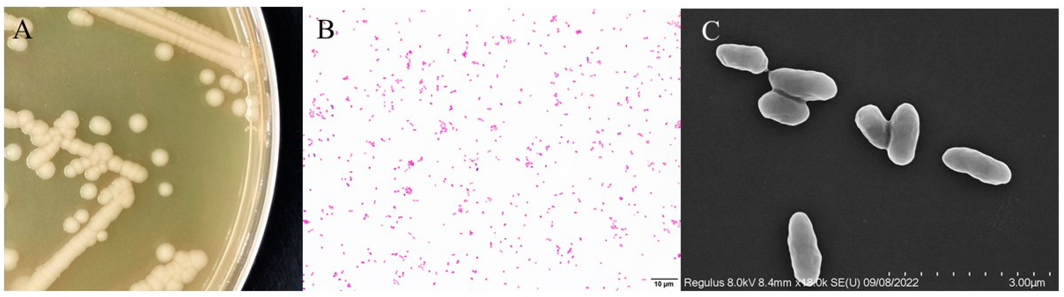
3.2. Morphological Features

The isolated HD-593 strain showed distinct growth characteristics on solid culture media. The colonies had a smooth, moist, and creamy white appearance with a characteristic odor (Figure 2A). Gram staining showed Gram-negative characteristics, indicated by red coloration under the microscope (Figure 2B). Scanning electron microscopy (SEM) further confirmed the bar-shaped morphology of the bacteria, with an average length of about 2 μm (Figure 2C).

3.3. Physiological and Biochemical Characterization of Bacteria

The HD-593 strain was identified as Escherichia coli utilizing the Biolog Microbial Automated Identification System (Biolog, Hayward, CA, USA). Table S2 details the specific metabolic responses and the decision results.

3.4. 16S rRNA Gene Sequences Analysis

The 16S rRNA gene of HD-593 is 1374 base pairs (GenBank accession number: PP155069). BLASTn analysis against the NCBI databases (http://blast.ncbi.nlm.nih.gov/, accessed on 5 November 2023) revealed high sequence similarity (>99%) between the HD-593 16S rRNA gene and those of three bacterial strains—KP868689.1, KT260578.1, and MH619508.1—all identified as E. coli. Phylogenetic analysis of the aligned sequences, employing the neighbor-joining method, was then conducted to elucidate the evolutionary relationships between HD-593 and the reference strains. As shown in Figure 3, strain HD-593 shares the same branch of the evolutionary tree as members of the genus E. coli.

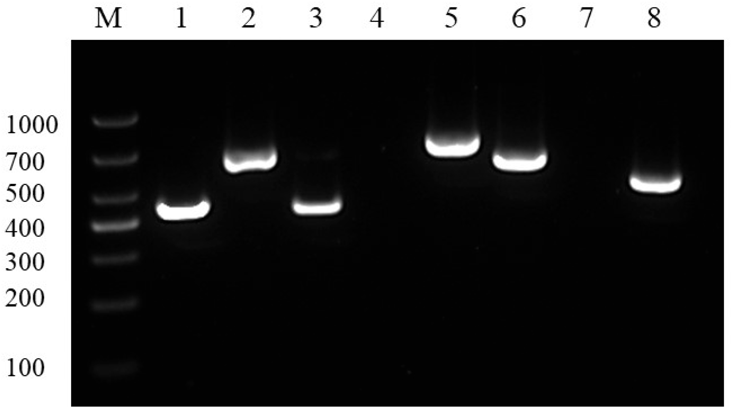
3.5. Drug Sensitivity and Drug Resistance Genes Tests
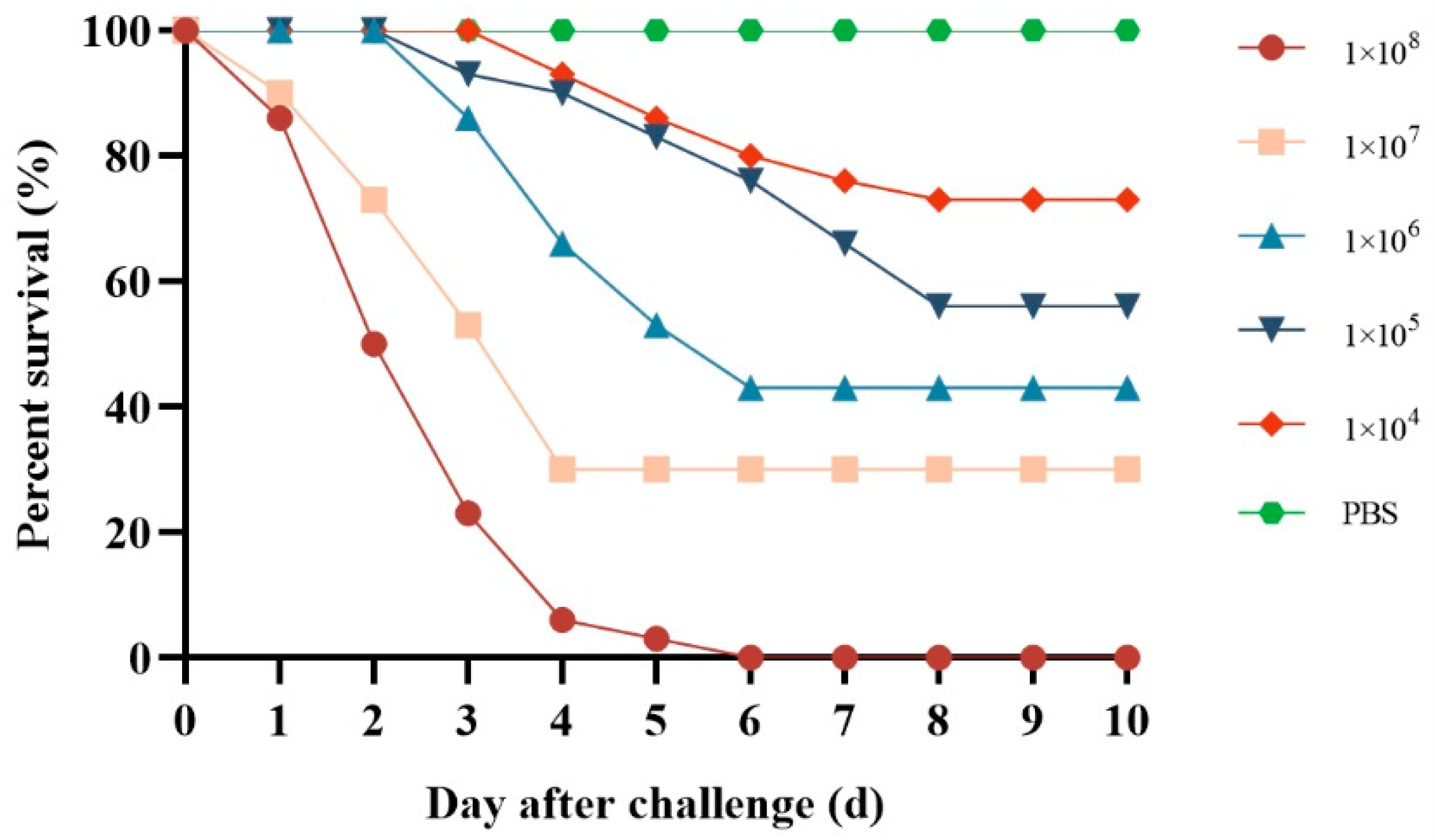
The drug susceptibility tests conducted on strain HD-593 are summarized in Table S3. The findings indicated that HD-593 exhibited resistance to ceftriaxone, enrofloxacin, doxycycline, sulfonamide, gentamicin, neomycin, florfenicol, carbenicillin, cefradine, erythromycin, penicillin, ampicillin, medithromycin, and streptomycin. Additionally, the isolate demonstrated moderate susceptibility to polymyxin and furazolidone.
According to the PCR profiles of the drug resistance genes, quinolone resistance genes (oqxA, oqxB), aminoglycoside resistance genes (aac(3)-II, aphA1), a β-lactam resistance gene (blaTEM), and an acylaminol resistance gene (floR) were present; however, the colistin resistance gene (mcr-4) and the macrolide resistance gene (ereA) were not detected (Figure 4).

3.6. Pathogenicity

The infected group exhibited mortality across all tested concentrations; however, there was no death in the control group (Figure 5). The group with the highest mortality rate was the one where P. sinensis had a bacterial load of 1 × 10⁸ CFU/g, reaching a mortality rate of 100%. The LD50 value of HD-593 was 6.53 × 10⁵ CFU/g P. sinensis weight. In the case of artificial infestation, the physical symptoms of the P. sinensis mirrored those of the naturally occurring disease, and E. coli was re-isolated from the artificially infested deceased P. sinensis.

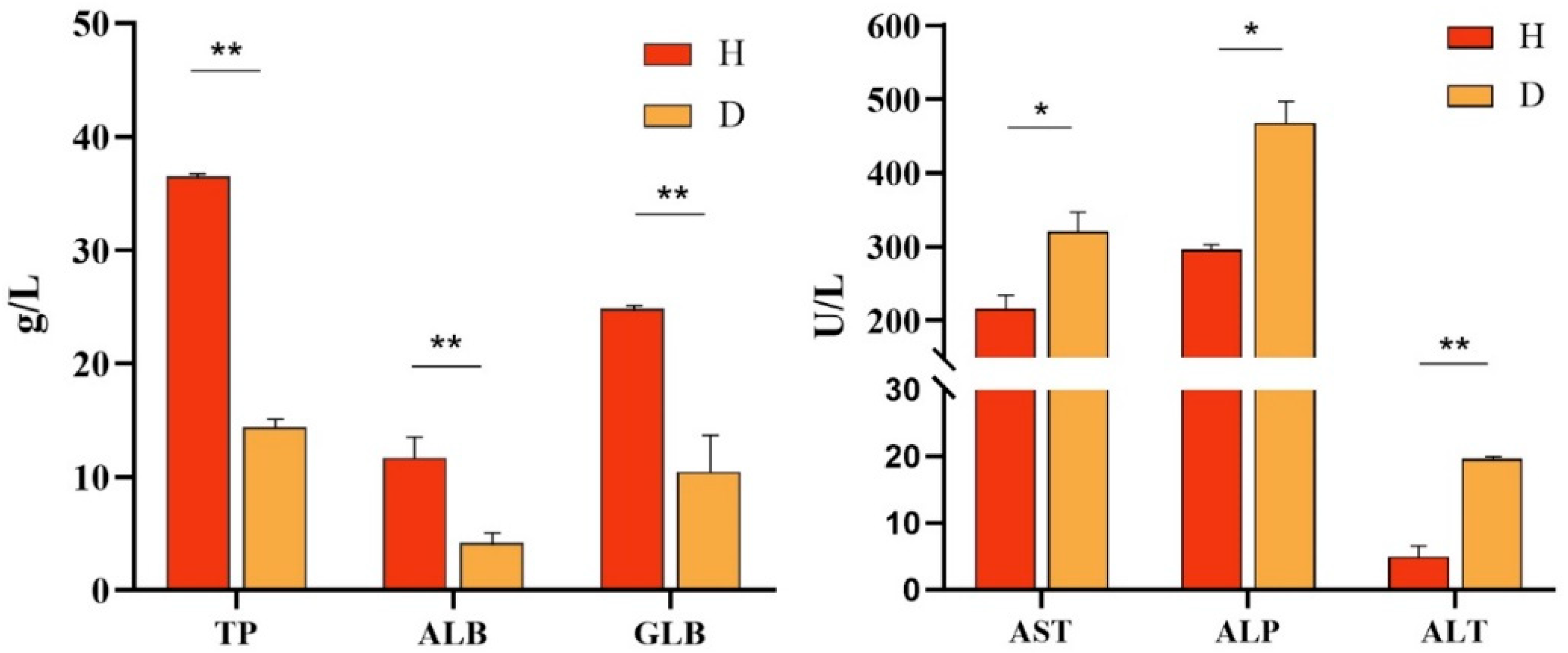
3.7. Serum Biochemistry Analysis

According to the serum biochemistry assays (Figure 6), the TP, ALB, and GLB levels in diseased P. sinensis were 14.3 g/L, 4.2 g/L, and 10.5 g/L, respectively, showing a marked decrease compared to those in healthy P. sinensis. Conversely, the levels of ASP, ALP, and ALT in diseased P. sinensis were 320.3 U/L, 468.3 U/L, and 19.6 U/L, respectively, which were significantly elevated compared to the healthy group.

4. Discussion

With the rapid development of the aquaculture industry, the need for the use of antibiotics to prevent and treat bacterial diseases is increasing. However, only 20% to 30% of the antibiotics added in aquaculture can be absorbed by the animal body, and the rest are discharged into the environment in the form of raw drugs or metabolites [15]. Antibiotics that remain in animals can cause food safety hazards [16]. Antibiotics that enter the environment are dispersed in water bodies and sediments, so that drug-resistant bacteria and drug-resistant genes in farms and the surrounding environment continue to increase, and even multiple-drug-resistant genes appear [17,18]. Unlike traditional environmental pollutants, drug-resistant genes are a new type of pollutant that is difficult to control. It can be horizontally transmitted to other bacteria through mobile genetic elements such as plasmids, integrons, and transposons, and has the characteristics of replicable transmission and environmental durability [19].
E. coli is an important indicator bacteria in food and environmental monitoring [20]. Functioning as a significant zoonotic pathogen, it is an obligate anaerobic Gram-negative bacillus and is now understood to be widely distributed in the intestinal tracts of both humans and animals, as well as prevalent in soil and water environments [21]. While the majority of E. coli strains are considered non-pathogenic to humans [22], certain variants can become pathogenic, leading to severe illnesses through intestinal infections [23]. Reports indicate the detection of E. coli in various animals, including chickens [24], pigs [25], and sheep [26]. Although there have been limited instances of E. coli isolation in aquatic animals [27], the transfer of pathogenic strains can occur in aquatic environments contaminated with E. coli. In a prior study, E. coli was found to induce intestinal and liver hemorrhage in crucian carp [28]. In this study, a strain of E. coli was isolated from diseased P. sinensis. The primary symptoms observed in the afflicted P. sinensis included enlarged liver, kidneys, and spleen, with no evidence of intestinal bleeding due to ingested food. These symptoms closely resembled the outcomes of the present experiment. Morphological characterization, bacterial biochemical identification, and molecular biology confirmed the identification of the isolated bacterium as E. coli.
The drug sensitivity tests showed that the isolated E. coli strains displayed resistance to ceftriaxone, enrofloxacin, doxycycline, sulfadiazine, gentamicin, neomycin, fosfomycin, carbenicillin, cefradine, erythromycin, penicillin, ampicillin, madicillin, and streptomycin. Additionally, they exhibited moderate susceptibility to furazolidone and polymyxin. Presently, antibiotics serve as the primary means for treating and preventing E. coli infections. However, the misuse of antibiotics has led to significant drug resistance issues and diminished efficacy. Furthermore, the E. coli HD-593 contained several resistance genes to these drugs, such as quinolone resistance genes (oqxA, oqxB), aminoglycoside resistance genes (aac(3)-II, aphA1), a β-lactam resistance gene (blaTEM), and an acylaminol resistance gene (floR). Moreover, there is a notable concern regarding drug residues, which not only hinders the effective control of E. coli disease but may also contribute to the emergence of superbugs, severely impacting the effectiveness of E. coli prevention and treatment [26,29].
Regression infection experiments successfully established the pathogenicity of the isolated strains in P. sinensis. Previous investigations determined the LD50 values of A. veronii and B. cereus in P. sinensis to be 4.17 × 10⁵ CFU/g and 6.80 × 10³ CFU/g, respectively [5,7]. In this study, pathogenicity tests revealed that the LD50 of E. coli on P. sinensis was 6.53 × 10⁵ CFU/g, indicating that the pathogenicity of E. coli HD-593 on P. sinensis was slightly lower than the above three other aquatic pathogens. However, the experimental design focused on acute infection models under controlled laboratory conditions, which may not fully replicate the multifactorial stressors (e.g., water quality fluctuations, co-infections) present in natural aquaculture settings.
Histopathological diagnosis stands as one of the foremost clinical approaches in contemporary medicine [5]. In a previous study, E. coli was identified as the causative agent for inflammatory cell infiltration in the liver and spleen of chickens [24]. Additionally, E. coli induced the shedding of intestinal villi and inflammatory cell infiltration in the spleen of crucian carp [28]. These findings align with the observations in the present study involving P. sinensis, where shedding of intestinal villi, necrosis of splenic cells, and infiltration of inflammatory cells in the liver were evident. The study suggests that E. coli infection in P. sinensis resulted in severe lesions in various tissues, including the liver, intestines, spleen, and kidneys, likely serving as a primary factor in the mortality of P. sinensis.
Animal serum plays a crucial role in their physiological and biochemical adjustments. In the sera of P. sinensis infected with A. veronii, there was a decrease in the levels of TP, ALB, and GLB. Meanwhile, the levels of AST, ALP, and ALT were elevated compared to non-infected P. sinensis [7]. In our study, a significant reduction in the proportions of serum biochemical molecules TP, ALB, and GLB was observed, indicating liver impairment, disease, and a weakened immune system. Concurrently, AST, ALP, and ALT levels were markedly elevated, suggesting inflammation in the liver and myocardium. These findings align with the results of the current experiment.

5. Conclusions

This study demonstrates that multidrug-resistant E. coli HD-593, isolated from diseased P. sinensis, poses a significant threat to aquaculture health and environmental safety. The strain exhibited resistance to 14 antibiotics and harbored critical resistance genes, highlighting risks of horizontal gene transfer and persistent environmental contamination. Pathogenicity testing confirmed its capacity to induce systemic infections in P. sinensis, with histopathological and serum biochemical analyses revealing severe multiorgan damage. These findings underscore the urgent need for antibiotic stewardship in aquaculture to mitigate the emergence of resistant pathogens. Future work should prioritize surveillance of resistance gene dissemination and explore alternatives to antibiotics, such as vaccines or probiotics, to safeguard both animal and public health.

Supplementary Materials

The following supporting information can be downloaded at https://www.mdpi.com/article/10.3390/vetsci12050473/s1, Table S1: Sequences of drug resistance genes primers; Table S2: Results of biochemical characterization of the bacterial strain HD-593; Table S3: Drug susceptibility tests of strain HD-593; Figure S1. Pathological observations of normal and diseased tissues in P. sinensis; Figure S2. Original gel image.

Author Contributions

Conceptualization, M.X., Y.M. and Y.Z.; methodology, M.X., X.H. and Y.M.; software, W.L., T.L., Z.X. and H.Z.; validation, N.J., W.L., Z.X. and C.Z.; formal analysis, C.Z., Y.W., H.Z. and T.L.; investigation, M.X., X.H., W.L. and N.J.; resources, X.H., Y.W., Z.X. and Y.M.; data curation, M.X., X.H., W.L. and N.J.; writing—original draft preparation, M.X., X.H. and Y.M.; writing—review and editing, N.J., C.Z. and W.L.; visualization, C.Z., Y.W., T.L. and H.Z.; supervision, Y.M. and Y.Z.; project administration, Y.F., Y.M. and Y.Z.; funding acquisition, M.X., Y.F., Y.M. and Y.Z. All authors have read and agreed to the published version of the manuscript.

Funding

This investigation has been funded by Central Public-interest Scientific Institution Basal Research Fund (NO. YFI20240603), the Scientific and Technological Innovation Project of Hubei Province (NO. 2023DJC102), the Central Public-interest Scientific Institution Basal Research Fund (NO. 2023TD46), and the National Freshwater Aquatic Germplasm Resource Center (NO. FGRC18537).

Institutional Review Board Statement

Ethical approval was issued by Animal Experimental Ethical Inspection of the Laboratory Animal Center, Yangtze River Fisheries Research Institute, Chinese Academy of Fishery Science YFI 2023-zhouyong-101 2023-10-08.

Informed Consent Statement

Not applicable.

Data Availability Statement

The data are contained within this paper and its Supplementary Materials.

Acknowledgments

We would like to thank Anhui Xijia Co., Ltd. and Hubei Hongwang Co., Ltd. for their support in carrying out this study.

Conflicts of Interest

The authors maintain that there are no recognized economic conflicts of interest or individual connections that they have that might have had an impact on the research presented in this paper.

References

  1. Ge, L.R.; Yu, Y.; Wen, X.X.; Xiao, H.W.; Liu, K.J.; Liu, Z.Y.; Liu, S.; Li, Q.; Wang, X.Q.; Deng, Z.F.; et al. Effects of dietary sodium butyrate on growth performance, immune function, and intestinal microflora of Chinese soft-shelled turtle (Pelodiscus sinensis). Front. Cell. Infect. Microbiol. 2023, 13, 1271912. [Google Scholar] [CrossRef] [PubMed]
  2. Lv, Z.; Hu, Y.Z.; Tan, J.; Wang, X.Q.; Liu, X.Y.; Zeng, C. Comparative Transcriptome Analysis Reveals the Molecular Immunopathogenesis of Chinese Soft-Shelled Turtle (Trionyx sinensis) Infected with Aeromonas hydrophila. Biology 2021, 10, 1218. [Google Scholar] [CrossRef] [PubMed]
  3. Zhou, L.; Lei, J.H.; Zhai, X.F.; Lu, N.N.; Shi, H.T.; Wang, J.C. Diversity of Underwater Vocalizations in Chinese Soft-Shelled Turtle (Pelodiscus sinensis). Animals 2023, 13, 812. [Google Scholar] [CrossRef] [PubMed]
  4. Chen, G.B.; Zhou, T.; Cao, J.Z.; Li, X.; Zhu, C.J.; Wang, L.; Zou, G.W.; Liang, H.W. Roles of estrogen receptors during sexual reversal in Pelodiscus sinensis. Mol. Biol. Rep. 2024, 51, 733. [Google Scholar] [CrossRef]
  5. Xiao, Z.D.; Cheng, M.M.; Hu, X.W.; Xue, M.Y.; Jiang, N.; Liu, W.; Fan, Y.D.; Meng, Y.; Xu, C.; Zhou, Y. Pathological changes of highly pathogenic Bacillus cereus on Pelodiscus sinensis. Vet. Q. 2023, 43, 1–10. [Google Scholar] [CrossRef]
  6. Xu, J.H.; Chen, H.X.; Zhang, W.R.; Xu, H.S. Evaluation of intraepithelial lymphocytes, goblet cells and immunoglobulin genes in the intestinal mucosal tissue of Pelodiscus sinensis after challenge with Aeromonas veronii bv. sobria and lipopolysaccharide. Fish. Sci. 2019, 85, 177–185. [Google Scholar] [CrossRef]
  7. Hu, X.W.; Xiao, Z.D.; Li, B.; Xue, M.Y.; Jiang, N.; Fan, Y.D.; Chen, P.; Qi, F.; Kong, X.H.; Zhou, Y. Isolation, Identification, and Characterization of Aeromonas veronii from Chinese Soft-Shelled Turtle (Trionyx sinensis). Microorganisms 2023, 11, 1304. [Google Scholar] [CrossRef]
  8. Liu, T.F.; Han, Y.W.; Chen, S.L.; Zhao, H.Y. Global characterization and expression analysis of interferon regulatory factors in response to Aeromonas hydrophila challenge in Chinese soft-shelled turtle (Pelodiscus sinensis). Fish Shellfish. Immunol. 2019, 92, 821–832. [Google Scholar] [CrossRef]
  9. Wang, R.H.; Yan, M.L.; Tan, Q.; Wang, J.L.; Luo, Y.S.; Chen, Z.Y. Isolation and genomic characterization of multi-drug resistant Morganella morganii strain from Chinese Soft-Shell Turtle Pelodiscus sinensis. Aquaculture 2025, 600, 742229. [Google Scholar] [CrossRef]
  10. Kaper, J.B.; Nataro, J.P.; Mobley, H.L.T. Pathogenic Escherichia coli. Nat. Rev. Microbiol. 2004, 2, 123–140. [Google Scholar] [CrossRef]
  11. Arbab, S.; Ullah, H.; Wang, W.W.; Zhang, J.Y. Antimicrobial drug resistance against Escherichia coli and its harmful effect on animal health. Vet. Med. Sci. 2022, 8, 1780–1786. [Google Scholar] [CrossRef] [PubMed]
  12. Singh, R.; Singh, A.P.; Kumar, S.; Giri, B.S.; Kim, K.H. Antibiotic resistance in major rivers in the world: A systematic review on occurrence, emergence, and management strategies. J. Clean. Prod. 2019, 234, 1484–1505. [Google Scholar] [CrossRef]
  13. Li, H.; Li, L.L.; Chi, Y.Y.; Tian, Q.W.; Zhou, T.T.; Han, C.H.; Zhu, Y.Q.; Zhou, Y.S. Development of a standardized Gram stain procedure for bacteria and inflammatory cells using an automated staining instrument. Microbiologyopen 2020, 9, e1099. [Google Scholar] [CrossRef]
  14. Arroyo, E.; Enríquez, L.; Sánchez, A.; Ovalle, M.; Olivas, A. Scanning electron microscopy of bacteria Tetrasphaera duodecadis. Scanning 2014, 36, 547–550. [Google Scholar] [CrossRef] [PubMed]
  15. Song, C.; Zhang, C.; Fan, L.M.; Qiu, L.P.; Wu, W.; Meng, S.L.; Hu, G.D.; Kamira, B.; Chen, J.Z. Occurrence of antibiotics and their impacts to primary productivity in fishponds around Tai Lake, China. Chemosphere 2016, 161, 127–135. [Google Scholar] [CrossRef]
  16. Ben, Y.J.; Fu, C.X.; Hu, M.; Liu, L.; Wong, M.H.; Zheng, C.M. Human health risk assessment of antibiotic resistance associated with antibiotic residues in the environment: A review. Environ. Res. 2019, 169, 483–493. [Google Scholar] [CrossRef]
  17. Obimakinde, S.; Fatoki, O.; Opeolu, B.; Olatunji, O. Veterinary pharmaceuticals in aqueous systems and associated effects: An update. Environ. Sci. Pollut. Res. 2017, 24, 3274–3297. [Google Scholar] [CrossRef]
  18. Jindal, P.; Bedi, J.; Singh, R.; Aulakh, R.; Gill, J. Phenotypic and genotypic antimicrobial resistance patterns of Escherichia coli and Klebsiella isolated from dairy farm milk, farm slurry and water in Punjab, India. Environ. Sci. Pollut. Res. 2021, 28, 28556–28570. [Google Scholar] [CrossRef]
  19. Poole, T.L.; Callaway, T.R.; Norman, K.N.; Scott, H.M.; Loneragan, G.H.; Ison, S.A.; Beier, R.C.; Harhay, D.M.; Norby, B.; Nisbet, D.J. Transferability of antimicrobial resistance from multidrug-resistant Escherichia coli isolated from cattle in the USA to E. coli and Salmonella Newport recipients. J. Glob. Antimicrob. Resist. 2017, 11, 123–132. [Google Scholar] [CrossRef]
  20. Wu, Y.; Zhu, P.P.; Xie, W.J.; Liu, Y.; Lu, H.; Tang, Q.; Han, C.Q. Study on E. coli Activity and Sterilization Effect Based on Fluorescence Spectroscopy. Spectrosc. Spectr. Anal. 2019, 39, 3619–3623. [Google Scholar] [CrossRef]
  21. Patel, J.; Yin, H.B.; Bauchan, G.; Mowery, J. Inhibition of Escherichia coli O157:H7 and Salmonella enterica virulence factors by benzyl isothiocyanate. Food Microbiol. 2020, 86, 103303. [Google Scholar] [CrossRef] [PubMed]
  22. Morcinek-Orlowska, J.; Walter, B.; Forquet, R.; Cysewski, D.; Carlier, M.; Mozolewski, M.; Meyer, S.; Glinkowska, M. Interaction networks of Escherichia coli replication proteins under different bacterial growth conditions. Sci. Data 2023, 10, 788. [Google Scholar] [CrossRef]
  23. Croxen, M.A.; Finlay, B.B. Molecular mechanisms of Escherichia coli pathogenicity. Nat. Rev. Microbiol. 2010, 8, 26–38. [Google Scholar] [CrossRef]
  24. Sarba, E.J.; Kelbesa, K.A.; Bayu, M.D.; Gebremedhin, E.Z.; Borena, B.M.; Teshale, A. Identification and antimicrobial susceptibility profile of Escherichia coli isolated from backyard chicken in and around ambo, Central Ethiopia. Bmc Vet. Res. 2019, 15, 85. [Google Scholar] [CrossRef] [PubMed]
  25. Zhao, Z.Q.; Liao, C.S.; Chang, S.K.; Ding, K.; Liu, Z.J.; Xue, Y. NDM-1-producing Escherichia coli isolated from pigs induces persistent infection with limited pathogenicity. Microb. Pathog. 2019, 135, 103620. [Google Scholar] [CrossRef] [PubMed]
  26. Gu, X.X.; Ma, X.; Wu, Q.; Tao, Q.X.C.; Chai, Y.J.; Zhou, X.; Han, M.L.; Li, J.; Huang, X.; Wu, T.Z.; et al. Isolation, identification, molecular typing, and drug resistance of Escherichia coli from infected cattle and sheep in Xinjiang, China. Vet. Med. Sci. 2023, 9, 1359–1368. [Google Scholar] [CrossRef]
  27. Amézquita-López, B.A.; Soto-Beltrán, M.; Lee, B.G.; Yambao, J.C.; Quiñones, B. Isolation, genotyping and antimicrobial resistance of Shiga toxin-producing Escherichia coli. J. Microbiol. Immunol. Infect. 2018, 51, 425–434. [Google Scholar] [CrossRef]
  28. Xue, L.G.; Luo, X.; Xing, J.H.; Wang, D.; Zhang, D.X. Isolation and pathogenicity evaluation of Escherichia coli O157:H7 from common carp, Cyprinus carpio. Microb. Pathog. 2023, 182, 106250. [Google Scholar] [CrossRef]
  29. Cengiz, M.; Sahinturk, P.; Hepbostanci, G.; Akalin, H.; Sonal, S. The in vitro activity of danofloxacin plus ceftiofur combination: Implications for antimicrobial efficacy and resistance prevention. Vet. Res. Forum 2022, 13, 149–153. [Google Scholar] [CrossRef]
Figure 1. Presentation of the diseased P. sinensis. (A): Boils on the backside (red arrows), (B): boils on the abdomen (red arrows), (C): anatomy of sick turtle, (D): enlargement of the liver, (E): no food and bleeding in the intestines.
Vetsci 12 00473 g001
Figure 2. Morphological features of HD-593 bacteria. (A): HD-593 bacteria growing in BHI solid medium. (B): HD-593 bacteria Gram stain (scale bar: 10 μm). (C): The scanning electronic micrograph of the HD-593 (scale bar: 3 μm).
Vetsci 12 00473 g002
Figure 3. The phylogenetic tree of the HD-593 (red triangle) isolate based on the neighbor-joining technique in MEGA 11. Each branch is annotated with a percentage, indicating the bootstrap values based on 1000 replicates. The scale bars represent the number of substitutions per locus.
Vetsci 12 00473 g003
Figure 4. Agarose gel electrophoresis of PCR products of the drug resistance genes. M: DL1000 marker, 1: blaTEM, 2: oqxA, 3: aac(3)-II, 4: mcr-4, 5: floR, 6: aphA1, 7: ereA, 8: oqxB. Original image—Figure S2.
Vetsci 12 00473 g004
Figure 5. Cumulative mortality of P. sinensis infected with E. coli HD-593 at different bacterial doses (1 × 10⁴–1 × 10⁸ CFU/g body weight). Groups (n = 30/group) were intraperitoneally injected with bacterial suspensions or PBS (control). Mortality was monitored for 10 days. Mortality reached 100% at the highest dose (1 × 10⁸ CFU/g). The LD50 value (6.53 × 10⁵ CFU/g) was calculated using the Reed–Muench method.
Vetsci 12 00473 g005
Figure 6. Serum biochemical parameters in healthy (H) and diseased (D) P. sinensis. Levels of total protein (TP), albumin (ALB), globulin (GLB), aspartate aminotransferase (AST), alkaline phosphatase (ALP), and alanine aminotransferase (ALT) were quantified. Values are mean ± SD (* p < 0.05; ** p < 0.01).
Vetsci 12 00473 g006
Disclaimer/Publisher’s Note: The statements, opinions and data contained in all publications are solely those of the individual author(s) and contributor(s) and not of MDPI and/or the editor(s). MDPI and/or the editor(s) disclaim responsibility for any injury to people or property resulting from any ideas, methods, instructions or products referred to in the content.

