Communication

Molecular Detection of Tick-Borne Bacterial Pathogens in Ticks and Rodents from the China–Vietnam Border

1
Chinese PLA Center for Disease Control and Prevention, Beijing 100071, China
2
Guangzhou Center for Disease Control and Prevention, Guangzhou 510440, China
3
College of Life Science and Technology, Beijing University of Chemical Technology, Beijing 100029, China
*
Authors to whom correspondence should be addressed.
These authors contributed equally to this work.
Vet. Sci. 2025, 12(3), 256; https://doi.org/10.3390/vetsci12030256
Submission received: 6 February 2025 / Revised: 3 March 2025 / Accepted: 4 March 2025 / Published: 10 March 2025

Simple Summary

Tick-borne bacterial pathogens are a threat to both animal and human health worldwide. In China, emerging and re-emerging tick-borne diseases have inspired investigations of ticks and tick-borne pathogens in recent years. The aim of this study was to evaluate tick-borne bacterial pathogens, including Rickettsia, Anaplasma, Ehrlichia, Candidatus Neoehrlichia, and Borrelia, in ticks and rodents from the China–Vietnam border, an area that has limited research on tick-borne pathogens. Four known tick-borne pathogens and two potentially novel pathogens were identified in this study. These results provide the first characterization of tick-borne bacterial pathogen diversity at the China–Vietnam border and are useful to researchers and individuals, not only from China but also from Vietnam, for the prevention of tick-borne diseases.

Abstract

Ticks and tick-borne diseases constitute a crucial focus for the health of both humans and animals worldwide. Although numerous studies on tick-borne diseases have been conducted in China, reports on tick-borne pathogens in ticks and rodents from the China–Vietnam border are scarce. In this study, we investigated tick-borne bacterial pathogens, including Rickettsia, Anaplasmataceae, and Borrelia, in nine rodents (Rattus norvegicus) and 88 ticks collected from cattle and rodents in Jingxi, a city at the China–Vietnam border. Through molecular detection and sequence analysis, four known tick-borne pathogens were identified. Specifically, Rickettsia japonica was detected in 46.3% (37/80) of Haemaphysalis cornigera; Anaplasma phagocytophilum and Candidatus Neoehrlichia mikurensis were identified in one Ixodes granulatus and one rodent, respectively; and Borrelia valaisiana was detected in two I. granulatus. Additionally, a potentially novel species of Rickettsia, provisionally named Rickettsia sp. JX, was detected in 41.3% (33/80) of Ha. cornigera, one Rhipicephalus microplus, three I. granulatus, and nine rodents, whereas a potentially novel species of Borrelia, tentatively named Borrelia sp. JX, was detected in one I. granulatus. To the best of our knowledge, this is the first report on tick-borne bacterial pathogens in ticks and rodents from the China–Vietnam border. These results expand the knowledge of the geographical distribution and vector diversity of tick-borne bacterial pathogens in China and are conducive to the evaluation of thee potential public health risk.

1. Introduction

Ticks are blood-sucking arthropods that parasitize vertebrates, such as livestock, wildlife, and humans, worldwide, ranking second only to mosquitoes as vectors of infectious diseases [1]. These arthropods undergo a three-stage life cycle (larva, nymph, adult), typically requiring a blood meal from different hosts at each stage. During the blood feeding process, ticks can transmit a variety of pathogens, including viruses, bacteria, and protozoa, thereby posing a significant threat to the health of both humans and animals [2,3]. The primary bacterial pathogens responsible for human diseases transmitted by ticks include Rickettsia, Anaplasma, Ehrlichia, Candidatus Neoehrlichia, and Borrelia. In recent years, climate warming and environmental changes have facilitated the geographic expansion of tick populations, resulting in an increase in tick-borne diseases, including emerging and re-emerging infectious diseases [4,5,6]. More than 33 emerging tick-borne pathogens have been detected in mainland China since the early 1980s [7,8,9,10,11,12,13]. However, the symptoms of tick-borne diseases (TBDs), such as fever, headache, fatigue, and myalgia, are analogous to those of influenza or the common cold, which may initially lead to misdiagnosis and delayed treatment [14,15,16].
Rickettsia species belong to the genus Rickettsia, which includes more than 70 species (including 30 candidate species). They have been categorized into four groups, the spotted fever group (SFG), the typhus group (TG), the ancestral group (AG), and the transitional group, on the basis of their phenotypic traits, encompassing ecological and epidemiological features, clinical information, and mouse serotyping results [17,18]. SFG rickettsiae (SFGR) are predominantly maintained and transmitted by ticks. In China, SFGR were mainly detected in Dermacentor silvarum, D. nuttalli, Ixodes persulcatus, Haemaphysalis longicornis, Rhipicephalus microplus, and Hyalomma asiaticum [11,19]. A previous study in Southwestern China showed an SFGR infection rate of 36.1% in ticks [20].
Anaplasma, Ehrlichia, and Candidatus Neoehrlichia are tick-borne bacterial pathogens in the family Anaplasmataceae. The genus Anaplasma comprises eight Anaplasma species, including A. phagocytophilum, A. ovis, A. bovis, A. marginale, A. platys, A. centrale, and two newly discovered species, A. odocoilei and A. capra [21]. A. capra, an emerging tick-borne pathogen, was initially detected in humans, goats, and I. persulcatus ticks in Northeastern China [22]. Moreover, A. phagocytophilum, which is the most frequently documented agent in Anaplasmataceae in China, was first detected in I. persulcatus from Heilongjiang Province in 1997, with a minimum infection rate of 0.8% [11,19]. The genus Ehrlichia includes six validated species, E. chaffeensis, E. muris, E. ewingii, E. ruminantium, E. canis, and E. minasensis, all of which have been detected in China [19,23,24,25]. The genus Candidatus Neoehrlichia is a new cluster in Anaplasmataceae. Until recently, six species were known in this genus, namely Candidatus N. mikurensis, Candidatus N. lotoris, Candidatus N. australis, Candidatus N. australis, Candidatus N. Tanzania, and Candidatus N. chilensis [26,27]. Candidatus N. mikurensis has been detected in D. silvarum, Ha. Concinna, Ha. longicornis, and I. persulcatus in China, with an infection rate of 1.6% reported in a prior study from Northeastern China [28].
Borrelia burgdorferi sensu lato complexes, which belong to the genus Borrelia, are the etiological agents of Lyme borreliosis. Although there are more than 20 genospecies in the B. burgdorferi sensu lato complexes, B. garinii, B. afzelii, and B. burgdorferi sensu stricto are the predominant pathogens responsible for Lyme borreliosis [19,29]. In China, ticks known to be vectors of Borrelia burgdorferi include I. granulatus, Hy. asiaticum, I. persulcatus, and Ha. concinna, with I. granulatus exhibiting the highest prevalence at 24% [29].
The China–Vietnam border is located in Southwestern China, which is mountainous and abundant in biological resources, offering an ecological and biological foundation for the survival and reproduction of ticks, rodents, and tick-borne pathogens. Notably, the ecological continuity of the China–Vietnam border creates shared habitats for ticks and hosts. In Northern Vietnam, pathogens such as Rickettsia spp., Anaplasma platys, and Ehrlichia canis have been detected in ticks and dogs [30,31]. However, at present, information regarding tick-borne pathogens in this region is limited. The objective of this study was to determine the existence and molecular characteristics of tick-borne bacterial pathogens in ticks and rodents from the China–Vietnam border.

2. Materials and Methods

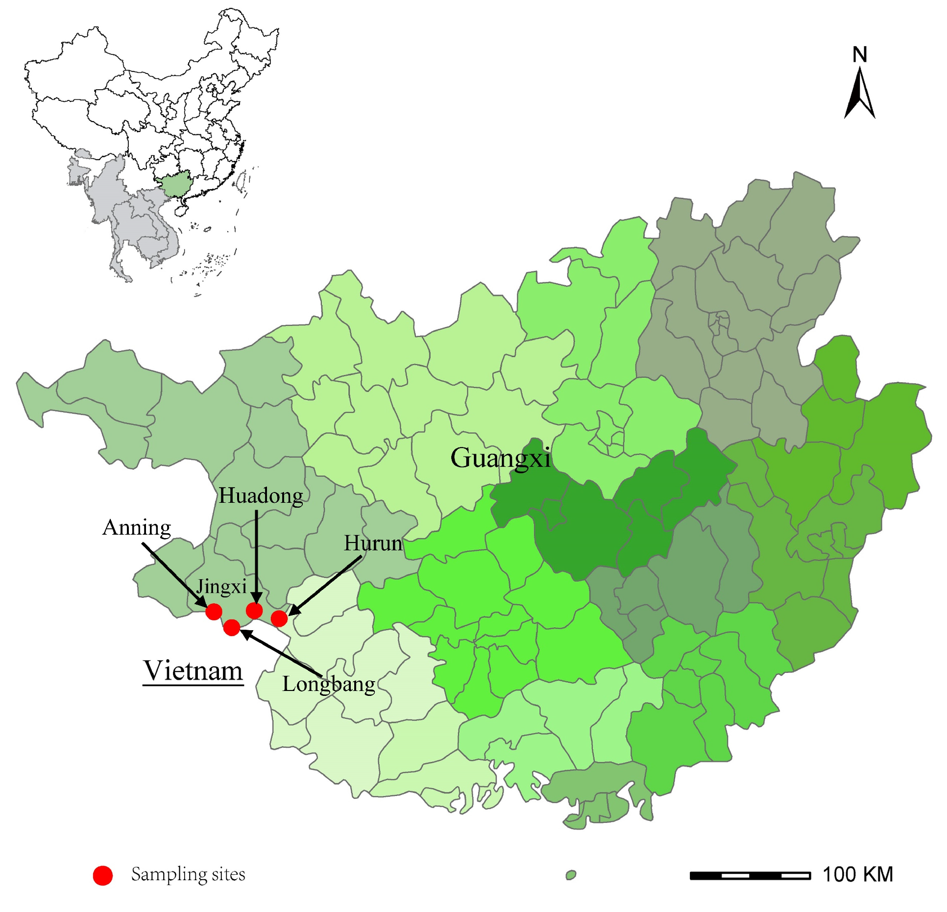
2.1. Sample Collection and Species Identification

The study area and sample sites were located in Jingxi City, Guangxi Province, Southwestern China (Figure 1). Jingxi is mainly characterized by karst plateau landforms at the border between China and Vietnam. In May 2020, ticks were initially collected from the body surfaces of cattle. To investigate tick-borne pathogens in rodents, rodents were captured using peanut bait, and ticks were collected from each rodent. All feeding ticks were removed from the hosts using steel forceps and placed in individual 5 mL tubes, each of which was hermetically sealed with breathable cotton. A total of 88 ticks and nine rodents were collected from four villages. All ticks were parasitic, with 85 specimens collected from 10 cattle and the remaining three obtained from a single rodent. The collected ticks were identified morphologically under a stereoscopic microscope according to existing morphological criteria and by molecular methods using the 16S ribosomal RNA and mitochondrial cytochrome c oxidase I genes, as previously described [32,33]. Spleen specimens were removed from each rodent after the identification of the species by morphology. All samples were stored at −80 °C to ensure their quality for further analysis.

2.2. DNA Extraction

Before DNA extraction, each tick was surface-sterilized in 75% ethanol and then rinsed twice with phosphate-buffered saline (PBS) to remove environmental contaminants. Individual ticks and 300 mg of each rodent tissue sample were singly homogenized in a 1.5 mL centrifuge tube with 2 mm stainless steel beads via an automated tissue homogenizer (TGrinder H24, Tiangen Biotech, Beijing, China). DNA extraction was performed on 200 µL of each homogenate using a DNeasy tissue kit (QIAGEN, Hilden, Germany), according to the manufacturer’s instructions.

2.3. Molecular Detection of Tick-Borne Bacterial Pathogens

The molecular identification of tick-borne bacterial pathogens was carried out using PCR assays. All samples of ticks and rodents were screened for spotted fever group Rickettsiae (SFGR), Anaplasmataceae, and Borrelia. The primers used in this study are listed in Table 1. Primer pair Rr190.70f/Rr190.602r, targeting the outer membrane protein A gene (ompA), and semi-nested primer pairs CS2d/CSEndr, RpCS877f/CSEndr, and CS2d/RpCS1258r, targeting the citrate synthase gene (gltA), were used for screened SFGR [34]. Semi-nested primer pairs Eh-out1/Eh-3-17, Eh-out1/Eh-out2, and Eh-out2f/Eh-3-17, targeting the 16S rRNA gene, were used for the detection of Anaplasmataceae and SFGR [35]. For the identification of Borrelia spp., conventional PCRs targeting the 16S rRNA gene and the flagellin gene (fla) were performed using the primer pairs 16S1/16S2 and FlaF/FlaR, respectively [36,37]. A negative control (distilled water) was included in each amplification. The PCR products were visualized on a 1.0% agarose gel stained with ethidium bromide under UV light.

2.4. Sequencing and Phylogenetic Analysis

PCR-positive products were purified using the EasyPure Quick Gel Extraction Kit (TransGen Biotech, Beijing, China) and sequenced using a commercial sequencing service (Tianyi Huiyuan, Beijing, China). The obtained nucleotide sequences were compared with those available in GenBank using nucleotide BLAST (https://blast.ncbi.nlm.nih.gov, accessed on 1 March 2025) (National Center for Biotechnology Information, NCBI, Bethesda, MD, USA), and multiple sequence alignment was performed using the ClustalW multiple alignment tool with the default parameters in MEGA 7.0 [38]. Phylogenetic trees were constructed via MEGA 7.0 based on the maximum likelihood method with the Kimura two-parameter method, and a bootstrap analysis was performed with 1000 replicates [39]

3. Results

3.1. Sample Collection

A total of 88 ticks were collected during the study. Morphological and molecular identification revealed that the ticks belonged to three species: 80 (90.9%) to Haemaphysalis cornigera, five (5.7%) to Rhipicephalus microplus, and three (3.4%) to Ixodes granulatus (Table 2). Ha. cornigera and R. microplus were collected from cattle, whereas I. granulatus was gathered from one of the nine rodents, all of which were identified as Rattus norvegicus (Rat. norvegicus). Specifically, 73 Ha. cornigera were obtained from Anning Village, along with three R. microplus from Longbang Village. Furthermore, two R. microplus and seven Ha. cornigera were obtained from Hurun Village, while Huadong Village yielded three I. granulatus and nine Rat. norvegicus.

3.2. Detection and Characterization of SFGR

Rickettsial DNA was detected in 74 ticks and nine Rat. norvegicus through the application of ompA gene primers, yielding an overall infection prevalence of 84.1% in ticks and 100% in Rat. norvegicus. Furthermore, the gltA and 16S rRNA genes were amplified and sequenced for all positive samples. Two SFGR species, namely R. japonica and a genetic variant of Rickettsia (tentatively designated as Rickettsia sp. JX), were identified through the analysis of partial fragments of the gltA, ompA, and 16S rRNA genes. Specifically, R. japonica was detected in 37 (46.3%) Ha. cornigera collected from cattle. Rickettsia sp. JX was identified in nine (100%) Rat. norvegicus and 37 (42.1%) ticks, including 33 (41.3%) Ha. cornigera, one R. microplus sampled from cattle, and three I. granulatus collected from Rat. norvegicus (Table 2).
The sequences of R. japonica derived from 37 Ha. cornigera were found to be identical across all three amplified genes. The determined ompA gene sequences showed 100% similarity to R. japonica strain LA16/2015 (CP047359) isolated from a human in Zhejiang Province, China, whereas the gltA and 16S rRNA gene sequences were 99.5% and 99.9% identical to the corresponding sequences of R. japonica strain LA16/2015, respectively. Phylogenetic analyses based on the above-mentioned three rickettsial genes revealed that the obtained R. japonica from Jingxi was clustered with the previous R. japonica strain LA16/2015, differing from other SFGR (Figure 2).
The sequences of Rickettsia sp. JX generated from 33 Ha. cornigera, one R. microplus, three I. granulatus, and nine Rat. norvegicus were identical to each other for each gene. The sequence analysis by BLAST showed that the sequences were closest to an unvalidated species, Rickettsia sp. TwKM01 (EF589609), with 100%, 99.7%, and 99.8% identity for the ompA, gltA, and 16S rRNA genes, respectively. However, the ompA sequence was 98% identical to the corresponding sequence of R. massiliae (CP000683) and 97.4% identical to R. aeschlimannii (U43800). The gltA sequence showed 99.4% similarity to R. massiliae (CP000683) and 97.4% similarity to R. aeschlimannii (MH267736). The 16S rRNA sequence shared 99.7% identity with R. massiliae (CP000683) and 99.5% identity with R. aeschlimannii (NR026042). The phylogenetic analyses suggested that Rickettsia sp. JX was closely related to Rickettsia sp. TwKM01, while it was distinct from other validated Rickettsia species and formed a separate clade.

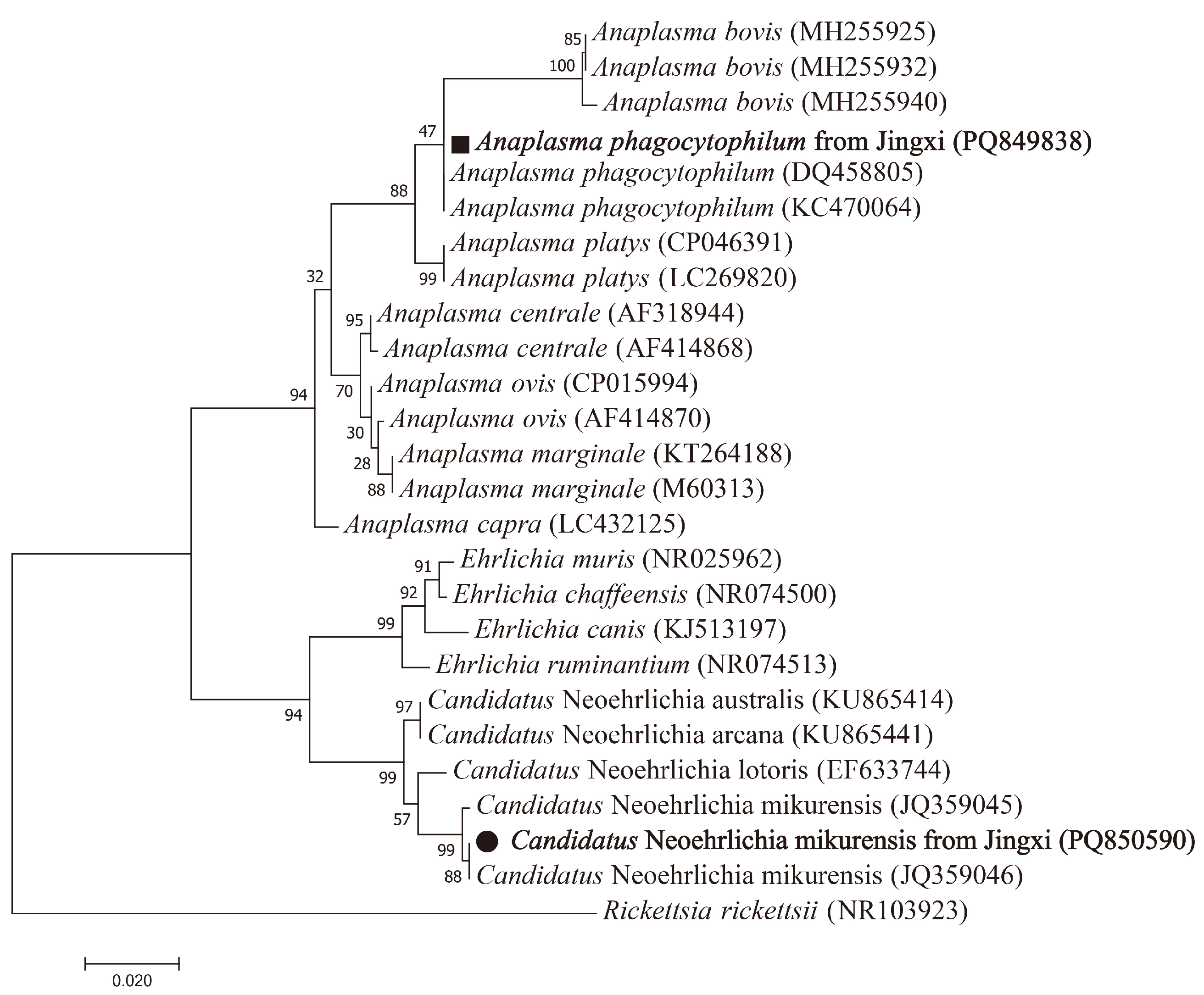
3.3. Detection and Characterization of Anaplasmataceae

In the investigation of Anaplasmataceae, one I. granulatus and one Rat. norvegicus were positive for the 16S rRNA gene. Through the analysis of the 16S rRNA gene, A. phagocytophilum and Candidatus N. mikurensis were identified in the above-mentioned I. granulatus and Rat. norvegicus, respectively. The sequence of A. phagocytophilum in this study was 100% identical to that of A. phagocytophilum from Zhejiang (DQ458805). The sequence of Candidatus N. mikurensis from Jingxi displayed 99.5% similarity to that of Candidatus N. mikurensis from Zhejiang (JQ359046) (Figure 3).

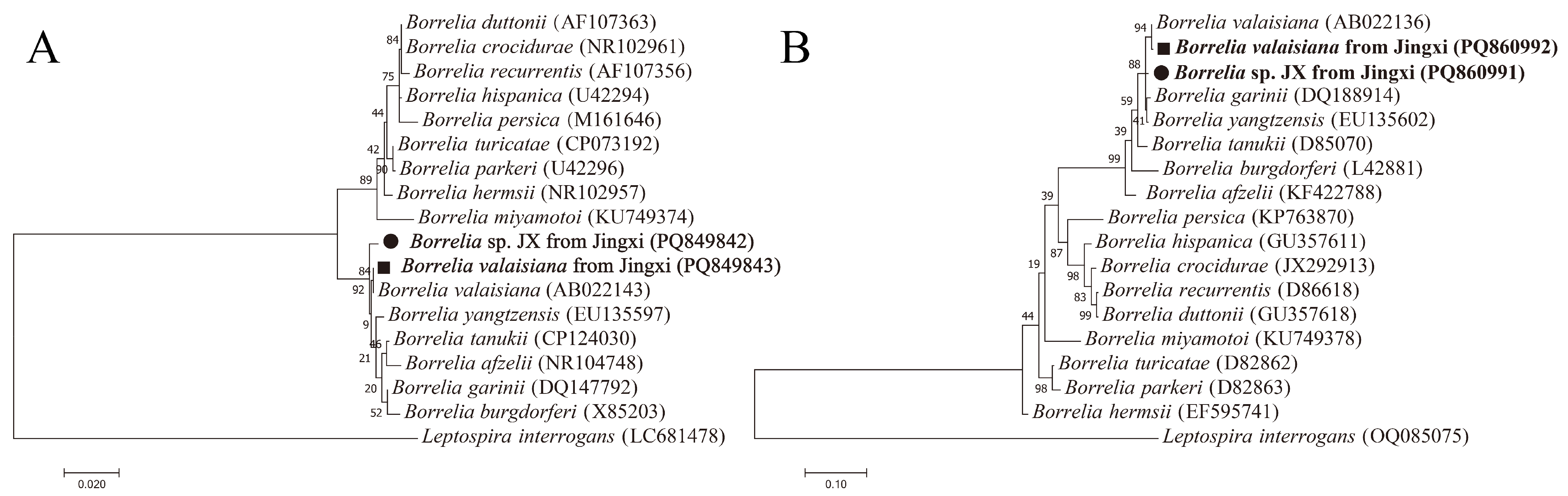
3.4. Detection and Characterization of Borrelia spp.

For Borrelia spp., all three I. granulatus obtained from Rat. norvegicus were positive for the 16S rRNA and fla genes. Two Borrelia species, namely B. valaisiana and a genetic variant of Borrelia (provisionally named Borrelia sp. JX), were determined through the analysis of the sequences of the 16S rRNA and fla genes.
The sequences of B. valaisiana obtained from two I. granulatus shared 100% identity with each other and had 99.9% and 99.8% identity in the 16S rRNA and fla genes, respectively, to the B. valaisiana detected in Zhejiang (AB022143, AB022136) (Figure 4).
The 16S rRNA gene sequence of Borrelia sp. JX from one I. granulatus was 99.5% and 99.1% identical to the corresponding sequences of B. valaisiana detected in Zhejiang (AB022143) and B. yangtzensis identified in Guizhou (EU135597), respectively. The fla gene sequence of Borrelia sp. JX showed 98.6% and 98.2% similarity to B. valaisiana discovered in Zhejiang (AB022136) and B. yangtzensis identified in Guizhou (EU135602), respectively. The phylogenetic analyses based on the 16S rRNA and fla genes indicated that Borrelia sp. JX was different from other validated Borrelia species and formed a separate clade.

3.5. Co-Infection in Individual Ticks and Rodents

The co-infection of Rickettsia sp. JX, A. phagocytophilum, and B. valaisiana was observed from one I. granulatus, while one Rat. norvegicus was co-infected by Rickettsia sp. JX and Candidatus N. mikurensis.

4. Discussion

Guangxi Province is located in the southwestern part of China, with a complex geographic environment, and possesses a border with Vietnam, extending over 1000 km. Due to the high forest cover, high species diversity, and frequent trade along the China–Vietnam border, the diversity of tick-borne pathogens deserves attention. In this study, we investigated the presence of Rickettsia, Anaplasmataceae, and Borrelia species in ticks and rodents collected from Jingxi, a city located at the China–Vietnam border. Through molecular detection and sequence analysis, two species of SFGR, specifically R. japonica and Rickettsia sp. JX, as well as A. phagocytophilum, Candidatus N. mikurensis, and two species of Borrelia, namely B. valaisiana and Borrelia sp. JX, were identified. Additionally, 80 (90.9%) ticks in this study were identified as Ha. cornigera, which has received limited study in China.
Rickettsia japonica was initially discovered in Japan in 1984 and has the capacity to cause Japanese spotted fever in humans, presenting with fever, headache, rash, eschar, and malaise [40]. Japanese spotted fever is predominant in Japan; however, cases have also been reported in China, South Korea, and Thailand [41,42,43]. In China, R. japonica has been detected in H. longicornis, H. hystricis, H. flava, H. taiwana, and R. microplus from the central, southeastern, and northeastern regions [19]. We identified R. japonica in Ha. cornigera from Jingxi. Thus, this study provides the first evidence of R. japonica in Ha. cornigera in Southwestern China. In accordance with the genetic criteria for the definition of novel rickettsial species, an isolate can be categorized as a potential new Rickettsia species if it shows no more than one of the following levels of nucleotide similarity with a validated Rickettsia species: ≥99.8%, ≥99.9%, and ≥98.8% for the 16S rRNA, gltA, and ompA genes, respectively [44]. Therefore, we regarded the newly detected Rickettsia sp. JX as a potential novel SFGR species. Rickettsia sp. JX recovered from Jingxi was closest in identity to Rickettsia sp. TwKM01, which was identified in R. haemaphysaloides from Taiwan Province, China [45]. However, in this study, Rickettsia sp. JX was detected in Rat. norvegicus and three species of ticks collected from Jingxi, including Ha. cornigera, R. microplus, and I. granulatus, which suggests that Rickettsia sp. JX is a major tick-borne pathogen prevalent in this region.
Anaplasma phagocytophilum is widely distributed globally, featuring a broad range of mammalian hosts and high genetic diversity. Moreover, six genera of ticks composed of 22 species serve as vectors of A. phagocytophilum in China [19]. In this study, A. phagocytophilum was confirmed in one I. granulatus with the 16S rRNA gene. Candidatus N. mikurensis has been detected mainly in Dermacentor silvarum, H. longicornis, and various rodents in China [11]. A previous study on rodents indicated that Rat. andamanensis and Rat. losea were the hosts of Candidatus N. mikurensis in Guangxi [46]. Additionally, in this study, Candidatus N. mikurensis was detected in Rat. Norvegicus in Guangxi, thereby expanding its host range in this region. Importantly, the findings from the present study align with an independent report of Candidatus N. mikurensis in Rat. norvegicus from Henan Province in Central China [47], collectively expanding the known host range and geographical distribution of this pathogen across distinct ecological regions.
Lyme borreliosis was first reported in America in 1975 and subsequently identified in Heilongjiang Province, China in 1986 [48]. Until recently, a total of nine genospecies of B. burgdorferi have been detected in China, among which B. garinii was the most predominant genospecies, followed by B. afzelii and B. valaisiana. In terms of the transmission of Lyme disease, I. granulatus, Hy. asiaticum, I. persulcatus, and H. concinna play significant roles in China [29]. Here, we identified B. valaisiana in two I. granulatus collected from Rat. Norvegicus. In China, B. valaisiana was initially detected in Zhejiang and was the prevalent genospecies in Southwestern China [36]. Additionally, a genetically related but distinct genospecies of B. valaisiana, namely the B. valaisiana-related genospecies, has been discovered in many areas of China [37,49,50]. Based on the 16S rRNA and fla genes, we detected a novel Borrelia sp. JX in one I. granulatus, belonging to the B. valaisiana-related genospecies. Although rodents are the major hosts of Borrelia species and the three Borrelia-positive ticks were gathered from rodents, no Borrelia species were detected in the nine Rat. norvegicus in the present study.
All Ha. cornigera and R. microplus with a high positivity rate for Rickettsia were collected from cattle; however, we did not obtain any blood samples from the cattle, which is one of the limitations of the study. Another limitation is that only the 16S rRNA gene was acquired for A. phagocytophilum and Candidatus N. mikurensis, affecting our comprehension of their genetic traits.

5. Conclusions

To our knowledge, this is the first study to investigate various tick-borne bacterial pathogens in ticks and rodents collected from the China–Vietnam border. This study provides information on the presence of R. japonica, A. phagocytophilum, Candidatus N. mikurensis, and B. valaisiana in the study region, which will promote the epidemiological and molecular understanding of tick-borne bacterial pathogens at the China–Vietnam border. Additionally, two potentially novel pathogens, namely Rickettsia sp. JX and Borrelia sp. JX, were identified for the first time in this region. These findings provide valuable insights for veterinarians and public health specialists to effectively control tick-borne diseases in this region. Although the sample size was not large, six species of tick-borne bacterial pathogens and co-infections were detected, indicating that the variety of tick-borne pathogens in this region is considerable and merits further investigation.

Author Contributions

Conceptualization, H.S. and S.Q.; methodology, H.L. and X.D.; formal analysis, H.L., W.X., X.D. and J.X.; investigation, H.W., Q.W., Y.W. and H.J.; resources, H.L.; data curation, H.L., W.X., X.D. and J.X. writing—original draft preparation, H.L. and W.X.; writing—review and editing, H.L., H.S. and S.Q.; funding acquisition, H.L. All authors have read and agreed to the published version of the manuscript.

Funding

This work was supported by the National Natural Science Foundation of China (82103895) and the National Key Research and Development Program of China (2019YFC1200505).

Institutional Review Board Statement

This study was approved by the Institutional Animal Care and Use Committee of the Chinese PLA Center for Disease Control and Prevention (IACUC-JKZX-20-26).

Informed Consent Statement

Not applicable.

Data Availability Statement

All relevant data are provided in the manuscript.

Conflicts of Interest

The authors declare no conflicts of interest.

References

  1. De la Fuente, J.; Estrada-Pena, A.; Rafael, M.; Almazan, C.; Bermudez, S.; Abdelbaset, A.E.; Kasaija, P.D.; Kabi, F.; Akande, F.A.; Ajagbe, D.O.; et al. Perception of Ticks and Tick-Borne Diseases Worldwide. Pathogens 2023, 12, 1258. [Google Scholar] [CrossRef]
  2. Dantas-Torres, F.; Chomel, B.B.; Otranto, D. Ticks and tick-borne diseases: A One Health perspective. Trends Parasitol. 2012, 28, 437–446. [Google Scholar] [CrossRef] [PubMed]
  3. Pustijanac, E.; Bursic, M.; Millotti, G.; Paliaga, P.; Ivesa, N.; Cvek, M. Tick-Borne Bacterial Diseases in Europe: Threats to public health. Eur. J. Clin. Microbiol. Infect. Dis. 2024, 43, 1261–1295. [Google Scholar] [CrossRef] [PubMed]
  4. Medlock, J.M.; Leach, S.A. Effect of climate change on vector-borne disease risk in the UK. Lancet Infect. Dis. 2015, 15, 721–730. [Google Scholar] [CrossRef] [PubMed]
  5. Bouchard, C.; Dibernardo, A.; Koffi, J.; Wood, H.; Leighton, P.A.; Lindsay, L.R. N Increased risk of tick-borne diseases with climate and environmental changes. Can. Commun. Dis. Rep. 2019, 45, 83–89. [Google Scholar] [CrossRef]
  6. Sonenshine, D.E. Range Expansion of Tick Disease Vectors in North America: Implications for Spread of Tick-Borne Disease. Int. J. Environ. Res. Public Health 2018, 15, 478. [Google Scholar] [CrossRef]
  7. Zhang, X.A.; Ma, Y.D.; Zhang, Y.F.; Hu, Z.Y.; Zhang, J.T.; Han, S.; Wang, G.; Li, S.; Wang, X.; Tang, F.; et al. A New Orthonairovirus Associated with Human Febrile Illness. N. Engl. J. Med. 2024, 391, 821–831. [Google Scholar] [CrossRef]
  8. Zhang, M.Z.; Bian, C.; Ye, R.Z.; Cui, X.M.; Yao, N.N.; Yang, J.H.; Chu, Y.L.; Su, X.L.; Wu, Y.F.; Ye, J.L.; et al. A series of patients infected with the emerging tick-borne Yezo virus in China: An active surveillance and genomic analysis. Lancet Infect. Dis. 2024. [Google Scholar] [CrossRef]
  9. Ma, J.; Lv, X.L.; Zhang, X.; Han, S.Z.; Wang, Z.D.; Li, L.; Sun, H.T.; Ma, L.X.; Cheng, Z.L.; Shao, J.W.; et al. Identification of a new orthonairovirus associated with human febrile illness in China. Nat. Med. 2021, 27, 434–439. [Google Scholar] [CrossRef]
  10. Jia, N.; Liu, H.B.; Ni, X.B.; Bell-Sakyi, L.; Zheng, Y.C.; Song, J.L.; Li, J.; Jiang, B.G.; Wang, Q.; Sun, Y.; et al. Emergence of human infection with Jingmen tick virus in China: A retrospective study. EBioMedicine 2019, 43, 317–324. [Google Scholar] [CrossRef]
  11. Fang, L.Q.; Liu, K.; Li, X.L.; Liang, S.; Yang, Y.; Yao, H.W.; Sun, R.X.; Sun, Y.; Chen, W.J.; Zuo, S.Q.; et al. Emerging tick-borne infections in mainland China: An increasing public health threat. Lancet Infect. Dis. 2015, 15, 1467–1479. [Google Scholar] [CrossRef] [PubMed]
  12. Luan, Y.; Gou, J.; Zhong, D.; Ma, L.; Yin, C.; Shu, M.; Liu, G.; Lin, Q. The Tick-Borne Pathogens: An Overview of China’s Situation. Acta Parasitol. 2023, 68, 1–20. [Google Scholar] [CrossRef]
  13. Yu, Z.; Wang, H.; Wang, T.; Sun, W.; Yang, X.; Liu, J. Tick-borne pathogens and the vector potential of ticks in China. Parasites Vectors 2015, 8, 24. [Google Scholar] [CrossRef] [PubMed]
  14. Wormser, G.P.; Dattwyler, R.J.; Shapiro, E.D.; Halperin, J.J.; Steere, A.C.; Klempner, M.S.; Krause, P.J.; Bakken, J.S.; Strle, F.; Stanek, G.; et al. The clinical assessment, treatment, and prevention of lyme disease, human granulocytic anaplasmosis, and babesiosis: Clinical practice guidelines by the Infectious Diseases Society of America. Clin. Infect. Dis. 2006, 43, 1089–1134. [Google Scholar] [CrossRef]
  15. Liu, H.B.; Wei, R.; Ni, X.B.; Zheng, Y.C.; Huo, Q.B.; Jiang, B.G.; Ma, L.; Jiang, R.R.; Lv, J.; Liu, Y.X.; et al. The prevalence and clinical characteristics of tick-borne diseases at One Sentinel Hospital in Northeastern China. Parasitology 2019, 146, 161–167. [Google Scholar] [CrossRef] [PubMed]
  16. Eilbert, W.; Matella, A. Tick-Borne Diseases. Emerg. Med. Clin. N. Am. 2024, 42, 287–302. [Google Scholar] [CrossRef]
  17. Diop, A.; El Karkouri, K.; Raoult, D.; Fournier, P.E. Genome sequence-based criteria for demarcation and definition of species in the genus Rickettsia. Int. J. Syst. Evol. Microbiol. 2020, 70, 1738–1750. [Google Scholar] [CrossRef]
  18. Xue, J.; Chen, S.S.; Jian, R.; Chen, G.Q.; Xie, G.; Du, L.; Guo, W.P. Molecular evidence of Rickettsia canadensis in ticks, Hebei, China. Infect. Genet. Evol. 2023, 115, 105506. [Google Scholar] [CrossRef]
  19. Zhao, G.P.; Wang, Y.X.; Fan, Z.W.; Ji, Y.; Liu, M.J.; Zhang, W.H.; Li, X.L.; Zhou, S.X.; Li, H.; Liang, S.; et al. Mapping ticks and tick-borne pathogens in China. Nat. Commun. 2021, 12, 1075. [Google Scholar] [CrossRef]
  20. Wang, Q.; Guo, W.B.; Pan, Y.S.; Jiang, B.G.; Du, C.H.; Que, T.C.; Zhan, L.; Wu, J.H.; Yu, M.H.; Cui, X.M.; et al. Detection of Novel Spotted Fever Group Rickettsiae (Rickettsiales: Rickettsiaceae) in Ticks (Acari: Ixodidae) in Southwestern China. J. Med. Entomol. 2021, 58, 1363–1369. [Google Scholar] [CrossRef]
  21. Rar, V.; Tkachev, S.; Tikunova, N. Genetic diversity of Anaplasma bacteria: Twenty years later. Infect. Genet. Evol. 2021, 91, 104833. [Google Scholar] [CrossRef] [PubMed]
  22. Li, H.; Zheng, Y.C.; Ma, L.; Jia, N.; Jiang, B.G.; Jiang, R.R.; Huo, Q.B.; Wang, Y.W.; Liu, H.B.; Chu, Y.L.; et al. Human infection with a novel tick-borne Anaplasma species in China: A surveillance study. Lancet Infect. Dis. 2015, 15, 663–670. [Google Scholar] [CrossRef]
  23. Li, J.; Liu, X.; Mu, J.; Yu, X.; Fei, Y.; Chang, J.; Bi, Y.; Zhou, Y.; Ding, Z.; Yin, R. Emergence of a Novel Ehrlichia minasensis Strain, Harboring the Major Immunogenic Glycoprotein trp36 with Unique Tandem Repeat and C-Terminal Region Sequences, in Haemaphysalis hystricis Ticks Removed from Free-Ranging Sheep in Hainan Province, China. Microorganisms 2019, 7, 369. [Google Scholar] [CrossRef]
  24. Dumler, J.S.; Barbet, A.F.; Bekker, C.P.; Dasch, G.A.; Palmer, G.H.; Ray, S.C.; Rikihisa, Y.; Rurangirwa, F.R. Reorganization of genera in the families Rickettsiaceae and Anaplasmataceae in the order Rickettsiales: Unification of some species of Ehrlichia with Anaplasma, Cowdria with Ehrlichia and Ehrlichia with Neorickettsia, descriptions of six new species combinations and designation of Ehrlichia equi and ‘HGE agent’ as subjective synonyms of Ehrlichia phagocytophila. Int. J. Syst. Evol. Microbiol. 2001, 51, 2145–2165. [Google Scholar] [CrossRef]
  25. Cabezas-Cruz, A.; Zweygarth, E.; Vancova, M.; Broniszewska, M.; Grubhoffer, L.; Passos, L.M.F.; Ribeiro, M.F.B.; Alberdi, P.; de la Fuente, J. Ehrlichia minasensis sp. nov., isolated from the tick Rhipicephalus microplus. Int. J. Syst. Evol. Microbiol. 2016, 66, 1426–1430. [Google Scholar] [CrossRef]
  26. Muller, A.; Monti, G.; Otth, C.; Sepulveda, P.; Bittencourt, P.; Nachum-Biala, Y.; Gutierrez, R.; Harrus, S. “Candidatus Neoehrlichia chilensis” sp. nov.: Molecular detection and characterization of a novel Anaplasmataceae in wild rodents from Valdivia, southern Chile. Transbound. Emerg. Dis. 2018, 65, 357–362. [Google Scholar] [CrossRef] [PubMed]
  27. Schwameis, M.; Auer, J.; Mitteregger, D.; Simonitsch-Klupp, I.; Ramharter, M.; Burgmann, H.; Lagler, H. Anaplasmataceae-Specific PCR for Diagnosis and Therapeutic Guidance for Symptomatic Neoehrlichiosis in Immunocompetent Host. Emerg. Infect. Dis. 2016, 22, 281–284. [Google Scholar] [CrossRef]
  28. Li, H.; Jiang, J.F.; Liu, W.; Zheng, Y.C.; Huo, Q.B.; Tang, K.; Zuo, S.Y.; Liu, K.; Jiang, B.G.; Yang, H.; et al. Human infection with Candidatus Neoehrlichia mikurensis, China. Emerg. Infect. Dis. 2012, 18, 1636–1639. [Google Scholar] [CrossRef] [PubMed]
  29. Che, T.L.; Jiang, B.G.; Xu, Q.; Zhang, Y.Q.; Lv, C.L.; Chen, J.J.; Tian, Y.J.; Yang, Y.; Hay, S.I.; Liu, W.; et al. Mapping the risk distribution of Borrelia burgdorferi sensu lato in China from 1986 to 2020: A geospatial modelling analysis. Emerg. Microbes Infect. 2022, 11, 1215–1226. [Google Scholar] [CrossRef]
  30. Nguyen, V.L.; Colella, V.; Iatta, R.; Bui, K.L.; Dantas-Torres, F.; Otranto, D. Ticks and associated pathogens from dogs in northern Vietnam. Parasitol. Res. 2019, 118, 139–142. [Google Scholar] [CrossRef]
  31. Do, T.; Bui, K.L.; Zafar, I.; Inpankaew, T.; Galon, M.E.; Ta, P.A.; Tran, K.T.; Hasan, T.; Shengwei, J.; Ma, Z.; et al. Molecular detection, risk factors, and phylogenetic analysis of tick-borne pathogens in dogs from northern Vietnam. Trop. Biomed. 2024, 41, 52–63. [Google Scholar] [CrossRef] [PubMed]
  32. Chen, Z.; Li, S.S.; Liu, J.Z. Illustrated keys to families and genera of the superfamily Ixodoidea under new taxonomic system. Zhongguo Ji Sheng Chong Xue Yu Ji Sheng Chong Bing Za Zhi 2011, 29, 302–304, 309. [Google Scholar]
  33. Chitimia, L.; Lin, R.Q.; Cosoroaba, I.; Wu, X.Y.; Song, H.Q.; Yuan, Z.G.; Zhu, X.Q. Genetic characterization of ticks from southwestern Romania by sequences of mitochondrial cox1 and nad5 genes. Exp. Appl. Acarol. 2010, 52, 305–311. [Google Scholar] [CrossRef]
  34. Parola, P.; Paddock, C.D.; Socolovschi, C.; Labruna, M.B.; Mediannikov, O.; Kernif, T.; Abdad, M.Y.; Stenos, J.; Bitam, I.; Fournier, P.E.; et al. Update on tick-borne rickettsioses around the world: A geographic approach. Clin. Microbiol. Rev. 2013, 26, 657–702. [Google Scholar] [CrossRef] [PubMed]
  35. Wen, B.; Cao, W.; Pan, H. Ehrlichiae and ehrlichial diseases in China. Ann. N. Y. Acad. Sci. 2003, 990, 45–53. [Google Scholar] [CrossRef]
  36. Masuzawa, T.; Takada, N.; Kudeken, M.; Fukui, T.; Yano, Y.; Ishiguro, F.; Kawamura, Y.; Imai, Y.; Ezaki, T. Borrelia sinica sp. nov., a lyme disease-related Borrelia species isolated in China. Int. J. Syst. Evol. Microbiol. 2001, 51, 1817–1824. [Google Scholar] [CrossRef] [PubMed]
  37. Ni, X.B.; Jia, N.; Jiang, B.G.; Sun, T.; Zheng, Y.C.; Huo, Q.B.; Liu, K.; Ma, L.; Zhao, Q.M.; Yang, H.; et al. Lyme borreliosis caused by diverse genospecies of Borrelia burgdorferi sensu lato in northeastern China. Clin. Microbiol. Infect. 2014, 20, 808–814. [Google Scholar] [CrossRef]
  38. Kumar, S.; Stecher, G.; Tamura, K. MEGA7: Molecular Evolutionary Genetics Analysis Version 7.0 for Bigger Datasets. Mol. Biol. Evol. 2016, 33, 1870–1874. [Google Scholar] [CrossRef]
  39. Hall, B.G. Building phylogenetic trees from molecular data with MEGA. Mol. Biol. Evol. 2013, 30, 1229–1235. [Google Scholar] [CrossRef]
  40. He, K.; Chen, S.; Yu, L.; Wei, D.; Xu, X. Japanese spotted fever complicated with pleural effusion in Zhejiang province, China: A case report and literature review. J. Infect. Dev. Ctries. 2024, 18, 1135–1140. [Google Scholar] [CrossRef]
  41. Li, H.; Zhang, P.H.; Du, J.; Yang, Z.D.; Cui, N.; Xing, B.; Zhang, X.A.; Liu, W. Rickettsia japonica Infections in Humans, Xinyang, China, 2014–2017. Emerg. Infect. Dis. 2019, 25, 1719–1722. [Google Scholar] [CrossRef] [PubMed]
  42. Chung, M.H.; Lee, S.H.; Kim, M.J.; Lee, J.H.; Kim, E.S.; Lee, J.S.; Kim, M.K.; Park, M.Y.; Kang, J.S. Japanese spotted fever, South Korea. Emerg. Infect. Dis. 2006, 12, 1122–1124. [Google Scholar] [CrossRef] [PubMed]
  43. Gaywee, J.; Sunyakumthorn, P.; Rodkvamtook, W.; Ruang-areerate, T.; Mason, C.J.; Sirisopana, N. Human infection with Rickettsia sp. related to R. japonica, Thailand. Emerg. Infect. Dis. 2007, 13, 657–659. [Google Scholar] [CrossRef]
  44. Fournier, P.E.; Dumler, J.S.; Greub, G.; Zhang, J.; Wu, Y.; Raoult, D. Gene sequence-based criteria for identification of new rickettsia isolates and description of Rickettsia heilongjiangensis sp. nov. J. Clin. Microbiol. 2003, 41, 5456–5465. [Google Scholar] [CrossRef]
  45. Tsui, P.Y.; Tsai, K.H.; Weng, M.H.; Hung, Y.W.; Liu, Y.T.; Hu, K.Y.; Lien, J.C.; Lin, P.R.; Shaio, M.F.; Wang, H.C.; et al. Molecular detection and characterization of spotted fever group rickettsiae in Taiwan. Am. J. Trop. Med. Hyg. 2007, 77, 883–890. [Google Scholar] [CrossRef]
  46. Xue, J.; Chen, S.S.; Jian, R.; Chen, G.Q.; Qin, X.; Lu, M.; Wang, W.; Xie, G.C.; Du, L.; Li, K.; et al. Great genetic diversity of vector-borne bacteria and protozoan in wild rodents from Guangxi, China. PLoS Negl. Trop. Dis. 2024, 18, e0012159. [Google Scholar] [CrossRef] [PubMed]
  47. Li, H.; Jiang, J.; Tang, F.; Sun, Y.; Li, Z.; Zhang, W.; Gong, Z.; Liu, K.; Yang, H.; Liu, W.; et al. Wide distribution and genetic diversity of “Candidatus Neoehrlichia mikurensis” in rodents from China. Appl. Environ. Microbiol. 2013, 79, 1024–1027. [Google Scholar] [CrossRef] [PubMed]
  48. Ai, C.X.; Wen, Y.X.; Zhang, Y.G.; Wang, S.S.; Qiu, Q.C.; Shi, Z.X.; Li, D.Y.; Chen, D.Q.; Liu, X.D.; Zhao, J.H. Clinical manifestations and epidemiological characteristics of Lyme disease in Hailin county, Heilongjiang Province, China. Ann. N. Y. Acad. Sci. 1988, 539, 302–313. [Google Scholar] [CrossRef]
  49. Chu, C.Y.; Liu, W.; Jiang, B.G.; Wang, D.M.; Jiang, W.J.; Zhao, Q.M.; Zhang, P.H.; Wang, Z.X.; Tang, G.P.; Yang, H.; et al. Novel genospecies of Borrelia burgdorferi sensu lato from rodents and ticks in southwestern China. J. Clin. Microbiol. 2008, 46, 3130–3133. [Google Scholar] [CrossRef]
  50. Hou, J.; Ling, F.; Chai, C.; Lu, Y.; Yu, X.; Lin, J.; Sun, J.; Chang, Y.; Ye, X.; Gu, S.; et al. Prevalence of Borrelia burgdorferi sensu lato in ticks from eastern China. Am. J. Trop. Med. Hyg. 2015, 92, 262–266. [Google Scholar] [CrossRef]
Figure 1. Map of the sampling sites in this study. The red dots are sampling sites in Jingxi City, Guangxi Province.
Vetsci 12 00256 g001
Figure 2. Phylogenetic trees of genus Rickettsia constructed using the maximum likelihood method in MEGA 7.0. (A) Phylogenetic tree based on ompA genes (500 bp). (B) Phylogenetic tree based on gltA genes (1099 bp). (C) Phylogenetic tree based on 16S rRNA genes (1397 bp). Scale bars indicate estimated evolutionary distances. The values at the nodes represent the percentage support from 1000 bootstrap replicates. Circles and squares indicate Rickettsia sp. JX and Rickettsia japonica identified in this study, respectively.
Vetsci 12 00256 g002
Figure 3. Phylogenetic tree based on 16S rRNA genes (735 bp) of Anaplasmataceae using the maximum likelihood method in MEGA 7.0. Scale bars indicate estimated evolutionary distances. The values at the nodes represent the percentage support from 1000 bootstrap replicates. Circles and squares indicate Candidatus N. mikurensis and A. phagocytophilum identified in this study, respectively.
Vetsci 12 00256 g003
Figure 4. Phylogenetic trees of genus Borrelia constructed using the maximum likelihood method in MEGA 7.0. (A) Phylogenetic tree based on 16S rRNA genes (1381 bp). (B) Phylogenetic tree based on fla genes (560 bp). Scale bars indicate estimated evolutionary distances. The values at the nodes represent the percentage support from 1000 bootstrap replicates. Circles and squares indicate Borrelia sp. JX and B. valaisiana identified in this study, respectively.
Vetsci 12 00256 g004
Table 1. Primers used in this study.
PathogensGenePrimer NameExpected Size (bp)Sequence (5′-3′)Reference
SFGRompARr190.70f532ATGGCGAATATTTCTCCAAAA
Rr190.602rAGTGCAGCATTCGCTCCCCCT
gltACS2d1256ATGACCAATGAAAATAATAAT[34]
RpCS1258rATTGCAAAAAGTACAGTGAACA
CSEndr750CTTATACTCTCTATGTACA
RpCS877fGGGGACCTGCTCACGGCGG
SFGR and Anaplasmataceae16S rRNAEh-out11438TTGAGAGTTTGATCCTGGCTCAGAACG
Eh-3-17GATAGCGGAATTCCTAGTGTAGAGGTG
Eh-out1660TTGAGAGTTTGATCCTGGCTCAGAACG[35]
Eh-out2TAAGGTGGTAATCCAGC
Eh-out2f890CACCTCTACACTAGGAATTCCGCTATC
Eh-3-17GATAGCGGAATTCCTAGTGTAGAGGTG
Borrelia. spp.16S rRNA16S11523ATAACGAAGAGTTTGATCCTGGC
16S2CAGCCGCACTTTCCAGTACG[36]
flaFlaF 588TTAGGTTTTCAATAGCATACTCAG
FlaR GCAGTTCAATCAGGTAACGG[37]
Table 2. The SFGR detected in ticks collected from the China–Vietnam border.
TickHostNo. of Ticks (%)Number of SFGR (%, 95% CI)
Rickettsia japonicaRickettsia sp. JXTotal
Ha. cornigeracattle80 (90.9)37 (46.3, 35.2–57.7)33 (41.3, 30.5–52.8)70 (87.5, 77.8–93.5)
R. micropluscattle5 (5.7)01 (20, 1.1–70.1)1 (20, 1.1–70.1)
I. granulatusR. norvegicus3 (3.4)03 (100, 31–100)3 (100, 31–100)
Total 8837 (42.1, 31.8–53.1)37 (42.1, 31.8–53.1)74 (84.1, 74.4–90.7)
Disclaimer/Publisher’s Note: The statements, opinions and data contained in all publications are solely those of the individual author(s) and contributor(s) and not of MDPI and/or the editor(s). MDPI and/or the editor(s) disclaim responsibility for any injury to people or property resulting from any ideas, methods, instructions or products referred to in the content.

