Article

Changes in the Microbiome in Yak Mastitis: Insights Based on Full-Length 16S rRNA Sequencing

1
College of Veterinary Medicine, Gansu Agricultural University, Lanzhou 730070, China
2
Institute of Animal Husbandry and Veterinary Medicine, Xizang Academy of Agriculture and Animal Husbandry Sciences, Lhasa 850009, China
3
College of Veterinary Medicine, Huazhong Agricultural University, Wuhan 430070, China
*
Author to whom correspondence should be addressed.
Vet. Sci. 2024, 11(8), 335; https://doi.org/10.3390/vetsci11080335
Submission received: 5 June 2024 / Revised: 3 July 2024 / Accepted: 23 July 2024 / Published: 25 July 2024

Abstract

:

Simple Summary

Mastitis is an infection of the mammary glands that affects yaks‘ health and milk quality. The various bacteria found in mastitis-affected yaks were examined using a technique for reading particular genetic information. We discovered that contaminated milk had higher concentrations of bacteria than healthy milk. Our findings highlight the complexity and irregularity in the milk microbiome of yaks. Understanding these bacterial communities may aid in developing more effective mastitis control measures and enhance the health and milk quality of yaks, thereby benefiting dairy producers and yak farmers.

Abstract

Mastitis is an inflammation of the mammary gland that can be caused by various factors, including biological, chemical, mechanical, or physical. Microbiological culture, DNA techniques, and high-throughput next-generation sequencing have been used to identify mastitis-causing pathogens in various animal species. However, little is known about microbiota and microbiome changes linked to yak milk mastitis. This study aimed to characterize the milk microbiota of healthy and mastitis-infected yaks using full-length 16S rRNA sequencing. The results showed that the bacterial microbiota comprises 7 phyla, 9 classes, 20 orders, 39 families, 59 genera, and 72 species. Proteobacteria and Firmicutes were the predominant microbial communities, with lower abundances of Bacteroidota, Actinobacteriota, Acidobacteriota, and other minor groupings also observed. Proteobacteria dominated the clinical and subclinical mastitis groups (95.36% and 89.32%, respectively), in contrast to the healthy group (60.17%). Conversely, Firmicutes were more common in the healthy group (39.7%) than in the subclinical and clinical mastitis groups (10.49% and 2.92%, respectively). The predominant organisms found in the healthy group were Leuconostoc mesenteroides, Lactococcus piscium, Carnobacterium maltaromaticum, and Lactococcus raffinolactis. Low abundances of Staphylococcus aureus species were found in both subclinical and clinical mastitis groups, with Moraxella osloensis and Psychrobacter cibarius dominating the subclinical mastitis group and Pseudomonas fluorescens dominating the clinical mastitis group. An alpha diversity study revealed that the healthy group had a higher microbial diversity than the clinical and subclinical mastitis groups. According to beta-diversity analysis, the principal coordinate analysis identified that mastitis-infected samples significantly differed from healthy ones. The milk microbiota of healthy yaks is more varied, and specific prominent taxa within various groups can act as marker microorganisms for mastitis risk. The genera Leuconostoc and Lactococcus are promising candidates for creating probiotics.

1. Introduction

Mastitis is a well-known inflammatory condition affecting dairy cattle udders and significantly affecting the quantity and quality of milk dairy herds produce and their general well-being [1]. Numerous bacteria that enter the teat canal and cause inflammation and harm to the mammary gland tissue are the primary triggers of this disorder [2]. Mastitis typically induces pain and discomfort in affected cows, which reduces their feed intake and milk output [3,4]. As a result, it is critical to understand the substantial impact of this condition on milk production. Additionally, changes in the somatic cell counts (SCC) and disruptions in the homeostasis of the bovine milk microbial community may indicate infection in the udders, resulting in modifications to the physical and chemical properties of the milk [5,6]. It becomes evident that mastitis poses significant challenges for dairy farmers worldwide due to its adverse effects on milk production levels and the long-term viability of their businesses.
Molecular methods have emerged as state-of-the-art technology for disease diagnosis, exceeding conventional methods due to the rapid advancement of high-throughput sequencing techniques in recent years. These techniques have demonstrated outstanding efficiency and dependability while offering a comprehensive knowledge of various microbes in complex ecosystems, including the udder microbiome [7,8]. Several microbiological techniques have identified specific bacterial taxa linked to healthy, subclinical, and clinical mastitis. Without the requirement for isolation or cultivation, these investigations have made it possible to identify every bacterium found in a sample fully and have contributed to understanding microbial communities’ functional profiles [9,10]. A comprehensive review of research conducted on milk microflora shows that typical taxa with various geographical origins are reliably identified in cows’ milk. In studies on the microbiota of bovine dairy products, Streptococcus, Pseudomonas, Bifidobacterium, Propionibacterium, Bacteroides, Corynebacterium, and Enterococcus are the leading taxa that are most frequently reported [11]. Furthermore, it has become easier to monitor changes in the populations of pathogenic bacteria that cause mastitis infections in cattle by applying metataxonomic approaches [12,13]. These findings contribute to understanding the interactions between bacteria in this environment and their impact on mammary gland health. Therefore, an in-depth understanding of the microbial composition of the udder microbiome is essential in preventing or curing this illness.
Yaks are an indigenous breed with properties enabling them to thrive in the harshest environments [14]. Yaks are significant to the local economy and culture due to their milk, meat, fiber (yak wool), and mobility [15]. The nutrient-rich milk of yaks is a vital nutritional resource for local populations who rely on this versatile animal for dairy products such as butter, cheese, yogurt, and traditional beverages [16]. However, little is known about the microbiota of its milk or udder. In this work, we comprehensively investigated bacterial microbiota based on full-length 16S rRNA sequencing to investigate yaks’ milk microbiota. By identifying different taxa of bacteria associated with healthy and mastitis-affected yaks, we intend to develop specialized probiotics that can effectively prevent or treat yak mastitis.

2. Materials and Methods

2.1. Sample Collection and Assessing Clinical Health Status

A total of 235 yak milk samples were collected in the Naqu district of Xizang. Based on comparable husbandry and management techniques, four homes were carefully chosen to guarantee the study’s validity and dependability. To ensure consistency within our sample population, we included only mature yaks between the ages of three and six that had undergone at least one lactation phase and had not received antibiotic treatment in the previous two months. The animals were separated into healthy and mastitis groups based on physical examination, assessment of udder condition, Lanzhou mastitis test (LMT), and somatic cell counts in milk samples. Out of all the samples collected, fifteen were chosen at random and taken for analysis. The samples included five from animals in good health with no signs of abnormal udder health, five from those with clinical mastitis and apparent abnormalities or inflammation, and five from those with subclinical mastitis showing no symptoms but still having a bacterial infection detectable through laboratory testing. Following the laboratory and field handbook instructions on bovine mastitis, milk samples were collected from each of the chosen animals as part of the mastitis detection screening procedure. The original milk was disposed of, and the teat-ends were carefully cleansed and properly sanitized with a 70% ethanol solution before being collected. The milk samples were transported to the laboratory and kept at −80 °C until further processing.

2.2. DNA Extraction

The DNA was extracted from yak milk samples using the TGuide S96 Magnetic Soil/Stool DNA Kit (Tiangen Biotech Co., Ltd., Beijing, China) following the manufacturer’s instructions. The DNA content of the samples was determined using the Qubit dsDNA HS Assay Kit and Qubit 4.0 Fluorometer (Invitrogen, Thermo Fisher Scientific, Hillsboro, OR, USA).

2.3. Amplicon Sequencing

The full-length 16S rRNA gene was amplified from the isolated genomic DNA of each sample using the universal primer sets 27F (AGRGTTTGATYNTGGCTCAG) and 1492R (TASGGHTACCTTGTTASGACTT). Sample-specific PacBio barcode sequences were incorporated into the forward and reverse primers to facilitate multiplexed sequencing. Barcoded primers were preferred over an alternative method involving a second PCR reaction to lessen the production of chimeras. The amplification procedure included 25 cycles of PCR using KOD One PCR Master Mix (TOYOBOLife Science, Tokyo, Japan), with the initial denaturation occurring at 95 °C for two minutes. Following this, denaturation occurred for 10 s at 98 °C, 30 s of annealing at 55 °C, 1 min and 30 s of extension at 72 °C per cycle, and an additional 2 min of annealing at 72 °C. After the PCR, amplicons were purified using Agencourt AMPure XP Beads (Beckman Coulter, Indianapolis, Indiana, USA), the findings were quantified using the Qubit 4.0 Fluorometer and dsDNA HS Assay Kit (Invitrogen, Thermo Fisher Scientific, Hillsboro, OR, USA). The exact number of amplicons was merged after each individual was quantified. The amplified DNA was used to build SMRTbell libraries using the SMRTbell Express Template Prep Kit 2.0 and the manufacturer’s instructions (Pacific Biosciences, Menlo Park, CA, USA). The purified SMRTbell libraries from the pooled and barcoded samples were sequenced on a single PacBio Sequel II 8M cell using the Sequel II Sequencing Kit 2.0.

2.4. Bioinformatic Analysis

The bioinformatics analysis of this work was performed using the BMK Cloud (Biomarker Technologies Co., Ltd., Beijing, China) to enhance its computing capabilities. The raw sequencing reads were filtered and demultiplexed using SMRT Link software (version 8.0; https://www.pacb.com/smrt-link/; accessed on 18 January 2024), with specific requirements minPasses ≥ 5 and minPredictedAccuracy ≥ 0.9, to obtain high-quality circular consensus sequencing (CCS) reads. Next, based on barcodes, the CCS reads were assigned to the proper samples using Lima software (version 1.7.0). To perform quality filtering and identify forward and reverse primers, CCS values that were either primer-deficient or fell outside the 1200–1650 bp length range were removed using the Cutadapt (version 3.4) quality control process [17]. Using the UCHIME method (version 8.1; https://drive5.com/usearch/manual/uchime_algo.html; accessed on 1 February 2024) to identify and eliminate chimeric reads produced high-quality readings. Reads showing ≥97% similarity were classified into the same operational taxonomic unit (OTU) using USEARCH  (version 10.0), and OTUs with an abundance of <0.005% were filtered out. The taxonomy annotation of the OTUs was performed with a 70% confidence level based on the naive Bayes classifier in QIIME2 [18] using the SILVA database [19]. The programs used to calculate and show the alpha diversity were R and QIIME2, respectively. Using QIIME, beta diversity was assessed to gauge how similar the microbial communities were across different samples. Principal coordinate analysis (PCoA), heat maps, unweighted partial graph modeling (UPGMA), and nonmetric multidimensional scaling (NMDS) were employed in the beta diversity analysis. Furthermore, we used linear discriminant analysis (LDA) effect size (LEfSe) to look for significant taxonomic differences across groups. A logarithmic LDA score of 4.0 was used as a threshold for discriminative features. Redundancy analysis (RDA) was used to examine the variations in microbiome composition associated with different parameters. The package vegan in R (version 3.1; https://cran.r-project.org/web/packages/vegan/index.html; accessed on 18 February 2024) was utilized for this purpose.

2.5. Statistical Analysis

The data were analyzed using a one-way analysis of variance (ANOVA) and the least significant difference (LSD) test in SPSS 22.0. The information was provided as the standard error (SEM) ± mean. The statistical significance levels were set at p < 0.05 or p < 0.01 to show significant or extremely significant differences between the groups. Utilizing the resources available on the Biomarker Technologies Co., Ltd. (Beijing, China) BMKCloud platform (www.biocloud.net; accessed on 3 March 2024), we investigated alpha/beta diversity indices, Venn diagrams, and classes of bacteria.

3. Results

3.1. Somatic Cell Counts

Our results show that 86 (36.6%) of the 235 yaks whose udder health was assessed had subclinical mastitis but they showed no abnormalities or inflammatory signs. Still, these yaks exhibited SCC > 200,000 cells/mL milk and tested positive for LMT. Following the National Mastitis Council requirements, only 10 (4.25%) instances had mammary gland irritation with obvious inflammatory signs in the udder or anomalies in the milk.

3.2. Taxonomic Identification of Milk Bacterial Community in Yak

We used 16S rRNA sequencing to evaluate the bacterial community’s diversity within each sample and group. All of the collected yak milk samples had 193,645 CCS reads that were obtained using the PacBio platform. The 193,191 high-quality, effective CCS reads were obtained after read-quality filtering; the total number of reads for the clinical mastitis, subclinical mastitis, and healthy groups were, respectively, 12,999 ± 243, 12,671 ± 471, and 12,967 ± 267 (Table S1). Based on a 97% sequence similarity, the average number of OTUs in all samples was 21 (range 9 to 45, SEM = 2). The rarefaction curves on the number of OTUs show that the sequencing depth utilized in this study sufficiently described the bacterial microbiota of the milk samples (Figure S1). The Venn diagram (Figure 1) shows that all three groups shared 23 OTUs; the groups with clinical mastitis, subclinical mastitis, and healthy skin had 1, 2, and 19 distinct OTUs, respectively. The examination of the milk microbiome profile revealed a variable distribution of 7 phyla, 9 classes, 20 orders, 39 families, 59 genera, and 72 species among numerous groups.

3.3. Characterization of the Bacterial Diversity in Yak Milk Samples

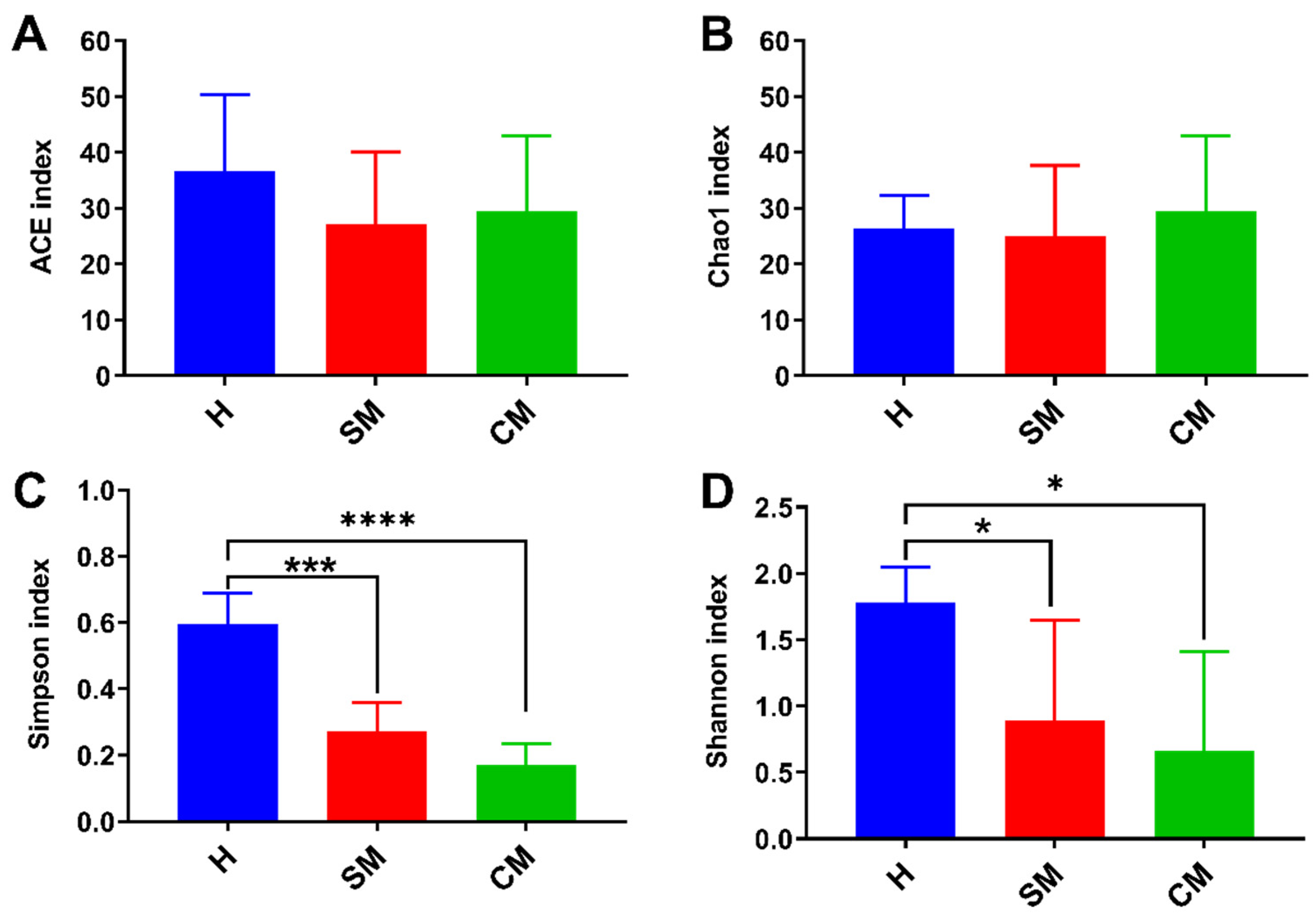
The α and β-diversity indices (ACE, Chao1, Shannon, and Simpson) and binary Jaccard, Bray–Curtis, unweighted, and weighted Unifrac metrics were used to compute the microbiota diversity values for the three groups. Alpha diversity is indicative of the richness and diversity of the microbiota. Comparisons between the Shannon and Simpson index results of the healthy group (H) and clinical mastitis (CM) group and between the healthy group and subclinical mastitis (SM) group both showed a significant difference (p < 0.001 and p < 0.05, respectively). The ACE and Chao1 indices did not significantly differ from one another in any of the comparisons (Figure 2). In addition; the β-diversity was measured using binary Jaccard PCoA to evaluate the similarities between microbial communities. All milk samples were found to be grouped into three clusters (Figure 3), with principal components PC1, PC2, and PC3 accounting for 23.23%, 16.92%, and 14.25% of the observed variance, respectively. These findings were based on Adonis [20], p = 0.001, R2 = 0.486. UPGMA analysis supported the findings, demonstrating that the clinical mastitis group matched the subclinical mastitis group more closely than the healthy group.

3.4. Taxonomic Bacterial Profiling of Milk Samples from Yak

Proteobacteria accounted for 81.61% of the most predominant phylum, followed by Firmicutes (17.71%). In lesser abundances, Bacteriodota, Actinobacteriota, Acidobacteriota, and more small groups were found (Figure 4A). Predominant was most prevalent in the group with clinical mastitis (95.36%), followed by the subclinical mastitis group (89.32%), and the healthy group (60.17%). Most firmicutes were observed in the subclinical mastitis group (10.49%) and the healthy group (39.7%). It was identified in lower abundance (2.92%) in the group with clinical mastitis. The clinical mastitis group had the highest abundance of Bacteroidota (1.69%), followed by the subclinical mastitis group (0.14%), whereas the healthy group had the lowest (0.005%). Actinobacteriota was more abundant in the healthy group (0.057%) than in the clinical mastitis group or subclinical mastitis groups. Acidobacteriota was only discovered in the group with clinical mastitis; it was not present in the other groups (Table S2).
The 59 taxa were identified overall by taxonomic classification of the yak milk microbiome, with varying abundance levels in each of the three groups. The Pseudomonas, Leuconostoc, and Lactococcus genera were found to be highly abundant in the healthy group (Figure 4B). There was an indication of Pseudomonas in all three groups. However, the clinical mastitis group had the highest percentage abundance (77.18%), followed by the subclinical mastitis group (49.03%), and the healthy group (56.93%). The subclinical mastitis group had the significantly highest abundance of the Enhydrobacter genus (27.94%) compared to the other groups. In terms of percentage abundance, the subclinical mastitis group had the highest percentage abundance of the Psychrobacter genus (12.15%), followed by the clinical mastitis group (11.95%). In comparison, the healthy group (0.52%) had the lowest. The Leuconostoc genus was discovered to be the most common (16.14%) in the healthy group, with a considerable abundance. This was followed by the subclinical group (3.82%) and the clinical mastitis group (0.98%). Brochothrix was found in abundance in the groups with subclinical mastitis (6.03%), clinical mastitis (1.12%), and healthy yaks (6.43%). The percentage abundance of Lactococcus in the healthy group was significantly higher (13.03%) than in the other groups. Carnobacterium was most abundant in the healthy group (3.56%) but less abundant in the groups with subclinical and clinical mastitis (0.04% and 0.03%, respectively). Rahnella1 was significantly higher (2.58%) in the healthy group and lower (0.002%) in the clinical mastitis group, but it was not detected in the subclinical mastitis group. Comparing the subclinical mastitis group to the clinical mastitis group, the percentage abundance of Chryseobacterium was higher in the mastitis group (1.54%). Weissella was also found in the groups with subclinical mastitis (0.23%), clinical mastitis (0.22%), and healthy yaks (0.31%).
The 72 different species of bacteria were identified in each of the three groups at the species level. As shown in Figure 4C, Pseudomonas fluorescens (56.93%), Leuconostoc mesenteroides (16.14%), Lactococcus piscium (12.33%), Brochothrix thermosphacta (6.43%), Carnobacterium maltaromaticum (3.56%), Rahnella aquatilis (2.58%), and Lactococcus raffinolactis (0.69%) comprised the majority in the healthy group. There were no indications of Staphylococcus aureus or Streptococcus dysgalactiae in the healthy group. In the subclinical mastitis group, Pseudomonas fluorescens (49.03%), Moraxella osloensis (27.94%), and Psychrobacter cibarius (12.12%) were the most common pathogens. Lactococcus piscium and Carnobacterium maltaromaticum abundance were less than 1%. Lactococcus raffinolactis and Rahnella aquatilis were not identified in the subclinical mastitis group. The species that dominated the clinical mastitis group were Pseudomonas fluorescens (77.18%), Moraxella osloensis (5.5%), Psychrobacter cibarius (11.94%), and Brochothrix thermosphacta (1.12%). Comparing the healthy group to the groups with clinical and subclinical mastitis, it was discovered that Leuconostoc mesenteroides, Carnobacterium maltaromaticum, and Lactococcus piscium were present in much higher amounts in the healthy group. Moraxella osloensis was found in significantly higher concentrations in the subclinical mastitis group than in the clinical and healthy groups.
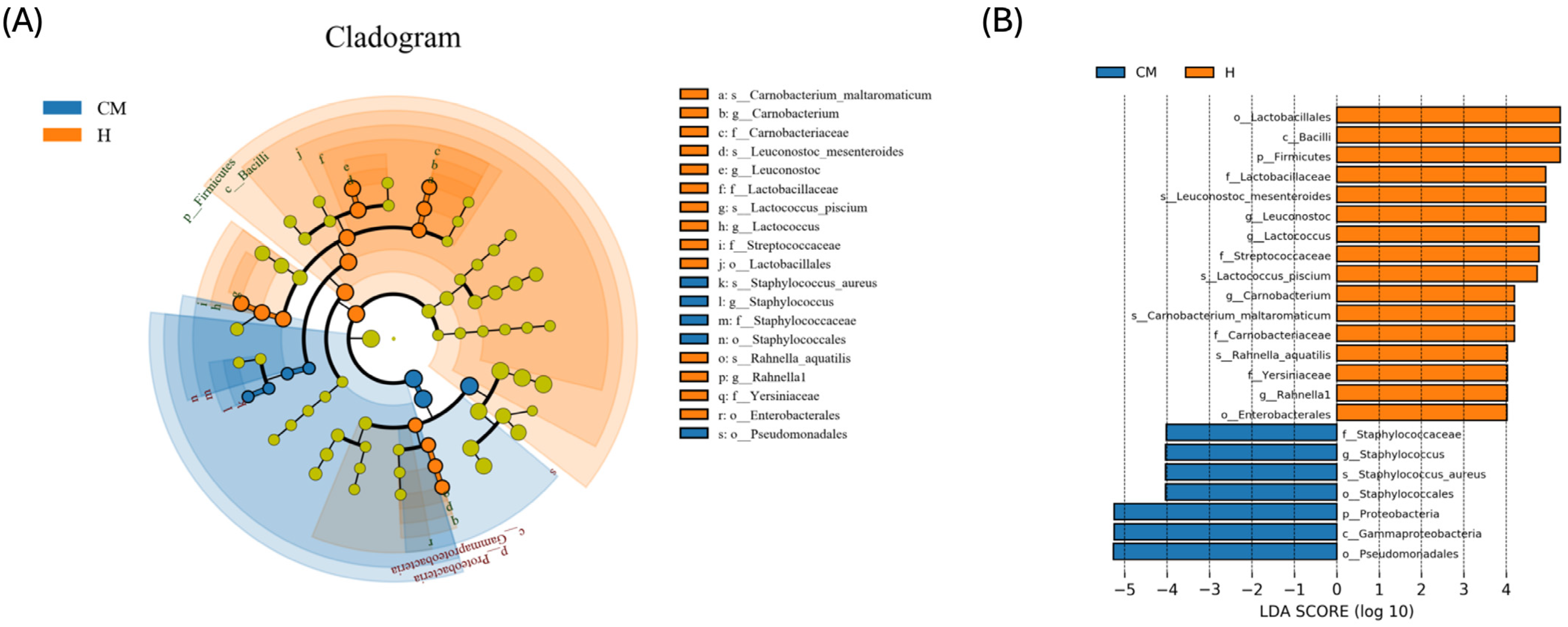
Since discriminant analysis is not able to detect every taxon, we used LEfSe and LDA to identify the main taxa of differences between groups (Figure 5). Specifically, the LEfSe analysis showed that five genera, four of which were endemic to the healthy group and one of which was unique to the clinical mastitis group—were effective biomarkers to differentiate between the treatment groups. g-Leuconostoc, g-Lactoccus, g-Carnobacterium, and g-Rahnella 1 were also demonstrated to be diagnostic of the healthy group in addition to these differential taxa. g- and s-Staphylococcus aureus identified the group with clinical mastitis. In contrast, the group with subclinical mastitis did not exhibit any statistically significant biomarkers.

4. Discussion

Mastitis is a significant concern for dairy farmers worldwide because it can result in lower milk yields, higher veterinary expenses, and even financial losses [21]. However, traditional culture-based methods of microbe identification have limitations because they can culture only approximately 1% of the total microbial population and identify microorganisms only at the genus level [22]. This problem can be resolved by using sequencing technologies like PacBio SMRT sequencing to identify and separate the microbiota of healthy yak samples from those suffering from mastitis. The present study identified and analyzed the differences in the bacterial community structure between healthy and mastitis-affected yaks using the full-length 16S rRNA sequence. These advanced techniques enabled a more thorough comprehension of the yak milk microbiota. They can aid in developing efficient mastitis control plans based on the etiological agents of the disease.
In this study, Proteobacteria and Firmicutes were the predominant bacterial phyla in the yak milk microbiome across all groups, which varied in relative abundance between milk samples from healthy and mastitis-affected yaks. These results are consistent with previous research on the microbiomes of mastitis in humans and cattle [23,24]. Microbiome diversity measurements indicated differences in microbial dysbiosis between healthy and diseased samples. The current study found significant variations in species richness and microbial diversity between milk samples from healthy yaks and those suffering from mastitis. Furthermore, beta diversity analysis showed that healthy yak milk samples’ microbial composition differed significantly from those affected by mastitis. Although significant differences in alpha diversity were observed between clinical and mastitis samples, some indicators did not show significant differences. These results highlight the complexity and variability of the data.
Clinical mastitis is a common inflammatory disease affecting dairy cattle’s mammary glands. In this study, Pseudomonas fluorescens and Chryseobacterium were the dominant bacterial species in yaks with clinical mastitis. Gram-negative Peudomonas fluorescens is a member of the Pseudomonadaceae family of bacteria. It is widely found in nature and is well-known for its capacity to produce a greenish fluorescent pigment [25]. Pseudomonas fluorescens’ ability to adapt to various ecological settings is one of its noteworthy traits [26]. It is crucial to the bioremediation processes to break down poisons and pollutants found in contaminated environments [27,28]. Its role in synthesizing secondary metabolites is also significant from a pharmacological perspective. It has been found that some strains can manufacture drugs that have antimicrobial or anticancer properties [29]. Although Pseudomonas fluorescens exhibits numerous advantageous traits in enhancing plant growth and combating pathogens and abiotic stresses, many species of Pseudomonas fluorescens have been associated with human and animal disease [30,31].
Furthermore, Chryseobacterium is a less common bacterium that has been linked to several animal diseases [32]. Its presence in clinical mastitis samples raises concerns regarding its potential function as a secondary invader or causative agent in mammary gland infection. Therefore, understanding the microbiological composition associated with clinical mastitis offers important information for creating disease treatment approaches.
Subclinical mastitis is characterized by inflammation of the mammary glands without any outward indications of infection. The quantity and quality of milk produced could decline due to this condition. In our study, the dominant bacterial species found in subclinical mastitis samples were Moraxella osloensis and Psychrobacter cibarius. Moraxella osloensis is a Gram-negative bacterium commonly found in animals and natural environments [32]. Moraxella osloensis can colonize human mucosal surfaces as well as skin. Although Moraxella osloensis is not commonly associated with clinical infections in humans or animals, it can potentially cause severe infections in immunocompromised individuals or have underlying medical conditions [33,34]. Its presence in subclinical mastitis samples suggests it may have an opportunistic pathogenic impact. Further research may be necessary to understand the specific infection processes and potential health risks. Moreover, Psychrobacter cibarius is a psychrophilic bacterial community member characterized by its ability to thrive in cold environments. Numerous cold settings, including soils and refrigerated foods, have isolated this bacterium [35]. Its existence in subclinical mastitis samples suggests adaptation to the udder environment despite its primary association with cold environments. Identifying these prominent bacterial species highlights the complexity of the microbial communities involved in subclinical mastitis. Thus, understanding their prevalence and activity in infected udders may help develop more accurate diagnostic techniques and focused treatment plans for this globally prevalent and economically significant illness affecting dairy cattle. Our results regarding Psychrobacter species in yak milk are consistent with those reported by Catozzi et al., who studied the microbiota of buffalo milk during asymptomatic and clinical mastitis and found significant differences in microbiota diversity between healthy and mastitis milk samples. According to their findings, relative abundance was less significant. This comparison highlights the potential impact of mastitis on the microbial composition of milk and supports the relevance of our findings for yak milk [36]. Additionally, Guo et al. studied the antibacterial activity of linalool against Pseudomonas fluorescens and highlighted the pathogenic potential of P. fluorescens in dairy products, providing a broader context for understanding the presence of P. fluorescens in yak milk [37].
Staphylococcus aureus is widely acknowledged as a significant causative agent of mastitis, which can significantly alter the composition and quality of milk, posing potential health hazards to consumers [38]. Our study revealed the relative abundance of Staphylococcus aureus species in subclinical and clinical mastitis. Persistent or recurrent mastitis may result from Staphylococcus aureus bacteria colonizing mammary gland tissue while the infection remains active [39]. These opportunistic bacteria exploit any weakness in the cow’s natural defenses, including teat injury and inadequate cleanliness during milking. Furthermore, studies show that some strains of Staphylococcus aureus possess virulence characteristics that enable them to evade the host’s immune system and cause chronic infections [40,41]. This ability prolongs inflammation and prevents the healing process in affected cows. Concerns regarding food safety are raised by identifying Staphylococcus aureus in mastitis milk, which further highlights the significance of following reasonable hygiene procedures during the milking and storage process [42]. Contamination can occur through various routes, such as unclean udders or equipment used for milking. This finding suggests that these bacteria may be present in the raw yak milk and their potential to function as opportunistic agents, disrupting metabolic processes, compromising host defense mechanisms, and hindering immunological development, all of which could lead to udder infections of various intensities.
Lactic acid bacteria (LAB) are a diverse group of microorganisms essential to food fermentation and preservation. The dairy industry extensively uses the genus Lactococcus because it can be used as a probiotic and in protective and starter cultures [43]. Four distract strains of bacteria were found in our study: Leuconostoc mesenteroides, Carnobacterium maltaromaticum, Lactococcus piscium, and Lactococcus raffinolactis. It is interesting to note that fermented foods frequently contain lactic acid bacteria of the type Leuconostoc mesenteroides. Numerous Enterococcus and Listeria species and other opportunistic pathogens are inhibited in their growth by this bacteriocin [44,45]. Extensive research has been conducted on this bacterium because of its potential as a probiotic and for improving food preservation [46,47]. Yak milk demonstrates its potential benefit during the production or storage of milk.
Furthermore, Carnobacterium maltaromaticum, a psychrotrophs lactic acid bacteria with strain-dependent spoilage and bioprotective characteristics, was also found in the present study [48]. Although their role in food spoiling may be disputed, these bacteria have been connected to beneficial traits such as flavor development during cheese ripening [49]. It has been reported that Carnobacterium colonizes the gut in an estrogen-dependent manner and collaborates with other microbes to increase the production of vitamin D in the intestinal tract, activating the host VDR and suppressing colorectal cancer [50].
Most labs are generally recognized as safe microorganisms and crucial for guaranteeing food safety. The psychrotrophic foodborne lactic acid bacterium Lactococcus piscium is essential to the biopreservation of various food products. This specific species has been found in many food sources, including meat, seafood, vegetables, and dairy products, all typically stored at low temperatures in packaging conditions [51]. The importance of L. piscium in food has been highlighted by applying non-cultivatable techniques to the microbial ecosystem characterization process. Food spoilage may be impacted differently by strain variations and different food matrices. Certain strains have been demonstrated to prevent food spoilage and improve sensory quality [52,53].
Furthermore, these beneficial bacteria support antimicrobial activities and raise the food’s safety for consumption by producing organic acids, antimicrobial peptides, and nutrient competition [54]. L. piscium shows a wide range of antimicrobial activity against strains of Salmonella, Staphylococcus aureus, Clostridium sporogenes, Listeria monocytogenes, and Escherichia coli [55,56]. It has been connected to anti-listeria activity dependent on cell-to-cell contact [57].
In addition to their direct impact on food safety, LABs also offer potential health benefits by enhancing digestion and promoting gut health through their ability to hydrolyze lactose and produce enzymes that facilitate nutrient absorption. This study also identified Lactococcus raffinolactis, which is widely distributed in various food products and materials derived from plants and animals. It is commonly found in fermented foods, including meat, fish, vegetables, milk, and dairy products [58]. This bacterium is vital to fermentation because of its ability to enzymatically transform complex compounds into simpler forms that promote flavor development [59]. Moreover, it has been found that L. raffinolactis is an essential component of several dairy products, such as cheese and yogurt. Its presence significantly affects the taste profiles of these products due to the metabolic activities it goes through during fermentation [60]. Our study highlights the diversity and richness of the microbial communities found in samples of healthy yak milk. Further investigation into these bacterial strains could offer valuable insights into their roles within dairy ecosystems, potentially enhancing food safety and quality control within the dairy industry.

5. Conclusions

For this study, we obtained 16S rRNA sequences to understand better the composition and diversity of bacterial communities associated with yak milk in healthy and mastitis-affected individuals. It has been found that the diversity of the milk microbiota has different levels and that the composition of the microbiota differs significantly between healthy yak and mastitis cases. Differential taxonomic abundance patterns within groups can function as indicators of mastitis susceptibility. More research should be conducted to investigate the potential of microbes such as Leuconostoc mesenteroides, Lactococcus piscium, Carnobacterium maltaromaticum, and Lactococcus raffinolactis species, which are prevalent in healthy yaks as probiotics for preventing mastitis.

Supplementary Materials

The following supporting information can be downloaded at: https://www.mdpi.com/article/10.3390/vetsci11080335/s1. Table S1. Summary of the full-length 16S rRNA sequencing data of milk samples in yak. Table S2. The percentage of taxa with top10 relative abundance and corresponding read at various taxonomic levels.

Author Contributions

L.Z. and H.M. designed the experiments. W.T. performed most of the experiments. J.Z. and J.H. analyzed the results. M.F.K. reviewed the manuscript with the contribution of all other authors. All authors have read and agreed to the published version of the manuscript.

Funding

This research was supported by the Central Government Guides Local Science and Technology Development Projects of China (XZ202301YD0015C), Youth Science and Technology Fund Project of Gansu Province (22JR5RA885), and Scientific Research Start-up Funds for Openly recruited Doctors of Gansu Agricultural University (GAU-KYQD-2021-09).

Institutional Review Board Statement

The animal study protocol was approved by the Gansu Agricultural University Institutional Animal Care (Approval Code: GSAU-Eth-VMC-2022-015).

Informed Consent Statement

Not Applicable.

Data Availability Statement

The original sequence data were submitted to the Sequence Read Archive (SRA) (NCBI, USA) with accession no. PRJNA1130414.

Acknowledgments

We are grateful to dairy farmers for their cooperation. The authors also extend their appreciation to Editlela.com for support regarding scholarly services.

Conflicts of Interest

The authors declare no conflict of interest.

References

  1. Ruegg, P.L. A 100-Year Review: Mastitis Detection, Management, and Prevention. J. Dairy Sci. 2017, 100, 10381–10397. [Google Scholar] [CrossRef]
  2. Gao, J.; Barkema, H.W.; Zhang, L.; Liu, G.; Deng, Z.; Cai, L.; Shan, R.; Zhang, S.; Zou, J.; Kastelic, J.P.; et al. Incidence of Clinical Mastitis and Distribution of Pathogens on Large Chinese Dairy Farms. J. Dairy Sci. 2017, 100, 4797–4806. [Google Scholar] [CrossRef]
  3. He, W.; Ma, S.; Lei, L.; He, J.; Li, X.; Tao, J.; Wang, X.; Song, S.; Wang, Y.; Wang, Y.; et al. Prevalence, Etiology, and Economic Impact of Clinical Mastitis on Large Dairy Farms in China. Vet. Microbiol. 2020, 242, 108570. [Google Scholar] [CrossRef]
  4. Puerto, M.A.; Shepley, E.; Cue, R.I.; Warner, D.; Dubuc, J.; Vasseur, E. The Hidden Cost of Disease: I. Impact of the First Incidence of Mastitis on Production and Economic Indicators of Primiparous Dairy Cows. J. Dairy Sci. 2021, 104, 7932–7943. [Google Scholar] [CrossRef]
  5. Derakhshani, H.; Fehr, K.B.; Sepehri, S.; Francoz, D.; De Buck, J.; Barkema, H.W.; Plaizier, J.C.; Khafipour, E. Invited Review: Microbiota of the Bovine Udder: Contributing Factors and Potential Implications for Udder Health and Mastitis Susceptibility. J. Dairy Sci. 2018, 101, 10605–10625. [Google Scholar] [CrossRef]
  6. Zhao, C.; Hu, X.; Bao, L.; Wu, K.; Zhao, Y.; Xiang, K.; Li, S.; Wang, Y.; Qiu, M.; Feng, L.; et al. Gut Dysbiosis Induces the Development of Mastitis through a Reduction in Host Anti-Inflammatory Enzyme Activity by Endotoxemia. Microbiome 2022, 10, 205. [Google Scholar] [CrossRef]
  7. Oultram, J.W.H.; Ganda, E.K.; Boulding, S.C.; Bicalho, R.C.; Oikonomou, G. A Metataxonomic Approach Could Be Considered for Cattle Clinical Mastitis Diagnostics. Front. Vet. Sci. 2017, 4, 36. [Google Scholar] [CrossRef]
  8. Hoque, M.N.; Istiaq, A.; Clement, R.A.; Sultana, M.; Crandall, K.A.; Siddiki, A.Z.; Hossain, M.A. Metagenomic Deep Sequencing Reveals Association of Microbiome Signature with Functional Biases in Bovine Mastitis. Sci. Rep. 2019, 9, 13536. [Google Scholar] [CrossRef]
  9. Hoque, M.N.; Istiaq, A.; Rahman, M.S.; Islam, M.R.; Anwar, A.; Siddiki, A.M.A.M.Z.; Sultana, M.; Crandall, K.A.; Hossain, M.A. Microbiome Dynamics and Genomic Determinants of Bovine Mastitis. Genomics 2020, 112, 5188–5203. [Google Scholar] [CrossRef]
  10. Cremonesi, P.; Severgnini, M.; Romanò, A.; Sala, L.; Luini, M.; Castiglioni, B. Bovine Milk Microbiota: Comparison among Three Different DNA Extraction Protocols To Identify a Better Approach for Bacterial Analysis. Microbiol. Spectr. 2021, 9, e00374-21. [Google Scholar] [CrossRef]
  11. Ashraf, A.; Imran, M. Causes, Types, Etiological Agents, Prevalence, Diagnosis, Treatment, Prevention, Effects on Human Health and Future Aspects of Bovine Mastitis. Anim. Health Res. Rev. 2020, 21, 36–49. [Google Scholar] [CrossRef]
  12. Pang, M.; Xie, X.; Bao, H.; Sun, L.; He, T.; Zhao, H.; Zhou, Y.; Zhang, L.; Zhang, H.; Wei, R.; et al. Insights into the Bovine Milk Microbiota in Dairy Farms with Different Incidence Rates of Subclinical Mastitis. Front. Microbiol. 2018, 9, 2379. [Google Scholar] [CrossRef]
  13. Alessandri, G.; Sangalli, E.; Facchi, M.; Fontana, F.; Mancabelli, L.; Donofrio, G.; Ventura, M. Metataxonomic Analysis of Milk Microbiota in the Bovine Subclinical Mastitis. FEMS Microbiol. Ecol. 2023, 99, fiad136. [Google Scholar] [CrossRef]
  14. Qiu, Q.; Zhang, G.; Ma, T.; Qian, W.; Ye, Z.; Cao, C.; Hu, Q.; Kim, J.; Larkin, D.M.; Auvil, L.; et al. The Yak Genome and Adaptation to Life at High Altitude. Nat. Genet. 2012, 44, 946–949. [Google Scholar] [CrossRef]
  15. Ayalew, W.; Chu, M.; Liang, C.; Wu, X.; Ping, Y. Adaptation Mechanisms of Yak (Bos grunniens) to High-Altitude Environmental Stress. Animals 2021, 11, 2344. [Google Scholar] [CrossRef]
  16. Gao, H.N.; Hu, H.; Wen, P.C.; Lian, S.; Xie, X.L.; Song, H.L.; Yang, Z.N.; Ren, F.Z. Yak Milk–Derived Exosomes Alleviate Lipopolysaccharide-Induced Intestinal Inflammation by Inhibiting PI3K/AKT/C3 Pathway Activation. J. Dairy Sci. 2021, 104, 8411–8424. [Google Scholar] [CrossRef]
  17. Martin, M. Cutadapt removes adapter sequences from high-throughput sequencing reads. EMBnet. J. 2011, 17, 10–12. [Google Scholar] [CrossRef]
  18. Bolyen, E.; Rideout, J.R.; Dillon, M.R.; Bokulich, N.A.; Abnet, C.C.; Al-Ghalith, G.A.; Alexander, H.; Alm, E.J.; Arumugam, M.; Asnicar, F.; et al. Reproducible, interactive, scalable and extensible microbiome data science using QIIME 2. Nat. Biotechnol. 2019, 37, 852–857. [Google Scholar] [CrossRef]
  19. Quast, C.; Pruesse, E.; Yilmaz, P.; Gerken, J.; Schweer, T.; Yarza, P.; Peplies, J.; Glöckner, F.O. The SILVA ribosomal RNA gene database project: Improved data processing and web-based tools. Nucleic Acids Res. 2012, 41, D590–D596. [Google Scholar] [CrossRef]
  20. Dixon, P. VEGAN, a package of R functions for community ecology. J. Veg. Sci. 2003, 14, 927–930. [Google Scholar] [CrossRef]
  21. Cobirka, M.; Tancin, V.; Slama, P. Epidemiology and Classification of Mastitis. Animals 2020, 10, 2212. [Google Scholar] [CrossRef]
  22. Franco-Duarte, R.; Černáková, L.; Kadam, S.; Kaushik, K.S.; Salehi, B.; Bevilacqua, A.; Corbo, M.R.; Antolak, H.; Dybka-Stępień, K.; Leszczewicz, M.; et al. Advances in Chemical and Biological Methods to Identify Microorganisms—From Past to Present. Microorganisms 2019, 7, 130. [Google Scholar] [CrossRef]
  23. Salman, M.M.; Nawaz, M.; Yaqub, T.; Mushtaq, M.H. Investigation of Milk Microbiota of Healthy and Mastitic Sahiwal Cattle. BMC Microbiol. 2023, 23, 304. [Google Scholar] [CrossRef]
  24. Khasapane, N.G.; Khumalo, Z.T.H.; Kwenda, S.; Nkhebenyane, S.J.; Thekisoe, O. Characterisation of Milk Microbiota from Subclinical Mastitis and Apparently Healthy Dairy Cattle in Free State Province, South Africa. Vet. Sci. 2023, 10, 616. [Google Scholar] [CrossRef]
  25. Ishii, H.; Kushima, H.; Koide, Y.; Kinoshita, Y. Pseudomonas Fluorescens Pneumonia. Int. J. Infect. Dis. 2024, 140, 92–94. [Google Scholar] [CrossRef]
  26. Gomez, P.; Hall, A.R.; Paterson, S.; Buckling, A. Rapid Decline of Adaptation of Pseudomonas Fluorescens to Soil Biotic Environment. Biol. Lett. 2022, 18, 20210593. [Google Scholar] [CrossRef]
  27. Ridl, J.; Suman, J.; Fraraccio, S.; Hradilova, M.; Strejcek, M.; Cajthaml, T.; Zubrova, A.; Macek, T.; Strnad, H.; Uhlik, O. Complete Genome Sequence of Pseudomonas alcaliphila JAB1 (=DSM 26533), a Versatile Degrader of Organic Pollutants. Stand. Genom. Sci. 2018, 13, 3. [Google Scholar] [CrossRef]
  28. Chautrand, T.; Depayras, S.; Souak, D.; Bouteiller, M.; Kondakova, T.; Barreau, M.; Ben Mlouka, M.A.; Hardouin, J.; Konto-Ghiorghi, Y.; Chevalier, S.; et al. Detoxification Response of Pseudomonas Fluorescens MFAF76a to Gaseous Pollutants NO2 and NO. Microorganisms 2022, 10, 1576. [Google Scholar] [CrossRef]
  29. Baker, S.; Nagendra, N.P.; Dhananjaya, B.L.; Mohan Kumar, K.; Yallappa, S.; Satish, S. Synthesis of Silver Nanoparticles by Endosymbiont Pseudomonas Fluorescens CA 417 and Their Bactericidal Activity. Enzym. Microb. Technol. 2016, 95, 128–136. [Google Scholar] [CrossRef]
  30. Zhu, L.; Qian, N.; Sun, Y.; Lu, X.; Duan, H.; Qian, L. Pseudomonas Fluorescens DN16 Enhances Cucumber Defense Responses against the Necrotrophic Pathogen Botrytis cinerea by Regulating Thermospermine Catabolism. Front. Plant Sci. 2021, 12, 645338. [Google Scholar] [CrossRef]
  31. Sokolov, S.; Fursova, K.; Shulcheva, I.; Nikanova, D.; Artyemieva, O.; Kolodina, E.; Sorokin, A.; Dzhelyadin, T.; Shchannikova, M.; Shepelyakovskaya, A.; et al. Comparative Analysis of Milk Microbiomes and Their Association with Bovine Mastitis in Two Farms in Central Russia. Animals 2021, 11, 1401. [Google Scholar] [CrossRef]
  32. Rahim, G.R.; Gupta, N. Dilemma in Identifying Chryseobacterium Species. Infect. Dis. 2018, 50, 878–879. [Google Scholar] [CrossRef]
  33. Tabbuso, T.; Defourny, L.; Lali, S.E.; Pasdermadjian, S.; Gilliaux, O. Moraxella Osloensis Infection among Adults and Children: A Pediatric Case and Literature Review. Arch. Pediatr. 2021, 28, 348–351. [Google Scholar] [CrossRef]
  34. Yamada, A.; Kasahara, K.; Ogawa, Y.; Samejima, K.; Eriguchi, M.; Yano, H.; Mikasa, K.; Tsuruya, K. Peritonitis Due to Moraxella Osloensis: A Case Report and Literature Review. J. Infect. Chemother. 2019, 25, 1050–1052. [Google Scholar] [CrossRef]
  35. Raghupathi, P.K.; Herschend, J.; Røder, H.L.; Sørensen, S.J.; Burmølle, M. Genome Sequence of Psychrobacter cibarius Strain W1. Genome Announc. 2016, 4, e00078-16. [Google Scholar] [CrossRef]
  36. Catozzi, C.; Sanchez Bonastre, A.; Francino, O.; Lecchi, C.; De Carlo, E.; Vecchio, D.; Martucciello, A.; Fraulo, P.; Bronzo, V.; Cuscó, A.; et al. The microbiota of water buffalo milk during mastitis. PLoS ONE 2017, 12, e0184710. [Google Scholar] [CrossRef]
  37. Guo, F.; Chen, Q.; Liang, Q.; Zhang, M.; Chen, W.; Chen, H.; Yun, Y.; Zhong, Q.; Chen, W. Antimicrobial activity and proposed action mechanism of linalool against Pseudomonas fluorescens. Front. Microbiol. 2021, 12, 562094. [Google Scholar] [CrossRef]
  38. Keim, K.C.; Horswill, A.R. Staphylococcus aureus. Trends Microbiol. 2023, 31, 1300–1301. [Google Scholar]
  39. Leijon, M.; Atkins, E.; Persson Waller, K.; Artursson, K. Longitudinal Study of Staphylococcus aureus Genotypes Isolated from Bovine Clinical Mastitis. J. Dairy Sci. 2021, 104, 11945–11954. [Google Scholar] [CrossRef]
  40. Cheung, G.Y.C.; Bae, J.S.; Otto, M. Pathogenicity and Virulence of Staphylococcus aureus. Virulence 2021, 12, 547–569. [Google Scholar]
  41. Howden, B.P.; Giulieri, S.G.; Wong Fok Lung, T.; Baines, S.L.; Sharkey, L.K.; Lee, J.Y.H.; Hachani, A.; Monk, I.R.; Stinear, T.P. Staphylococcus aureus Host Interactions and Adaptation. Nat. Rev. Microbiol. 2023, 21, 380–395. [Google Scholar] [CrossRef]
  42. Haag, A.F.; Fitzgerald, J.R.; Penadés, J.R. Staphylococcus aureus in Animals. Microbiol. Spectr. 2019, 7. [Google Scholar] [CrossRef]
  43. Gaudu, P.; Yamamoto, Y.; Jensen, P.R.; Hammer, K.; Lechardeur, D.; Gruss, A. Genetics of Lactococci. Microbiol. Spectr. 2019, 7. [Google Scholar] [CrossRef]
  44. Chang, J.Y.; Lee, H.J.; Chang, H.C. Identification of the Agent from Lactobacillus plantarum KFRI464 That Enhances Bacteriocin Production by Leuconostoc citreum GJ7. J. Appl. Microbiol. 2007, 103, 2504–2515. [Google Scholar] [CrossRef]
  45. Zhang, H.; HuangFu, H.P.; Wang, X.; Zhao, S.S.; Liu, Y.; Lv, H.; Qin, G.Y.; Tan, Z. Antibacterial Activity of Lactic Acid Producing Leuconostoc mesenteroides QZ1178 against Pathogenic Gallibacterium anatis. Front. Vet. Sci. 2021, 8, 630294. [Google Scholar] [CrossRef]
  46. Ogier, J.C.; Casalta, E.; Farrokh, C.; Saïhi, A. Safety Assessment of Dairy Microorganisms: The Leuconostoc Genus. Int. J. Food Microbiol. 2008, 126, 286–290. [Google Scholar] [CrossRef]
  47. Li, Y.; Liu, Y.; Cao, C.; Zhu, X.Y.; Wang, C.; Wu, R.; Wu, J. Extraction and Biological Activity of Exopolysaccharide Produced by Leuconostoc mesenteroides SN-8. Int. J. Biol. Macromol. 2020, 157, 36–44. [Google Scholar] [CrossRef]
  48. de Andrade Cavalari, C.M.; Imazaki, P.H.; Pirard, B.; Lebrun, S.; Vanleyssem, R.; Gemmi, C.; Antoine, C.; Crevecoeur, S.; Daube, G.; Clinquart, A.; et al. Carnobacterium maltaromaticum as Bioprotective Culture against Spoilage Bacteria in Ground Meat and Cooked Ham. Meat Sci. 2024, 211, 109441. [Google Scholar] [CrossRef]
  49. Iskandar, C.F.; Borges, F.; Taminiau, B.; Daube, G.; Zagorec, M.; Remenant, B.; Leisner, J.J.; Hansen, M.A.; Sørensen, S.J.; Mangavel, C.; et al. Comparative Genomic Analysis Reveals Ecological Differentiation in the Genus Carnobacterium. Front. Microbiol. 2017, 8, 357. [Google Scholar] [CrossRef]
  50. Li, Q.; Chan, H.; Liu, W.X.; Liu, C.A.; Zhou, Y.; Huang, D.; Wang, X.; Li, X.; Xie, C.; Liu, W.Y.Z.; et al. Carnobacterium maltaromaticum Boosts Intestinal Vitamin D Production to Suppress Colorectal Cancer in Female Mice. Cancer Cell 2023, 41, 1450–1465. [Google Scholar] [CrossRef]
  51. Saraoui, T.; Leroi, F.; Björkroth, J.; Pilet, M.F. Lactococcus piscium: A Psychrotrophic Lactic Acid Bacterium with Bioprotective or Spoilage Activity in Food—A Review. J. Appl. Microbiol. 2016, 121, 907–918. [Google Scholar] [CrossRef]
  52. Rahkila, R.; Nieminen, T.; Johansson, P.; Säde, E.; Björkroth, J. Characterization and Evaluation of the Spoilage Potential of Lactococcus piscium Isolates from Modified Atmosphere Packaged Meat. Int. J. Food Microbiol. 2012, 156, 50–59. [Google Scholar] [CrossRef]
  53. Chaillou, S.; Chaulot-Talmon, A.; Caekebeke, H.; Cardinal, M.; Christieans, S.; Denis, C.; Hélène Desmonts, M.; Dousset, X.; Feurer, C.; Hamon, E.; et al. Origin and Ecological Selection of Core and Food-Specific Bacterial Communities Associated with Meat and Seafood Spoilage. ISME J. 2015, 9, 1105–1118. [Google Scholar] [CrossRef]
  54. Saraoui, T.; Fall, P.A.; Leroi, F.; Antignac, J.P.; Chéreau, S.; Pilet, M.F. Inhibition Mechanism of Listeria monocytogenes by a Bioprotective Bacteria Lactococcus piscium CNCM I-4031. Food Microbiol. 2016, 53, 70–78. [Google Scholar] [CrossRef]
  55. Matamoros, S.; Pilet, M.F.; Gigout, F.; Prévost, H.; Leroi, F. Selection and Evaluation of Seafood-Borne Psychrotrophic Lactic Acid Bacteria as Inhibitors of Pathogenic and Spoilage Bacteria. Food Microbiol. 2009, 26, 638–644. [Google Scholar] [CrossRef]
  56. Fall, P.A.; Leroi, F.; Cardinal, M.; Chevalier, F.; Pilet, M.F. Inhibition of Brochothrix thermosphacta and Sensory Improvement of Tropical Peeled Cooked Shrimp by Lactococcus piscium CNCM I-4031. Lett. Appl. Microbiol. 2010, 50, 357–361. [Google Scholar] [CrossRef]
  57. Saraoui, T.; Leroi, F.; Chevalier, F.; Cappelier, J.M.; Passerini, D.; Pilet, M.F. Bioprotective Effect of Lactococcus piscium CNCM I-4031 against Listeria monocytogenes Growth and Virulence. Front. Microbiol. 2018, 9, 1564. [Google Scholar] [CrossRef]
  58. Jung, M.Y.; Lee, C.; Seo, M.J.; Roh, S.W.; Lee, S.H. Characterization of a Potential Probiotic Bacterium Lactococcus raffinolactis WiKim0068 Isolated from Fermented Vegetable Using Genomic and in Vitro Analyses. BMC Microbiol. 2020, 20, 136. [Google Scholar] [CrossRef]
  59. Boucher, I.; Vadeboncoeur, C.; Moineau, S. Characterization of Genes Involved in the Metabolism of α-Galactosides by Lactococcus raffinolactis. Appl. Environ. Microbiol. 2003, 69, 4049–4056. [Google Scholar] [CrossRef]
  60. Kimoto-Nira, H.; Aoki, R.; Mizumachi, K.; Sasaki, K.; Naito, H.; Sawada, T.; Suzuki, C. Interaction between Lactococcus lactis and Lactococcus raffinolactis during Growth in Milk: Development of a New Starter Culture. J. Dairy Sci. 2012, 95, 2176–2185. [Google Scholar] [CrossRef]
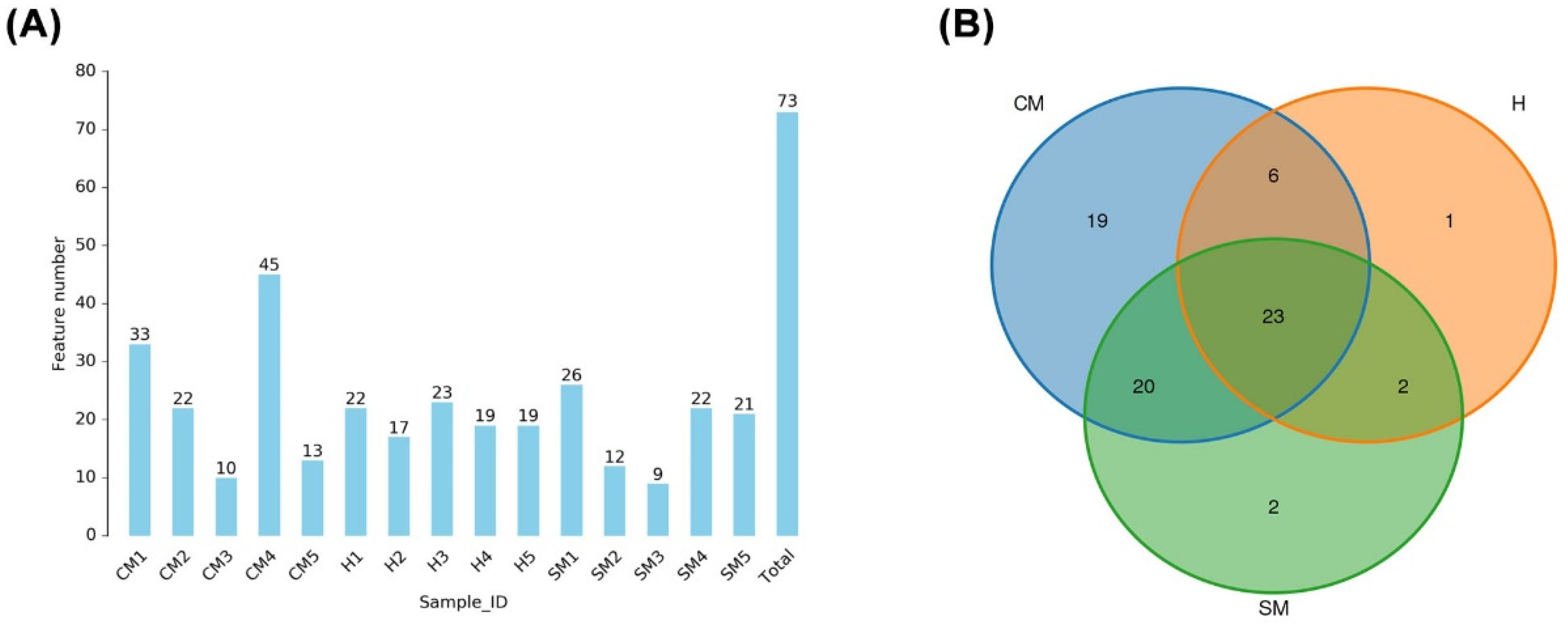
Figure 1. Operational taxonomic units (OTUs) of different samples. (A) Distribution of OTU numbers within each group and sample. (B) OTU Venn diagrams for the three groupings.
Vetsci 11 00335 g001
Figure 2. The microbiota alpha diversity was measured using the ACE (A), Chao1 (B), Simpson (C), and Shannon (D) Indices. Significant differences with p < 0.05 are indicated by an asterisk (*); highly significant differences with p < 0.001 are marked by three asterisks (***) and p < 0.00001 are marked as (****). H: healthy group, SM: subclinical mastitis group, CM: clinical mastitis group.
Vetsci 11 00335 g002
Figure 3. Microbiota beta diversity in the yak milk of the healthy (H), subclinical mastitis (SM), and clinical mastitis (CM) groups was assessed using. (A) PCoA score plot. (B) Binary-Jaccard method with arithmetic mean (UPGMA) analysis.
Vetsci 11 00335 g003
Figure 4. The relative abundance of the milk bacterium community for healthy (H), subclinical (SM), and clinical mastitis (CM) groups. (A) Phylum; (B) genus; (C) species.
Vetsci 11 00335 g004
Figure 5. The differentially abundant taxa of healthy (H) yaks and those with clinical mastitis (CM) were identified using linear discriminant analysis effect size (LEfSe) coupled with linear discriminant analysis (LDA) scores. The evolutionary distribution of differentiated biomarkers: (A) the cladogram; (B) LDA values greater than 4 were the threshold for a meaningful difference.
Vetsci 11 00335 g005
Disclaimer/Publisher’s Note: The statements, opinions and data contained in all publications are solely those of the individual author(s) and contributor(s) and not of MDPI and/or the editor(s). MDPI and/or the editor(s) disclaim responsibility for any injury to people or property resulting from any ideas, methods, instructions or products referred to in the content.