
Congenital Anomalies in American Crocodile (Crocodylus acutus, Cuvier, 1807) Embryos from a Farm Breeder in Colombia

 ,
,  ,
,  ,
, 
Abstract
Simple Summary
Abstract
1. Introduction
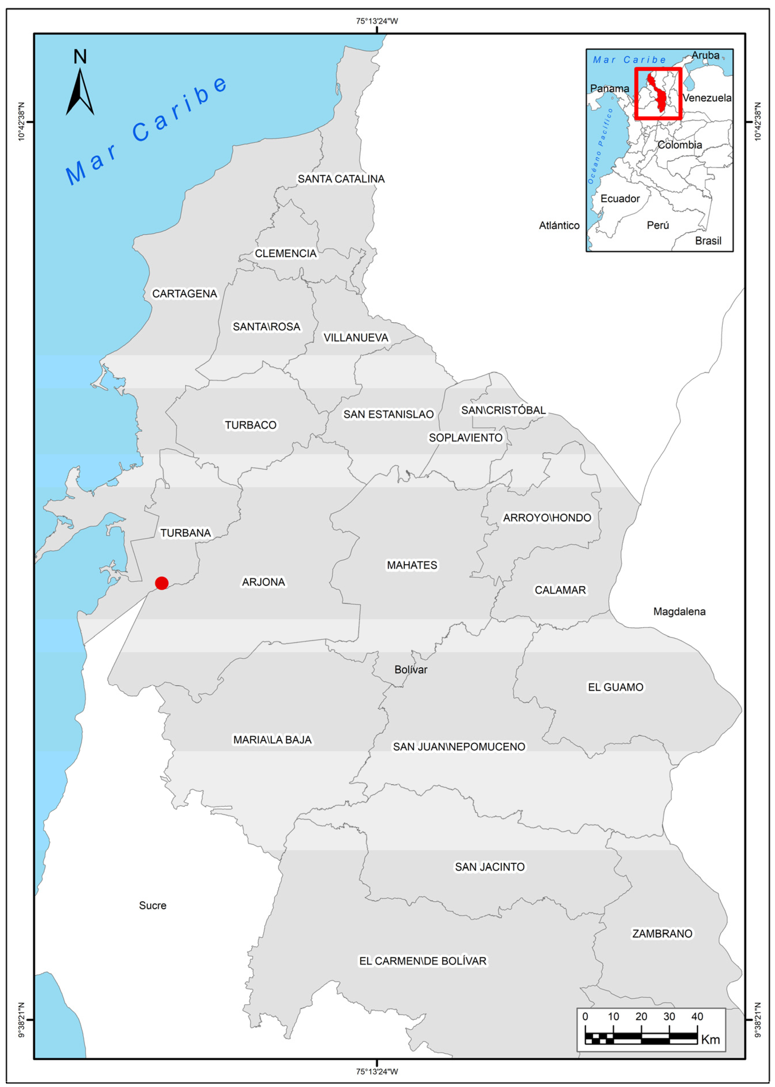
2. Materials and Methods
3. Results
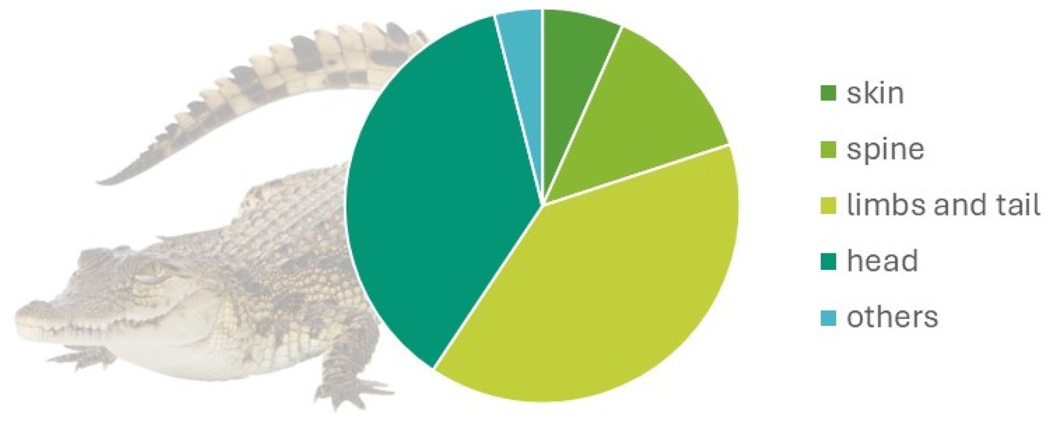
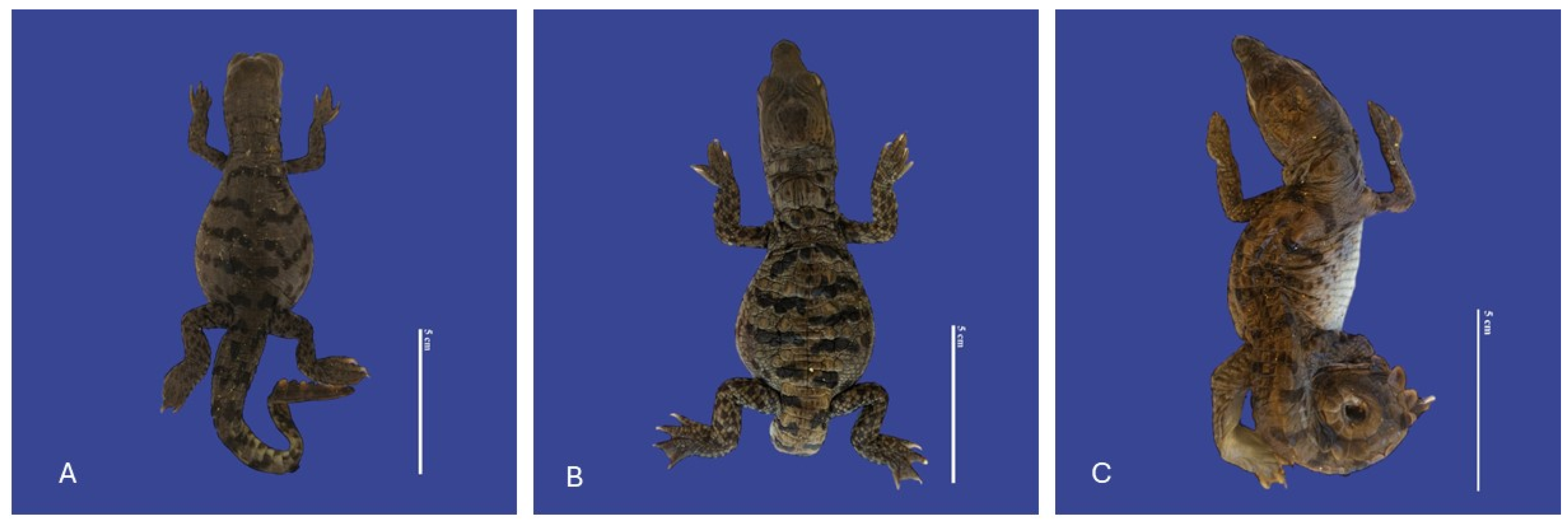
3.1. Skin Malformations
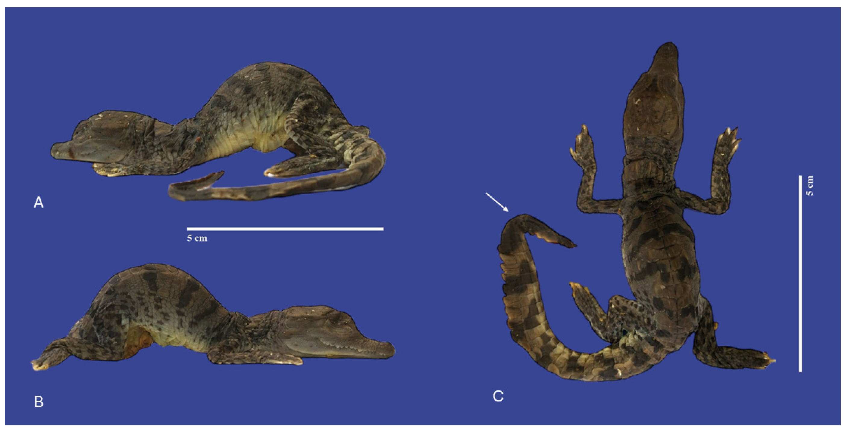
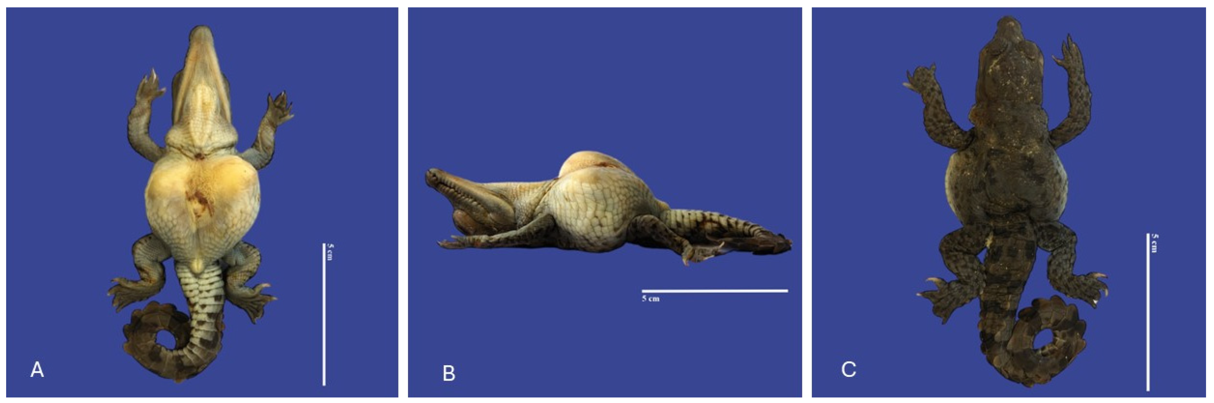
3.2. Spine Malformations
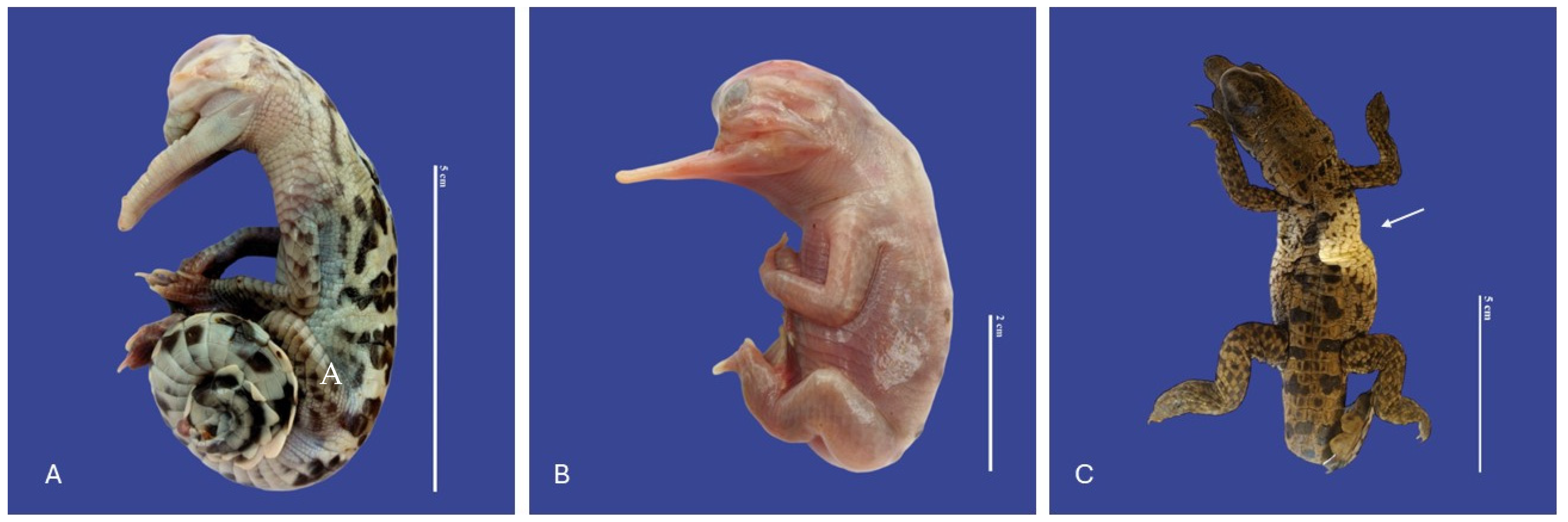
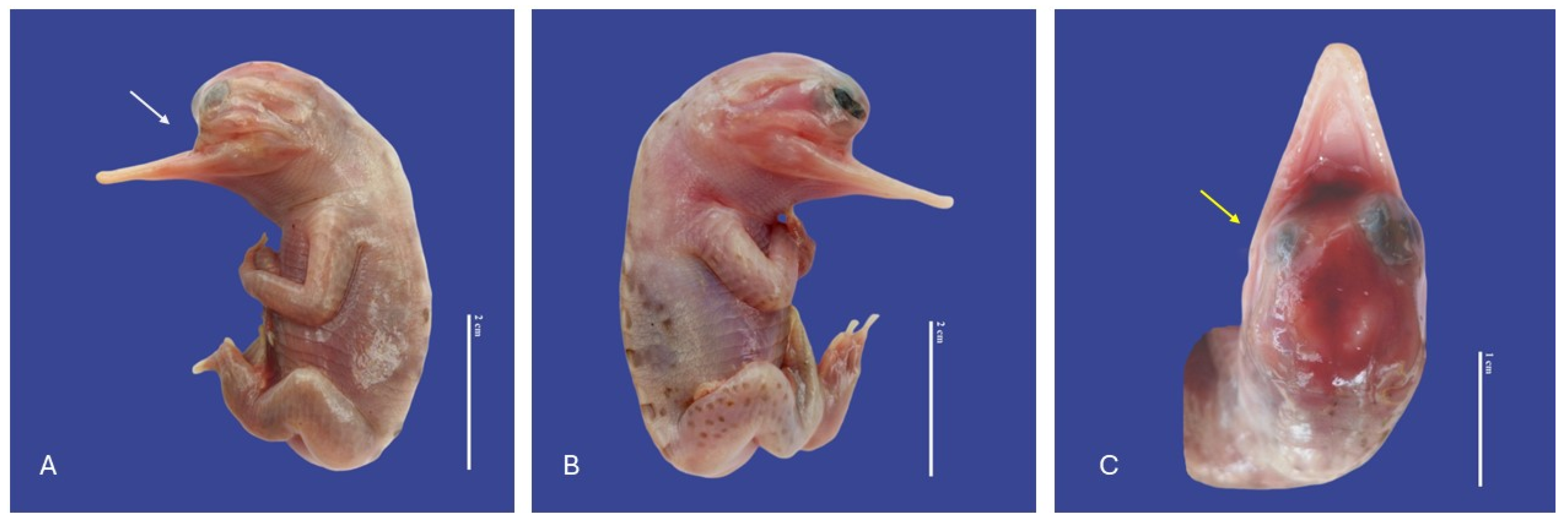
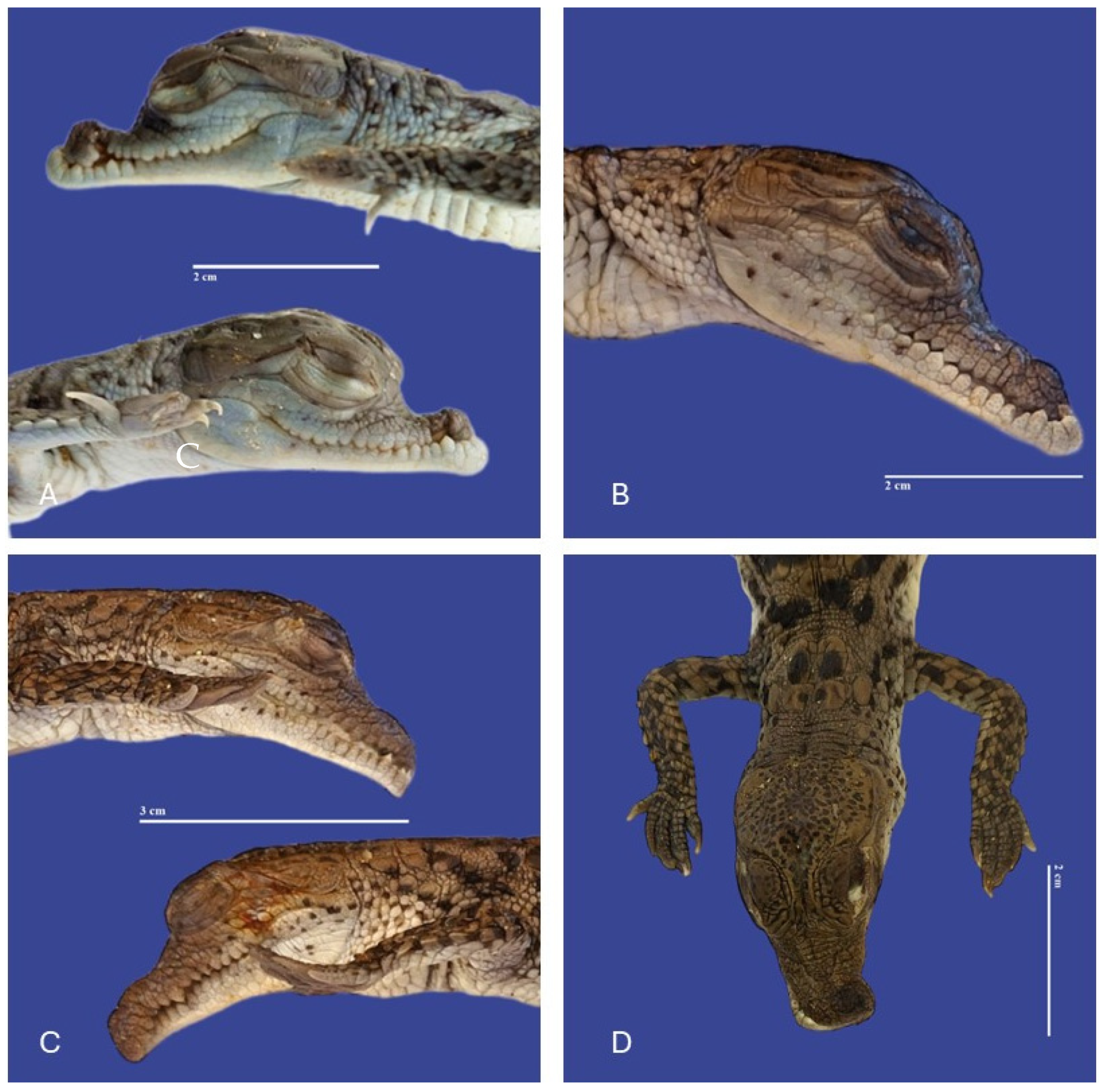
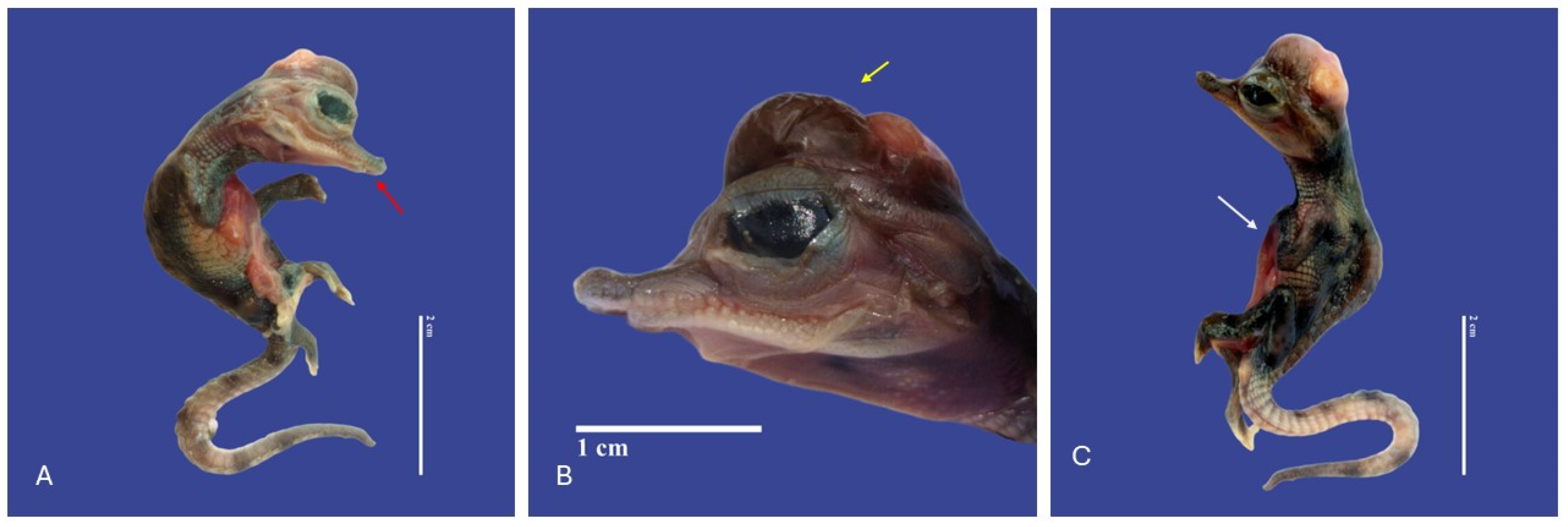
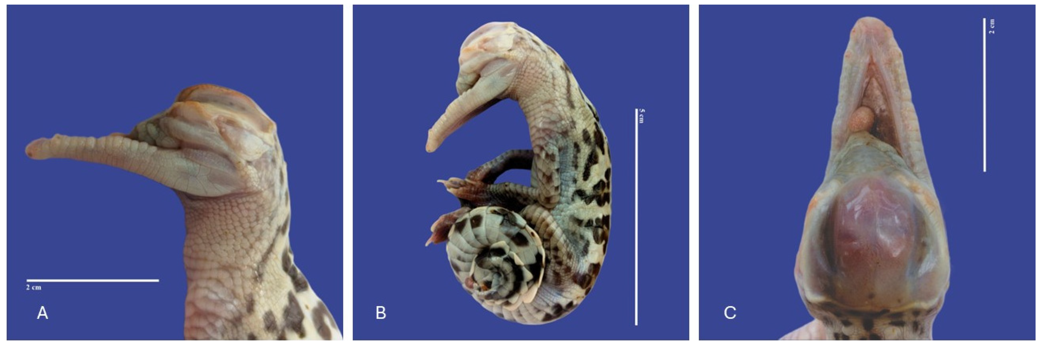
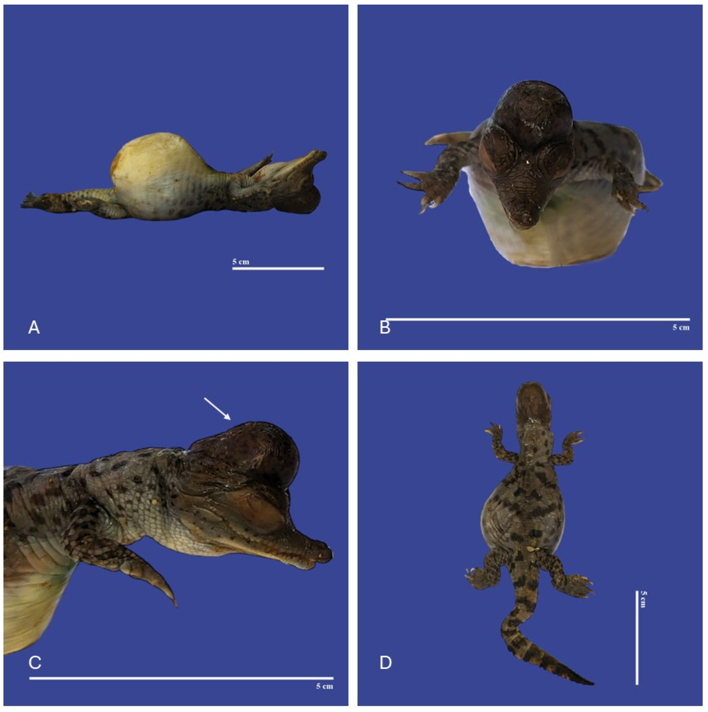
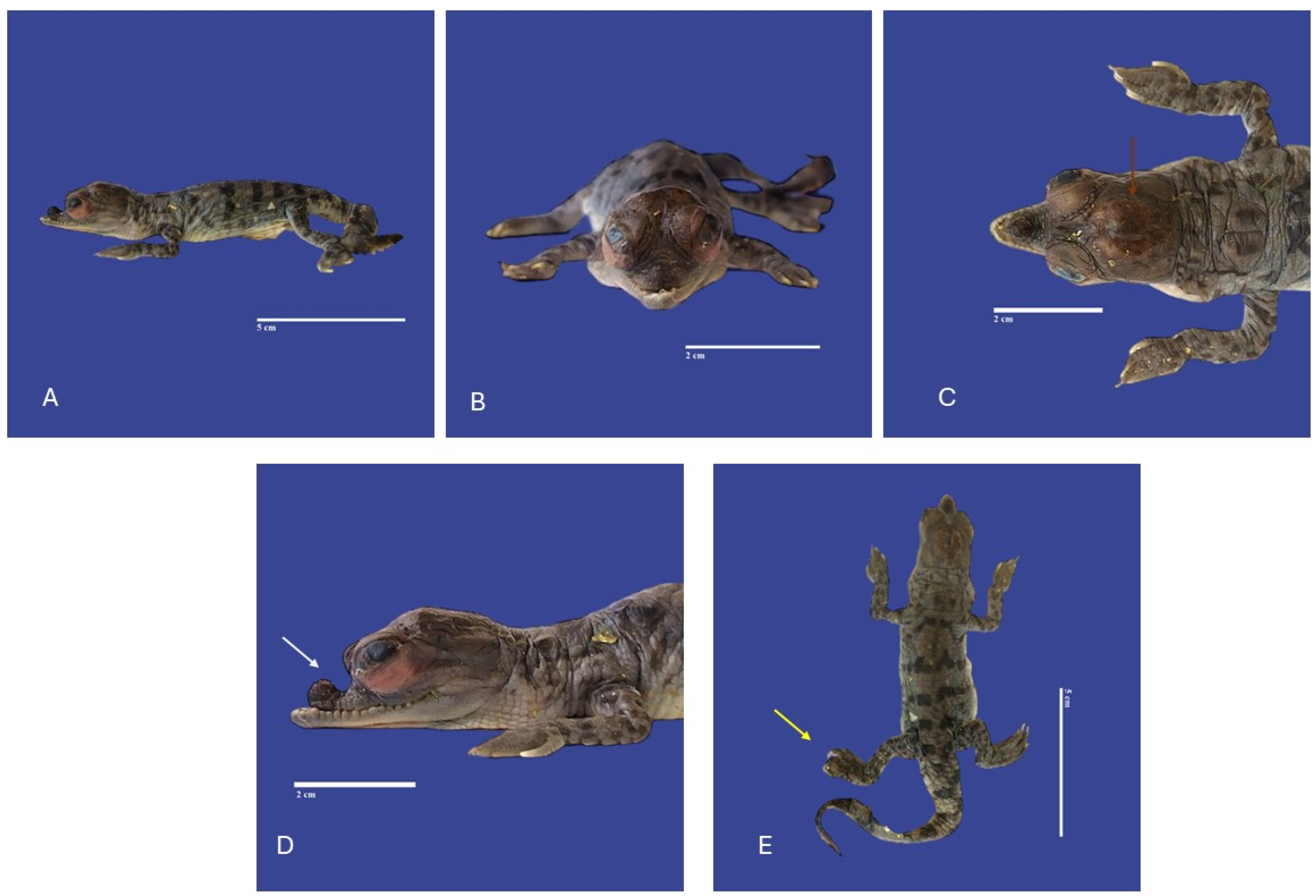
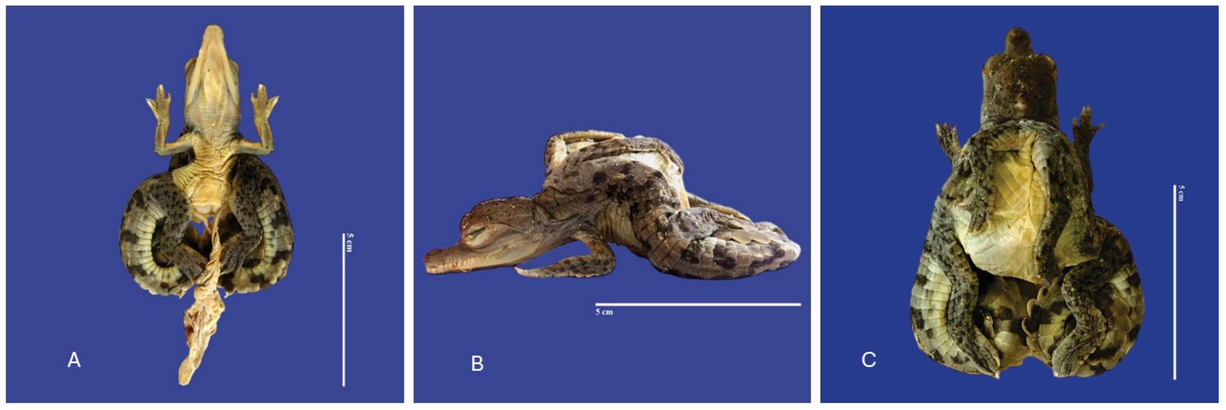
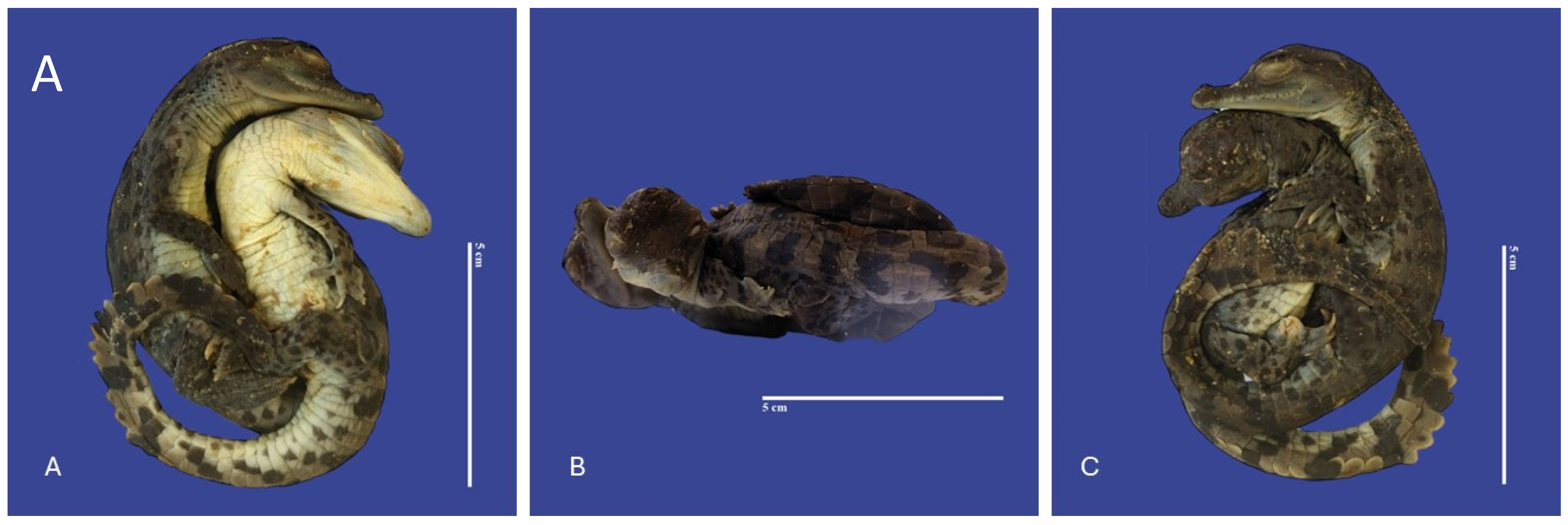
3.3. Head
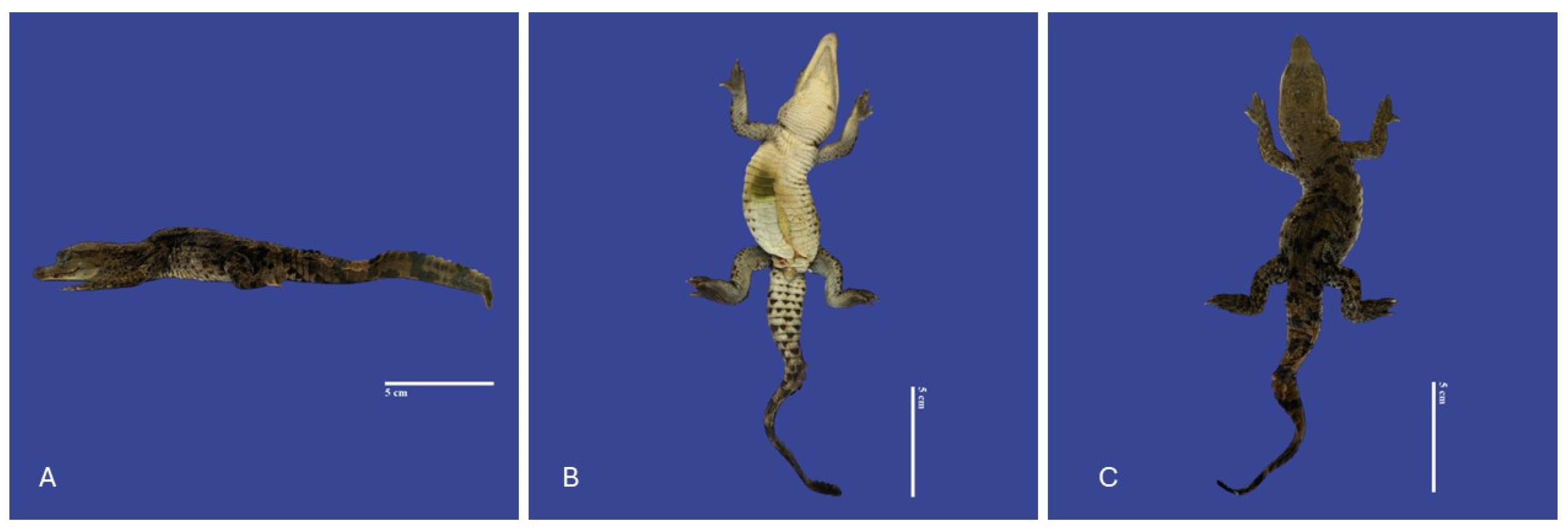
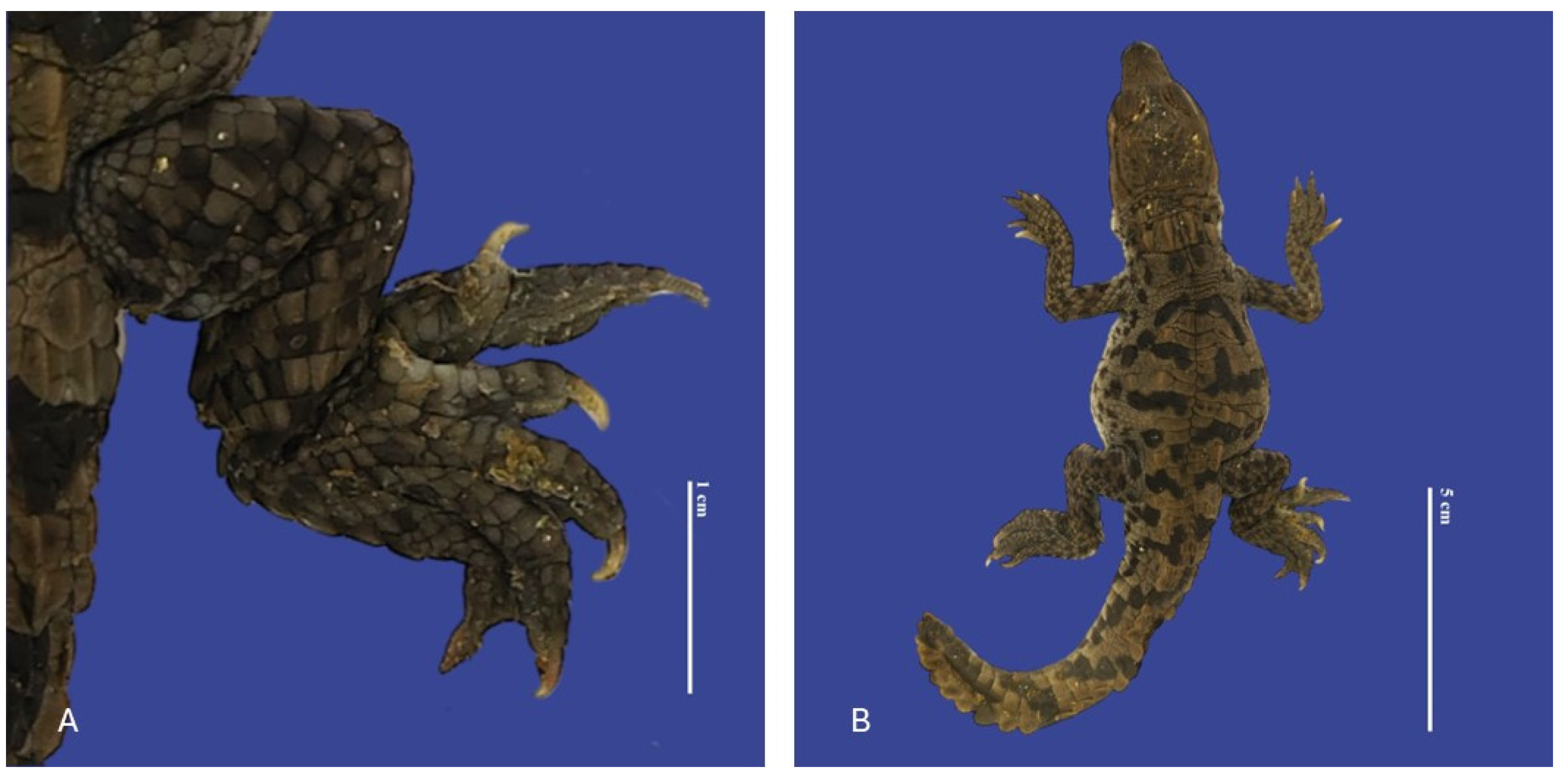
3.4. Limbs and Tail Malformations
3.5. Other Malformations
4. Discussion
5. Conclusions
Author Contributions
Funding
Institutional Review Board Statement
Informed Consent Statement
Data Availability Statement
Conflicts of Interest
References
- Balaguera-Reina, S.A.; Jennings, N.D.; Godfrey, S.T.; Brandt, L.A.; Daykin, B.; Squires, M.A.; Mazzotti, F.J. Hematology and Biochemistry Reference Intervals for American Crocodiles (Crocodylus acutus) in South Florida. Front. Vet. Sci. 2022, 9, 919488. [Google Scholar] [CrossRef] [PubMed]
- American Crocodile (Crocodylus acutus). Available online: https://www.inaturalist.org/taxa/26085-Crocodylus-acutus (accessed on 30 December 2023).
- Rainwater, K.L.; Wiederhold, N.P.; Sutton, D.A.; Garner, M.M.; Maguire, C.; Sanders, C.; Gibas, C.; Cano, J.F.; Guarro, J.; Stchigel, A.M. Novel Paranannizziopsis Species in a Wagler’s Viper (Tropidolaemus wagleri), Tentacled Snakes (Erpeton tentaculatum), and a Rhinoceros Snake (Rhynchophis boulengeri) in a Zoological Collection. Med. Mycol. 2019, 57, 825–832. [Google Scholar] [CrossRef] [PubMed]
- Ellis, T.M. Tolerance of Sea Water by the American Crocodile, Crocodylus acutus. J. Herpetol. 1981, 15, 187–192. [Google Scholar] [CrossRef]
- Ogden, J.C. Status and Nesting Biology of the American Crocodile, Crocodylus acutus, (Reptilia, Crocodilidae) in Florida. J. Herpetol. 1978, 12, 183–196. [Google Scholar] [CrossRef]
- Platt, S.G.; Thorbjarnarson, J.B. Nesting Ecology of the American Crocodile in the Coastal Zone of Belize. Copeia 2000, 2000, 869–873. [Google Scholar] [CrossRef]
- Barragán Lara, R.; Grajales, J.G.; Martínez Ramírez, E. Nest Temperature Assessment in an American Crocodile (Crocodylus acutus) Population on the Central Coast of Oaxaca, Mexico. J. Therm. Biol. 2021, 99, 103012. [Google Scholar] [CrossRef] [PubMed]
- Burkhart, J.G.; Ankley, G.; Bell, H.; Carpenter, H.; Fort, D.; Gardiner, D.; Gardner, H.; Hale, R.; Helgen, J.C.; Jepson, P.; et al. Strategies for Assessing the Implications of Malformed Frogs for Environmental Health. Environ. Health Perspect. 2000, 108, 83–90. [Google Scholar] [CrossRef] [PubMed]
- Briggs-Gonzalez, V.S.; Basille, M.; Cherkiss, M.S.; Mazzotti, F.J. American Crocodiles (Crocodylus acutus) as Restoration Bioindicators in the Florida Everglades. PLoS ONE 2021, 16, e0250510. [Google Scholar] [CrossRef] [PubMed]
- Platt, S.G.; Thorbjarnarson, J.B. Status and Conservation of the American Crocodile, Crocodylus acutus, in Belize. Biol. Conserv. 2000, 96, 13–20. [Google Scholar] [CrossRef]
- Rodgers, E.M.; Schwartz, J.J.; Franklin, C.E. Diving in a Warming World: The Thermal Sensitivity and Plasticity of Diving Performance in Juvenile Estuarine Crocodiles (Crocodylus porosus). Conserv. Physiol. 2015, 3, cov054. [Google Scholar] [CrossRef]
- Brien, M.L.; Gienger, C.M.; Browne, C.A.; Read, M.A.; Joyce, M.J.; Sullivan, S. Patterns of Human–Crocodile Conflict in Queensland: A Review of Historical Estuarine Crocodile (Crocodylus porosus) Management. Wildl. Res. 2017, 44, 281–290. [Google Scholar] [CrossRef]
- Garcês, A.; Pires, I.; Rodrigues, P. Teratological Effects of Pesticides in Vertebrates: A Review. J. Environ. Sci. Health Part B 2020, 55, 75–89. [Google Scholar] [CrossRef] [PubMed]
- Garcês, A.; Pires, I. Teratological Effects of Pesticides in Reptiles—A Review. In Bird and Reptile Species in Environmental Risk Assessment Strategies; Liwszyc, G., Larramendy, M.L., Eds.; The Royal Society of Chemistry: London, UK, 2023; pp. 97–109. ISBN 978-1-83916-710-2. [Google Scholar]
- Bárcenas-Ibarra, A.; de la Cueva, H.; Rojas-Lleonart, I.; Abreu-Grobois, F.A.; Lozano-Guzmán, R.I.; Cuevas, E.; García-Gasca, A. First Approximation to Congenital Malformation Rates in Embryos and Hatchlings of Sea Turtles. Birth Defects Res. A Clin. Mol. Teratol. 2015, 103, 203–224. [Google Scholar] [CrossRef] [PubMed]
- Sant’Anna, S.S.; Grego, K.F.; Lorigados, C.A.B.; Fonseca-Pinto, A.C.B.C.; Fernandes, W.; Sá-Rocha, L.C.; Catão-Dias, J.L. Malformations in Neotropical Viperids: Qualitative and Quantitative Analysis. J. Comp. Pathol. 2013, 149, 503–508. [Google Scholar] [CrossRef] [PubMed]
- MacGeady, T.; Quinn, P.J.; Fiztpatrick, E.; Ryan, M.; Kilroy, D.; Lonergan, P. Veterinary Embryology, 2nd ed.; Wiley-Blackwell: Hoboken, NY, USA, 2017. [Google Scholar]
- Seifer, R. Teratology. In Encyclopedia of Infant and Early Childhood Development; Haith, M.M., Benson, J.B., Eds.; Academic Press: San Diego, CA, USA, 2008; pp. 333–343. ISBN 978-0-12-370877-9. [Google Scholar]
- Bellairs, A.A. Cleft palate, microphthalmia and other malformations in embryos of lizards and snakes. Proc. Zool. Soc. Lond. 1965, 144, 239–252. [Google Scholar] [CrossRef]
- Palmieri, C.; Selleri, P.; Di Girolamo, N.; Montani, A.; Della Salda, L. Multiple Congenital Malformations in a Dicephalic Spur-Thighed Tortoise (Testudo graeca ibera). J. Comp. Pathol. 2013, 149, 368–371. [Google Scholar] [CrossRef]
- Martín-del-Campo, R.; Calderón-Campuzano, M.F.; Rojas-Lleonart, I.; Briseño-Dueñas, R.; García-Gasca, A. Congenital Malformations in Sea Turtles: Puzzling Interplay between Genes and Environment. Animals 2021, 11, 444. [Google Scholar] [CrossRef] [PubMed]
- Wu, T.H.; Rainwater, T.R.; Platt, S.G.; McMurry, S.T.; Anderson, T.A. Organochlorine Contaminants in MoreletÕs Crocodile (Crocodylus moreletii) Eggs from Belize. Chemosphere 2000, 40, 671–678. [Google Scholar] [CrossRef]
- Vyas, R. Case of Polydactyly Limb in Juvenile Mugger Crocodile (Crocodylus palustris). Russ. J. Herpetol. 2018, 25, 139–142. [Google Scholar] [CrossRef]
- Huchzermeyer, F.W. Chapter 4—Diseases of Eggs and Hatchlings. In Crocodiles Biology Husbandry and Disease; CAB International: Wallingford, UK, 2003; pp. 139–156. [Google Scholar] [CrossRef]
- Charruau, P.; Niño-Torres, C. A Third Case of Amelia in Morelet’s Crocodile from the Yucatan Peninsula. Dis. Aquat. Org. 2014, 109, 263–267. [Google Scholar] [CrossRef]
- Brandon-Pliego, J.; Vannini, F. Anomalies and Growth of a Crocodylus acutus in Puerto Escondido, Oaxaca, Mexico. Crocodile Spec. Group Newsl. 2009, 28, 1–20. [Google Scholar]
- Boete, E.E.; Sogbe, E. Enfermedades en caimanes del Orinoco (Crocodylus intermedius) y caimanes de la costa (Crocodylus acutus) mantenidos en zoocriaderos venezolanos. Rev. Cient. 1991, 10, 328–338. [Google Scholar]
- Singh, L.A.K.; Bustard, H.R. Congenital Defects in the Gharial Gavialis Gangeticus (Gmelin). Br. J. Herpetol. 1982, 6, 215–219. [Google Scholar]
- Sepúlveda, M.S.; Wiebe, J.J.; Honeyfield, D.C.; Rauschenberger, H.R.; Hinterkopf, J.P.; Johnson, W.E.; Gross, T.S. Organochlorine Pesticides and Thiamine in Eggs of Largemouth Bass and American Alligators and Their Relationship with Early Life-Stage Mortality. J. Wildl. Dis. 2004, 40, 782–786. [Google Scholar] [CrossRef]
- González, L.M.; Margalida, A.; Mañosa, S.; Sánchez, R.; Oria, J.; Molina, J.I.; Caldera, J.; Aranda, A.; Prada, L. Causes and Spatio-Temporal Variations of Non-Natural Mortality in the Vulnerable Spanish Imperial Eagle Aquila adalberti during a Recovery Period. Oryx 2007, 41, 495–502. [Google Scholar] [CrossRef]
- Rauschenberger, R.H. Developmental Mortality in American Alligators (Alligator mississippiensis) Exposed to Organochlorine Pesticides. Ph.D. Thesis, University of Florida, Gainesville, FL, USA, 2004. [Google Scholar]
- Bell, B.; Spotila, J.R.; Congdon, J. High Incidence of Deformity in Aquatic Turtles in the John Heinz National Wildlife Refuge. Environ. Pollut. 2006, 142, 457–465. [Google Scholar] [CrossRef]
- Board on Environmental Studies and Toxicology; National Research Council; Division on Earth and Life Studies; Commission on Life Sciences; Committee on Animals as Monitors of Environmental Hazard. Animals as Sentinels of Environmental Health Hazards: Committee on Animals as Monitors of Environmental Hazards, Board on Environmental Studies and Toxicology, Commission on Life Sciences, National Re; National Academies Press: Washington, DC, USA, 1991; ISBN 978-0-309-59489-9. [Google Scholar]
- Tavalieri, Y.E.; Galoppo, G.H.; Canesini, G.; Luque, E.H.; Muñoz-de-Toro, M.M. Effects of Agricultural Pesticides on the Reproductive System of Aquatic Wildlife Species, with Crocodilians as Sentinel Species. Mol. Cell. Endocrinol. 2020, 518, 110918. [Google Scholar] [CrossRef]


| Individual Collected | Lake | Nest | Eggs Incubated | Eggs Broken | Total Eggs Laid | Infertile Eggs | Collection Date | Hatching Date | Hatches | Dead Embryos |
|---|---|---|---|---|---|---|---|---|---|---|
| N53 | 2 | 53 | 36 | 0 | 36 | 0 | 2 February 2023 | 25 April 23 | 20 | 16 |
| N64 | 195 | 64 | 27 | 0 | 27 | 0 | 4 February 2023 | 25 April 23 | 13 | 14 |
| N96A | 9 | 96 | 17 | 0 | 17 | 16 | 8 February 2023 | 29 April 23 | 1 | 0 |
| N96B | 9 | 96 | 17 | 0 | 17 | 16 | 8 February 2023 | 29 April 23 | 1 | 0 |
| N97 | 11 | 97 | 41 | 0 | 41 | 25 | 8 February 2023 | 2 May 2023 | 8 | 8 |
| N105 | 8 | 105 | 17 | 0 | 17 | 3 | 9 February 2023 | 2 May 2023 | 1 | 13 |
| N116 | 3 | 116 | 47 | 0 | 47 | 2 | 9 February 2023 | 25 April 23 | 33 | 12 |
| N121 | 8 | 121 | 21 | 9 | 30 | 0 | 10 February 2023 | 5 May 2023 | 2 | 19 |
| N156 | 15 | 156 | 23 | 1 | 24 | 4 | 12 February 2023 | 5 May 2023 | 4 | 15 |
| N162 | 10 | 162 | 32 | 0 | 32 | 5 | 13 February 2023 | 2 May 2023 | 18 | 9 |
| N167 | 2 | 167 | 48 | 0 | 48 | 1 | 13 February 2023 | 4 May 2023 | 28 | 19 |
| N167G1 | 2 | 167 | 48 | 0 | 48 | 1 | 13 February 2023 | 4 May 2023 | 28 | 19 |
| N167G2 | 2 | 167 | 48 | 0 | 48 | 1 | 13 February 2023 | 4 May 2023 | 28 | 19 |
| N181A | 10 | 181 | 39 | 0 | 39 | 0 | 14 February 2023 | 5 May 2023 | 19 | 20 |
| N181B | 10 | 181 | 39 | 0 | 39 | 0 | 14 February 2023 | 5 May 2023 | 19 | 20 |
| N200 | 7 | 200 | 22 | 3 | 25 | 0 | 15 February 2023 | 5 May 2023 | 15 | 7 |
| N202 | 7 | 202 | 19 | 0 | 19 | 0 | 15 February 2023 | 5 May 2023 | 15 | 4 |
| N205 | 12 | 205 | 26 | 0 | 26 | 5 | 15 February 2023 | 4 May 2023 | 12 | 9 |
| N206 | 11 | 206 | 33 | 0 | 33 | 29 | 15 February 2023 | 8 May 2023 | 2 | 2 |
| N222 | 8 | 222 | 11 | 0 | 11 | 0 | 16 February 2023 | 7 May 2023 | 8 | 3 |
| N241 | 7 | 241 | 29 | 0 | 29 | 0 | 17 February 2023 | 7 May 2023 | 18 | 11 |
| N254 | 15 | 254 | 25 | 14 | 39 | 3 | 18 February 2023 | 4 May 2023 | 16 | 6 |
| N264A | 5 | 264 | 21 | 1 | 22 | 4 | 18 February 2023 | 11 May 2023 | 9 | 8 |
| 265B | 9 | 265 | 30 | 0 | 30 | 2 | 18 February 2023 | 13 May 2023 | 15 | 13 |
| N281 | 195 | 281 | 15 | 0 | 15 | 1 | 19 February 2023 | 10 May 2023 | 14 | 0 |
| N283 | 195 | 283 | 30 | 0 | 30 | 5 | 19 February 2023 | 8 May 2023 | 14 | 11 |
| N291 | 6 | 291 | 28 | 0 | 28 | 15 | 20 February 2023 | 11 May 2023 | 8 | 5 |
| N292 | 5 | 292 | 32 | 0 | 32 | 32 | 27 March 2023 | 9 May 2023 | 0 | 0 |
| N315 | 1 | 315 | 27 | 0 | 27 | 10 | 21 February 2023 | 9 May 2023 | 8 | 9 |
| N333 | 9 | 333 | 23 | 0 | 23 | 5 | 22 February 2023 | 17 May 2023 | 6 | 12 |
| N334 | 8 | 334 | 9 | 0 | 9 | 0 | 22 February 2023 | 13 May 2023 | 8 | 1 |
| N352C | 9 | 352 | 38 | 0 | 38 | 5 | 23 February 2023 | 15 May 2023 | 24 | 9 |
| N366 | 11 | 366 | 38 | 2 | 40 | 37 | 31 March 2023 | 13 May 2023 | 0 | 1 |
| N367 | 10 | 367 | 37 | 0 | 37 | 7 | 24 February 2023 | 17 May 2023 | 19 | 11 |
| N368 | 10 | 368 | 35 | 0 | 35 | 1 | 24 February 2023 | 17 May 2023 | 25 | 9 |
| N385 | 3 | 385 | 30 | 0 | 30 | 1 | 24 February 2023 | 17 May 2023 | 15 | 14 |
| N402 | 16 | 402 | 7 | 0 | 7 | 7 | 1 April 2023 | 14 May 2023 | 0 | 0 |
| N404 | 2 | 404 | 31 | 0 | 31 | 3 | 25 February 2023 | 17 May 2023 | 16 | 12 |
| N404A | 2 | 404 | 31 | 0 | 31 | 3 | 25 February 2023 | 17 May 2023 | 16 | 12 |
| N405 | 3 | 405 | 30 | 0 | 30 | 28 | 25 February 2023 | 17 May 2023 | 1 | 1 |
| N405A | 3 | 405 | 30 | 0 | 30 | 28 | 25 February 2023 | 17 May 2023 | 1 | 1 |
| N420 | 11 | 420 | 37 | 0 | 37 | 29 | 26 February 2023 | 17 May 2023 | 5 | 3 |
| N442 | 7 | 422 | 36 | 0 | 36 | 1 | 27 February 2023 | 15 May 2023 | 35 | 0 |
| N443 | 6 | 443 | 26 | 0 | 26 | 13 | 28 February 2023 | 17 May 2023 | 7 | 6 |
| N453 | 1 | 453 | 30 | 0 | 30 | 29 | 1 March 2023 | 18 May 2023 | 0 | 1 |
| N455 | 4 | 455 | 37 | 1 | 38 | 0 | 1 March 2023 | 23 May 2023 | 27 | 10 |
| N457 | 8 | 457 | 26 | 0 | 26 | 0 | 1 March 2023 | 21 May 2023 | 22 | 4 |
| N459A | 6 | 459 | 20 | 0 | 20 | 0 | 1 March 2023 | 20 May 2023 | 17 | 3 |
| N459 | 6 | 459 | 20 | 0 | 20 | 0 | 1 March 2023 | 20 May 2023 | 17 | 3 |
| N486 | 7 | 486 | 30 | 0 | 30 | 1 | 3 March 2023 | 21 May 2023 | 24 | 5 |
| N489 | 195 | 489 | 20 | 0 | 20 | 1 | 3 March 2023 | 28 May 2023 | 16 | 3 |
| N492 | 12 | 492 | 23 | 0 | 23 | 5 | 3 March 2023 | 21 May 2023 | 8 | 10 |
| N496 | 4 | 496 | 40 | 0 | 40 | 36 | 3 March 2023 | 20 May 2023 | 0 | 4 |
| N502 | 2 | 502 | 19 | 0 | 19 | 19 | 5 March 2023 | 22 May 2023 | 0 | 0 |
| N504A | 4 | 504 | 33 | 2 | 35 | 10 | 5 March 2023 | 23 May 2023 | 15 | 8 |
| N504 | 4 | 504 | 33 | 2 | 35 | 10 | 5 March 2023 | 23 May 2023 | 15 | 8 |
| N506 | 8 | 506 | 26 | 2 | 28 | 1 | 5 March 2023 | 23 May 2023 | 11 | 14 |
| N509 | 7 | 509 | 26 | 0 | 26 | 2 | 6 March 2023 | 23 May 2023 | 9 | 15 |
| N509A | 7 | 509 | 26 | 0 | 26 | 2 | 6 March 2023 | 23 May 2023 | 9 | 15 |
| N531 | 4 | 531 | 15 | 11 | 26 | 0 | 8 March 2023 | 13 May 2023 | 5 | 10 |
| N551 | 7 | 551 | 15 | 0 | 15 | 6 | 14 March 2023 | 31 May 2023 | 0 | 9 |
| Body Region | Teratology | Number |
|---|---|---|
| Skin | Ephitheliogenesis imperfecta | 5 |
| Depigmentation | 3 | |
| Leucism | 2 | |
| Spine | Scoliosis | 14 |
| Lordosis | 3 | |
| Kyphosis | 2 | |
| Campylorrachis scoliosa | 1 | |
| Head | Anencephaly/prosencephalic hypoplasia | 1 |
| Laterognathia | 12 | |
| Meningoencephalocete | 3 | |
| Brachygnatia | 1 | |
| Microcephaly | 4 | |
| Maxillary micrognathia | 3 | |
| External hydrocephalus | 1 | |
| Maxillary macrognathia | 7 | |
| Acrania | 3 | |
| Acephaslostomia | 1 | |
| Maxilla Agnathia | 1 | |
| Brachycephalic skull | 1 | |
| Eye and nose | Congenital cataract | 2 |
| Anophthalmia | 1 | |
| Exophthalmia | 7 | |
| Microphthalmia | 1 | |
| Atresia | 2 | |
| Limbs | Micromelia | 1 |
| Polydactyly | 2 | |
| Carpal flexure | 1 | |
| Ankylodactylia | 2 | |
| Malrotation | 1 | |
| Arthrogryposis | 1 | |
| Tail | Curled tail | 17 |
| Kinked tail | 18 | |
| Acaudia | 16 | |
| Brachyuria | 3 | |
| Others | Achondroplasia (Dwarfism) | 4 |
| Distended celomic cavity | 1 | |
| Ectopia cordis | 1 | |
| Yolk sac retention | 47 | |
| Gastroschisis | 1 | |
| Monozygotic symmetrical twins | ||
| Thoracopagus, sternopagus, xiphopagus twins | ||
| Cephalothoracopagus twins |
Disclaimer/Publisher’s Note: The statements, opinions and data contained in all publications are solely those of the individual author(s) and contributor(s) and not of MDPI and/or the editor(s). MDPI and/or the editor(s) disclaim responsibility for any injury to people or property resulting from any ideas, methods, instructions or products referred to in the content. |
© 2024 by the authors. Licensee MDPI, Basel, Switzerland. This article is an open access article distributed under the terms and conditions of the Creative Commons Attribution (CC BY) license (https://creativecommons.org/licenses/by/4.0/).
Share and Cite
Serrano, O.S.; Garcês, A.; Pires, I.; Calderón Mateus, J.A.; Olivera, J.M.; Dávila, J.J. Congenital Anomalies in American Crocodile (Crocodylus acutus, Cuvier, 1807) Embryos from a Farm Breeder in Colombia. Vet. Sci. 2024, 11, 317. https://doi.org/10.3390/vetsci11070317
Serrano OS, Garcês A, Pires I, Calderón Mateus JA, Olivera JM, Dávila JJ. Congenital Anomalies in American Crocodile (Crocodylus acutus, Cuvier, 1807) Embryos from a Farm Breeder in Colombia. Veterinary Sciences. 2024; 11(7):317. https://doi.org/10.3390/vetsci11070317
Chicago/Turabian StyleSerrano, Oscar Sierra, Andreia Garcês, Isabel Pires, John Alexander Calderón Mateus, Juan Medina Olivera, and Jhesteiner Julio Dávila. 2024. "Congenital Anomalies in American Crocodile (Crocodylus acutus, Cuvier, 1807) Embryos from a Farm Breeder in Colombia" Veterinary Sciences 11, no. 7: 317. https://doi.org/10.3390/vetsci11070317
APA StyleSerrano, O. S., Garcês, A., Pires, I., Calderón Mateus, J. A., Olivera, J. M., & Dávila, J. J. (2024). Congenital Anomalies in American Crocodile (Crocodylus acutus, Cuvier, 1807) Embryos from a Farm Breeder in Colombia. Veterinary Sciences, 11(7), 317. https://doi.org/10.3390/vetsci11070317