Article

Virulence of Shigatoxigenic and Enteropathogenic Escherichia coli O80:H2 in Galleria mellonella Larvae: Comparison of the Roles of the pS88 Plasmids and STX2d Phage

1
Veterinary Bacteriology, Department of Infectious and Parasitic Diseases, Faculty of Veterinary Medicine, Center for Fundamental and Applied Research for Animals and Health (FARAH), University of Liège, B-4000 Liege, Belgium
2
Department of Bacteriology, Faculty of Medical Science, Kyushu University, Fukuoka 812-8582, Japan
3
Belgium National Reference Center of STEC (NRC STEC), Universitair Ziekenhuis Brussel (UZ Brussel), Vrije Universiteit Brussel (VUB), B-1090 Brussels, Belgium
*
Author to whom correspondence should be addressed.
Current address: Department of Process Transfer and Development, Eurogentec, B-4100 Seraing, Belgium.
Current address: Veterinary Surgeon, F-57140 Norroy-le-Veneur, France.
§
Current address: Molecular Biology, AZ Sint-Lucas & Volkskliniek, B-9000 Ghent, Belgium.
These authors contributed equally to this work.
Vet. Sci. 2023, 10(7), 420; https://doi.org/10.3390/vetsci10070420
Submission received: 26 May 2023 / Revised: 23 June 2023 / Accepted: 24 June 2023 / Published: 29 June 2023

Abstract

:

Simple Summary

Following the “Replacement/Reduction/Refinement” policy, insects are proposed to replace mammals and birds as experimental models to study the virulence of bacterial pathogens and to identify their virulence properties. The aim of this study was to assess in larvae of the Galleria mellonella moth the virulence of the Shigatoxigenic and enteropathogenic Escherichia coli O80:H2 and the respective roles of two virulence properties: the pS88 plasmid-encoded invasiveness properties and the phage-encoded Shiga toxin 2d. The objectives were to compare: (i) the virulence of bovine Shigatoxigenic and enteropathogenic E. coli O80:H2; (ii) the roles of the pS88 plasmid and Shiga toxin 2d-encoding phage; (iii) the virulence of E. coli O80:H2 and O80:non-H2. The results and the conclusions are: (i) E. coli O80:H2 and O80:non-H2 are lethal at log5 and log6 concentrations; (ii) the pS88 plasmids are partially responsible for the virulence of E. coli O80:H2; (iii) the phage-encoded Stx2d toxin is entirely responsible for the virulence of the Shigatoxigenic Escherichia coli O80:H2; (iv) the virulence properties of E. coli O80:non-H2 could not be identified. As a general conclusion, G. mellonella larvae represent a useful model to study the virulence of bacterial pathogens but are limited in identifying their virulence properties.

Abstract

The invasiveness properties of Shigatoxigenic and enteropathogenic Escherichia coli (STEC and EPEC) O80:H2 in humans and calves are encoded by genes located on a pS88-like ColV conjugative plasmid. The main objectives of this study in larvae of the Galleria mellonella moth were therefore to compare the virulence of eight bovine STEC and EPEC O80:H2, of two E. coli pS88 plasmid transconjugant and STX2d phage transductant K12 DH10B, of four E. coli O80:non-H2, and of the laboratory E. coli K12 DH10B strains. Thirty larvae per strain were inoculated in the last proleg with 10 μL of tenfold dilutions of each bacterial culture corresponding to 10 to 106 colony-forming units (CFUs). The larvae were kept at 37 °C and their mortality rate was followed daily for four days. The main results were that: (i) not only the STEC and EPEC O80:H2, but also different E. coli O80:non-H2 were lethal for the larvae at high concentrations (from 104 to 106 CFU) with some variation according to the strain; (ii) the Stx2d toxin and partially the pS88 plasmid were responsible for the lethality caused by the E. coli O80:H2; (iii) the virulence factors of E. coli O80:non-H2 were not identified. The general conclusions are that, although the Galleria mellonella larvae represent a useful first-line model to study the virulence of bacterial pathogens, they are more limited in identifying their actual virulence properties.

1. Introduction

Escherichia coli (E. coli) is a Gram-negative bacterial species and a member of the human and animal intestinal microbiota. Although most E. coli strains are harmless to their hosts, some strains have acquired genes encoding virulence properties and can cause disease in both humans and animals: they are generically called pathogenic E. coli [1].
Pathogenic E. coli are classified into two main groups: diarrheagenic E. coli or DEC and extraintestinal E. coli or ExPEC. ExPEC comprise different invasive E. coli (septicemic or SePEC, neonatal meningitis-associated or NMEC) and uropathogenic E. coli (UPEC) whereas DEC are subdivided into six main pathotypes: enterotoxigenic (ETEC), enteroinvasive (EIEC), enteropathogenic (EPEC), Shigatoxigenic (STEC), enteroaggregative (EAEC), and Diffusely Adherent (DAEC) E. coli [1,2,3,4].
Moreover, “hybrid” pathogenic E. coli have been described that combine the properties of different pathotypes. One recent dramatic example is the E. coli O104:H4 that caused a short-lived outbreak of diarrhea and hemolytic–uremic syndrome in humans in Germany in 2011 and combined the typical properties of EAEC and STEC [5,6]. Nevertheless, the most frequent examples are still today the enterohemorrhagic E. coli (EHEC) that emerged in humans in the year 1980 and produced both the Attaching-Effacing (AE) lesion on enterocytes typical of EPEC and one or two Shiga toxins (Stx) typical of STEC [1,3]. Since the EHEC nomenclature is considered obsolete by EFSA [7], they will be named “Attaching-Effacing STEC” (AE-STEC) in this manuscript, as previously proposed [6]. The AE lesion is encoded by genes grouped together on one chromosomal pathogenicity island (Locus of Enterocyte Effacement or LEE) while the Stx are encoded by stx phage-located genes [1,3,4]. Two Stx families have been described: Stx1 with three subtypes, (Stx1a, Stx1c, Stx1d) and Stx2 with up to 11 subtypes so far (Stx2a to Stx2k) [8,9,10,11].
The most frequent and pathogenic AE-STEC serotypes in humans are O26:H11, O103:H2, O111:H-, O121:H9, O145:H- and O157:H7, and most frequently the contamination sources are meat, dairy, or vegetable foods contaminated with feces of ruminants, especially cattle, that are healthy carriers in their intestines. Nevertheless, other so-called “minor” serotypes can emerge from time to time and become epidemiologically important in some countries [3,12].
Serotype O80:H2 represents one of these “minor” serotypes that has been emerging since 2010 in France and other Western European countries in humans suffering (bloody) diarrhea, hemolytic–uremic syndrome, and bacteremia [13,14,15,16,17,18]. These AE-STEC O80:H2 combine not only the pathogenic properties of EPEC and STEC, but also of SePEC since they can invade the blood stream and colonize internal organs. The invasiveness properties are encoded by genes located on a pS88-like ColV conjugative plasmid [15,16,18,19].
In addition to their presence in humans, not only AE-STEC, but also EPEC O80:H2 have been emerging since 2009 in young calves in Belgium suffering from diarrhea and only very occasionally from septicemia [20,21]. Genetic analysis confirmed that calf AE-STEC and EPEC O80:H2 are highly related to human AE-STEC O80:H2 by their virulotypes, including the presence of the eaeξ intimin-encoding gene on the LEE and of a pS88-like ColV plasmid [21]. Ca. 80% of the bovine AE-STEC harbor the stx2d gene like the great majority of human AE-STEC O80:H2, while the remaining bovine and human AE-STEC O80:H2 harbor the stx1a or the stx2a gene [17,21,22]. Nevertheless, a difference in the virulotypes of AE-STEC and EPEC O80:H2 exists: the majority of stx2d AE-STEC harbor the etsC and iucC genes associated with the pS88 plasmid, but not the cma and iha chromosomal genes, while the EPEC and the stx1a and stx2a AE-STEC have the opposite gene profile [21]. Phylogenetically, the different AE-STEC and EPEC O80:H2 form different closely related sub-lineages in the single nucleotide polymorphism (SNP)-based phylogenetic tree and the bovine and human AE-STEC are intermixed in the same sub-lineages. The stx2d-positive AE-STEC group together, while the other AE-STEC are more closely related to EPEC [21].
No in vivo study has so far been performed to compare the respective role(s) of the AE lesion, Stx toxin, and pS88-encoded properties in the pathogenicity of AE-STEC and EPEC O80:H2 in humans and calves. Following the 3R policy (Replacement, Reduction and Refinement) [23], insects have been proposed as infectious models to replace mammals and birds for in vivo testing of not only virulence, but also therapy and prophylaxis of bacterial and viral pathogens [24,25]. Of them, larvae of the Galleria mellonella moth are frequently used to study bacterial species, including different pathotypes of E. coli [25,26,27,28]. Besides their low cost and the possibility of testing multiple groups of larvae in a short time, G. mellonella larvae possess an innate immune system similar to mammals and can be maintained at 37 °C, like the bacterial pathogens of mammals [25,29]. So, it was recently observed that the STX2d phage and the type 3 secretion system (T3SS) contribute to the virulence of AE-STEC O80:H2 and EPEC O127:H6, respectively, in G. mellonella larvae [25,30,31], while the contribution of the T3SS to the virulence of AE-STEC O157:H7 is less clear [32,33]. However, no study has been performed yet to assess the role of the pS88 plasmids of AE-STEC and EPEC O80:H2.
Therefore, the aim of this study in G. mellonella larvae was to compare the virulence of: (i) Belgian calf AE-STEC and EPEC O80:H2 harboring different stx genes and pS88 plasmids; (ii) laboratory E. coli K12 pS88 plasmid transconjugant and STX2d phage transductant; (iii) Belgian bovine AE-STEC and EPEC O80:H2 and E. coli O80:non-H2.

2. Materials and Methods

2.1. Bacterial Strains

The virulence of 11 wild-type Belgian bovine E. coli O80 strains was studied in Galleria mellonella larvae (Table 1): eight AE-STEC and EPEC O80:H2 strains with different virulotypes isolated from diarrheic calves at ARSIA (“Association Régionale de Santé et d’Identification animale”) and three E. coli O80:H45 and O80:H6 strains isolated from healthy adult cattle [21,34]. The genomes of six AE-STEC and EPEC O80:H2 and of the three E. coli O80:H45 and O80:H6 strains were previously sequenced and analyzed. The remaining two AE-STEC O80:H2 were identified by PCR, as previously described [21], and were genome sequenced for confirmation (see Section 2.2). One of the AE-STEC O80:H2 (EH3320/SES3090) had been previously tested in larvae of G. mellonella [30] and was the O80:H2 positive control.
The laboratory E. coli K12 DH5α strain and one E. coli O80:H26 strain of the laboratory serotype collection were the non-O80:H2 negative and positive controls, also based on previously published results in larvae of G. mellonella [30]. One E. coli O78:H4 strain also of the laboratory serotype collection was added as a control strain after genome sequencing and preliminary testing in G. mellonella larvae (see Section 3.1).
Laboratory E. coli K12 DH10B and MG1655 strains were used in the pS88 plasmid conjugation and the former was also used in the STX phage transduction. The laboratory E. coli K12 strains harbor the genes for the production of the O16 surface antigen although they are phenotypically rough [35].

2.2. Genome Sequencing

The genomes of the two not-yet-sequenced AE-STEC O80:H2 and of the E. coli O80:H26 and O78:H4 were sequenced and analyzed to confirm the presence or absence of stx, eae, pS88-located and cma/iha genes using Virulence Finder 2.0 (https://cge.food.dtu.dk/services/VirulenceFinder/; accessed on 23 March 2023) and their O:H serotypes using SeroType Finder 2.0 (https://cge.food.dtu.dk/services/SerotypeFinder/; accessed on 23 March 2023), as previously described [21,34]. Sequencing data were submitted as NCBI BioProject PRJNA973567. The Genbank accession numbers are SAMN35130762 (O78C), SAMN35130763 (EH3161), SAMN35130764 (SES5320) and SAMN35130765 (SES5363).

2.3. Construction of DH10B Transconjugant and Transductant

The pS88 plasmid conjugation strategy was based on the presence of the ColV-encoding cva operon on the pS88 plasmid of bovine AE-STEC and EPEC O80:H2 [21,22,23,24,25,26,27,28,29,30,31,32,33,34,35,36]. The recipient strain was the E. coli K12 DH10B strain harboring the recombinant plasmid pAuto-ColV-Switch1.0 carrying a synthetic ColV-encoding locus (received from Syngulon Company, Seraing, Belgium; https://syngulon.com). The cvaC ColV peptide-encoding gene is, however, disrupted by the insertion of an ampicillin resistance cassette (AmpR) while the other genes of the locus remain functional. Therefore, the E. coli K12 DH10B strain is unable to produce the ColV peptide but remains immune to ColV since the cvi gene is intact.
To choose the donor strain, all 33 Belgian bovine AE-STEC and EPEC O80:H2 previously identified [21] were phenotypically tested for their antibiotic resistance profile by the disk diffusion assay [37] against 12 antibiotics on Mueller–Hinton (AxonLab, Machelen, Belgium) agar plates: ampicillin/amoxicillin (20 μg), amoxicillin/clavulanic acid (20 μg + 10 μg), cefoxitin (30 μg), ceftiofur (30 μg) and cefquinome (30 μg) (β lactams), kanamycin (30 μg) and gentamicin (30 μg) (aminosides), nalidixic acid (30 μg) and enrofloxacin (5 μg) (<fluoro>quinolones), florfenicol (30 μg) (phenicols), doxycycline (30 μg) (tetracyclins), and trimethoprim/sulfamethoxazole (1.25 μg + 23.75 μg) (AxonLab, Machelen, Belgium). According to the EUCAST CASFM and CASFMVET 2020 reference of values (https://www.sfm-microbiologie.org/2020/10/02/casfm-eucast-v1-2-octobre-2020/; https://www.sfm-microbiologie.org/2020/09/09/casfm-veterinaire-2020/; accessed on 15 February 2021), nine strains were sensitive to ampicillin. Of them, the AE-STEC EH2282 strain (Table 1) isolated in 1987 [20] was resistant to only three of the tested antibiotics (kanamycin, doxycycline, trimethoprim/sulfamethoxazole) compared to the other ampicillin-sensitive strains and was therefore chosen as the donor strain.
The pS88 plasmid conjugation was performed on Luria–Bertani (LB; VWR Chemicals, Leuven, Belgium) agar plates [38] by mixing 100 μL of an 8 h growth of the donor strain in 5 mL of LB broth and 500 μL of an 8 h growth of the recipient strain in LB broth with 100 μg/mL ampicillin. After overnight incubation at 37 °C, full loops of the macrocolony were streaked on 10 LB agar plates with 100 μg/mL ampicillin and incubated overnight at 37 °C. Isolated colonies were transferred to two 96-well microtiter plates containing 200 μL LB broth with 100 μg/mL ampicillin and grown overnight at 37 °C. The following day the colonies were transferred onto LB agar plates with 100 μg/mL ampicillin covered with a layer of the ColV-sensitive E. coli K12 MG1655 strain harboring the pKK223-3 plasmid [39] carrying an ampicillin resistance-encoding gene (received from Syngulon Company, Seraing, Belgium; https://syngulon.com). The colonies inhibiting the growth of the E. coli K12 MG1655 strain were sub-cultured in LB broth with 100 μg/mL ampicillin of which two mL were transferred into two CRYO tubes (Greiner Bio-One, Frickhausen, Germany) with two mL of sterile 80% glycerol that were stored at −20 °C and −80 °C, respectively, until further use.
These colonies were subsequently grown on LB agar plates with 100 μg/mL ampicillin and tested with PCR for the serotype O80-, H2-, and O16-encoding genes, the cvaC wild-type and AmpR cassette-inserted genes and the pS88 plasmid-located hlyF gene (Table 2), using the FASTGENE2x Optima Hotstart kit (Nippon Genetics, Filter service, Eupen, Belgium) after DNA extraction from one colony by boiling [34].
STX2d phage transductants were constructed using the E. coli K12 DH10B strain free of the recombinant plasmid pAuto-ColV-Switch1.0 (received from the Syngulon Company, Seraing, Belgium; https://syngulon.com) as the recipient strain, as previously described [30]. STX2d phages were induced by UV radiation and isolated from the three bovine stx2d AE-STEC O80:H2 EH3155, EH3160 and EH3320/SES3090 strains. The transductant candidates were confirmed with a stx2d gene quantitative (q) PCR.

2.4. In Vivo Assay: The Galleria Mellonella Model

At first, the growth curve of all 17 E. coli strains tested in larvae of G. mellonella (eight E. coli O80:H2, two E. coli O80:H45, one E. coli O80:H6, the two E. coli O80:H26 and O78:H4, the two E. coli K12 DH5a and DH10B strains, and the two E. coli K12 DH10B pS88 plasmid-conjugated and STX2d phage-transduced strains) was followed by comparing the optical density of an LB broth culture at a wavelength of 600 nm (OD600) and the number of colony-forming units (CFU) after plating 10 μL on LB agar and overnight growth at 37 °C. An OD600 between 0.2 and 0.35 corresponded to a concentration of 108 CFU/mL depending on the strain. Then, 10 μL of an overnight culture of each strain in LB broth at 37 °C was transferred to fresh LB broth that was incubated at 37 °C until reaching the appropriate OD600 corresponding to a concentration of 108 CFU/mL.
Thirty larvae per strain divided into three groups of 10 larvae (Animal Confort, Loncin, Belgium) were inoculated in the last proleg, as previously described [30], with 10 μL of each of the tenfold dilutions of the bacterial culture corresponding to 10 to 106 CFU using an automatic injector (ColeParmer, Vernon Hills, IL, USA). In addition, 30 larvae were injected with 10 μL of PBS as a control group. The larvae were kept at 37 °C and their mortality rate was followed daily for four days. In parallel, back-titration was performed to confirm the actual inoculation dose by streaking 10 μL of each dilution on LB agar plates and incubating them overnight at 37 °C.

2.5. Statistical Analysis

All statistical analyses were performed using R software with “Rcmdr v2.6-0” and “survival” packages (https://www.john-fox.ca/RCommander/index.html; accessed on 1 April 2023) after pooling the different groups of larvae per strain, virulotype, or serotype. Kaplan–Meier curves were created to assess the survival of the larvae according to the inoculation doses of each bacterial strain. Log-rank tests were carried out to highlight significant differences in survival rates between the groups of larvae inoculated with the different concentrations (log1 to log6) of each E. coli strain and the PBS group. Hazard ratios (HRs) with a 95% confidence interval (HR-95%) were calculated to give the relative measure of the risk factor for death for the larvae when comparing the same concentration of two different E. coli strains or groups of strains, the reference and the test strains: when the HR-95% values included the value 1, the risk factor for death was the same with both strains; when both HR-95% values were >1 or <1, the risk factor for death was higher or lower, respectively, with the test strain(s) compared to the reference strain(s). The significant thresholds of 0.05, 0.01, and 0.001 were applied for all statistical analysis.

3. Results

3.1. Virulotypes and Serotypes of the Four Genome Sequenced E. coli Strains

The genome sequencing and analysis confirmed the virulotypes and serotypes of the two AE-STEC O80:H2 SES5320 and SES5363 strains using Virulence Finder 2.0 and SeroType Finder 2.0 (Table 1 and Table 3). The eaeξ and stx1a genes and several genes located on the pS88 plasmid, including the specific hlyF gene, were detected. The pS88 plasmid-located etsC/iucC genes were detected in the AE-STEC O80:H2 SES5363 strain, but not in the AE-STEC O80:H2 SES5320 strain. Conversely, the chromosomal cma/iha genes were detected in the AE-STEC O80:H2 SES5320 strain, but not in the AE-STEC O80:H2 SES5363 strain. Therefore, three stx1a AE-STEC, three stx2d AE-STEC, and two EPEC O80:H2 strains with different pS88 plasmid and chromosomal gene profiles (Table 1 and Table 3) were tested in G. mellonella larvae.
The serotypes of the E. coli O80:H26 and O78:H4 of the laboratory serotype collection were also confirmed (Table 1 and Table 3). The stx genes and the LEE-located genes were not detected, identifying them as neither AE-STEC nor EPEC. Conversely, the pS88 plasmid-located genes, including the etsC/iucC genes, were detected in the E. coli O78:H4 strain, but not in the E. coli O80:H26 strain (Table 1 and Table 3), whereas the cma/iha chromosomal genes were not detected. Since the E. coli O78:H4 was pathogenic for larvae of G. mellonella in preliminary testing and harbored one pS88-like plasmid, this strain was added as a non-O80 pS88 plasmid-positive control.

3.2. Identification of Transconjugant and Transductant

After growth in LB broth with 100 μg/mL ampicillin in microtiter plates, 50 colonies were transferred onto LB agar plates with 100 μg/mL ampicillin covered with a layer of the ColV-sensitive ampicillin-resistant E. coli K12 MG1655 strain. Eleven of these 50 colonies inhibited the growth of the E. coli K12 MG1655 strain and gave two amplification fragments (314 bp and 1555bp; Table 2) with the PCR for the cvaC wild-type and AmpR cassette-inserted genes, respectively. Of the 11 transconjugant candidates, two also tested positive with the PCR for the O16-encoding genes and the pS88 plasmid-located hlyF gene and negative with the PCR for the serotype O80- and H2-encoding genes. These two transconjugant candidates (F5 and D4) were sub-cultured on two LB agar plates with 100 μg/mL ampicillin. Ten colonies from each agar plate were chosen and re-tested with the same PCR. Of these 20 colonies, 12 once more gave unambiguous PCR results and 1 colony from the F5 transconjugant candidate was chosen for testing in G. mellonella larvae.
The STX2d phages isolated from the AE-STEC O80:H2 EH3155 and EH3160 strains, but not from the EH3320/SES3090 strain, produced plaque lysis on the E. coli K12 DH10B strain. Either phage and the E. coli K12 DH10B strain were mixed in LB broth and incubated overnight at 37 °C. The transductant candidates were recovered by centrifugation, re-suspended in LB broth, and spread on STEC agar plates. After overnight incubation at 37 °C, one colony from each plate was randomly picked up and confirmed by qPCR targeting the stx2d gene. These colonies were sub-cultured three times on STEC agar to confirm the stability of the transduced phages. At each stage, the transductant candidates were confirmed by the qPCR targeting the stx2d gene. E. coli K12 DH10B transductant from AE-STEC O80:H2 EH3160 strain was chosen for studies in G. mellonella larvae.

3.3. Virulence of E. coli Strains in G. mellonella Larvae

3.3.1. E. coli Non-O80:H2 Control Strains

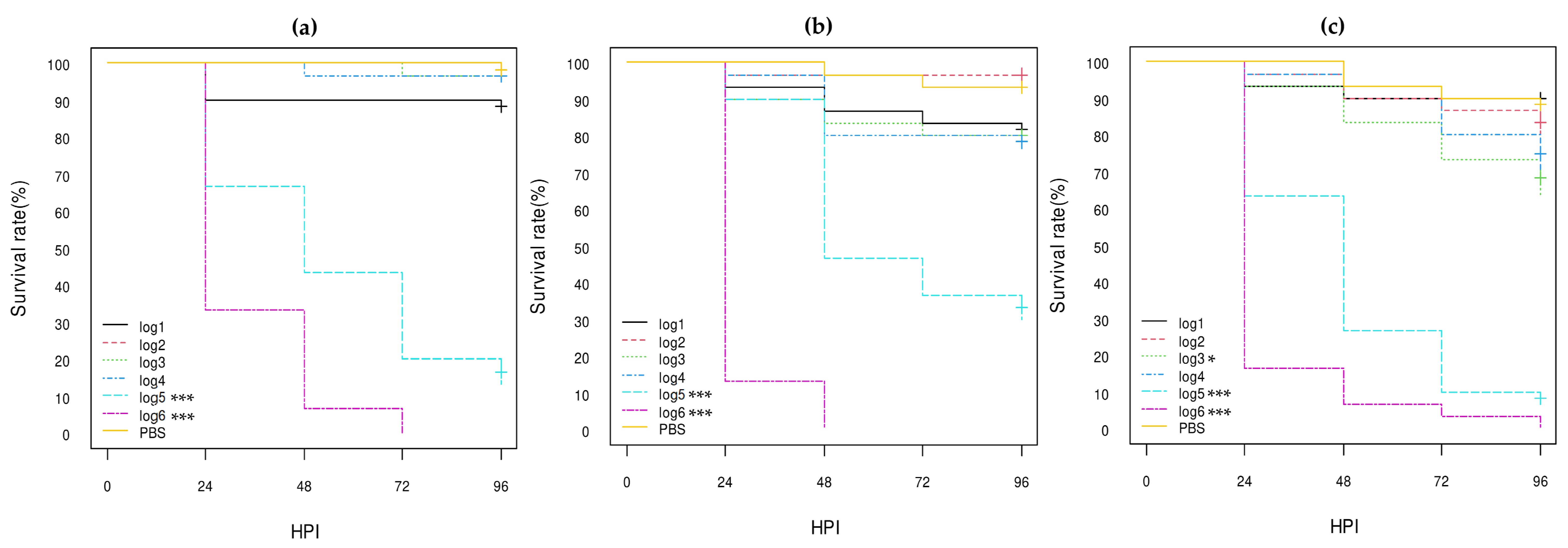
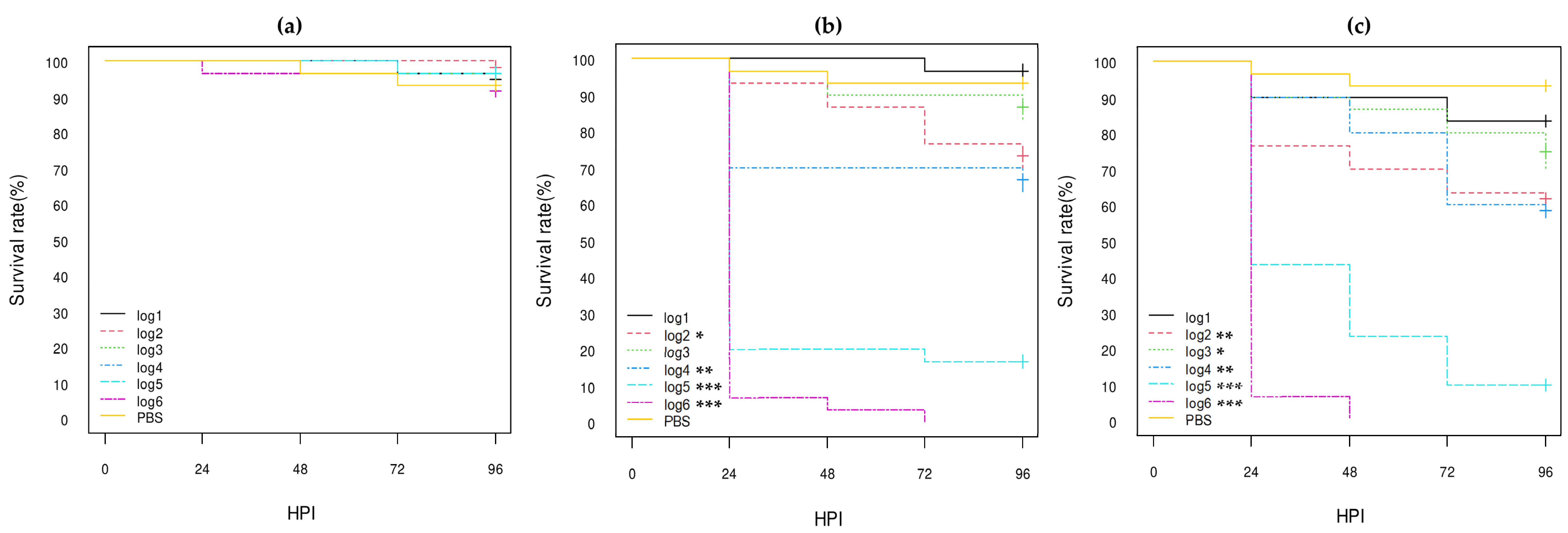
Of these three control strains (E. coli K12 DH5α, E. coli O80:H26, and E. coli O78:H4), the E. coli O78:H4 strain was the most highly virulent, killing almost all larvae within 24 h post-inoculation (HPI) even at log1 concentration (p-value < 0.001), while more than 80% of larvae inoculated with the E. coli K12 DH5α strain still survived at 96 HPI at log6 concentration (Figure 1; Table S1). The E. coli O80:H26 strain was also virulent, killing about half and 75% of the larvae at log4 and log5 concentrations, respectively, at 96 HPI and all larvae at log6 concentration at 72 HPI (p-value < 0.001; Figure 1; Table S1). The E. coli K12 DH5α and O80:H26 strains gave similar results to those previously observed [30].

3.3.2. Comparison of the E. coli O80:H2 Strains Belonging to Different Virulotypes

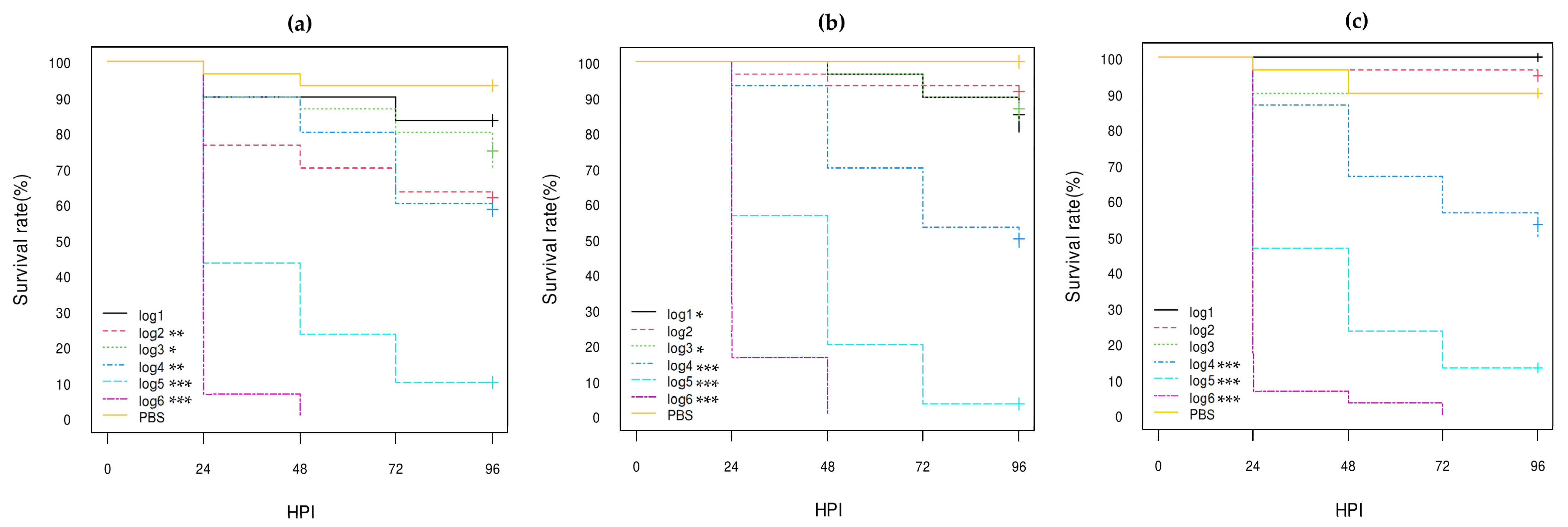
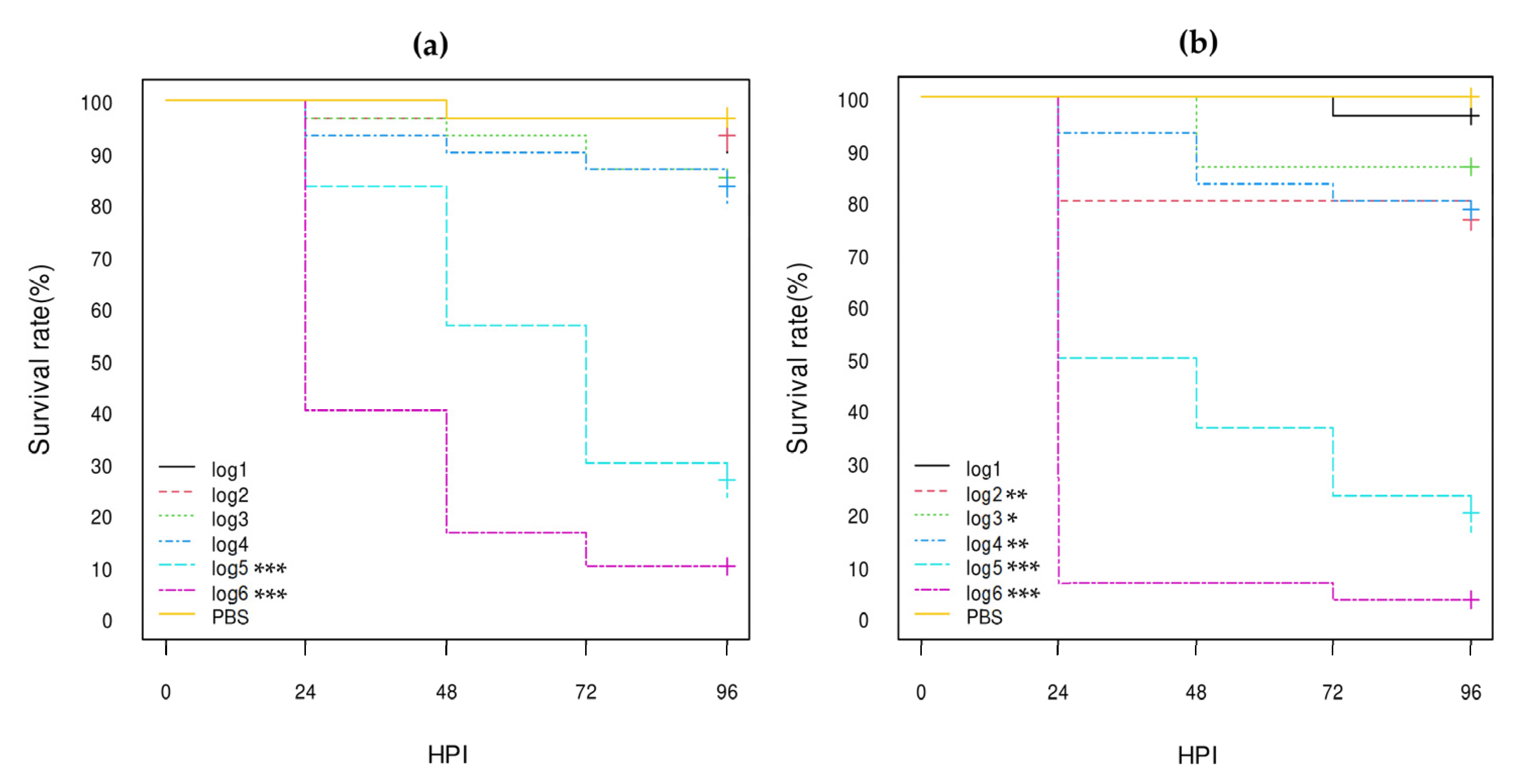
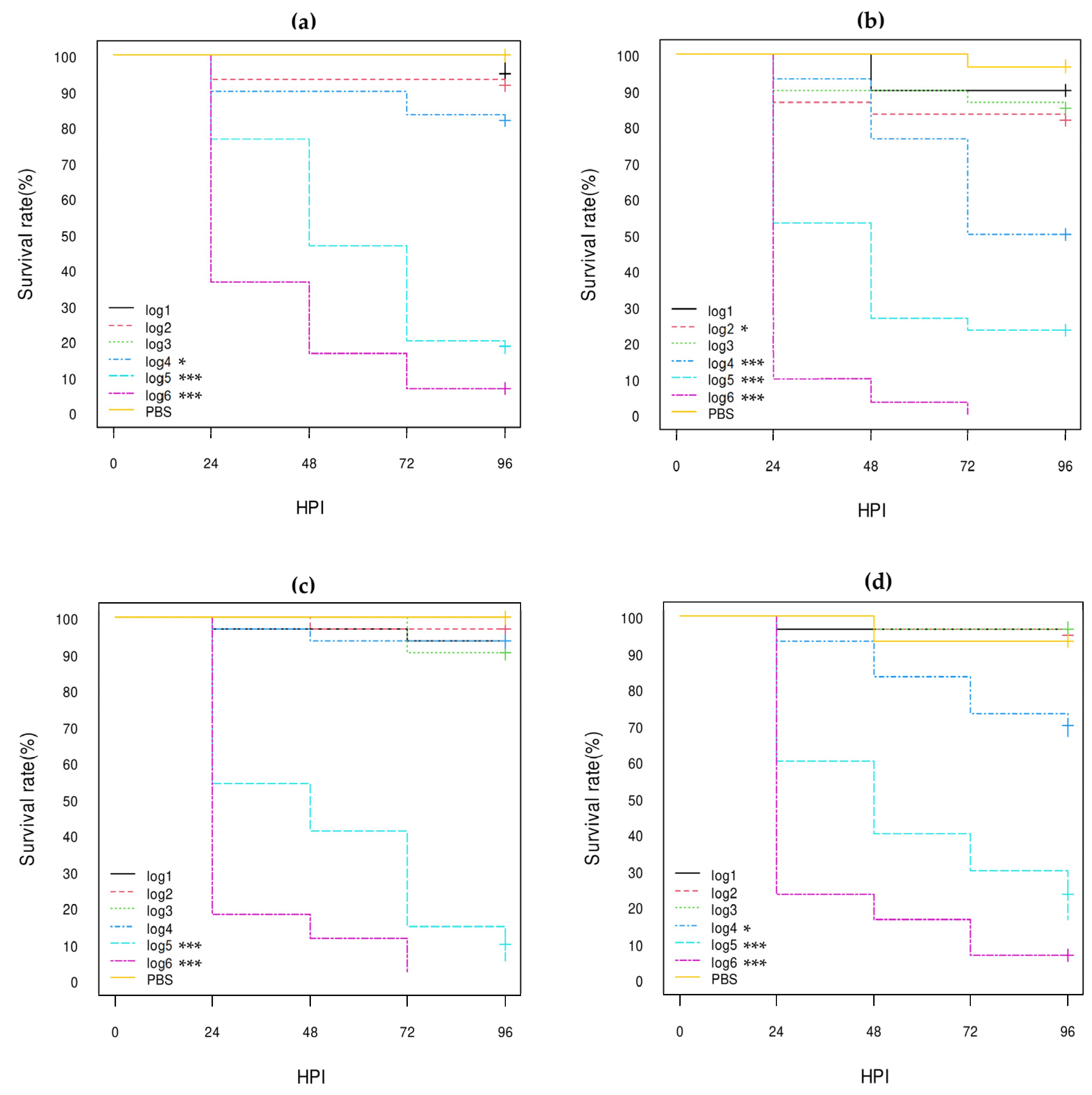
According to the log-rank analysis of Kaplan–Meier curves (Figure 2, Figure 3 and Figure 4; Table S1), the log5 and log6 concentrations of all eight E. coli O80:H2 strains gave significantly different results from the PBS group results with less than 40% survival at 96 HPI (p-value < 0.001). The stx2d AE-STEC EH3320/SES3090 strain gave similar results to those previously obtained [30]. Although all three stx2d AE-STEC were also significantly more lethal compared to the PBS group at the log4 concentration (p-value < 0.001) with ca. 40% death at 96 HPI (Figure 3; Table S1), this was not the case for the stx1a AE-STEC and at least for one EPEC (Figure 2 and Figure 4; Table S1). Conversely, the results with the lowest concentrations (log1 to log3) were much more heterogeneous, even within the same virulotype (Figure 2, Figure 3 and Figure 4; Table S1), and were not further analyzed. Therefore, the hazard ratios (HRs) and the confidence intervals 95% (HR-95%) were statistically analyzed only for the log5 and log6 concentrations.
At the highest bacterial concentration (log6), almost all larvae were dead at 96 HPI regardless of the virulotype (Figure 2, Figure 3 and Figure 4). However, the stx2d AE-STEC strains significantly killed more larvae and more quickly than the EPEC (Figure 3 and Figure 4) with all larvae dead at 72 HPI while a few larvae were still alive at 96 HPI with the EPEC. The HR-95% of the stx2d AE-STEC vs. EPEC strains was statistically significant: between 0.44 and 0.88 (p-value < 0.01) (Table S2). Conversely, the results with stx1a vs. stx2d AE-STEC and with stx1a AE-STEC vs. EPEC were not significantly different (Figure 2 and Figure 3; Table S2).
At the log5 concentration, the stx2d AE-STEC strains significantly killed more larvae and more quickly than the EPEC and the stx1a AE-STEC, with a survival rate lower than 20% at 96 HPI vs. 10–40% for the different stx1a AE-STEC and EPEC strains (Figure 2, Figure 3 and Figure 4). The HC-95% of the stx1a AE-STEC vs. stx2d AE-STEC was higher than 1 (between 1.17 and 2.19 with a p-value < 0.01), while the HC-95% of the stx2d AE-STEC vs. EPEC was lower than 1 (between 0.40 and 0.83 with a p-value < 0.01) (Table S2). Conversely, the results of the stx1a AE-STEC vs. EPEC were not significant, similar to the log6 concentration.
Since some results could be due to intra-group variations as consequence of different genetic backgrounds, the results of the three stx2d AE-STEC, the three stx1a AE-STEC, and the two EPEC were intra-virulotype compared (Table S2). Only the results of the two EPEC strains were statistically different at the log6 concentration. The EPEC EH3322/SES3122 strain harboring a pS88 plasmid carrying the etsC and iucC genes (pS88++) killed more larvae and more rapidly than the EH3308/SES2973 strain whose pS88 plasmid does not carry these two genes (pS88--) (Figure 4). The HR-95% of the EPEC EH3308/SES2973 pS88-- strain vs. the EPEC EH3322/SES3122 pS88++ strain was statistically significant: between 1.09 and 3.23 (p-value < 0.05) (Table S2).

3.3.3. Comparison of the Role of the pS88 Plasmid and of the STX2d Phage

According to the results described above, the etsC/iucC-positive pS88 plasmid may play a role in the pathogenicity of some E. coli O80:H2 strains in G. mellonella larvae. The virulence of one etsC/iucC-positive pS88 plasmid-conjugated E. coli K12 DH10B strain was therefore compared with the virulence of one STX2d phage-transduced E. coli K12 DH10B, of the E. coli K12 DH10B recipient, and of the AE-STEC O80:H2 EH2282 and EH3160 plasmid and phage donor strains (Table 1).
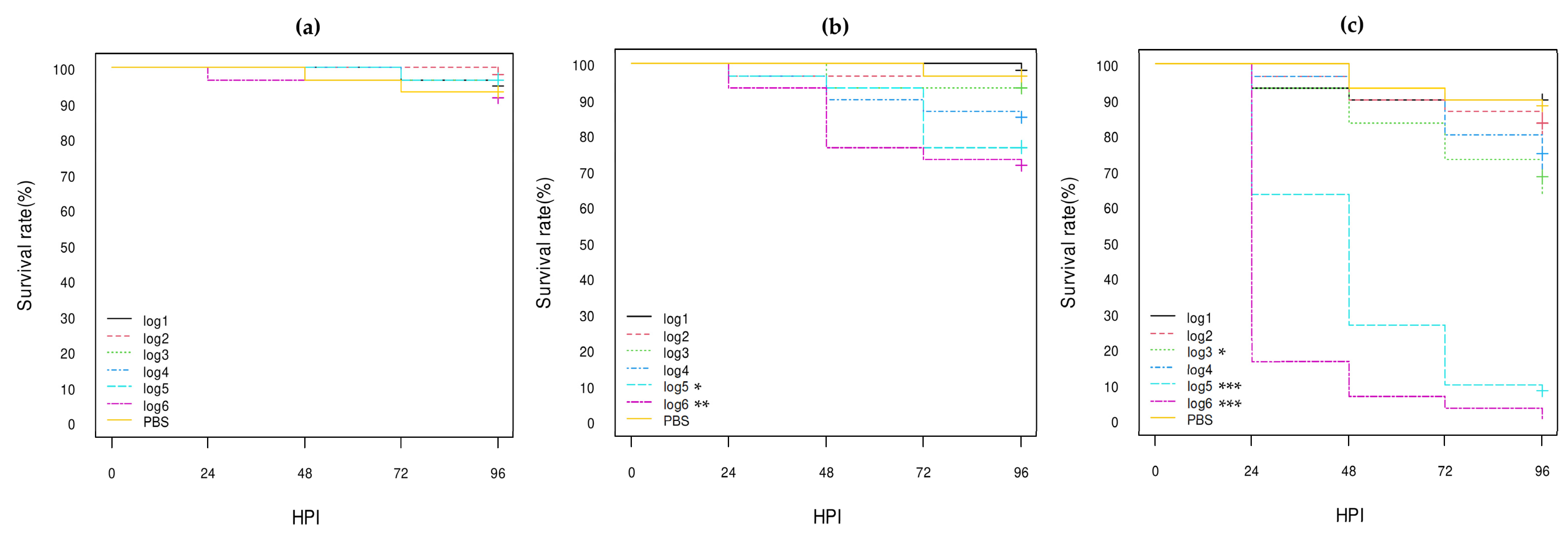
The log-rank analysis of the Kaplan–Meier curves confirmed that the acquisition by conjugation of the etsC/iucC-positive pS88 plasmid significantly increased the lethality of the E. coli K12 DH10B recipient strain (ca. 25% at 96 HPI), both at log5 (p-value < 0.05) and log6 (p-value < 0.01) concentrations (Figure 5; Table S1). However, the HR-95% of the transconjugant vs. the E. coli K12 DH10B recipient strain was not statistically significant, neither at log6 nor at log5 (Table S3). Conversely, the HR-95% of the transconjugant vs. the stx1a AE-STEC EH2282 donor strain was highly significant (between 5.74 and 29.37) at log6 (p-value < 0.001) and between 4.32 and 23.82 at log5 (p-value < 0.001) concentrations (Table S3). Nevertheless, the HR-95% of the E. coli K12 DH10B recipient strain vs. the stx1a AE-STEC EH2282 donor strain was even more highly significant: between 12.78 and 153.50 at log6 (p-value < 0.001) and between 10.57 and 586.30 at log5 (p-value < 0.001) concentrations (Table S3).
The log-rank analysis of the Kaplan–Meier curves also confirmed that the acquisition by transduction of the STX2d phage significantly increased the lethality of the E. coli K12 DH10B recipient strain, at log4 (p-value < 0.01), log 5 (p-value < 0.001), and log 6 (p-value < 0.001) concentrations. The lethality of the transduced E. coli K12 DH10B strain was very similar to the lethality of the stx2d AE-STEC EH3160 donor strain (Figure 6; Table S1): less than 20% of the larvae inoculated with the log5 concentration survived at 96 HPI with both transduced and donor strains while all larvae were dead at 72 HPI and at 48 HPI, respectively, at the log6 concentration (Figure 6). Moreover, the HR-95% of the STX2d-transduced E. coli K12 DH10B and of the stx2d AE-STEC EH3160 donor strains vs. the E. coli K12 DH10B recipient strain were similar and also highly significant with HR-95% values between ca. 8 and more than 400 at log5 and between ca. 17 and more than 350 at log6 (Table S3). Conversely, the HR-95% of the STX2d-transduced E. coli K12 DH10B vs. the stx2d AE-STEC EH3160 donor strains was not statistically significant (Table S3).

3.3.4. Comparison with Other E. coli O80 Serotypes

In addition to AE-STEC and EPEC O80:H2, the virulence in G. mellonella larvae of three pS88 plasmid-negative, non-AE-STEC, non-EPEC E. coli O80:H6 and O80:H45 strains isolated from healthy cattle (Table 1 and Table 3) was assessed for comparison with all E. coli O80:H2 strains. The control E. coli O80:H26 strain was also included in this comparison.
According to the log-rank analysis of the Kaplan–Meier curves (Figure 7; Table S1), the log5 (p-value < 0.001) and log6 (p-value < 0.001) concentrations of all three E. coli O80:H6 and O80:H45 strains gave significantly different results from the PBS group results with less than 30% and 10% survival at 96 HPI, respectively, while the results with the lower concentrations (log1 to log4) were not consistently statistically significant with more than 70% survival at 96 HPI (Figure 7; Table S1). The results of the E. coli O80:H26 strain were similar to the log5 and log6 concentrations, but the log4 concentration was also statistically significant (p-value < 0.001) (Figure 7; Table S1). Moreover, the HR-95% ratios between these three O80 serotypes were not statistically significant with the exception of the HR-95% of the E. coli O80:H6 strain vs. the E. coli O80:H26 strain that was between 1.11 and 3.12 (p-value < 0.05) at the log6 concentration (Table S4).
As for the comparison between the results of E. coli O80:H6, O80:H26, and O80:H45 strains on the one hand and the stx1a AE_STEC, stx2d AE-STEC, and EPEC O80:H2 on the other hand, some statistically significant differences were observed, especially between the E. coli O80:H6 strain and the AE-STEC O80:H2 strains (Table S4). For instance, the HR-95% ratios of the E. coli O80:H6 strain vs. the stx1a and stx2d AE-STEC O80:H2 strains were statistically significant at log6: between 0.41 and 0.98 (p-value < 0.05) and between 0.31 and 0.76 (p-value < 0.01), respectively. At log5, the HR-95% of the E. coli O80:H6 strain vs. the stx2d AE-STEC O80:H2 strains was also statistically significant: between 0.38 and 0.93 (p-value < 0.05). In addition, the HR-95% of the E. coli O80:H45 strain vs. the stx2d AE-STEC O80:H2 strains was statistically significant at log6 (between 0.49 and 0.97; p-value < 0.05), but not at log5 (Table S4). Conversely, the HR-95% ratios of any E. coli O80:non-H2 strain vs. the EPEC O80:H2 strains were not statistically significant (Table S4).

4. Discussion

The emerging AE-STEC O80:H2 are recognized as a multiple hybrid pathotype responsible for enteritis, hemolytic–uremic syndrome, and bacteremia or septicemia in humans and/or young calves [13,15,21]. They indeed harbor not only phage-located stx genes with a large majority of stx2d gene and/or LEE pathogenicity island-located genes, but also pS88-like ColV plasmids carrying invasiveness-encoding genes [15,21,22]. However, the respective role(s) of the Stx toxins, AE lesion, and pS88-encoded properties in their pathogenicity have not been confirmed by in vivo studies yet. Following the 3R policy (Replacement, Reduction and Refinement) [23], insects have been recommended and have been used for some years as a first step for in vivo testing of pathogenic bacterial species [24,25]. Thus, the main purpose of this study was to assess larvae of the G. mellonella moth [24,25,27] as a model to study and compare the virulence of different AE-STEC and EPEC O80:H2 isolated from calves in Belgium [21], and the respective roles of the pS88-like ColV plasmid and STX2d phage.
According to the statistical analysis results, all eight calf stx1a AE-STEC, stx2d AE-STEC, and EPEC O80:H2 were pathogenic for G. mellonella larvae at log5 and log6 concentrations compared to the PBS group (Figure 2, Figure 3 and Figure 4; Table S1). They were also more pathogenic than the E. coli K12 DH5α and DH10B strains, but less than the E. coli O78:H4 strain (Figure 1, Figure 5 and Figure 6; Table S1). Moreover, all three stx2d AE-STEC were already lethal for larvae at log4 and more rapidly and intensively at log5 and log6 than the stx1a AE-STEC and EPEC (Figure 2, Figure 3 and Figure 4; Tables S1 and S2).
The reason for the stx2d AE-STEC being more virulent to G. mellonella larvae may be related to the production of the Stx2d toxin. Indeed, the lethality rate of larvae with STX2d phage-transduced E. coli K12 DH10B strain was significantly higher compared to the E. coli K12 DH10B recipient strain and was similar to the AE-STEC O80:H2 EH3160 phage donor strain (Figure 6; Table S3), as previously reported with the STX2d phage-transduced E. coli K12 DH5α strain [30]. The HR-95% ratios of the STX2d phage-transduced E. coli K12 DH10B strain and of the EH3160 phage donor strains were also similar and significantly higher when compared with the E. coli K12 DH10B recipient strain. These results confirmed that the Stx2d toxin plays an important role in the pathogenicity of stx2d AE-STEC O80:H2 in G. mellonella larvae, as previously suggested [30].
Although the HR-95% between stx1a AE-STEC and stx2d AE-STEC was statistically significant at log5, a role for the Stx1a toxin in the lethality of larvae cannot be totally excluded, since the difference was not statistically significant at log6 (Table S2). The reasons for these conflicting results may be several: (i) low numbers of CFUs of one stx1a AE-STEC actually inoculated, especially at log5 (3 × 10⁴ CFU instead of 105 CFU after back-titration; data not shown); (ii) slower multiplication of stx1a AE-STEC compared to stx2d AE-STEC as a consequence of different general genetic backgrounds of the strains; (iii) delay in the production of the Stx1a toxin compared to the Stx2d toxin; (iv) less efficacious action of the Stx1a toxin in G. mellonella larvae compared to the Stx2d toxin. Whatever the actual reason, counting the number of CFUs of stx1a AE-STEC and stx2d AE-STEC in dead larvae, testing STX1a phage transductant, and/or following the expression of the stx1a and stx2d genes may answer these questions.
Two other specific properties of the AE-STEC and EPEC O80:H2 are the production of the AE lesion and the invasiveness properties encoded by pS88-like plasmid-located genes [15,18,19,21]. The role of the T3SS responsible for the development of the AE lesion was not assessed in his study but is indeed partially responsible for the lethality of the human EPEC E2348/69 (serotype O127:H6) in G. mellonella larvae compared to a mutant in one of the encoding genes, as observed by others [31]. However, the situation seems different for AE-STEC O157:H7 [32,33], possibly because the Stx toxins play a more important role ([30], this study).
As far as the pS88 plasmids are concerned, a role of the etsC/iucC-positive pS88 plasmid can be hypothesized, according to the statistical analysis of the pathogenicity in G. mellonella larvae of the two EPEC O80:H2 (Figure 4; Tables S1 and S2). Indeed, the EPEC EH3322/SES3122 strain harboring one etsC/iucC-positive pS88 plasmid (Table 1 and Table 3) was almost twice as lethal as the EPEC EH3308/SES2973 strain harboring a pS88 plasmid not carrying these two genes at log6 (Table S2). However, the etsC/iucC-positive pS88 plasmid transconjugant was not statistically more lethal than the E. coli K12 DH10B recipient strain, although the HR-95% lower values were borderline at both log6 (between 0.92 and 12.60) and log5 (between 0.95 and 63.07) concentrations (Table S3). Nevertheless, this transconjugant was far from being as lethal as the stx1a AE-STEC O80:H2 plasmid donor strain (Figure 5; Tables S1 and S3). The etsC gene is a member of one operon coding for an ABC transporter system while the iucC gene is a member of an operon encoding the aerobactin siderophore [43]. Although both ets and iuc genes are markers of ExPEC, especially of Avian Pathogenic E. coli (APEC) and of NMEC, the contribution of the ets genes in the pathogenicity of E. coli in larvae of G. mellonella is still unknown, while contradictory results have been obtained for the iuc genes [19,44]. Comparison with the results obtained with one etsC/iucC-negative pS88 plasmid transconjugant would represent a first step in the understanding of their role.
The role of the pS88 plasmids could actually depend more on the general genetic background of the E. coli tested than on the pS88 plasmid virulotype. For instance, the difference observed between the two EPEC O80:H2 strains was not observed between the three stx2d AE-STEC or between the three stx1a AE-STEC (Tables S1 and S2), possibly because the Stx toxins were also here more important virulence factors in G. mellonella larvae than the properties encoded by the pS88 plasmid. Moreover, the great majority of AE-STEC and EPEC O80:H2 harboring one etsC/iucC-positive pS88 plasmid group together in a single nucleotide polymorphism-based phylogenetic tree were negative for the iha and cma chromosomal genes (Table 3), and vice versa [21]. Nevertheless, no role for the cma and iha genes in the pathogenicity of E. coli O80:H2 in this intrahemocoelic inoculation model of larvae of G. mellonella can be proposed at this stage. Indeed, the cma gene codes for a colicin degrading the glycan chain of the murein precursor of other E. coli cells [45] while the iha gene encodes an outer membrane protein, conferring adherence to epithelial cells in culture [46]. Moreover, the iha gene is located on the pathogenicity island SPLE1, and its absence can also mean the absence of other genes located on SPLE1 [46,47].
Another striking observation was the human EPEC O127:H6 that was already lethal for G. mellonella larvae at a concentration of 5 × 10³ CFU, like the stx2d AE-STEC O80:H2, while not harboring any stx genes or pS88 plasmid [31]. Therefore, testing different pS88 plasmid-cured E. coli O80:H2 strains would also help to determine its actual role in G. mellonella larvae, as already published in a mammalian model with the S88 NMEC strain. The S88 strain cured of the pS88 plasmid loses almost all virulence in a neonatal rat model of infection while reintroduction of the plasmid restores full virulence [19]. The role of any bacterial property can indeed also depend on the animal model. Testing the S88 strain and its plasmid-cured derivative would also help to confirm G. mellonella larvae as an in vivo model to study the virulence of different E. coli pathotypes.
Still another example of the importance of the genetic background of the strains was the E. coli O78:H4 control strain that harbors an etsC/iucC-positive pS88 plasmid (Table 1 and Table 3). This E. coli O78:H4 strain, however, had the highest lethality and pathogenicity to G. mellonella larvae in spite of being neither AE-STEC nor EPEC (Figure 1; Table S1). Septicemia-associated E. coli serotype O78 are frequently isolated from mammals and poultry and one of the important virulence factors is the production of the O78 lipopolysaccharide that displayed anti-complement properties in chickens [4,48]. Since G. mellonella larvae have an innate immune system similar to mammals and birds, including the presence of complement-like proteins [25,26,27,28,29], any E. coli property conferring enhanced resistance to complement may also increase its virulence in G. mellonella larvae.
Another reason could be the availability of and the access to iron in G. mellonella larvae and the challenge model. To the authors’ knowledge, there are no data published about the former in G. mellonella. About the latter, APEC and NMEC produce several iron-chelation systems, including two encoded by pS88-located genes, the salmochelin (iroBCDEN genes) and the aerobactin (iuc and iutA genes). Although they both can contribute to the virulence of APEC in poultry [44,49], neither is important for the pathogenicity of NMEC strain S88 in the neonatal rat model [19]. Nevertheless, the situation may be different in G. mellonella larvae, since the EPEC O80:H2 strain harboring an iucC-positive pS88 plasmid was statistically more lethal for G. mellonella larvae than the EPEC strain harboring an iucC-negative pS88 plasmid (Figure 4; Tables S1 and S2).
The role of other genes located on plasmids or on the chromosome, including on pathogenicity islands and/or phages, in the virulence of different E. coli strains is beyond any doubt [1,50] but was here illustrated by the results of the four pS88-negative E. coli O80:H6, O80:H26, and O80:H45 strains in G. mellonella larvae. These four E. coli O80:non-H2 strains were indeed statistically significantly lethal for larvae at log5 and log6 compared to the PBS group, like all the E. coli O80:H2 strains (Figure 2, Figure 3, Figure 4 and Figure 7; Table S1). Nevertheless, the E. coli O80:H26 strain was already statistically significantly lethal for the larvae at the log4 concentration, as previously reported [30], and like the stx2d AE-STEC O80:H2, killed half of the larvae at 96 HPI vs. less than 25% for the other three O80:non-H2 strains (Figure 7; Table S1).
The E. coli O80:H26 actually appeared to be the most lethal and the E. coli O80:H6 the least lethal of the E. coli O80:non-H2 serotypes for G. mellonella larvae. The E. coli O80:H26 was, for instance, statistically more lethal than the E. coli O80:H6 to G. mellonella larvae with the HR-95% of the E. coli O80:H6 vs. the E. coli O80:H26 statistically significantly > 1 (Table S4). Moreover, the HR-95% ratios of all AE-STEC and EPEC O80:H2 vs. the E. coli O80:H26 strains were not statistically significant (Table S4). Conversely, the HR-95% of the stx2d and stx1a AE-STEC O80:H2 vs. the E. coli O80:H6 strain were significantly < 1 at the log5 and/or log6 concentrations (Table S4), while the HR-95% of the stx2d but not of the stx1a AE-STEC O80:H2 vs. the E. coli O80:H45 strains was significantly < 1 but only at the log6 concentration (Table S4).
All these results confirmed the necessary role of the general genetic background in the virulence of E. coli strains to G. mellonella larvae. Unfortunately, at this stage, no correlation between the general virulotypes of these four E. coli O80:non-H2 strains could be made, although some putative virulence genes related to ExEPEC strains (Table 3) were detected. It is however striking that none of these genes could be detected in the E. coli O80:H26 strain (Table 3) that was the most virulent of the E. coli O80:non-H2 strains. This represented either our shortage of knowledge on the actual role of several genes of E. coli in virulence in G. mellonella larvae, or the current limits of the Virulence Finder tool.

5. Conclusions

The general conclusions of this study in G. mellonella larvae are that: (i) not only the AE-STEC and EPEC O80:H2 but also different E. coli O80:non-H2 strains are lethal at high concentrations (log5 and log6 CFU); (ii) the pS88 plasmids, especially the etsC/iucC-positive pS88 plasmids, are partly responsible for the lethality of the EPEC O80:H2; (iii) the Stx2d toxins are entirely responsible for the lethality of the stx2d AE-STEC O80:H2; and (iv) the identity of the virulence factor(s) responsible for the lethality of the E. coli O80:non-H2 strains is unknown at this stage.
Identification of these different virulence factors and understanding their respective role(s) are beyond the purpose of this study but could be the goals of future studies with different mutants engineered by, for instance, plasmid curing and allelic exchanges and/or with an in vivo imaging system using bioluminescence or fluorescence microscopy strains [19,31,32,33,51,52,53]. In parallel, comparative in vivo studies with mammalian and avian models are needed to further assess insects, especially larvae of the G. mellonella moth, as an in vivo challenge model to study and elucidate bacterial virulence.

Supplementary Materials

The following supporting information can be downloaded at: https://www.mdpi.com/article/10.3390/vetsci10070420/s1, Table S1: Log-rank analysis results of the lethality rates of all E. coli O80 and non-O80 strains tested in G. mellonella larvae according to the inoculated concentration (log1 to log6 CFU) vs. the PBS injected larvae; Table S2: Interpretation of hazard ratios (HRs) and confidence intervals 95% (HR-95%) in the comparison of the log6 and log5 concentrations of E. coli O80:H2 strains tested in G. mellonella larvae according to the pathotype (stx1a AE-STEC, stx2d AE-STEC, EPEC) and to the detection (++) or not (--) of the pS88 plasmid-located etsC and iucC genes; Table S3: Interpretation of hazard ratios (HRs) and confidence intervals 95% (HR-95%) in the comparison of the log6 and log5 concentrations of laboratory E. coli K12 DH10B, E. coli O80:H2, pS88 plasmid DH10B transconjugant, and STX2d phage DH10B transductant strains tested in G. mellonella larvae; Table S4: Interpretation of hazard ratios (HRs) and confidence intervals 95% (HR-95%) in the comparison of the log6 and log5 concentrations of E. coli O80:H2 and O80:non-H2 strains tested in G. mellonella larvae.

Author Contributions

Conceptualization, R.I., J.G.M. and D.T.; methodology, R.I., F.L, C.A. and D.T.; validation, R.I., F.L. and J.G.M.; formal analysis, R.I. and F.L.; investigation, R.I., M.A., K.N., A.H., C.K. and K.D.R.; resources, R.I., K.N., A.H., C.K. and K.D.R.; data curation, R.I., F.L., A.H., C.K., K.N. and K.D.R.; writing—original draft preparation, R.I., F.L. and J.G.M.; writing—review and editing, C.A., K.N., T.H. and D.T.; visualization, R.I., J.G.M. and D.T.; supervision, T.H., J.G.M. and D.T.; project administration, J.G.M.; funding acquisition, J.G.M. and D.T. All authors have read and agreed to the published version of the manuscript.

Funding

This research was funded by the University of Liège, Belgium, “Crédits sectoriels de la Recherche en Sciences de la Santé” 2020–2022 and 2021–2023. Rie Ikeda is a 2020 DVM graduate of the Osaka Prefecture University (OPU), Osaka, Japan, benefiting from a “Bourse d’Etudes internationales” of “Wallonie-Bruxelles International” (WBI), Belgium between November 2020 and October 2022 and from a grant from the University of Liege, Belgium in 2022–2023 (“Crédits sectoriels de la Recherche en Sciences de la Santé” 2021–2023).

Institutional Review Board Statement

Not applicable.

Informed Consent Statement

Not applicable.

Data Availability Statement

The data were submitted to NCBI GenBank under accession numbers SAMN35130762; SAMN35130763; SAMN35130764; SAMN35130765.

Acknowledgments

The authors wish to thank Jean-Noël Duprez (Bacteriology, Department of Infectious Diseases, Faculty of Veterinary Medicine, ULiège, Liège, Belgium, for his skillful technical help; Marc Saulmont of ARSIA (“Association Régionale de Santé et d’Identification Animales”) for providing the two additional AE-STEC O80:H2; and the company SYNGULON, Seraing, Belgium (https://syngulon.com/) for providing different laboratory E. coli K12 strains. M.A. was a trainee veterinary student from Osaka Prefecture University, Osaka, Japan, and C.K. was a trainee veterinary student from University of Liege.

Conflicts of Interest

The authors declare no conflict of interest and the funders had no role in the design of the study; in the collection, analyses, or interpretation of data; in the writing of the manuscript; or in the decision to publish the results.

References

  1. Kaper, J.B.; Nataro, J.P.; Mobley, H.L. Pathogenic Escherichia coli. Nat. Rev. Microbiol. 2004, 2, 123–140. [Google Scholar] [CrossRef] [PubMed]
  2. Russo, T.A.; Johnson, J.R. Proposal for a New Inclusive Designation for Extraintestinal Pathogenic Isolates of Escherichia coli: ExPEC. J. Infect. Dis. 2000, 181, 1753–1754. [Google Scholar] [CrossRef] [PubMed] [Green Version]
  3. Tozzoli, R.; Scheutz, F. Diarrheagenic Escherichia coli Infections in Humans. In Pathogenic Escherichia coli: Molecular and Cellular Microbiology; Morabito, S., Ed.; Caister Academic Press: Norfolk, UK, 2014; pp. 1–18. ISBN 978-1-908230-37-9. [Google Scholar]
  4. Mainil, J.G.; Fairbrother, M.J. Pathogenic Escherichia coli in Domestic Mammals and Birds. In Pahogenic Escherichia coli: Molecular and Cellular Microbiology; Morabito, S., Ed.; Caister Academic Press: Norfolk, UK, 2014; pp. 19–43. ISBN 978-1-908230-37-9. [Google Scholar]
  5. Borgatta, B.; Kmet-Lunaček, N.; Rello, J.E. coli O104:H4 Outbreak and Haemolytic-Uraemic Syndrome. Med. Intensiv. 2012, 36, 576–583. [Google Scholar] [CrossRef] [PubMed] [Green Version]
  6. Piérard, D.; De Greve, H.; Haesebrouck, F.; Mainil, J. O157:H7 and O104:H4 Vero/Shiga Toxin-Producing Escherichia coli Outbreaks: Respective Role of Cattle and Humans. Vet. Res. 2012, 43, 13. [Google Scholar] [CrossRef] [Green Version]
  7. EFSA BIOHAZ Panel; Koutsoumanis, K.; Allende, A.; Alvarez-Ordóñez, A.; Bover-Cid, S.; Chemaly, M.; Davies, R.; De Cesare, A.; Herman, L.; Hilbert, F.; et al. Pathogenicity Assessment of Shiga Toxin-Producing Escherichia coli (STEC) and the Public Health Risk Posed by Contamination of Food with STEC. EFSA J. 2020, 18, 5967. [Google Scholar] [CrossRef] [Green Version]
  8. Bai, X.; Fu, S.; Zhang, J.; Fan, R.; Xu, Y.; Sun, H.; He, X.; Xu, J.; Xiong, Y. Identification and Pathogenomic Analysis of an Escherichia coli Strain Producing a Novel Shiga Toxin 2 Subtype. Sci. Rep. 2018, 8, 6756. [Google Scholar] [CrossRef]
  9. Gill, A.; Dussault, F.; McMahon, T.; Petronella, N.; Wang, X.; Cebelinski, E.; Scheutz, F.; Weedmark, K.; Blais, B.; Carrillo, C. Characterization of Atypical Shiga Toxin Gene Sequences and Description of Stx2j, a New Subtype. J. Clin. Microbiol. 2022, 60, e0222921. [Google Scholar] [CrossRef]
  10. Martin, C.C.; Svanevik, C.S.; Lunestad, B.T.; Sekse, C.; Johannessen, G.S. Isolation and Characterisation of Shiga Toxin-Producing Escherichia coli from Norwegian Bivalves. Food Microbiol. 2019, 84, 103268. [Google Scholar] [CrossRef] [Green Version]
  11. Yang, X.; Bai, X.; Zhang, J.; Sun, H.; Fu, S.; Fan, R.; He, X.; Scheutz, F.; Matussek, A.; Xiong, Y. Escherichia coli Strains Producing a Novel Shiga Toxin 2 Subtype Circulate in China. Int. J. Med. Microbiol. 2020, 310, 151377. [Google Scholar] [CrossRef]
  12. Karmali, M.A.; Mascarenhas, M.; Shen, S.; Ziebell, K.; Johnson, S.; Reid-Smith, R.; Isaac-Renton, J.; Clark, C.; Rahn, K.; Kaper, J.B. Association of Genomic O Island 122 of Escherichia coli EDL 933 with Verocytotoxin-Producing Escherichia coli Seropathotypes That Are Linked to Epidemic and/or Serious Disease. J. Clin. Microbiol. 2003, 41, 4930–4940. [Google Scholar] [CrossRef] [Green Version]
  13. Soysal, N.; Mariani-Kurkdjian, P.; Smail, Y.; Liguori, S.; Gouali, M.; Loukiadis, E.; Fach, P.; Bruyand, M.; Blanco, J.; Bidet, P.; et al. Enterohemorrhagic Escherichia coli Hybrid Pathotype O80:H2 as a New Therapeutic Challenge. Emerg. Infect. Dis. 2016, 22, 1604–1612. [Google Scholar] [CrossRef] [PubMed] [Green Version]
  14. Nüesch-Inderbinen, M.; Morach, M.; Cernela, N.; Althaus, D.; Jost, M.; Mäusezahl, M.; Bloomberg, G.; Stephan, R. Serotypes and Virulence Profiles of Shiga Toxin-Producing Escherichia coli Strains Isolated during 2017 from Human Infections in Switzerland. Int. J. Med. Microbiol. 2018, 308, 933–939. [Google Scholar] [CrossRef] [PubMed] [Green Version]
  15. Cointe, A.; Bizot, E.; Delannoy, S.; Fach, P.; Bidet, P.; Birgy, A.; Weill, F.-X.; Lefèvre, S.; Mariani-Kurkdjian, P.; Bonacorsi, S. Emergence of New ST301 Shiga Toxin-Producing Escherichia coli Clones Harboring Extra-Intestinal Virulence Traits in Europe. Toxins 2021, 13, 686. [Google Scholar] [CrossRef] [PubMed]
  16. Gigliucci, F.; van Hoek, A.H.A.M.; Chiani, P.; Knijn, A.; Minelli, F.; Scavia, G.; Franz, E.; Morabito, S.; Michelacci, V. Genomic Characterization of HlyF-Positive Shiga Toxin-Producing Escherichia coli, Italy and the Netherlands, 2000–2019. Emerg. Infect. Dis. 2021, 27, 853–861. [Google Scholar] [CrossRef] [PubMed]
  17. Wijnsma, K.L.; Schijvens, A.M.; Rossen, J.W.A.; Kooistra-Smid, A.M.D.; Schreuder, M.F.; van de Kar, N.C.A.J. Unusual Severe Case of Hemolytic Uremic Syndrome Due to Shiga Toxin 2d-Producing E. coli O80:H2. Pediatr. Nephrol. 2017, 32, 1263–1268. [Google Scholar] [CrossRef] [Green Version]
  18. De Rauw, K.; Thiry, D.; Caljon, B.; Saulmont, M.; Mainil, J.; Piérard, D. Characteristics of Shiga toxin Producing- and Enteropathogenic Escherichia coli of the Emerging Serotype O80:H2 Isolated from Humans and Diarrheic Calves in Belgium. Clin. Microbiol. Infect. 2019, 25, 111.e5–111.e8. [Google Scholar] [CrossRef] [Green Version]
  19. Peigne, C.; Bidet, P.; Mahjoub-Messai, F.; Plainvert, C.; Barbe, V.; Médigue, C.; Frapy, E.; Nassif, X.; Denamur, E.; Bingen, E.; et al. The Plasmid of Escherichia coli Strain S88 (O45:K1:H7) That Causes Neonatal Meningitis Is Closely Related to Avian Pathogenic E. coli Plasmids and Is Associated with High-Level Bacteremia in a Neonatal Rat Meningitis Model. Infect. Immun. 2009, 77, 2272–2284. [Google Scholar] [CrossRef] [Green Version]
  20. Thiry, D.; Saulmont, M.; Takaki, S.; De Rauw, K.; Duprez, J.-N.; Iguchi, A.; Piérard, D.; Mainil, J.G. Enteropathogenic Escherichia coli O80:H2 in Young Calves with Diarrhea, Belgium. Emerg. Infect. Dis. 2017, 23, 2093–2095. [Google Scholar] [CrossRef] [Green Version]
  21. Habets, A.; Crombé, F.; Nakamura, K.; Guérin, V.; De Rauw, K.; Piérard, D.; Saulmont, M.; Hayashi, T.; Mainil, J.G.; Thiry, D. Genetic Characterization of Shigatoxigenic and Enteropathogenic Escherichia coli O80:H2 from Diarrhoeic and Septicaemic Calves and Relatedness to Human Shigatoxigenic E. coli O80:H2. J. Appl. Microbiol. 2021, 130, 258–264. [Google Scholar] [CrossRef]
  22. Cointe, A.; Birgy, A.; Mariani-Kurkdjian, P.; Liguori, S.; Courroux, C.; Blanco, J.; Delannoy, S.; Fach, P.; Loukiadis, E.; Bidet, P.; et al. Emerging Multidrug-Resistant Hybrid Pathotype Shiga Toxin-Producing Escherichia coli O80 and Related Strains of Clonal Complex 165, Europe. Emerg. Infect. Dis. 2018, 24, 2262–2269. [Google Scholar] [CrossRef] [Green Version]
  23. Lewis, D.I. Animal Experimentation: Implementation and Application of the 3Rs. Emerg. Top Life Sci. 2019, 3, 675–679. [Google Scholar] [CrossRef]
  24. Harnish, J.M.; Link, N.; Yamamoto, S. Drosophila as a Model for Infectious Diseases. Int. J. Mol. Sci. 2021, 22, 2724. [Google Scholar] [CrossRef] [PubMed]
  25. Ahlawat, S.; Sharma, K.K. Lepidopteran Insects: Emerging Model Organisms to Study Infection by Enteropathogens. Folia Microbiol. 2023, 68, 181–196. [Google Scholar] [CrossRef] [PubMed]
  26. Cutuli, M.A.; Petronio Petronio, G.; Vergalito, F.; Magnifico, I.; Pietrangelo, L.; Venditti, N.; Di Marco, R. Galleria mellonella as a Consolidated in vivo Model Hosts: New Developments in Antibacterial Strategies and Novel Drug Testing. Virulence 2019, 10, 527–541. [Google Scholar] [CrossRef] [PubMed] [Green Version]
  27. Pereira, M.F.; Rossi, C.C.; da Silva, G.C.; Rosa, J.N.; Bazzolli, D.M.S. Galleria mellonella as an Infection Model: An In-Depth Look at why it Works and Practical Considerations for Successful Application. Pathog. Dis. 2020, 78, ftaa056. [Google Scholar] [CrossRef] [PubMed]
  28. Antoine, C.; Laforêt, F.; Blasdel, B.; Fall, A.; Duprez, J.-N.; Mainil, J.; Delcenserie, V.; Thiry, D. In vitro Characterization and in vivo Efficacy Assessment in Galleria mellonella Larvae of Newly Isolated Bacteriophages against Escherichia coli K1. Viruses 2021, 13, 2005. [Google Scholar] [CrossRef] [PubMed]
  29. Wojda, I.; Staniec, B.; Sułek, M.; Kordaczuk, J. The Greater Wax Moth Galleria mellonella: Biology and Use in Immune Studies. Pathog. Dis. 2020, 78, ftaa057. [Google Scholar] [CrossRef]
  30. Habets, A.; Antoine, C.; Wagemans, J.; Vermeersch, M.; Laforêt, F.; Diderich, J.; Lavigne, R.; Mainil, J.; Thiry, D. Impact of Shiga-Toxin Encoding Gene Transduction from O80:H2 Shiga Toxigenic Escherichia coli (STEC) on Non-STEC Strains. Sci. Rep. 2022, 12, 21587. [Google Scholar] [CrossRef]
  31. Chen, R.Y.; Keddie, B.A. The Galleria mellonella-Enteropathogenic Escherichia coli Model System: Characterization of Pathogen Virulence and Insect Immune Responses. J. Insect. Sci. 2021, 21, 7. [Google Scholar] [CrossRef]
  32. Morgan, J.K.; Ortiz, J.A.; Riordan, J.T. The Role for TolA in Enterohemorrhagic Escherichia coli Pathogenesis and Virulence Gene Transcription. Microb. Pathog. 2014, 77, 42–52. [Google Scholar] [CrossRef]
  33. De la Cruz, M.A.; Morgan, J.K.; Ares, M.A.; Yanez-Santos, J.A.; Riordan, J.T.; Giron, J.A. The Two-Component Sytem CpxRA Negatively Regulates the Locus of Enterocyte Effacement of Enterohemorrhagic Escherichia coli Involving σ32 and Lon Protease. Front. Cell. Infect. Microbiol. 2016, 6, 11. [Google Scholar] [CrossRef] [PubMed] [Green Version]
  34. Ikeda, R.; Nakamura, K.; Saulmont, M.; Habets, A.; Duprez, J.-N.; Korsak, N.; Hayashi, T.; Thiry, D.; Mainil, J.G. Escherichia coli O80 in Healthy Cattle: Absence of Shigatoxigenic and Enteropathogenic E. coli O80:H2 and (Phylo) Genomics of Non-Clonal Complex 165 E. coli O80. Microorganisms 2023, 11, 230. [Google Scholar] [CrossRef] [PubMed]
  35. Liu, D.; Reeves, P.R. Escherichia coli K12 Regains Its O Antigen. Microbiology 1994, 140 Pt 1, 49–57. [Google Scholar] [CrossRef] [PubMed] [Green Version]
  36. Gilson, L.; Mahanty, H.K.; Kolter, R. Four Plasmid Genes Are Required for Colicin V Synthesis, Export, and Immunity. J. Bacteriol. 1987, 169, 2466–2470. [Google Scholar] [CrossRef] [Green Version]
  37. Bauer, A.W.; Kirby, W.M.; Sherris, J.C.; Turck, M. Antibiotic Susceptibility Testing by a Standardized Single Disk Method. Am. J. Clin. Pathol. 1966, 45, 493–496. [Google Scholar] [CrossRef]
  38. Pohl, P. Introduction à la Plasmidologie: Données actuelles sur sa Signification en Thérapeutique et en Pathologie. J. Pharm. Belg. 1981, 36, 165–174. [Google Scholar]
  39. Brosius, J.; Holy, A. Regulation of Ribosomal RNA Promoters with a Synthetic Lac Operator. Proc. Natl. Acad. Sci. USA 1984, 81, 6929–6933. [Google Scholar] [CrossRef] [Green Version]
  40. Iguchi, A.; Iyoda, S.; Seto, K.; Morita-Ishihara, T.; Scheutz, F.; Ohnishi, M. Pathogenic E. coli Working Group in Japan Escherichia coli O-Genotyping PCR: A Comprehensive and Practical Platform for Molecular O Serogrouping. J. Clin. Microbiol. 2015, 53, 2427–2432. [Google Scholar] [CrossRef] [Green Version]
  41. Banjo, M.; Iguchi, A.; Seto, K.; Kikuchi, T.; Harada, T.; Scheutz, F.; Iyoda, S. Pathogenic E. coli Working Group in Japan Escherichia coli H-Genotyping PCR: A Complete and Practical Platform for Molecular H Typing. J. Clin. Microbiol. 2018, 56, e00190-18. [Google Scholar] [CrossRef] [Green Version]
  42. Vandekerchove, D.; Vandemaele, F.; Adriaensen, C.; Zaleska, M.; Hernalsteens, J.-P.; De Baets, L.; Butaye, P.; Van Immerseel, F.; Wattiau, P.; Laevens, H.; et al. Virulence-Associated Traits in Avian Escherichia coli: Comparison between Isolates from Colibacillosis-Affected and Clinically Healthy Layer Flocks. Vet. Microbiol. 2005, 108, 75–87. [Google Scholar] [CrossRef]
  43. Johnson, T.J.; Siek, K.E.; Johnson, S.J.; Nolan, L.K. DNA Sequence of a ColV Plasmid and Prevalence of Selected Plasmid-Encoded Virulence genes among Avian Escherichia coli Strains. J. Bacteriol. 2006, 188, 755–758. [Google Scholar] [CrossRef] [PubMed] [Green Version]
  44. Ling, J.; Pan, H.; Gao, Q.; Xiong, L.; Zhou, Y.; Zhang, D.; Gao, S.; Liu, X. Aerobactin Synthesis Genes iucA and iucC Contribute to the Pathogenicity of Avian Pathogenic Escherichia coli O2 Strain E058. PLoS ONE 2013, 8, e57794. [Google Scholar] [CrossRef] [PubMed]
  45. Braun, V.; Helbig, S.; Patzer, S.I.; Pramanik, A.; Römer, C. Import and Export of Bacterial Protein Toxins. Int. J. Med. Microbiol. 2015, 305, 238–242. [Google Scholar] [CrossRef] [PubMed]
  46. Tarr, P.I.; Bilge, S.S.; Vary, J.C., Jr.; Jelacic, S.; Habeeb, R.L.; Ward, T.R.; Baylor, M.R.; Besser, T.E. Iha: A Novel Escherichia coli O157:H7 Adherence-conferring Molecule Encoded on a Recently Acquired Chromosomal Island of Conserved Structure. Infect. Immun. 2000, 68, 1400–1407. [Google Scholar] [CrossRef] [Green Version]
  47. Schmidt, H.; Hensel, M. Pathogenicity Islands in Bacterial Pathogenesis. Clin. Microbiol. Rev. 2004, 17, 14–56. [Google Scholar] [CrossRef] [Green Version]
  48. Mellata, M.; Dho-Moulin, M.; Dozois, C.M.; Curtiss, R.; Brown, P.K.; Arné, P.; Brée, A.; Desautels, C.; Fairbrother, J.M. Role of Virulence Factors in Resistance of Avian Pathogenic Escherichia coli to Serum and in Pathogenicity. Infect. Immun. 2003, 71, 536–540. [Google Scholar] [CrossRef] [Green Version]
  49. Caza, M.; Lépine, F.; Milot, S.; Dozois, C.M. Specific Roles of the iroBCDEN Genes in Virulence of an Avian Pathogenic Escherichia coli O78 Strain and in Production of Salmochelins. Infect. Immun. 2008, 76, 3539–3549. [Google Scholar] [CrossRef] [Green Version]
  50. Gyles, C.; Boerlin, P. Horizontally Transferred Genetic Elements and their Role in Pathogenesis of Bacterial Disease. Vet. Pathol. 2014, 51, 328–340. [Google Scholar] [CrossRef]
  51. Datsenko, K.A.; Wanner, B.L. One-Step Inactivation of Chromosomal Genes in Escherichia coli K-12 Using PCR Products. Proc. Natl. Acad. Sci. USA 2000, 97, 6640–6645. [Google Scholar] [CrossRef] [Green Version]
  52. Hutchens, M.; Luker, G.D. Applications of Bioluminescence Imaging to the Study of Infectious Diseases. Cell. Microbiol. 2007, 9, 2315–2322. [Google Scholar] [CrossRef] [Green Version]
  53. Witcomb, L.A.; Czupryna, J.; Francis, K.P.; Frankel, G.; Taylor, P.W. Non-Invasive Three-Dimensional Imaging of Escherichia coli K1 Infection Using Diffuse Light Imaging Tomography Combined with Micro-Computed Tomography. Methods 2017, 127, 62–68. [Google Scholar] [CrossRef] [PubMed]
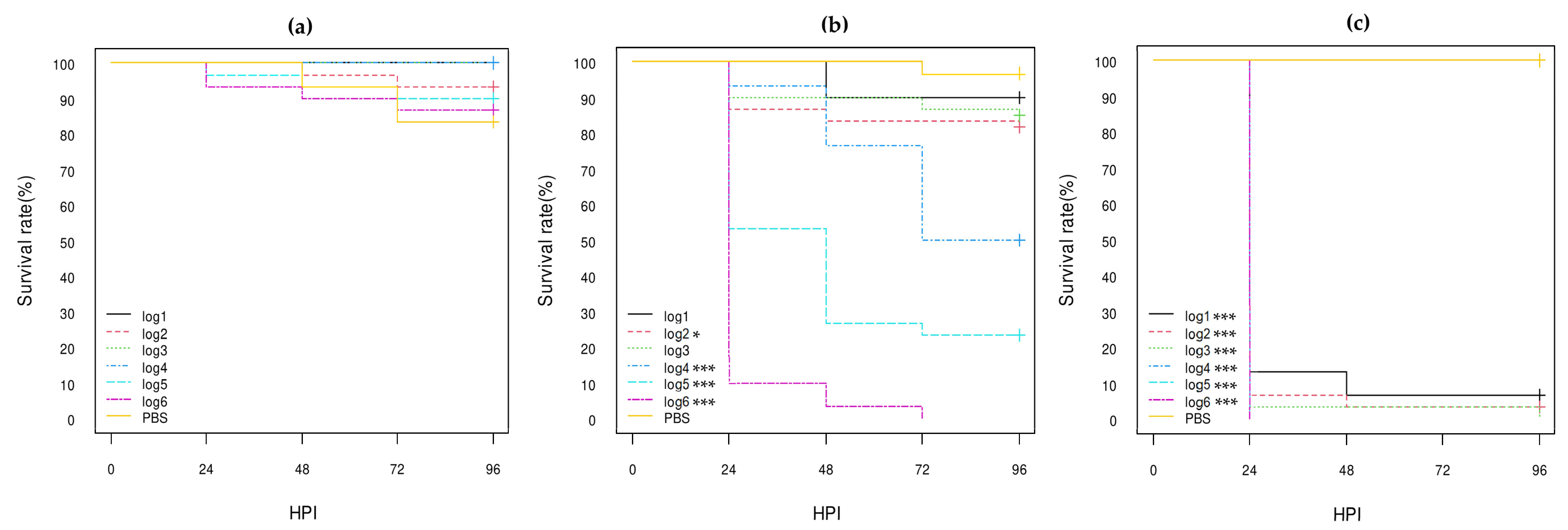
Figure 1. Kaplan–Meier survival curves of control E. coli strains: (a) K12-DH5α; (b) serotype O80:H26 (EH3161 strain); (c) serotype O78:H4 (pS88++). HPI = Hour Post-Inoculation. * p-value statistically significant at the threshold 0.05. *** p-value statistically significant at the threshold 0.001. pS88++: pS88 plasmid carrying the etsC/iucC genes.
Vetsci 10 00420 g001
Figure 2. Kaplan–Meier survival curves of stx1a AE-STEC O80:H2 strains: (a) SES5320 strain (pS88--); (b) SES5363 strain (pS88++); (c) EH2282 strain (pS88++). HPI = Hour Post-Inoculation. * p-value statistically significant at the threshold 0.05. *** p-value statistically significant at the threshold 0.001. pS88++: pS88 plasmid carrying the etsC/iucC genes. pS88--: pS88 plasmid not carrying the etsC/iucC genes.
Vetsci 10 00420 g002
Figure 3. Kaplan–Meier survival curves of stx2d AE-STEC O80:H2 strains: (a) EH3160 strain (pS88++); (b) EH3307/SES2959 strain (pS88--); (c) EH3320/SES3090 strain (pS88++). HPI = Hour Post-Inoculation. * p-value statistically significant at the threshold 0.05. ** p-value statistically significant at the threshold 0.01. *** p-value statistically significant at the threshold 0.001. pS88++: pS88 plasmid carrying the etsC/iucC genes. pS88--: pS88 plasmid not carrying the etsC/iucC genes.
Vetsci 10 00420 g003
Figure 4. Kaplan–Meier survival curves of EPEC O80:H2 strains: (a) EH3308/SES2973 strain (pS88--); (b) EH3322/SES3122 strain (pS88++). HPI = Hour Post-Inoculation. * p-value statistically significant at the threshold 0.05. ** p-value statistically significant at the threshold 0.01. *** p-value statistically significant at the threshold 0.001. pS88++: pS88 plasmid carrying the etsC/iucC genes. pS88--: pS88 plasmid not carrying the etsC/iucC genes.
Vetsci 10 00420 g004
Figure 5. Kaplan–Meier survival curves of: (a) K12-DH10B recipient strain; (b) K12-DH10B pS88 plasmid-conjugated strain (pS88++); (c) stx1a AE-STEC O80:H2 EH2282 pS88 plasmid donor strain (pS88++). HPI = Hour Post-Inoculation. * p-value statistically significant at the threshold 0.05. ** p-value statistically significant at the threshold 0.01. *** p-value statistically significant at the threshold 0.001. pS88++: pS88 plasmid carrying the etsC/iucC genes.
Vetsci 10 00420 g005
Figure 6. Kaplan–Meier survival curves of: (a) K12 DH10B recipient strain; (b) K12 DH10B STX2d phage-transduced strain; (c) stx2d AE-STEC O80:H2 EH3160 STX2d phage donor strain. HPI = Hour Post-Inoculation. * p-value statistically significant at the threshold 0.05. ** p-value statistically significant at the threshold 0.01. *** p-value statistically significant at the threshold 0.001.
Vetsci 10 00420 g006
Figure 7. Kaplan–Meier survival curves of E. coli O80:non-H2 strains: (a) serotype O80:H6 SES6039 strain; (b) serotype O80:H26 (EH3161 strain); (c) serotype O80:H45 SES5725 strain; (d) serotype O80:H45 SES6156 strain. HPI = Hour Post-Inoculation. * p-value statistically significant at the threshold 0.05. *** p-value statistically significant at the threshold 0.001.
Vetsci 10 00420 g007
Table 1. Serotypes and virulotypes of the wild-type E. coli strains used in this study.
Isolate References 1Serotypestx Geneseae GenepS88 Plasmid Virulotype
(iucC/etsC Genes)
Reference of Genome Sequencing
SES5320O80:H21aξnd/nd 2This study
SES53631aξd/d
EH2282 3,41aξd/d[21]
EH3160 42dξd/d
EH3307/SES29592dξnd/nd
EH3320/SES3090 52dξd/d
EH3308/SES2973ndξnd/nd
EH3322/SES3122ndξd/d
SES6039O80:H6ndnd--- 6[34]
SES5725O80:H45ndnd---
SES6156ndnd---
Serotype collectionO80:H26 5ndnd---This study
O78:H4ndndd/d
1 EH reference in [21]; SES reference is a laboratory new reference. 2 d: gene detected; nd: gene not detected. 3 AE-STEC O80:H2 EH2282 strain was erroneously referred to as stx2a instead of stx1a in Figure 1 of reference [21]. 4 AE-STEC O80:H2 EH2282 strain was the donor strain for the pS88 plasmid conjugation; AE-STEC O80:H2 EH3160 strain was the donor strain for STX2d phage transduction. 5 AE-STEC O80:H2 EH3320/SES3090 and serotype collection E. coli O80 strains were previously tested in G. mellonella [30]. 6 --- = no pS88 plasmid.
Table 2. Target genes, primer sequences and amplified fragment lengths of the PCR applied on the DH10B pS88 plasmid transconjugant candidates.
PCRTarget GenesPrimersAmplified Fragment LengthReference
Serotype O80wzyO80Og80-F: 5′-TGGTGTTGATTCCACTAGCGT-3′
Og80-R: 5′-CGAGAGTACCTGGTTCCCAAA-3′
285 bp[40]
Serotype H2fliCH2Hg2-F: 5′-TGATCCGACACTTCCTGATG-3′
Hg2-R: 5′-CCGTCATCACCAATCAACGC-3′
228 bp[41]
Serotype O16wzxO16Og16-F: 5′-GGTTTCAATCTCACAGCAACTCAG-3′
Og16-R: 5′-GTTAGAGGGATAATAGCCAAGCGG-3′
302 bp[40]
Colicin V_wtcvaC_wtCvaC-F: 5′-TATGAGAACTCTGACTCTAAAT-3′
CvaC-R: 5′-ATTTATAAACAAACATCACTAA-3′
314 bp[42]
Colicin V_M 1cvaC_M1555 bp 1
Avian hemolysinhlyFHlyF-F: 5′-GGCGATTTAGGCATTCCGATACTC-3′
HlyF-R: 5′-ACGGGGTCGCTAGTTAAGGAG-3′
599 bp[19]
1 The cvaC gene is disrupted by the insertion of an ampicillin resistance cassette (see Section 2.3).
Table 3. Virulence genes detected after genome analysis with Virulence Finder 2.0 of the E. coli O80:H2, O80:H6, O80:H26, O80:H45 and O78:H4 strains.
E. coli Serotypes and StrainsO80:H2 1,2O80:H6 3O80:H26 4O80:H45 3O78:H4 4
Genes Detected after Genome AnalysisSES5320SES5363EH2282 5EH3307/
SES2959
EH3320/
SES3090
EH3160EH3308/
SES2973
EH3322/
SES3122
SES6039EH3161SES5725SES6156O78C
Phage-located genesstx1a+ 6++
stx2d +++
LEE-located geneseaeξ++++++++
espA/B/F/P++++++++
tir++++++++
pS88 plasmid-located genescia++++++++ +
cvaA++++++++ +
hlyF++++++++ +
iroN++++++++ +
iss+++++++++ +++
ompT++++++++ +++
sitA++++++++ +++
etsC ++ ++ + +
iucC ++ ++ + +
Chromosome-located genescma+ ++ +
iha+ ++ +
1 all but stx1a AE-STEC SES5320 and SES5363 strains were previously sequenced [21]. The Genbank accession numbers are SAMN35130764 (SES5320) and SAMN35130765 (SES5363). 2 EH numbers are listed in [21]; SES numbers are the new laboratory collection references. 3 all three E. coli O80:H6 and O80:H45 strains were previously sequenced [34]. 4 The Genbank accession numbers are SAMN35130763 (EH3161) and SAMN35130762 (O78C). 5 stx1a AE-STEC EH2282 strain was erroneously referred to as stx2a instead of stx1a in Figure 1 of reference [21]. 6 +: gene detected; empty box: gene not detected.
Disclaimer/Publisher’s Note: The statements, opinions and data contained in all publications are solely those of the individual author(s) and contributor(s) and not of MDPI and/or the editor(s). MDPI and/or the editor(s) disclaim responsibility for any injury to people or property resulting from any ideas, methods, instructions or products referred to in the content.