Article

Development of Yorkshire Terrier Dentition

1
Waltham Petcare Science Institute, Waltham-on-the-Wolds, Leicestershire LE14 4RT, UK
2
Interuniversity Institute for Biostatistics and Statistical Bioinformatics (I-Biostat), Data Science, Hasselt University, 3500 Hasselt, Belgium
*
Author to whom correspondence should be addressed.
Vet. Sci. 2023, 10(7), 406; https://doi.org/10.3390/vetsci10070406
Submission received: 22 March 2023 / Revised: 5 June 2023 / Accepted: 15 June 2023 / Published: 21 June 2023
(This article belongs to the Section Veterinary Microbiology, Parasitology and Immunology)

Abstract

:

Simple Summary

Several problems, particularly in smaller breed dogs, have been associated with tooth development, e.g., over-crowding, missing teeth, and persistent deciduous teeth (where a juvenile tooth remains in the mouth at the same time as its permanent counterpart). This opportunistic study of 61 Yorkshire terrier puppies aimed to determine the average age of deciduous tooth loss and permanent tooth eruption, the incidence of persistent deciduous teeth, and potential influencing factors such as body weight. The age at which deciduous teeth were lost, and permanent teeth erupted, varied according to body weight; dogs weighing less than 3 kg lost their deciduous teeth and gained their permanent teeth later than dogs weighing more than 3 kg. The sequence of tooth loss and eruption (i.e., incisors first followed by canines, premolars, and then molars) was disrupted in dogs < 3 kg. Persistent deciduous teeth were common, affecting 69% of puppies. Those with lower body weights (<3 kg) had greater proportions and were more likely to require surgical extractions by a veterinarian. This study highlights the importance of regular checks by a veterinary professional during the active phases of tooth development.

Abstract

The development of dentition in dogs has been associated with several problems including tooth over-crowding, missing permanent dentition, and persistent deciduous teeth (PDT). Information on dentition development in different breeds is lacking. This study of 61 Yorkshire terriers aimed to determine the (i) average age at deciduous tooth exfoliation, (ii) average age at permanent tooth eruption, (iii) PDT incidence, and influencing factors such as body weight. The ages of exfoliation of deciduous teeth and eruption of permanent dentition were influenced by body weight and tooth type. These dentition changes tended to occur later in dogs ≤ 3 kg versus dogs > 5 kg. Generally, incisors were exfoliated first, followed by premolars and then canines. At a body weight of 4.5 kg, the middle of the data range, the estimated age at loss of deciduous teeth (with 95% confidence intervals) was 21.9 (21.1, 22.9) weeks for incisors, 26.1 (24.9, 27.4) weeks for canines, and 23.9 (22.9, 24.9) weeks for premolar. The estimated age at eruption of permanent dentition was 22.3 (21.6, 23.0) weeks for incisors, 23.8 (23.0, 24.6) weeks for canines, 24.7 (24.0, 25.5) weeks for premolars, and 26.4 (25.5, 27.3) for molar teeth. However, this sequence was disrupted in dogs ≤ 3 kg. Yorkshire terriers had a high incidence of PDT. At a body weight of 4.5 kg, the estimated proportion of PDT was: incisors 0.86% (0.32, 2.31), canines 15.62% (7.62, 29.37) and premolars 3.57% (1.62, 7.66). Canines constituted the most frequently retained tooth type, with 89.1% retained in dogs ≤ 3 kg compared to 12.0% in dogs > 5 kg. This information will enable veterinarians to provide personalised advice regarding the oral care requirements for Yorkshire terriers and highlights the need to regularly monitor this breed between the ages of two and seven months, during the active phases of tooth development.

1. Introduction

Dogs have two sets of teeth over the course of their lifetimes (i.e., they are diphyodonts). The deciduous teeth begin to erupt at three to six weeks of age, and by twelve weeks of age, most breeds of dog will have their full complement of 28 teeth [1,2,3]. As the permanent teeth develop, they place pressure on the apex of the deciduous tooth, causing the resorption of the deciduous tooth root. This process continues until the deciduous root is sufficiently resorbed, the tooth is exfoliated, and the permanent tooth occupies its position. The permanent dentition begins to erupt at about three months of age and most breeds of dog will have their full complement of 42 permanent teeth by seven months of age [1,2,3]. A number of factors are thought to affect tooth eruption including genetics, infections, and trauma [1,2,3].
Several problems have been associated with the development of dentition, such as delayed eruption, over-crowding, missing underlying permanent dentition, and PDT [3]. A deciduous tooth is termed persistent if it exists in the mouth at the same time as its permanent counterpart [4]. PDT is particularly common in small and toy breeds of dog [4,5,6]. A prevalence rate of 5.7% was reported in a UK study of 11,647 Chihuahuas, increasing to 11.3% in individuals less than two years of age [7]. The most frequent cause of PDT is an incorrect eruption path of the permanent tooth [4]. PDT and the subsequent tooth over-crowding create regions in the mouth where plaque can accumulate, which can lead to the development of periodontal disease [4,8]. They can also cause orthodontic issues; for example, the mandibular canines can impinge on the palate [4,8,9]. These problems have been reported to occur soon after the permanent canines start to erupt [8]. Abnormal tooth eruption can also cause malocclusion (misalignment of teeth during bite), which can be mild to severe and may require correction [9]. Treatment options include the extraction of the offending tooth, removal of the crown combined with endodontic therapy, and orthodontics to move the teeth into the correct position. These tooth eruption anomalies are thought to be genetic due to the pattern of occurrence within different breeds of dog.
There is a lack of published information on variation in the age and sequence of permanent tooth eruption, and incidence of PDT, between different breeds of dog. An opportunity arose to study a population of Yorkshire terriers, a susceptible breed to dental issues, that were being acquired by the Waltham Petcare Science Institute. This breed was selected as a representative breed of the toy dog category based on its popularity, medical predispositions, behaviour, and genetic diversity. The primary objectives of the study were to determine (i) the average age when deciduous teeth are exfoliated, (ii) the average age when permanent teeth erupt, (iii) and the incidence of PDT in Yorkshire terriers. A secondary objective was to investigate the influence of factors such as body weight and litter. This information will enable veterinarians to provide personalised advice regarding the oral care requirements for Yorkshire terriers.

2. Materials and Methods

2.1. Study Cohort

A total of 61 Yorkshire terriers (14 litters) were acquired, between September 2012 and April 2016, from registered external breeders by the Waltham Petcare Science Institute. There were no inclusion or exclusion criteria for the selection of breeders other than being based in the United Kingdom and their willingness to supply pure-bred Yorkshire terrier puppies. All puppies included in the study had genetic DNA tests (Wisdom Panel™, Mars Petcare, Portland, OR, USA) conducted on them which confirmed that they were representative of the global Yorkshire terrier pet population. The pure-bred Yorkshire terrier puppies were from 11 bitches, each of which had mated with one of eight dogs. The puppies were housed in pairs in environmentally enriched kennels and provided with a comprehensive socialisation programme. Thirty of the dogs were female and 31 were male. The female dogs were intact and the male dogs were castrated between 24.4 and 29.4 weeks of age. The average weight of the dogs at 28 weeks of age was 4.54 kg (range 1.53 to 7.55 kg). There were 21 dogs ≤ 3.0 kg, 24 dogs between >3.0 kg and ≤5.0 kg, and 16 dogs > 5.0 kg. Body weight was not recorded for five dogs (one litter) at 28 weeks of age as they had been re-homed. The dogs were fed a commercial dry diet (Royal Canin® Yorkshire terrier 29 Junior) from weaning up to 14 weeks of age. From 14 to 28 weeks of age, 32 dogs remained on the dry diet, 8 were transitioned onto a commercial wet diet (Cesar® puppy with chicken and rice with a carrot topping), and 21 were fed a simultaneous offering of the two.

2.2. Study Design

The status of each dog’s primary and permanent dentition was assessed and recorded at 12, 16, 20, 24, and 28 weeks of age (±2 weeks). The various tooth states included erupted (E), not erupted (N), partially erupted (P), not visible (X), and lost/exfoliated (L). The information was captured using a modified dental chart and later entered into a bespoke Microsoft® Access® database. Dogs received regular mouth handling from 8 to 10 weeks of age so that their dentition could be visualised without the need for anaesthesia. Six people performed assessments, and all were trained by recognised European Specialists in Veterinary Dentistry (Peter Southerden, Eastcott Dental Referrals, Swindon, Wiltshire, UK and Lisa Milella, The Veterinary Dental Surgery, Byfleet, Surrey, UK). Any PDT identified were surgically extracted at 32 weeks of age, or sooner if deemed necessary by the European Specialists in Veterinary Dentistry operating under the Veterinary Surgeons Act.

2.3. Data Handling

The variables recorded as part of the study are summarised in Table 1. Since oral assessments were performed at four-weekly intervals, and so the exact date of loss/eruption was unknown, the data is effectively “interval censored”. For the purposes of these analyses, the average at the censoring interval was used to estimate age at exfoliation/eruption.
The deciduous teeth data was utilised to determine the age at loss of deciduous dentition. The average age, in weeks, between the last time point at which the tooth status was ‘L’ and the first time point at which the tooth status was ‘E’ or ‘P’ was determined. If the tooth had not been exfoliated at the last age recorded, it was labelled as censored. The data were assigned as missing if the status was always ‘X’, ‘X’ occurred before ‘L’, ‘X’ was last measured after ‘E’ or ‘P’, or ‘E’ or ‘P’ were recorded after ‘L’.
The permanent teeth sub-data set was utilised to determine the age at the eruption of permanent dentition. The average age, in weeks, between the last time point at which the tooth status was ‘N’ and the first time point at which the tooth status was ‘E’ or ‘P’ was determined. The last age recorded was assigned if the tooth status was never ‘E’, and the value was labelled as censored. The data was assigned as missing if the status was always ‘X’, if ‘X’ occurred before ‘E’ or ‘P’, if ‘X’ was the last measure after ‘N’, or if there were later measurements labelled as ‘N’ after an ‘E’ or ‘P’.

2.4. Statistical Modelling

Data were analysed using the statistical software R 3.5 using lme4, multcomp, and survival packages [10,11,12,13].
Survival regression models were used for both age at loss of deciduous dentition and age at eruption of permanent dentition. The age at loss/eruption was used as the response variable, each dog was used as a frailty term, and body weight, tooth type (or tooth number), and their interaction were considered fixed effects. The effect of body weight was added as a linear and quadratic effect. The interaction of body weight and tooth type was initially added on both linear and quadratic body-weight terms, but the model was then simplified if required. Both tooth type models contained the main effects of tooth type and body weight in both linear and quadratic terms, and all interactions were dropped from the permanent eruption but not the deciduous loss tooth type model. The tooth number deciduous loss model contained the main effects of tooth number and body weight, in linear and quadratic terms, and the interaction between the tooth number and the linear effect of weight. The tooth number permanent eruption model contained both the main effects of tooth number and body weight in linear and quadratic terms as well as all interactions. The models used assumed Weibull distributions for the log(age) values and normal distributions for the frailty terms. Multiplicity correction was carried out using a single-step correction method based on the joint normal distribution of the linear function of the model parameters [11].
Using mixed logistic regression models, the events of PDT and surgical extraction were used as response variables. Litter and dog nested in litter were included as the random effects. Body weight, tooth type, and their interaction were included as fixed effects. For both analyses, the random litter effect was dropped from the model as it was not significant (p > 0.05). The effect of body weight was added as a linear and quadratic effect. The interaction of body weight and tooth type was initially added on both linear and quadratic body weight terms, but the model was then simplified and only the interaction term with the linear body weight effect was retained. The final models contained a significant interaction effect of body weight and tooth type (p < 0.05).

3. Results

The development of dentition in Yorkshire terriers from 14 litters was investigated. The age at exfoliation of 1708 deciduous teeth (28 teeth, 61 dogs) and the age at eruption of 2562 permanent teeth (42 teeth, 61 dogs) were recorded based on assessments every 4 weeks from 12 to 28 weeks of age. In addition, the incidence of PDT was determined and, if surgical extraction was necessary, a record was made, and these data were included in the analysis. There were missing data points for four dogs due to difficulties assessing their teeth for behavioural reasons: two dogs had data missing for one time point, one dog had two missing time points, and one dog had three missing time points. Five dogs were removed from the trial at 28 weeks of age, due to being re-homed, and so were omitted from the persistent teeth analysis.

3.1. Age at Exfoliation of Deciduous Dentition

The initial inspection of the data suggested that the age at exfoliation of deciduous dentition varied according to litter; however, this was confounded by body weight. There was also a relationship between the average age of exfoliation of deciduous teeth and body weight (Figure 1). Preliminary exploration of the data using arbitrary body weight groups showed that, on average, dogs ≤ 3 kg exfoliated their primary dentition later than dogs > 3 kg (Table 2). Various tooth types were also exfoliated at different ages. Generally, the incisors were exfoliated first, followed by the premolars and canines (Table 2).
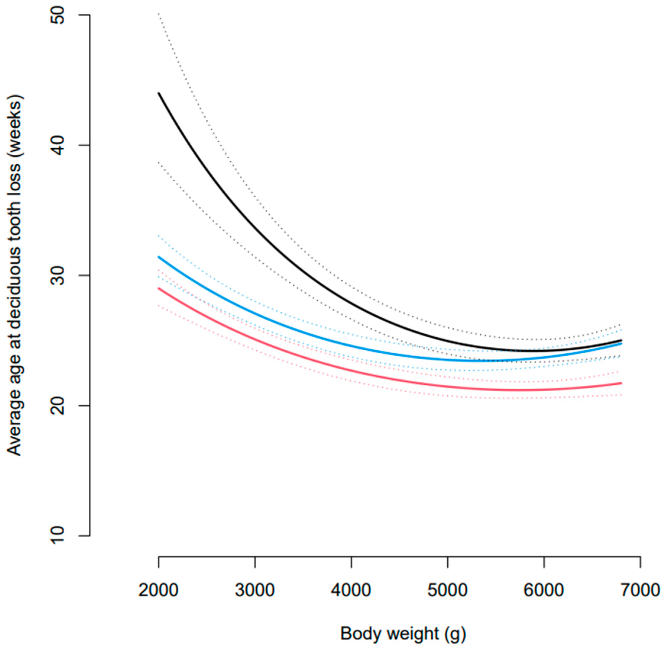
Statistical modelling of the data showed that the effect of body weight on the average age at exfoliation of deciduous dentition differed based on tooth type (Figure 2). At body weights of 2 kg and 4.5 kg, all tooth types significantly differed in terms of the age at which they were exfoliated (all p < 0.0001). However, at 7 kg the incisors significantly differed to the canines and premolars (both p < 0.00001), but the premolars and canines did not significantly differ (p = 0.87). According to the statistical model, at a body weight of 4.5 kg, the middle of the data range, the estimated age at loss of deciduous teeth (with 95% confidence intervals) was 21.9 (21.1, 22.9) weeks for incisors, 26.1 (24.9, 27.4) weeks for canines, and 23.9 (22.9, 24.9) weeks for premolars.
The estimates for the mean age at deciduous tooth loss for each tooth in a dog weighing 4.5 kg showed that the mandibular first (701, 801) and second incisors (702, 802) were the first teeth to be exfoliated (Supplementary Table S1). The mandibular third incisors (703, 803) and then the maxillary first (501, 601) and second incisors (502, 602) closely followed this. The mandibular and maxillary third and fourth premolars (507,508, 607, 608, 707, 708, 807, 808) were the next teeth to be exfoliated, followed by the maxillary third incisors (503, 603). The mandibular and maxillary second premolars (506, 606, 706, 806) and mandibular canines (704, 804) were exfoliated next, and the final teeth to be exfoliated were the maxillary canines (504, 604).

3.2. Age at Eruption of Permanent Dentition

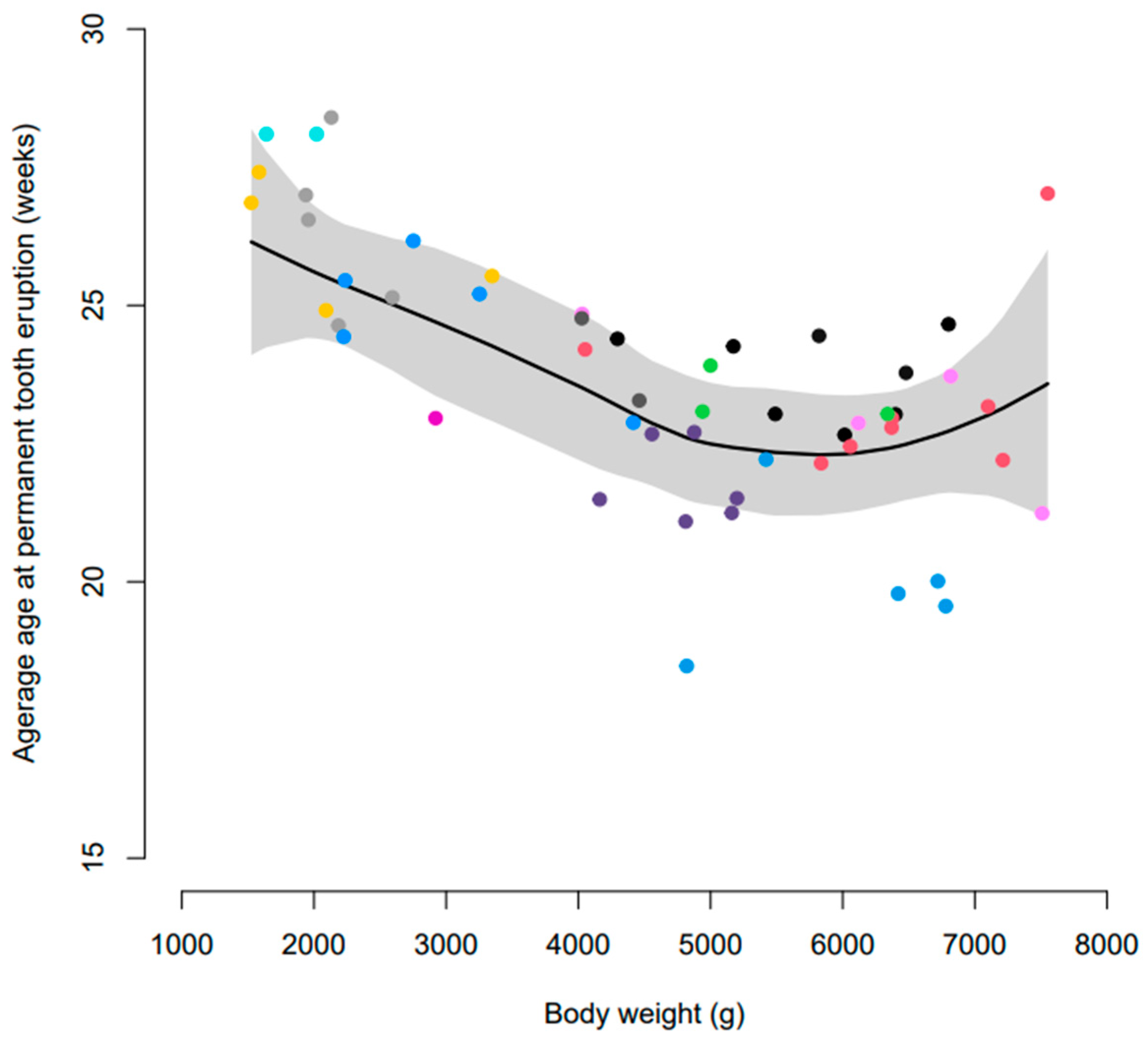
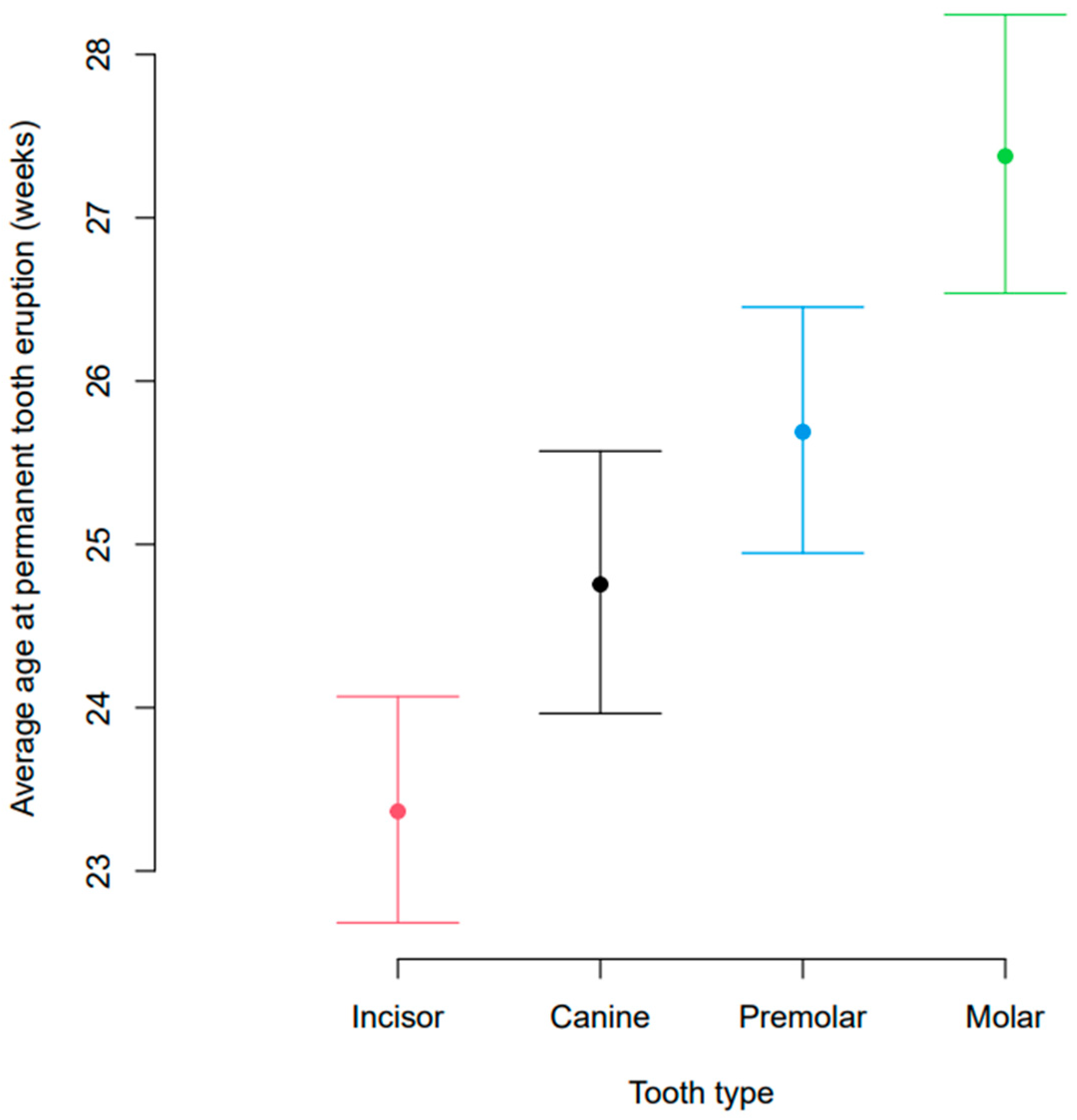
Initial assessment of the data suggested that the age at eruption of permanent dentition was subject to dog-to-dog variation. There was also a relationship between body weight and age at eruption of permanent dentition (Figure 3). Generally, the eruption of permanent dentition was later in dogs ≤ 3 kg compared to those >3 kg (Table 3). Overall, incisors erupted first, followed by canines, premolars, and molars in dogs > 3 kg (Figure 4). However, in dogs ≤ 3 kg, the eruption sequence changed to incisors, premolars, molars, and then canines (Table 3).
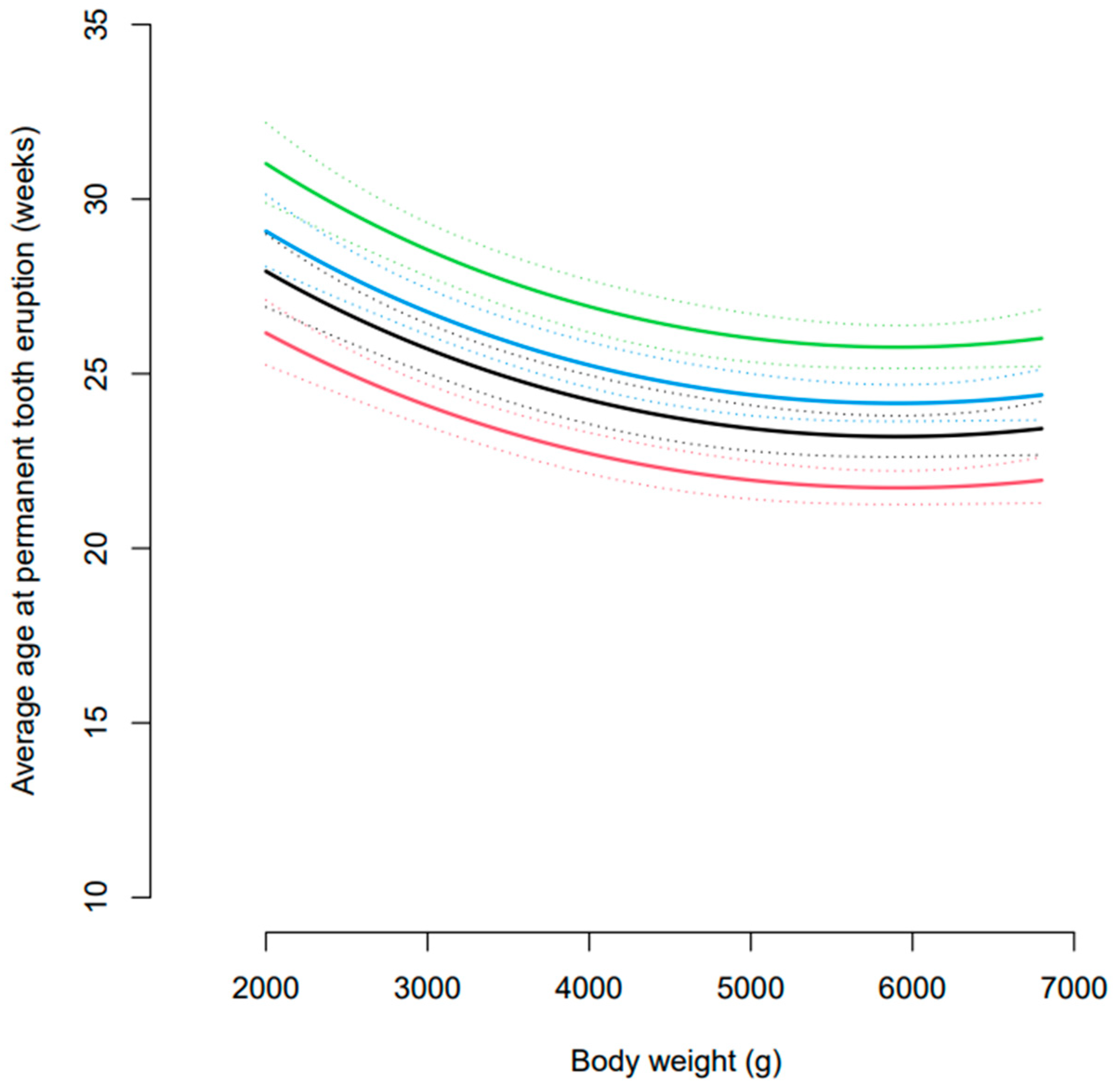
Statistical modelling of the data showed a significant effect of body weight and of tooth type on the estimated age at tooth eruption (Figure 5). At a body weight of 4.5 kg, the middle of the data range, the estimated age at eruption of permanent dentition was 22.3 (21.6, 23.0) weeks for incisors, 23.8 (23.0, 24.6) weeks for canines, 24.7 (24.0, 25.5) weeks for premolars, and 26.4 (25.5, 27.3) for molar teeth. The age at tooth eruption significantly differed between all four tooth types (all p < 0.0001) and these differences were constant across the different body weights. There was no significant interaction between body weight and tooth type in the final statistical model, and therefore, the comparison by tooth type was the same for all body weights.
On the basis of statistical model estimates, the first permanent teeth to erupt, at a body weight of 4.5 kg, were the maxillary and mandibular first incisors (101, 201, 301, 401) (Supplementary Table S2). The maxillary and mandibular second incisors (102, 202, 302, 402), mandibular third incisors (303, 403), and the maxillary fourth premolars (108, 208) followed this. Next to erupt were the mandibular first molars (309, 409), maxillary third premolars (107, 207), maxillary and mandibular canines (104, 204, 304, 404), and mandibular fourth premolars (308, 408). These were followed by the mandibular third premolars (307, 407), maxillary first premolars (105, 205) and first molars (109, 209), and maxillary third incisors (103, 203). Subsequently, the mandibular and maxillary second premolars (106, 206, 306, 406), second molars (110, 210, 310, 410), and mandibular first premolars (305, 405) erupted. The final teeth to erupt were the mandibular third molars (311, 411).

3.3. Incidence of PDT

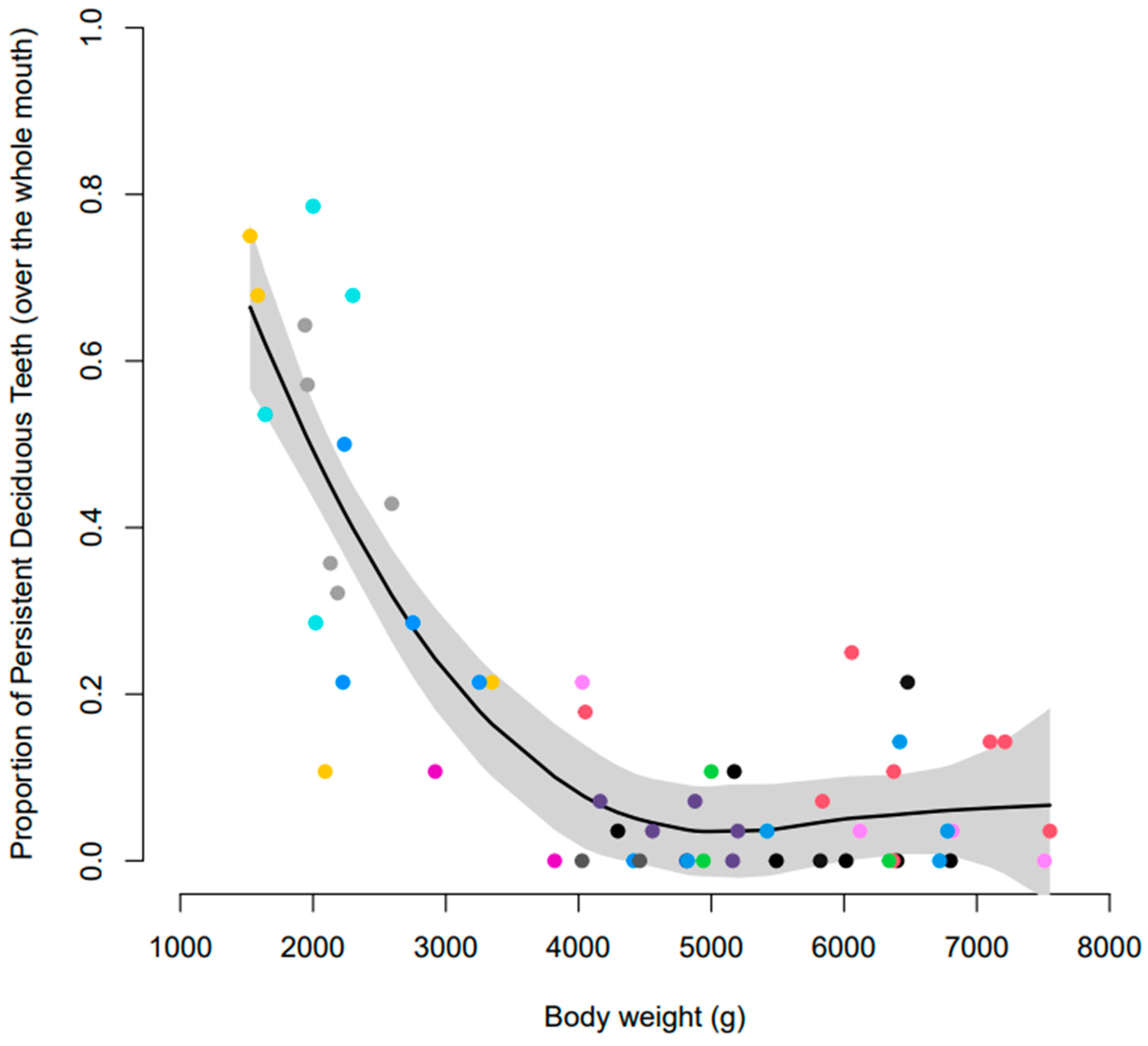
Preliminary assessment of the data indicated that 42 of the 61 Yorkshire terriers (69%) had one or more PDT. There was dog-to-dog variability in the number of PDT, varying from 1 to 22. There was also an effect of body weight wherein dogs with lower body weights (≤3 kg) had a greater proportion of PDT than those with higher body weights (>3 kg) (Figure 6; Table 4). The proportion of PDT that were not naturally exfoliated, and required surgical extraction, was also greater for dogs with lower body weights than heavier dogs (Table 5). There were also differences between tooth types; canine teeth were most likely to be retained, followed by premolar and then incisor teeth (Table 4 and Table 5). There were 22 dogs that were recorded as having overshot jaws (class 2 malocclusion) and 10 with undershot jaws (class 3 malocclusion) at one or more points throughout the study. The proportion of dogs requiring surgical extractions was similar between those with malocclusions recorded (62.5%) versus those with normal occlusions (58.6%).
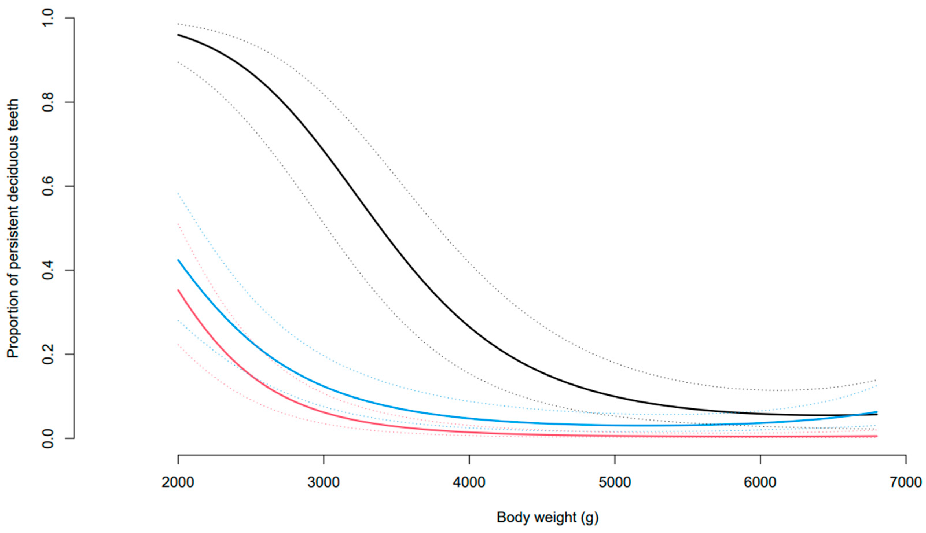
Statistical modelling showed that the effect of body weight on the proportion of PDT differed significantly by tooth type (Figure 7). At a body weight of 4.5 kg, the middle of the data range, the estimated proportion of PDT in each tooth type was: incisors 0.86% (0.32, 2.31), canines 15.62% (7.62, 29.37), and premolars 3.57% (1.62, 7.66). The odds of a canine being retained were 21.3 (8.9, 51.1) times that of an incisor and 5.0 (2.7, 9.4) times that of a premolar (both p < 0.00001) being retained. The odds of a persistent premolar being retained were 4.3 (2.0, 9.1) times that of an incisor (p = 0.0002). When only those that required surgical extraction are considered, the estimated proportions were 0.24%, (0.05, 1.22) for incisors, 6.63% (2.19, 18.36) for canines, and 2.23% (0.72, 6.73) for premolars.
Dogs with lower body weights (2.0 kg) had an increased estimated proportion of PDT for all types of teeth: incisors 35.29% (20.19, 54.05), canines 95.97% (87.34, 98.91), and premolars 42.42% (25.67, 61.15). When considering heavier dogs (7 kg), the estimated proportion of PDT decreased for all tooth types: incisors 0.61% (0.11, 3.25), canines 5.99% (1.72, 18.87), and premolars 7.6% (2.91, 18.46). At a body weight of 2 kg, the canine teeth were significantly more likely to be retained than the incisors and premolars (both p < 0.00001), but the incisor and premolar teeth did not significantly differ (p = 0.39). At a body weight of 7 kg, the canines and premolars were significantly more likely to be retained than the incisor teeth (p = 0.0035 and p < 0.00001, respectively), but the premolars and canines did not significantly differ (p = 0.85).

4. Discussion

To our knowledge, this is the first study to report on the age at exfoliation, age at eruption, and prevalence of PDT in Yorkshire terriers. Our findings suggest that several different factors could affect tooth development such as litter and body weight.
Visual inspection of the data indicated litter-to-litter variability in the proportion of PDT. Although litter was confounded by body weight, a role for genetics in the development of dentition in this breed of dog cannot be ruled out. No published studies were identified which had investigated the role of genetics in tooth development in dogs. However, studies of human twins have suggested a strong genetic basis for the development of dentition [14,15,16,17]. A genome-wide association study of the number of permanent teeth erupted between ages six and fourteen, using records from women in the Danish National Birth cohort, identified loci that were strongly associated with tooth eruption [18]. A population-based genome-wide association study of individuals from the Avon Longitudinal Study of Parents and Children and the 1966 Northern Finland Birth Cohort identified loci associated with time to first tooth eruption and number of teeth at one year of age [19,20]. Some publications suggest that heritability is higher for tooth development than for tooth eruption [21,22]. These studies have not determined whether the developments of primary and permanent dentition have different or similar mechanisms. Furthermore, the mutations identified may simply be markers of the stage of overall maturation rather than being specifically associated with dental development. Further work is required to determine the role of genetics in the development of canine dentition. Longitudinal and cross-sectional studies of humans have also reported differences in tooth emergence time among different races [23,24,25,26,27,28,29,30,31,32,33,34,35]. There is a lack of information regarding differences between breeds of dog in their age at tooth emergence and loss. Again, further research is required to determine whether there is a breed effect on the age and sequence of tooth eruption.
Body weight could explain the patterns observed across the litters in the age at exfoliation of deciduous teeth, age at eruption of permanent dentition, and prevalence of PDT. This study has shown that the age of deciduous tooth loss and permanent tooth eruption in dogs negatively correlates with body weight. It would be interesting to understand whether smaller size and body weight are consequences of tooth development issues (e.g., affecting food intake) or whether these issues arise due to genetic predispositions to smaller size consequently impacting dental development (e.g., tooth over-crowding). These findings show some similarity with human studies that have shown a negative linear correlation between the time of first deciduous tooth eruption and birth weight [36,37]. Delayed tooth eruption has also been reported in low-body-weight infants and premature infants [38,39]. Infant age and weight have also been shown to independently influence the timing of tooth eruption [40,41,42]. In contrast, other studies were non-conclusive about the relationship between body weight, or birth height, and age at first dental eruption [43,44,45]. Dogs with a body weight ≤ 3 kg also had a greater proportion of PDT than those >3 kg. The different sequence of permanent tooth eruption in dogs ≤ 3 kg when compared with larger dogs is likely to be directly related to the higher incidence of PDT in Yorkshire terriers.
Tooth over-crowding may also play a role in the development of dentition in dogs. Human studies have shown that tooth eruption is earlier when there is adequate space within the jaw bone [28]. This theory may also hold true for dogs, as toy and small breed dogs have been reported to have more crowded adult dentition than larger breeds [46,47,48]. A contributing factor in this may be the reported proportionately larger mandible first molar relative to mandibular height in small versus large dogs [49]. Toy/small- and medium/large-breed dogs also significantly differ in the oral biometrics of their upper mandibles (Pesci et al., personal communication). The total mesio-distal length:tooth arch ratio has been shown to be higher in smaller breeds, indicating that there is less space between teeth. This finding concurs with a study of jaws and teeth from a collection of 250 dogs [50]. Toy/small-breed dogs also have significantly larger teeth relative to their arch perimeter compared with medium/large dogs (Pesci et al., personal communication). It is possible that the selective breeding of smaller dogs has selected for mutations in genetic loci that regulate body size. One hypothesis is that this disconnect between tooth and jaw size may suggest that the genetic factors that influence body size are independent from those that regulate the size of dentition. A study of monozygotic and dizygotic twins indicated a strong genetic influence on tooth dimensions in humans [51].
In total, 69% of the Yorkshire terriers had one or more PDT and 76% of these required surgical extractions under general anaesthesia based on the recommendation of European Specialists in Veterinary Dentistry. This was higher than observed in other studies with the exception of what has been reported for toy poodles. A retrospective analysis of medical records of 759 dogs aged 7–18 months reported an overall incidence of 19.7% [52]. In a study of 259 dogs (representing 23 breeds), based on a retrospective analysis of radiographs, the overall prevalence was 5.4% with all cases falling within the 7-month to 2-year age category [53]. The latter study reported breed differences in the prevalence of PDT; 74.4% of the cases observed were toy poodles, 11.2% Chihuahuas, and 10% were other small dogs. A study of health problems in 11,647 Chihuahuas aged 0.1–18.6 years showed an overall PDT prevalence of 5.7%, rising to 11.3% in dogs aged less than two years [7]. An examination of 251 mongrels, including 143 strays kept in animal protection offices in Tokyo, identified 46 dogs (18.3%) less than 0.5 years old with PDT [54]. In this study of Yorkshire terriers, canines were the most likely teeth to be retained. Others have also reported canines as the most frequently affected teeth in dogs. At an average body weight of 4.5 kg, 15.6% of canines were retained, and this number rose to 96.0% for dogs < 2 kg [53]. Human studies indicate that the most common types of persistent primary teeth are the mandibular second molars and maxillary canines [55,56]. PDT can cause the permanent teeth to erupt in abnormal positions, which can result in an abnormal jaw position, the over-crowding of teeth, accidental bites into the palate, and an increased predisposition to periodontitis. To prevent complications, once the permanent tooth has erupted, its deciduous counterpart should be surgically extracted.
Several human studies reported that the mandibular permanent teeth erupted earlier than maxillary opposing teeth, sometimes with the exception of the premolars and molars [29,57,58,59]. In Yorkshire terriers, there was no clear trend, with teeth simultaneously erupting on the maxillae and mandibles. For all body weights, the first permanent teeth to erupt were the mandibular and maxillary incisors, followed by the maxillary fourth premolars. Amongst the last teeth to erupt were the mandibular and maxillary first molars. This finding is consistent with the existing literature on the sequence of tooth eruption in dogs [1,2,3]. Humans differ in that the first permanent teeth to emerge are the molars followed by the central incisors and then the lateral incisors [57,58,60,61,62,63,64]. On the maxilla, these are followed by the first premolars and then the canine/second premolars [57,59,60,61,62]. On the mandible, the first premolars/canines are the next teeth to erupt after the incisors, followed by the second premolars [58,60,61,62,63]. The last teeth to emerge are the second molars [57,58,60,61,62,63].
One of the main strengths of this study was the ability to follow the same individuals of a single breed of dog from 8 to 28 weeks of age. In addition, the dogs were exposed to the same environmental factors. However, the study was also subject to limitations. For behavioural reasons, it was sometimes difficult to visualise and capture the dentition statuses of some of the Yorkshire terriers. Furthermore, the dogs were only assessed every four weeks and, therefore, a precise timepoint of loss and eruption could not be determined. Sometimes, it was difficult to distinguish between deciduous and permanent teeth during the stages of mixed dentition. The high incidence of PDT would also have confounded the estimates of age for deciduous tooth loss. As puppies were not acquired until eight weeks of age, and most had their full complement of deciduous teeth, it was not possible to determine the time of eruption for their primary dentition. It was not possible to differentiate the effect of body weight from litter in this study as all the puppies within a litter were of similar sizes and body weights. Additionally, the birth weights of the puppies were not known and their weights at 28 weeks of age may have been influenced by other factors. Finally, this study was relatively small-scale compared to the large data sets available in the human field. Despite this, there are some valuable insights about the impact of dog weight and/or litter on the development of Yorkshire terrier dentition.
Tooth eruption is a complex process and this study has shown that it may be influenced by several factors such as body weight and/or litter. The time of exfoliation of deciduous teeth and eruption of permanent dentition in Yorkshire terriers is variable. The eruption of permanent dentition occurs later in small compared to large Yorkshire terriers, and smaller dogs are also more prone to PDT. This study suggests that the sequence and timing of permanent tooth eruption in Yorkshire terriers may be heritable, and this can have important implications for the risk of PDT, malocclusion, tooth over-crowding, and periodontal disease. For this reason, dogs affected by tooth development disorders should be excluded from future breeding.

5. Conclusions

This study has provided information about the age at exfoliation, age at eruption, and prevalence of PDT in Yorkshire terriers, and indicates that litter and body weight can impact these events. It has highlighted the importance of educating owners about potential tooth development problems in Yorkshire terriers and dogs with lower body weight (e.g., less than 3 kg). It is important for Yorkshire terriers to be regularly checked by veterinary professionals between two and seven months of age as their dentition is developing. If PDT are observed, veterinarians and owners must discuss the implications and consider the benefits of surgical extractions. There is need for further research, in a variety of dog breeds, to provide improved guidance to veterinary professionals regarding the “normal” time of eruption and other potential risk factors.

Supplementary Materials

The following supporting information can be downloaded at: https://www.mdpi.com/article/10.3390/vetsci10070406/s1, Table S1: Average age at deciduous tooth loss for each tooth at a body weight of 4.5 kg with lower confidence intervals (LCI) and upper confidence intervals (UCI); Table S2: Estimated age at eruption of permanent dentition at a body weight of 4.5 kg with lower confidence intervals (LCI) and upper confidence intervals (UCI).

Author Contributions

Conceptualization, C.W., L.J.H. and N.D.; study design, C.W. and I.P.; co-ordination, I.P.; acquisition and analysis of data, C.W.; statistical analysis, F.S.; maintenance of data integrity, I.P.; data interpretation, C.W. and F.S.; writing—original draft preparation, C.W.; writing—review and editing, C.W., I.P., L.J.H., N.D. and F.S. All authors have read and agreed to the published version of the manuscript.

Funding

This research was funded by Mars Petcare.

Institutional Review Board Statement

The study was approved by the Waltham Petcare Science Institute Ethical Review Body. Initial approval for studies with the Yorkshire terriers was granted on 28 September 2012 and further details of the oral health aspect of the study were provided and approved on 11 January 2013.

Informed Consent Statement

Not applicable.

Data Availability Statement

Data are not publicly available but can be obtained from the corresponding author upon reasonable request.

Acknowledgments

The authors acknowledge the significant contributions of Waltham’s Procedure Technicians and Judith Allsopp for providing technical expertise and performing all visual oral assessments.

Conflicts of Interest

C.W., I.P., L.J.H. and N.D. are employees in company Mars Petcare, a provider of oral care products and veterinary services for dogs. F.S. has no conflict of interest to declare.

References

  1. Wiggs, R.; Lobprise, H. Pedodontics. In Veterinary Dentistry, Principles and Practice; Raven-Lippincott: Philadelphia, PA, USA, 1997; pp. 167–185. [Google Scholar]
  2. Harvey, C.; Emily, P. Function, formation, and anatomy of oral structures in carnivores. In Small Animal Dentistry; Mosby-Year Book: St. Louis, MO, USA, 1993. [Google Scholar]
  3. Hale, F. Juvenille Veterinary Dentistry. Vet. Clin. N. Am. Small Anim. Pract. 2005, 35, 789–817. [Google Scholar] [CrossRef]
  4. Hobson, P. Extraction of retained primary canine teeth in the dog. J. Vet. Dent. 2005, 22, 132–137. [Google Scholar] [CrossRef]
  5. Niemiec, B.A. Oral pathology. Top. Companion Anim. Med. 2008, 23, 59–71. [Google Scholar] [CrossRef] [Green Version]
  6. Niemiec, B.A. Conditions common in small and toy breed dogs. In Breed Predispositions to Dental and Oral Disease in Dogs; Wiley Online Library: New York, NY, USA, 2021; pp. 1–38. [Google Scholar]
  7. O’Neill, D.G.; Packer, R.M.A.; Lobb, M.; Church, D.B.; Brodbelt, D.C.; Pegram, C. Demography and commonly recorded clinical conditions of Chihuahuas under primary veterinary care in the UK in 2016. BMC Vet. Res. 2020, 16, 42. [Google Scholar] [CrossRef] [Green Version]
  8. Harvey, C.; Emily, P. Occlusion, occlusive abnormalities and orthodontic treatment. In Small Animal Dentistry; Mosby-Year Book: St. Louis, MO, USA, 1993. [Google Scholar]
  9. Baxter, C. Should that tooth be extracted? Part 1: Problems associated with deciduous dentition in puppies. UK Vet. Companion Anim. 2009, 14, 58–61. [Google Scholar] [CrossRef]
  10. Bates, D.; Mächler, M.; Bolker, B.; Walker, S. Fitting Linear Mixed-Effects Models Using lme4. J. Stat. Softw. 2015, 67, 48. [Google Scholar] [CrossRef]
  11. Hothorn, T.; Bretz, F.; Westfall, P. Simultaneous inference in general parametric models. Biom. J. 2008, 50, 346–363. [Google Scholar] [CrossRef] [Green Version]
  12. Therneau, T.M. A package for Survival Analysis in S Version 2.38, ed. 2015. Available online: https://CRAN.R-project.org/package=survival (accessed on 14 June 2023).
  13. Therneau, T.M.; Grambsch, P.M. The cox model. In Modeling Survival Data: Extending the Cox Model; Springer: Berlin/Heidelberg, Germany, 2000; pp. 39–77. [Google Scholar]
  14. Garn, S.M.; Lewis, A.B.; Kerewsky, R.S. Genetic, Nutritional, and Maturational Correlates of Dental Development. J. Dent. Res. 1965, 44, 228–242. [Google Scholar] [CrossRef]
  15. Townsend, G.; Richards, L.; Messer, L.B.; Hughes, T.; Pinkerton, S.; Seow, K.; Gotjamanos, T.; Gully, N.; Bockmann, M. Genetic and environmental influences on dentofacial structures and oral health: Studies of Australian twins and their families. Twin Res. Hum. Genet. 2006, 9, 727–732. [Google Scholar] [CrossRef]
  16. Pelsmaekers, B.; Loos, R.; Carels, C.; Derom, C.; Vlietinck, R. The genetic contribution to dental maturation. J. Dent. Res. 1997, 76, 1337–1340. [Google Scholar] [CrossRef]
  17. Hughes, T.; Bockmann, M.; Seow, K.; Gotjamanos, T.; Gully, N. Strong genetic control of emergence of human primary incisor. J. Dent. Res. 2007, 86, 1160–1165. [Google Scholar] [CrossRef] [Green Version]
  18. Geller, F.; Feenstra, B.; Zhang, H.; Shaffer, J.R.; Hansen, T.; Esserlind, A.-L.; Boyd, H.A.; Nohr, E.A.; Timpson, N.J.; Fatemifar, G.; et al. Genome-wide association study identifies four loci associated with eruption of permanent teeth. PLoS Genet. 2011, 7, e1002275. [Google Scholar] [CrossRef] [Green Version]
  19. Pillas, D.; Hoggart, C.J.; Evans, D.M.; O’Reilly, P.F.; Sipilä, K.; Lähdesmäki, R.; Millwood, I.Y.; Kaakinen, M.; Netuveli, G.; Blane, D.; et al. Genome-wide association study reveals multiple loci associated with primary tooth development during infancy. PLoS Genet. 2010, 6, e1000856. [Google Scholar] [CrossRef]
  20. Fatemifar, G.; Hoggart, C.J.; Paternoster, L.; Kemp, J.P.; Prokopenko, I.; Horikoshi, M.; Wright, V.J.; Tobias, J.H.; Richmond, S.; Zhurov, A.I.; et al. Genome-wide association study of primary tooth eruption identifies pleiotropic loci associated with height and craniofacial distances. Hum. Mol. Genet. 2013, 22, 3807–3817. [Google Scholar] [CrossRef] [Green Version]
  21. Liu, H.; Deng, H.; Cao, C.F.; Ono, H. Genetic analysis of dental traits in 82 pairs of female-female twins. Chin. J. Dent. Res. Off. J. Sci. Sect. Chin. Stomatol. Assoc. 1998, 1, 12–16. [Google Scholar]
  22. Liu, H.; Deng, H.; Cao, C. Genetic analysis of tooth development and eruption in 82 pairs of female-female twins. Zhonghua Kou Qiang Yi Xue Za Zhi = Zhonghua Kouqiang Yixue Zazhi = Chin. J. Stomatol. 1999, 34, 159–161. [Google Scholar]
  23. Hagg, U.; Taranger, J. Timing of tooth emergence. A prospective longitudinal study of Swedish urban children from birth to 18 years. Swed. Dent. J. 1986, 10, 195–206. [Google Scholar]
  24. Mugonzibwa, E.A.; Kuijpers-Jagtman, A.M.; Laine-Alava, M.T.; Hof, M.A.V. Emergence of permanent teeth in Tanzanian children. Community Dent. Oral Epidemiol. 2002, 30, 455–462. [Google Scholar] [CrossRef]
  25. Nystrom, M.; Kleemola-Kujala, E.; Evalahti, M.; Peck, L.; Kataja, M. Emergence of permanent teeth and dental age in a series of Finns. Acta Odontol. Scand. 2001, 59, 49–56. [Google Scholar] [CrossRef]
  26. Sharma, K.; Mittal, S. Permanent tooth emergence in Gujjars of Punjab, India. Anthropol. Anz. Bericht. Uber Die Biol.-Anthropol. Lit. 2001, 59, 165–178. [Google Scholar] [CrossRef]
  27. Nonaka, K.; Ichiki, A.; Miura, T. Changes in the eruption order of the first permanent tooth and their relation to season of birth in Japan. Am. J. Phys. Anthropol. 1990, 82, 191–198. [Google Scholar] [CrossRef]
  28. Brown, T. Tooth emergence in Australian Aboriginals. Ann. Hum. Biol. 1978, 5, 41–54. [Google Scholar] [CrossRef]
  29. Gaur, R.; Singh, N.Y. Emergence of permanent teeth among the meiteis of Manipur, India. Am. J. Hum. Biol. Off. J. Human. Biol. Counc. 1994, 6, 321–328. [Google Scholar] [CrossRef]
  30. Garcia-Godoy, F.; D’Iaz, A.N.; del Valle, J.M.; Arana, E.J. Timing of permanent tooth emergence in a Southeastern Dominican schoolchildren population sample. Community Dent. Oral Epidemiol. 1982, 10, 43–46. [Google Scholar] [CrossRef]
  31. Chaillet, N.; Nystrom, M.; Demirjian, A. Comparison of dental maturity in children of different ethnic origins: International maturity curves for clinicians. J. Forensic Sci. 2005, 50, 1164–1174. [Google Scholar] [CrossRef]
  32. Höuffding, J.; Maeda, M.; Yamaguchi, K.; Tsuji, H.; Kuwabara, S.; Nohara, Y.; Yoshida, S. Emergence of permanent teeth and onset of dental stages in Japanese children. Community Dent. Oral Epidemiol. 1984, 12, 55–58. [Google Scholar] [CrossRef]
  33. Kanagaratnam, S.; Schluter, P.J. The age of permanent tooth emergence in children of different ethnic origin in the Auckland region: A cross-sectional study. N. Z. Dent. J. 2012, 108, 55–61. [Google Scholar]
  34. Kochhar, R.; Richardson, A. The chronology and sequence of eruption of human permanent teeth in Northern Ireland. Int. J. Paediatr. Dent. 1998, 8, 243–252. [Google Scholar] [CrossRef]
  35. Pahel, B.T.; Vann, W.F., Jr.; Divaris, K.; Rozier, R.G. A Contemporary Examination of First and Second Permanent Molar Emergence. J. Dent. Res. 2017, 96, 1115–1121. [Google Scholar] [CrossRef]
  36. Sajjadian, N.; Shajari, H.; Jahadi, R.; Barakat, M.G.; Sajjadian, A. Relationship between birth weight and time of first deciduous tooth eruption in 143 consecutively born infants. Pediatr. Neonatol. 2010, 51, 235–237. [Google Scholar] [CrossRef] [Green Version]
  37. Billewicz, W.Z.; McGregor, I.A. Eruption of permanent teeth in West African (Gambian) children in relation to age, sex and physique. Ann. Hum. Biol. 1975, 2, 117–128. [Google Scholar] [CrossRef] [PubMed]
  38. Viscardi, R.; Rombery, B.; Abrams, R. Delayed primary tooth eruption in premature infant: Relationship to neonatal factors. Pediatr. Dent. 1994, 16, 23–28. [Google Scholar]
  39. Fadavi, S.; Punwani, I.C.; Adeni, S.; Vidyasagar, D. Eruption pattern in the primary dentition of premature low-birth-weight children. ASDC J. Dent. Child. 1992, 59, 120–122. [Google Scholar]
  40. Lawoyin, T.; Lawoyin, D.; Lawoyin, J. Epidemiological study of some factors related to deciduous tooth eruption. Afr. Dent. J. 1996, 10, 19–23. [Google Scholar] [PubMed]
  41. Correa-Faria, P.; Leite-Faria, L.; Viana, A.N.; Marques, L.S.; Ferreira, F.O.; Ramos-Jorge, M.L. Factors associated with number of erupted primary teeth in Brazilian children: A cross-sectional study. J. Dent. Child. 2013, 80, 111–114. [Google Scholar]
  42. Soliman, N.L.; El-Zainy, M.A.; Hassan, R.M.; Aly, R.M. Relationship of deciduous teeth emergence with physical growth. Indian J. Dent. Res. 2012, 23, 236–240. [Google Scholar]
  43. Shuper, A.; Sarnat, H.; Mimouni, F.; Mimouni, M.; Varsano, I. Deciduous tooth eruption in Israeli children. A cross-sectional study. Clin. Pediatr. 1985, 24, 342–344. [Google Scholar] [CrossRef]
  44. Haddad, A.; Correa, M. The relationship between the number of erupted teeth and the child’s height and weight: A cross-sectioanl study. J. Pediatr. Dent. 2005, 29, 357–362. [Google Scholar] [CrossRef] [PubMed]
  45. Kutesa, A.; Nkamba, E.M.; Muwazi, L.; Buwembo, W.; Rwenyonyi, C.M. Weight, height and eruption times of permanent teeth of children aged 4-15 years in Kampala, Uganda. BMC Oral Health 2013, 13, 15. [Google Scholar] [CrossRef] [Green Version]
  46. Lobprise, H. Canine periodontal disease overview. Vet. Tech. 2006, 27, 168–174. [Google Scholar]
  47. Tutt, C.; Deeprose, J.; Crossley, D. BSAVA Manual of Canine and Feline Dentistry; British Small Animal Veterinary Association: Gloucestershire, UK, 2007. [Google Scholar]
  48. Albuquerque, C.; Morinha, F.; Requicha, J.; Martins, T.; Dias, I.; Guedes-Pinto, H.; Bastos, E.; Viegas, C. Canine periodontitis: The dog as an important model for periodontal studies. Vet. J. 2012, 191, 299–305. [Google Scholar] [CrossRef]
  49. Gioso, M.A.; Shofer, F.; Barros, P.S.; Harvey, C.E. Mandible and mandibular first molar tooth measurements in dogs: Relationship of radiographic height to body weight. J. Vet. Dent. 2001, 18, 65–68. [Google Scholar] [CrossRef] [PubMed]
  50. McKeown, M. Craniofacial variability and its relationship to disharmony of the jaws and teeth. J. Anat. 1975, 119 Pt 3, 579–588. [Google Scholar]
  51. Potter, R.H.; Nance, W.E.; Yu, P.L.; Davis, W.B. A twin study of dental dimension. II. Independent genetic determinants. Am. J. Phys. Anthropol. 1976, 44, 397–412. [Google Scholar] [CrossRef]
  52. Borissov, I.; Sivrev, D.; Milev, N. Incidence of some teeth and occlusion abnormlities in dogs: A retrospective study. Bulg. J. Vet. Med. 2004, 7, 245–250. [Google Scholar]
  53. Butkovic, V.; Šehič, M.; Stanin, D.; Šimpraga, M.; Capak, D.; Kos, J. Dental diseases of dogs: A retrospective study of radiological data. Acta Vet. Brno 2001, 70, 203–208. [Google Scholar] [CrossRef] [Green Version]
  54. Isogai, H.; Isogai, E.; Okamoto, H.; Shirakawa, H.; Nakamura, F.; Matsumoto, T.; Watanabe, T.; Miura, H.; Aoi, Y.; Kagota, W.; et al. Epidemiological Study on Periodontal Diseases and some Other Dental Disorders in Dogs. Jpn. J. Vet. Sci. 1989, 51, 1151–1162. [Google Scholar] [CrossRef]
  55. Aktan, A.M.; Kara, I.; Şener, I.; Bereket, C.; Çelik, S.; Kırtay, M.; Çiftçi, M.E.; Arıcı, N. An evaluation of factors associated with persistent primary teeth. Eur. J. Orthod. 2012, 34, 208–212. [Google Scholar] [CrossRef] [Green Version]
  56. Nordquist, I.; Lennartsson, B.; Paulander, J. Primary teeth in adults—A pilot study. Swed. Dent. J. 2005, 29, 27–34. [Google Scholar] [PubMed]
  57. Dashash, M.; Al-Jazar, N. Timing and sequence of emergence of permanent teeth in Syrian schoolchildren. J. Investig. Clin. Dent. 2017, 9, e12311. [Google Scholar] [CrossRef] [PubMed]
  58. Friedrich, R.E.; Kamalwand, N.; Jurgen, S.W.; Scheuer, H.A. Die Durchbruchszeiten der bleibenden Dentition bei Jungen in Teheran (Iran) [Eruption times of permanent teeth in male children and adolescents of Tehran (Iran)]. Arch. Fur Kriminol. 2007, 219, 145–168. (In German) [Google Scholar]
  59. Khatskevich, G.A.; Bogomolova, I.A. Sroki prorezyvaniia postoiannykh zubov u shkol’nikov Sankt-Peterburga [Time of permanent teeth eruption in schoolchildren of Saint-Petersburg]. Stomatologiia 2004, 83, 53–57. (In Russian) [Google Scholar] [PubMed]
  60. Friedrich, R.E.; Habib, S.; Scheuer, H.A. Die Durchbruchszeiten der permanenten Dentition bei Kindern und Jugendlichen in Latakia (Syrien) [Eruption times of permanent teeth in children and adolescents in Latakia (Syria)]. Arch. Fur Kriminol. 2009, 223, 84–97. (In German) [Google Scholar]
  61. Friedrich, R.E.; Katerji, H.; Wedl, J.S.; Scheuer, H.A. Die Zahndurchbruchszeiten der bleibenden Dentition bei Jungen und Madchen in Paderborn, Westfalen [Eruption times of permanent teeth in children and adolescents of Paderborn, Westphalia, Germany]. Arch. Fur Kriminol. 2006, 217, 20–35. (In German) [Google Scholar]
  62. Friedrich, R.E.; Leist, A.; Scheuer, H.A. Die Durchbruchszeiten der permanenten Dentition bei Kindern und Jugendlichen im Saarland [Eruption times of permanent teeth in children and adolescents in the German state of Saarland]. Arch. Fur Kriminol. 2008, 222, 73–104. (In German) [Google Scholar]
  63. Kaur, B.; Singh, R. One year follow-up study of stature, weight, emergence of dentition, and sexual maturation of well-nourished indian girls from birth to 20 years. Am. J. Hum. Biol. Off. J. Human. Biol. Counc. 1994, 6, 425–436. [Google Scholar] [CrossRef]
  64. Agarwal, K.N.; Gupta, R.; Faridi, M.M.; Kalra, N. Permanent dentition in Delhi boys of age 5–14 years. Indian Pediatr. 2004, 41, 1031–1035. [Google Scholar]
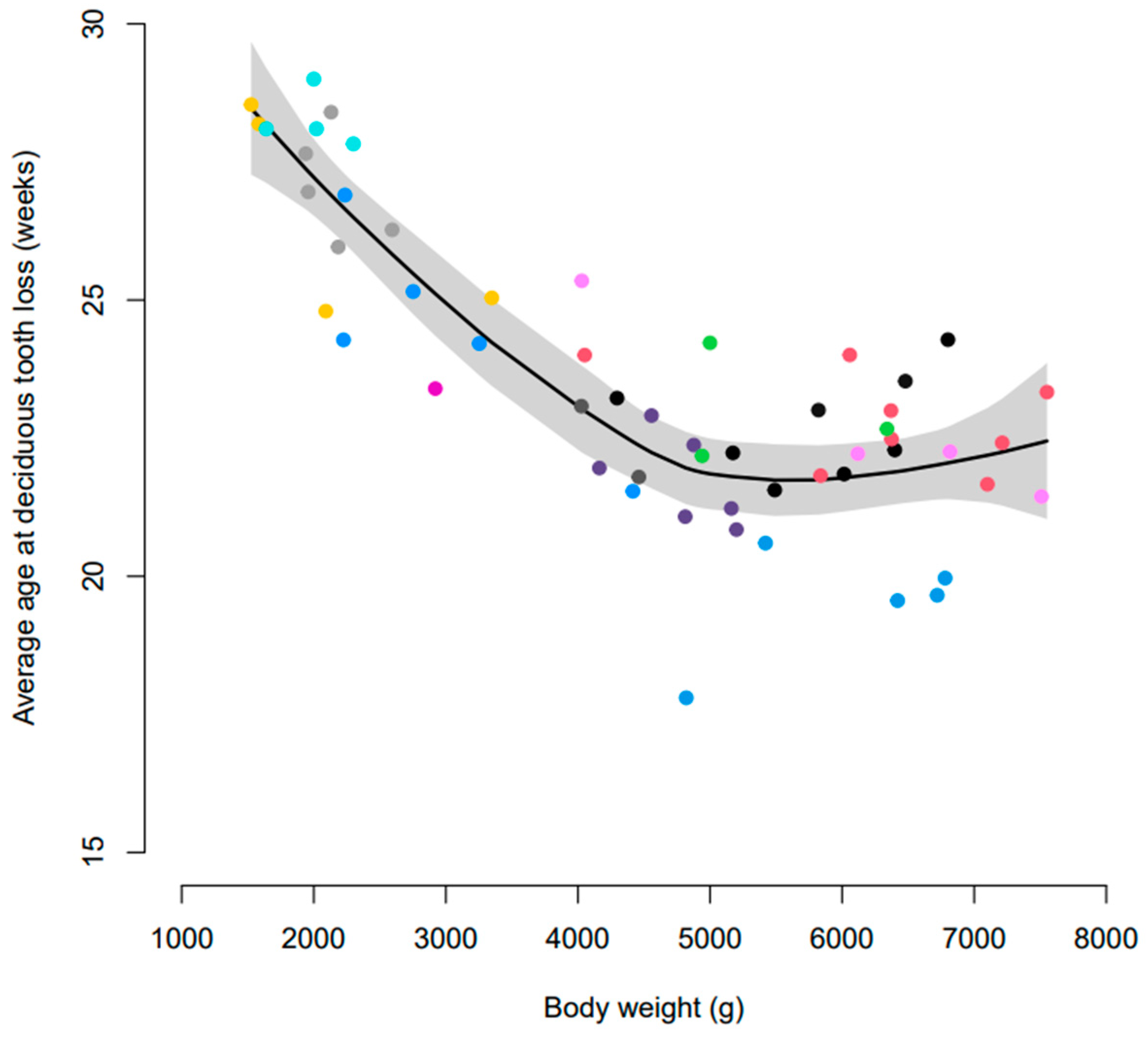
Figure 1. Smoothed average age representation of deciduous tooth loss (mouth average) according to body weight at 28 weeks of age. Coloured dots represent individual litters and the shaded area represents 95% confidence intervals.
Vetsci 10 00406 g001
Figure 2. Estimated average age of deciduous tooth loss for incisors (red), canines (black), and premolar (blue) teeth across a range of body weights seen in the cohort. Dotted lines represent pointwise 95% confidence intervals.
Vetsci 10 00406 g002
Figure 3. Smoothed average age at eruption of permanent dentition according to body weight. Coloured dots represent individual litters and the shaded area represents built-in 95% confidence intervals.
Vetsci 10 00406 g003
Figure 4. Average age at eruption of permanent dentition for each of the different tooth types (incisor, canine, premolar, and molar). The bars indicate 95% confidence intervals.
Vetsci 10 00406 g004
Figure 5. Estimated average age at eruption of permanent dentition for incisors (red), canines (black), premolars (blue), and molars (green) teeth across a range of body weights. The dotted lines represent 95% confidence intervals.
Vetsci 10 00406 g005
Figure 6. Smoothed mean proportion of PDT according to body weight. Coloured dots represent individual litters and the shaded area represents 95% confidence intervals.
Vetsci 10 00406 g006
Figure 7. Estimated mean proportion of PDT by body weight and tooth type: incisor (red), canine (black), and premolar (blue). Dotted lines represent 95% confidence intervals.
Vetsci 10 00406 g007
Table 1. Variables recorded as part of the study.
VariableDescription
LitterDogs’ litter group
AgeDogs’ age (weeks) at time of measurement
Body weightDogs’ weight (kg) at end of study (28 weeks of age)
Tooth typeIncisor, canine, premolar, molar
Persistent deciduous tooth
indicator
Defined as whether deciduous teeth were present in the mouth at the same time as their permanent counterparts. Not persistent—0; persistent—1
Surgery indicatorDeciduous dentition, which was not naturally exfoliated by 32 weeks of age and required surgical intervention. No surgery—0; surgery—1
Table 2. Average age in weeks (standard deviation) of exfoliation of deciduous dentition by tooth type and body weight.
Tooth TypeBody Weight
≤3 kg>3 kg and ≤5 kg >5 kg
Incisor25.8 (2.8)20.8 (2.5)20.6 (2.5)
Canine28.3 (0.6)25.5 (2.8)23.8 (2.7)
Premolar26.9 (2.2)23.6 (2.7)23.0 (2.8)
Table 3. Average age in weeks (standard deviation) of eruption of permanent dentition by tooth type and body weight.
Tooth TypeBody Weight
≤3 kg>3 kg and ≤5 kg >5 kg
Incisor24.4 (2.3)21.2(2.4)21.1 (2.5)
Canine26.8 (1.7)22.7 (1.6)22.7 (1.6)
Premolar26.1 (2.6)24.1 (3.4)22.9 (3.0)
Molar26.4 (2.9)25.7 (2.9)24.4 (3.4)
Table 4. Proportion of PDT (standard deviation) by tooth type and body weight.
Tooth TypeBody Weight
≤3 kg>3 kg and ≤5 kg >5 kg
Incisor0.349 (0.477)0.017 (0.128)0.013 (0.115)
Canine0.891 (0.312)0.250 (0.433)0.120 (0.325)
Premolar0.411 (0.492)0.061 (0.240)0.087 (0.281)
Table 5. Proportion of PDT (standard deviation), by tooth type and body weight, that required surgical extraction.
Tooth TypeBody Weight
≤3 kg>3 kg and ≤5 kg >5 kg
Incisor0.188 (0.390)0.011 (0.105)0.003 (0.058)
Canine0.641 (0.480)0.167 (0.373)0.080 (0.271)
Premolar0.234 (0.424)0.044 (0.206)0.080 (0.271)
Disclaimer/Publisher’s Note: The statements, opinions and data contained in all publications are solely those of the individual author(s) and contributor(s) and not of MDPI and/or the editor(s). MDPI and/or the editor(s) disclaim responsibility for any injury to people or property resulting from any ideas, methods, instructions or products referred to in the content.