Review

Non-Human Primate Models of Orthopoxvirus Infections

Pathology Unit, German Primate Center, Leibniz Institute for Primate Research, Kellnerweg 4, 37077 Göttingen, Germany
*
Author to whom correspondence should be addressed.
Vet. Sci. 2014, 1(1), 40-62; https://doi.org/10.3390/vetsci1010040
Submission received: 23 April 2014 / Revised: 4 June 2014 / Accepted: 5 June 2014 / Published: 10 June 2014
(This article belongs to the Special Issue Animal Models of Disease)

Abstract

:
Smallpox, one of the most destructive diseases, has been successfully eradicated through a worldwide vaccination campaign. Since immunization programs have been stopped, the number of people with vaccinia virus induced immunity is declining. This leads to an increase in orthopoxvirus (OPXV) infections in humans, as well as in animals. Additionally, potential abuse of Variola virus (VARV), the causative agent of smallpox, or monkeypox virus, as agents of bioterrorism, has renewed interest in development of antiviral therapeutics and of safer vaccines. Due to its high risk potential, research with VARV is restricted to two laboratories worldwide. Therefore, numerous animal models of other OPXV infections have been developed in the last decades. Non-human primates are especially suitable due to their close relationship to humans. This article provides a review about on non-human primate models of orthopoxvirus infections.

1. Introduction

The genus Orthopoxvirus (OPXV), as part of the Poxviridae, includes, among others, the species variola virus (VARV), monkeypox virus (MPXV), cowpox virus (CPXV), vaccinia virus (VACV), and mousepox virus. The causative agent of smallpox, VARV, was one of the most dangerous viruses known to mankind, being responsible for the death of 300 to 500 million people. Fortunately, smallpox has been successfully eradicated by a worldwide vaccination campaign under the leadership of the World Health Organization (WHO) [1]. Because of the inferior safety profile of the smallpox vaccine in immune compromised persons, pregnant women, or persons with atopic dermatitis, routine smallpox vaccinations were stopped in the 1980s following a recommendation from WHO. Only military personnel, selected healthcare, and laboratory workers still get the vaccine. Consequently, the number of people with lacking immunity, not only against smallpox, but also against other zoonotic OPXV infections is increasing [2], in humans, as well as in animals [3,4,5,6,7]. In the Democratic Republic of Congo there is a massive (20-fold) increase in human monkeypox incidence [8]. Additionally, it is feared that smallpox could be used as a biological weapon [9,10,11]. There is, thus far, no pharmaceutic treatment available and vaccines are partly unsafe, therefore, more research concerning orthopoxvirus infections is very important. Because of its high risk potential, research with VARV is limited to two BSL4 containments worldwide (Atlanta and Novosibirsk) and is, therefore, highly restricted. Suitable animal models are needed in which an orthopoxvirus causes a disease course, morbidity, and mortality, similar to human disease. Furthermore, the route of infection and transmission should be mimicked, and the infectious dose should be similar to that of humans. Unfortunately, no animal model fulfills all these criteria [12,13]. Nevertheless, these models can help to get an insight into the pathogenesis, and are a good tool to test the efficacy of antiviral compounds and of new vaccines. Furthermore, these animal models are important to fulfill the requirements of the animal efficacy rule. This rule was promulgated by the Food and Drug Administration (FDA) and demands a testing of medical countermeasures in at least two different animal models when clinical trials in humans are unethical or impossible [14].
In the last decades, considerable progress has been made in development of new animal models for OPXV infections [12,15,16,17,18,19]. CPXV, MPXV, VACV, VARV, ectromelia virus, rabbitpox virus, and camelpox virus have been used in animal models. Small animal models, other than non-human primates (NHP) models, have the advantage that large numbers of animals are available. Furthermore, maintenance costs are lower compared to monkeys. However, small animal models have limitations: Disease pathology, a shortened time course of disease, pharmacokinetic behavior of compounds, and tissue distribution can vary from human conditions [20]. Therefore, animals, which are closely related to humans, and whose physiological and pathological reactions are, therefore, more comparable to humans, are more suitable. NHP are the next relatives to humans, recapitulate human condition as closely as possible, and are, therefore, very appropriate to evaluate new vaccines, treatments, or pathogenesis [21]. Thus, NHP are the gold-standard for OPXV models, as their resemblance to humans allows the best predicative value for effects or side effects of new therapeutics or vaccines in humans [13].
In the following, we will review OPXV models in NHP, arranged by virus species and inoculation route (see Table 1).

2. Monkeypox

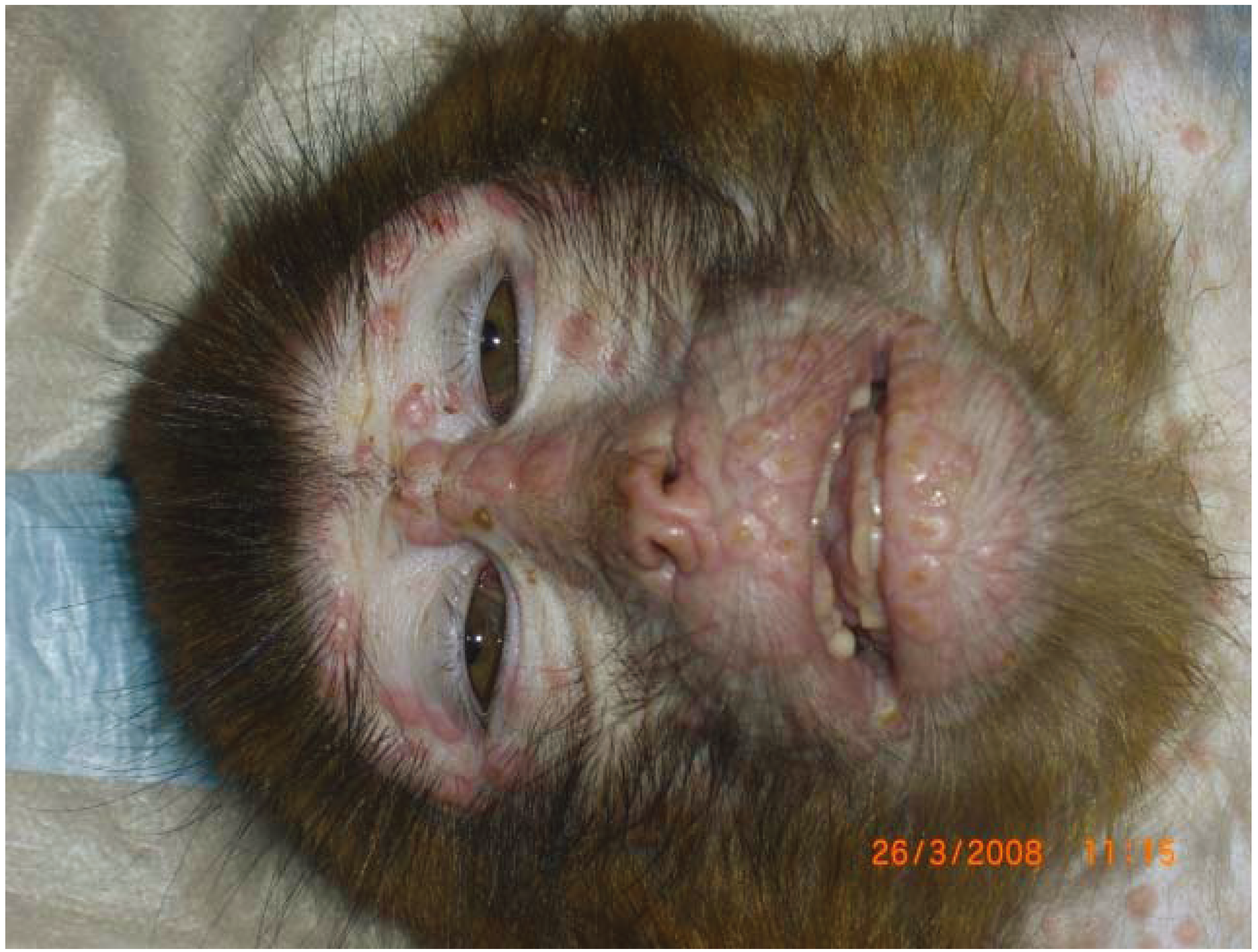
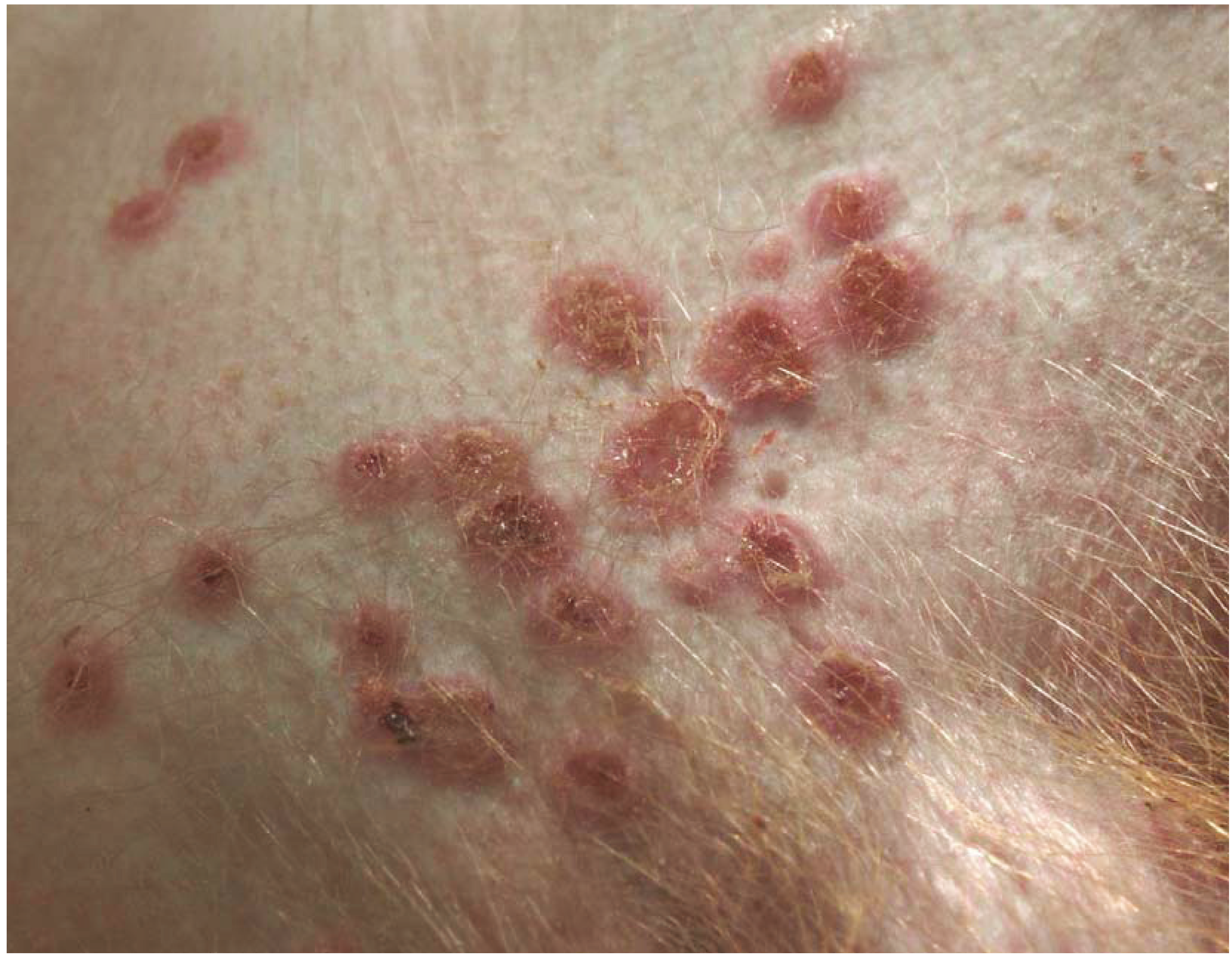
MPXV is an Orthopoxvirus which causes a zoonotic disease characterized by symptoms similar to smallpox but with a lethality rate of 1%–8% [2,22]. In all probability, some monkeypox infections were misinterpreted as smallpox because of the lack of laboratory testing [23]. One clinical symptom, which allows a differentiation from smallpox, is lymphadenopathy, which could lead to the conclusion that there is a more effective immune response [24,25]. MPXV was first detected in 1957 in captive primates in Denmark [26]. Not only are several species of NHP (rhesus macaques (see Figure 1 and Figure 2), cynomolgus macaques, and chimpanzees), but also a broad spectrum of mammals, are susceptible to the disease [15,16]. In 1960, a naturally occurring MPXV outbreak was reported in cynomolgus and rhesus macaques [27]. Human monkeypox emerged in the 1970s in Western and Central Africa and today still leads to outbreaks in the rural villages near the rainforests [8]. The natural host is still unknown, but, with the utmost probability, African squirrels and/or other rodents play an important role [15]. Only once, the virus could be found in a carcass of a Funiscirus squirrel in Democratic Republic of the Congo [28]. Probably, the virus is transmitted via eating infected bushmeat, via saliva/respiratory excretions, or direct contact with crust material. However, the exact transmission route for MPXV is unclear. [12,15,22,24]. In 2003, the disease was accidentally introduced into the U.S. due to infected wild rodents imported from Africa, which transmitted the virus to prairie dogs (Cynomys ludovicianus) and then, via the prairie dogs, to approximately 40 humans [29,30,31]. Remarkably, the prairie dogs were the only infection source for the humans [15]. Discontinuation of vaccination after eradication of smallpox, inadequate health infrastructure and bushmeat consumption has given rise to increasing susceptibility to MPXV infection in the African population [23,32].
Studies with NHP are useful for understanding monkeypox disease. Additionally, NHP models of MPXV are a good alternative to VARV models, because they are less dangerous for laboratory workers and researchers. Compared to other MPXV susceptible species, such as prairie dogs, NHP are more suitable due to the close relationship to humans. Furthermore, they get better accustomed to the research conditions than other susceptible species, such as prairie dogs [15].
NHP can be infected with MPXV experimentally via different techniques, which are described below.
Figure 1. Monkeypox virus infected Macaca mulatta (M. mulatta) with multifocal severe papular dermatitis.
Vetsci 01 00040 g001
Figure 2. Skin of Monkeypox virus infected M. mulatta with multiple vesicular and erosive to ulcerative skin lesions.
Vetsci 01 00040 g002

2.1. Intramuscular

In the late 1960s, Wenner and colleagues developed a model in which they inoculated 105 plaque-forming unit (PFU) of MPXV intramuscularly in cynomolgus and rhesus macaques [33]. Animals developed a vesicular to pustular rash and became diseased with a systemic viral infection. Disadvantages of this model are skin and muscle necrosis at the inoculation site [16].
In a study conducted in 1971, six baboons (Papio cynocephalus) were challenged intramuscularly with MPXV and all became infected. Typical lesions like vesicular pustules on the extremities, face, lips, and buccal mucosa, happened after around eight days post infection. One single animal succumbed to the disease. Challenging surviving animals with MPXV after first experiment proved the acquired immunity and the possibility to protect monkey colonies in captivity by immunizing [34].

2.2. Skin Scarification

In a following study, Heberling and Kalter challenged baboons with MPXV via skin scarification and showed an increased resistance to MPXV with ageing [35]. Baboons aged approximately one year developed typical pox lesions, fever, and lymphadenopathy, but did not die. The younger animals (approximately three-months old) showed a similar disease course, but five of ten animals succumbed to the disease.

2.3. Intravenous

Though the intravenous infection of MPXV causes a fulminant disease with severe lesions, which are very similar to smallpox, this model has several disadvantages: Key events like infection of the upper respiratory tract, primary viremia phase, and prodromal phases are skipped. Additionally, the route of transmission does not resemble the natural route of transmission of smallpox, which happens through close contact or inhalated aerosols. Furthermore, the infectious dose is much higher than the natural infection dose [36]. However, intravenous application of MPXV produces a course of disease that is appropriate to evaluate the efficacy and benefits of anti-OPXV therapeutics and vaccines.
Huggins and colleagues infected eight male cynomolgus macaques with a Zaire strain of MPXV via intravenous inoculation and proved the efficacy of the antiviral drug Tecovirimat (previously known as ST-246) dosed with 300 mg/kg/day. Tecovirimat was able to protect animals from disease and death. The infection with MPXV in the control group triggered a vesiculopustular rash accompanied by fever, elevated white blood cell count, lymphadenopathy, splenomegaly, and pulmonary edema [20]. Because of the fact that high drug doses at early times post infection were given, a follow-up study was conducted in which lower doses at later times were compared. The researchers conclude that 400 mg, once daily for 14 days after clinical diagnosis, can be an effective treatment for smallpox and monkeypox in humans [37].
To visualize viral infection, researchers inserted a gene encoding green fluorescent protein (GFP) into MPXV Zaire-79 and infected cynomolgus macaques intravenously. This way, initial lesions could be detected under fluorescent light one to two days earlier. Fluorescence was most intense in lesions of the oral cavity and weakest in skin of the palms and soles, which is protected by a thick layer of keratin [38].
Hooper and colleagues challenged rhesus macaques intravenously with MPXV after immunizing with a DNA vaccine consisting of four vaccinia virus genes. Unvaccinated control animals died on days seven, ten, and fourteen, showing hemorrhages in multiple organs, lymphadenopathy, and vesiculopustular rashes on the face and the hands. With a subunit vaccine, immunized animals did not develop severe disease and survived. This is a promising study, because a subunit vaccine does not have the feared side effects of the present smallpox vaccine [39]. In a following study, they tried to boost immune response to the gene-based vaccine using adjuvants like granulocyte macrophage-colony stimulating factor (GM-CSF) and Escherichia coli heat-labile enterotoxin. Unvaccinated control animals became severely ill, four of the five had to be euthanized, whereas all five animals vaccinated with gene-based vaccine with adjuvants survived without shedding virus, but one developed severe disease. Control animals which were vaccinated with modified Virus Ankara (MVA) shed virus, three of five became heavily ill [40].
To get more information about innate immunity in MPXV infection, Song and colleagues challenged rhesus macaques intravenously with MPXV and analyzed the changes in natural killer (NK) cell numbers, NK cell proliferation, chemokine receptor expression and cellular functions. They found that NK cell frequency and the absolute number in the blood and in the lymphoid tissues increase following MPXV infection. Interestingly, MPXV infection induces a reduced migrating capacity of NK cells and a reduced degranulation capacity. Thus, cytotoxic effects of cytokines were not reached [41].
Dyall and colleagues challenged cynomolgus macaques of both sexes intravenously, as well as intrabronchially, with 5 × 10⁶ or 5 × 10⁵ PFU of MPXV and observed progression and regression of the disease course with molecular imaging like positron emission tomography (PET) and computed tomography (CT). Animals that were infected intravenously did not show any signs of lung consolidation or inflammation in molecular imaging contrary to those infected intrabronchially. In these animals, PET/CT imaging revealed areas of necrosis, mixed inflammation, oedema, and some fibrosis in the lung. For both routes, clear manifestation of lymphadenopathy and manifestation in the axillary lymph nodes could be shown [42].
Earl and colleagues challenged cynomolgus macaques intravenously with MPXV at four, six, ten, and thirty days after immunization with MVA and Dryvax. The animals were protected against disease outbreak if they were challenged at least six days after immunization. The researchers found that a single dose of MVA induces a rapid immune response suggesting that it might be a countermeasure against a potential smallpox virus outbreak. A further result was that MVA produces a more rapid immune response compared to the licensed Dryvax vaccine [43].
To examine the question whether human immunodeficiency virus type 1 (HIV-1) infected creatures are protected by smallpox vaccination, researchers infected fourteen rhesus macaques with simian immunodeficiency virus (SIV) and immunized them with different combinations of MVA, NYVAC, and Dryvax. One and six months after immunization, the animals which had CD4+ cell counts <300 mm3 were challenged intravenously with 5 × 10⁷ PFU of MPXV. All challenged monkeys showed a severe disease course with typical pox lesions on skin and the lining of the oropharynx. This lack of protection could be the consequence of the inability of the SIV-infected macaques to switch vaccinia-specific immunoglobulin (Ig) from IgM to IgG [44]. A further vaccination study with Dryvax conducted by Edghill-Smith and colleagues found that vaccinia-specific B-cell responses are indispensable for protection from lethal monkeypox disease, whereas CD4+ and CD8+ T cells do not play an important role. They immunized rhesus macaques with Dryvax and challenged them intravenously with a dose of 5 × 10⁷ PFU of MPV [45].
Another study examined the use of subunit recombinant vaccine against MPXV in a rhesus macaque model. They immunized the monkeys with plasmid DNA alone, in combination with the equivalent recombinant proteins, or only with the proteins. Thirty-five weeks after the beginning of immunization, the animals were challenged intravenously with 5 × 10⁷ PFU of MPXV. Immunization only with DNA did not lead to protection against MPXV challenge; the animals had to be euthanized due to severe disease. Animals which were immunized with proteins, developed mild to severe disease but survived. Animals that were immunized with a combination of DNA and proteins had only few lesions, which healed within days and showed only mild disease symptoms [46].
The integration of human cytokine IL15 into the genome of Wyeth strain of VACV confers long-term protection to cynomolgus macaques [47]. The researchers immunized the monkeys and challenged them three years later intravenously with 107 PFU of MPXV. By day 27 post challenge, all skin lesions of Wyeth-IL15 immunized animals were healed, whereas all unvaccinated control animals succumbed to the disease.

2.4. Intratracheal/Aerosolization

The transmission via aerosol is supposed to play an important role even if primary monkeypox infection is transmitted by direct cutaneous or mucosal contact [22,48]. Stittelaar and colleagues challenged cynomolgus macaques intratracheally with MPXV after vaccination with MVA and Elstree. One single animal developed a light course of disease but did not die. Unvaccinated control animals developed severe disease showing fibrinonecrotic bronchopneumonia, necrotizing dermatitis, glossitis, and splenitis with lymphoid depletion [49]. In a following study, the researchers again inoculated cynomolgus macaques intratracheally with a lethal dose of MPXV to evaluate the efficacy of post-exposure antiviral treatments and vaccination. In contrast to post-exposure vaccination with Elstree, antiviral treatment with cidofovir or with a nucleoside phosphonate analogue resulted in a reduced number of skin lesions and a reduced mortality rate. Untreated challenged control animals showed severe skin lesions, dyspnea and low blood oxygen saturation and died within 15 days after being challenged with MPXV [50].
One disadvantage of intratracheal models of MPXV infection is the huge amount of virus needed which does not resemble the natural infection. Intratracheal infection models do not take into consideration the physiological inhalation procedure, which is skipped by depositing virus directly into airways.
To infect monkeys more efficiently, researchers developed new aerosolization methods: Zaucha and colleagues established an aerosolized MPXV infection in cynomolgus monkeys using a head-only esposure chamber with a collision nebulizer. They exposed monkeys to four different doses of aerosolized MPXV, trying to imitate the natural route of transmission for human VARV infection. Fever, lymphadenopathy and depression were present around day six, post exposure. The animals died nine to seventeen days post exposure suffering from fibrinonecrotic bronchopneumonia. The pathogenesis of this route of infection is comparable to smallpox infection, which also starts in the respiratory mucosa, spreads to local lymph nodes, and is also followed by viremia [51].
To further characterize the pathogenesis of MPXV, Nalca and colleagues exposed cynomolgus macaques in the same way like described by Zaucha. Blood analysis revealed that complete blood count (CBC) and clinical chemistry of MPXV infected animals deviate from normal values—comparable to human monkeypox cases. Main pathological findings and cause of death were primary fibrinonecrotic bronchopneumonia, comparable to the results of Zaucha. Furthermore, they interestingly found that heavy males were more resistant [21].
Barnewall and colleagues performed a MPXV infection study with six cynomolgus macaques, also using the above described head-only aerosol exposure system. The most prominent pathological lesion was bronchopneumonia. They also found that weight and/or sex may be important for disease course. Furthermore they compared two sampling methods of MPXV aerosols and concluded that gelatin filters and impingers delivered comparable results [52]. One disadvantage of above described head-only chamber is the fact that each monkey inhales different doses of virus, which aggravates comparability [15].
To compare the protective effects of Imvamune and Acam2000 vaccines, Hatch and colleagues challenged cynomolgus macaques with a target dose of 105 PFU of MPXV. They used a flexible system, in which the challenge aerosol is delivered by a nebulizer over a modified veterinary anesthesia mask. Acam2000 protected the animals completely even if applicated only once, whereas Imvamune showed protection from severe disease and death only if applicated twice (prime and boost immunization) [53].

2.5. Microsprayer-Technique and Intrabronchial

A new challenge technique tries to refine the virus delivery. Goff and colleagues inserted a microsprayer attached to a bronchoscope into the trachea of cynomolgus macques and challenged the animals via a high-pressure syringe, which facilitates precise dosage. They could lower the dose of virus compared to the intravenous challenge dose and the infection resulted in a fatal disease course. The animals showed marked lobar fibrinonecrotic pneumonia and more cutaneous lesion compared to other aerosol models. This means a better comparability to smallpox. One disadvantage of this technique is the possible mechanical damage of the respiratory tissue causing an inflammation at the inoculation site [36].
Compared to the aerosol models, intrabronchial models have the advantage that inoculation via bronchoscope allows a measurable and exact deposit of virus. Furthermore, the technique is easy to conduct and does not require special expensive equipment.
Estep and colleagues proposed that monkeypox inhibitor of complement enzymes (MOPICE) is an important virulence factor in Central African strains of MPXV. To verify that, they inoculated rhesus monkeys intrabronchially with 2 × 10⁵ PFU of MPXV-Zaire (n = 4) and with a recombinant MPXV-Zaire (n = 4), which does not express MOPICE. Both animals groups developed similar disease symptoms: typical pox skin lesions, fever, and respiratory symptoms. Interestingly, the animals which were infected with the knockout clade, showed more severe disease courses: One animal died at day 17 post infection due to MPXV-related disease complications, two animals had very low oxygen saturation levels at 14 and 28 days post infection, respectively, and two animals showed a delayed wound healing process. The knockout group had higher peak viral loads than the wild type infected group. The results suggest that MOPICE is not an important virulence factor, but that it is important to generate a successful adaptive immune response [54].
To find out if intrabronchial exposure to MPXV produces a disease course, which is comparable to human MPXV infection, researchers infected cynomolgus macaques with MPXV by the intravenous and the intrabronchial inoculation route. Although both routes produced typical pox-like disease, they concluded that the intrabronchial model is more adequate than the intravenous model: The whole disease course is delayed and, therefore, allows a deeper insight into pathogenesis [55].
To study host-pathogen interaction in bronchoalveolar lavage fluids (BALF), Brown and colleagues infected rhesus macaques intrabronchially with 2 × 10⁵ PFU of MPXV and flushed lungs of infected animals with PBS. They found an increase in inflammatory and interestingly a decrease in structural and metabolic proteins after infection and conclude that inflammation is not the only contributor to disease course. Structural and metabolic proteins also seem to play in important role in pathogenesis of MPXV [56].

2.6. Intranasal

Saijo and colleagues showed that vaccination with Lister, as well as with LC16m8, protected cynomolgus macaques against a lethal MPXV infection. They infected the animals intranasally with MPXV strain Liberia or Zaire. Unvaccinated control animals featured loss of appetite, diarrhea, and papulovesicular skin lesions, but none of the animals died [57]. In a further study, they inoculated Congo Basin and West African strains of MPXV subcutaneously, respectively intranasally into cynomolgus monkeys. They found that the virulence of Congo Basin strain is much higher than of West African one, which could be explained by the difference in organ tropism and, thus, in the sites of virus replication. Congo Basin caused more organ dysfunction than West African and replicated more efficiently [58].

2.7. Subcutaneous

Subcutaneous infection seems to play in important role in MPXV infections [15] and is therefore a good model to test antiviral therapeutics. Early studies revealed that subcutaneous injection of MPXV in rhesus and cynomolgus macaques only caused locally restricted skin lesions or mild generalized disease without death [15,27,59]. Recent studies have obtained different results: In a vaccination study with Lister and LC16m8, cynomolgus macaques were challenged intranasally and subcutaneously. In non-immunized control animals, subcutaneous injection of MPXV has led to generalized, severe disease that required euthanasia. Histopathological investigations revealed alterations in the lymphoid system, respiratory, digestive and urinary tract and the skin [57]. A subsequent study compared Congo Basin and West African strains of MPXV. Three of four monkeys, which were subcutaneously infected with Congo Basin strain, showed a sharp decrease of body weight and succumbed to the disease. In the group that was subcutaneously infected with West African strain, one of three animals died [58].

Summary Monkeypox

Although MPXV has only been detected in African non-human primates, Asian macaques like rhesus and cynomolgus macques, which are not natural hosts, are the most often used species for MPXV models. This may be due to the fact that these species are widely spread in animal experiments, easy available and have been well investigated [15]. Both species are susceptible to MPXV, but rhesus macaques seem to be less susceptible than cynomolgus macaques, as observed in an US outbreak in 1960 [15,60]. Concerning the appropriate inoculation route, none mimics sufficiently natural infection with VARV or MPXV. The exact transmission route for MPXV is still not clear, which makes it more difficult to evaluate transmission routes. Additionally, the inoculum dose in natural infections may be lower than those inoculated experimentally. Intravenous models of MPXV infection skip infection of respiratory tissue and incubation phase and, therefore, do not mimic the natural route of transmission. Nevertheless, they cause systemic disease with mortality rates up to 100%. This makes the intravenous model interesting for vaccine and therapeutical studies concerning smallpox [15].
Respiratory NHP challenge models of MPXV have a slowed course of disease compared to intravenous models. They often cause fibrinonecrotic bronchopneumonia, which resembles human MPXV disease course. In addition, mortality rates of 100% rates are not reached in respiratory challenge models [15,36,51,55,57], which also resembles human MPXV. Thus, respiratory models are appropriate to get further insight into pathogenesis of human MPXV. Different techniques can be used to inoculate virus into the respiratory tract. Application via bronchoscope is easy, inexpensive and allows an exact deposition of the virus. However, there is the potential risk to damage tissue and to cause non-virus related inflammation [15]. This problem does not occur if virus is aerosolized. However, there is the disadvantage that quite high doses of virus are needed and it is not guaranteed that each monkey gets exact the same amount of virus.
Subcutaneous models closely mimic natural transmission of MPXV and are, therefore, a good tool to examine pathogenesis of human MPXV infections. Contrary to this, they do not mimic the transmission route for VARV, which happens via respiratory tract, and are therefore less suitable for VARV research.

3. Smallpox

Development of animal models for smallpox is very difficult, because natural VARV is restricted to humans. Additionally, it is unclear to what extent resultant pathology of animals compares with that of naturally happening smallpox disease in humans [61]. Due to their close relationship to humans, WHO and the U.S. National Academy of Sciences recommended the use of NHP models for smallpox research [62]. The VARV NHP models are reserved for in vivo experiments with antiviral drugs or vaccines that have already fulfilled the criteria required by FDA. Additionally, the research community hopes to get a deeper insight into the, thus far, unexplored pathogenesis of smallpox.
In 1906, Brinckerhoff and Tyzzer unsuccessfully tried to infect cynomolgus and pig-tailed macaques with VARV [61,63]. In the 1950s and 1960s, researchers still failed to develop efficient NHP models of smallpox. They challenged wild-caught monkeys with VARV via inhalation of a cloud of dried particles, but, beyond pulmonary lesions, they could detect no pathological changes in other tissues. The resulting mild disease did not resemble fulminant disease course of smallpox in humans [64]. Westwood challenged rhesus macaques via aerosol to VARV and succeeded in provoking a more pronounced clinical disease [65,66]. Ultimately, VARV studies with NHP were discontinued due to unsatisfying results. Sociopolitical changes and the fear of bioterrorism in the early 21st century reintensified VARV studies with NHP. First experiments again failed to produce lethal disease course: Cynomolgus macaques were challenged via aerosol with high doses of VARV strains. They only showed mild clinical signs without dying [67]. But a later experiment with two VARV strains (Harper and India 7124) resulted in a fulminant lethal disease course of cynomolgus macaques which were infected intravenously or intravenously in combination with aerosol exposure [68]. Disease course appeared to be dose dependent, as high doses administered intravenously (109 PFU) caused hemorrhagic diathesis ending in acute deaths whereas lower doses resulted in ordinary smallpox. A disadvantage regarding testing antiviral drugs is the fact that intravenous inoculation of VARV skips the incubation period which happens in natural human smallpox. Nevertheless, this model is the first that could produce a lethal VARV infection in cynomolgus macaques. It can be used as a model for hemorrhagic smallpox.
To get further insight into the molecular features of smallpox infection, researchers infected cynomolgus macaques intravenously or intravenously in combination with aerosol exposure with VARV (strain Harper and India 7124) and analyzed in serial blood samples the host gene expression in PBMCs (peripheral blood mononuclear cells). The animals had a severe clinical disease course developing fever, typical pox skin lesions, visceral and mucosal hemorrhage, and showed an upregulation of inflammatory cytokines [62].
To get more knowledge about the progression of smallpox disease and the differences between the ordinary and the hemorrhagic disease course, Wahl-Jensen and colleagues challenged cynomolgus macaques intravenously with VARV doses which either caused hemorrhagic or ordinary disease. They performed temporal pathology analysis and took serial samples. In the ordinary smallpox model, lymphoid and myeloid hyperplasia was observed. Only a few VARV particles in tissue could be detected compared to the hemorrhagic model. In hemorrhagic smallpox model lymphocytolysis and hematopoietic necrosis, as well marked antigen accumulation occurred. Furthermore, hemorrhagic disease was always accompanied by secondary bacterial infections [69].
Huggins and colleagues infected eight cynomolgus monkeys intravenously with 108 PFU of VARV (strain Harper). Two control animals, which did not receive drugs, had to be euthanized on day twelve. Their disease course resembled human smallpox infection, especially disease endpoints like lesions and death. Three animals were treated with Tecovirimat (300 mg/kg/day) immediately after infection and three animals 24 h post infection. Both groups were protected from disease and death [20].
A study conducted by Mucker and colleagues also evaluated the efficacy of Tecovirimat. They infected 18 male cynomolgus macaques intravenously with 108 PFU of VARV and showed that treatment with Tecovirimat two, as well as four, days post infection protected the animals from death, effected a significant reduction in total skin lesions, reduced viremia and virus shedding in the oropharynx. All non-treated control animals succumbed to the disease. The researchers conclude that Tecovirimat is an effective drug against smallpox disease [70].

Summary Smallpox

Due to fear of bioterrorism, efforts to develop smallpox NHP models have been intensified since the beginning of the 21st century leading to NHP models in which pathogenesis and antiviral drugs, especially Tecovirimat, have been investigated. Though intravenous inoculation of the virus does not represent the natural infection route, it causes a clinical course, which resembles the latter stage of human disease, and is, therefore, suitable to prove the efficacy of therapeutics. Working with smallpox virus to test the efficacy of antiviral drugs and vaccines against smallpox has the advantage that the original causative agent of the disease is used, even though NHP are not natural hosts. Because of its high zoonotic potential, research with this extremely dangerous virus is restricted to two biosafety-level four laboratories worldwide (CDC, USA and State Research Center of Virology and Biotechnology, Russia) and is, therefore, available to only a very small research community.

4. Cowpox

CPXV, which belongs to the OPXV, is a noteworthy virus. It has the broadest host range of all OPXV, the largest genetic repertoire and produces an array of gene products, which manipulate the immune system [7,71]. Cowpox is an emerging hazard. During the last decades, reports about CPXV infections in humans, as well as in animal are increasing [72,73,74,75,76]. As smallpox immunization has been stopped, human population is now getting more vulnerable to CPXV infections. Human cowpox is a zoonotic infection, which usually causes self-limiting, painful skin lesions, particularly located on the hands, face, or trunk. In immunocompetent people, they are healing within several weeks, leaving scars. Further symptoms are fever, depression, and lymphadenopathy. Ocular cowpox infection can, rarely, happen [77,78,79]. However, severe complications can occur in immunocompromised persons or persons with atopic dermatitis [80,81,82,83,84]. Unlike smallpox, which is transmitted via the respiratory route, cowpox has to be transmitted through direct contact with skin or mucosal lesions.
The name cowpox originates from infected domestic cattle, which transmitted the disease to milkers. The name is misleading [85], as transmission by cows is not the main transmission route any more. Nowadays, CPXV exists in many host species in Western Eurasia [86]. Asymptomatic carriers are wild infected rodents. A survey conducted by Kinnunen and colleagues, in which they screened wild rodents from Germany, Finland, and Siberia’s Baikal region for OPV antibodies and for the presence of OPXV DNA, revealed a seroprevalence up to 33% and the presence of poxvirus antigen in three rodents [87]. Cats are infected by feeding on rodents that are carrying the virus [88]. Zoo animals and exotic animals like Llamas are also infected [4,86,89,90]. CPXV outbreaks in NHP in European zoos and sanctuaries have occurred [91,92].
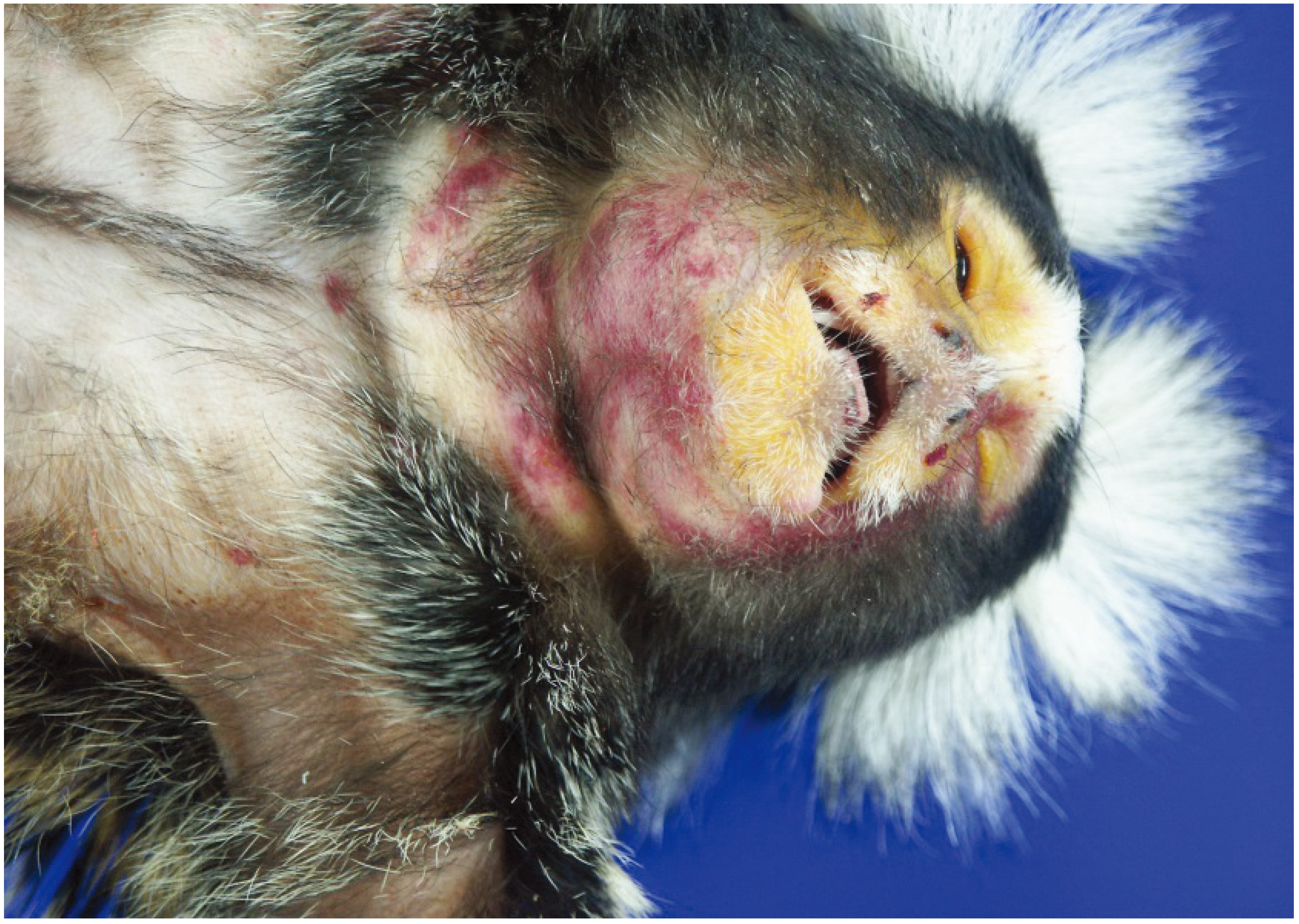
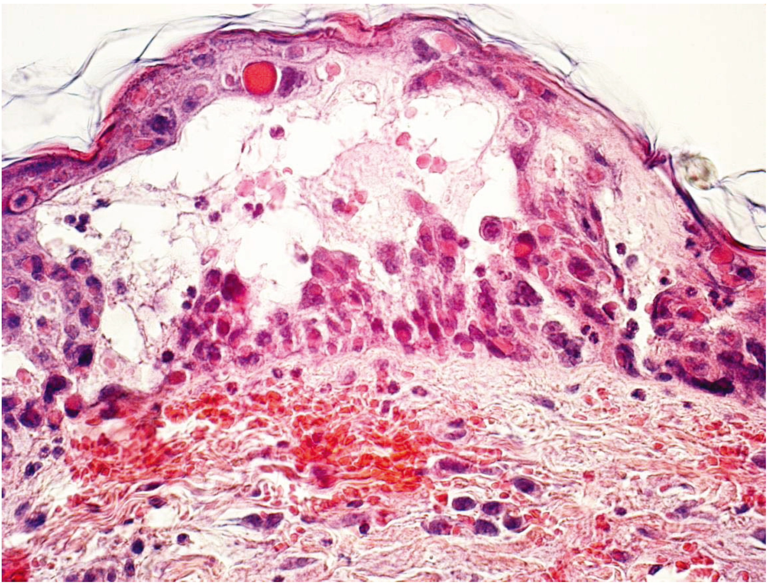
Common marmosets (Callithrix jacchus) showed a fulminant disease course [91,92]. In a private husbandry in Germany, 30 of 80 New World Monkeys died within a week after onset of symptoms. They showed fever, depression, severe erosive-ulcerative lesions of the oral membranes, and lymphadenopathy. In this case, owners and animal caretakers did not become ill. Researchers from the German Primate Center and the Robert Koch Institute isolated an OPXV with close homology to CPXV and named it calpox, according to its host Callithrix jacchus. Pathologic examination showed hemorrhagic skin lesions on the face, scrotal region, soles and palms, facial oedema, and focal erosions and ulcerations of the oral mucous membranes (see Figure 3). Histopathologically, the hemorrhagic-to-pustular skin lesions were accompanied by vesiculation, epidermal acanthosis, and acantholysis and necrosis (see Figure 4). Eosinophilic intracytoplasmic inclusions bodies could be detected in degenerated keratinocytes. Transmission Electron Microscopy revealed particles with characteristic pox-like features. Kramski and colleagues succeeded in developing a common marmoset model for calpoxvirus infection. They infected marmosets via the intravenous and the intranasal route and produced a lethal disease course, in which the animals died within three days after onset of symptoms. The animals showed characteristic pox-like lesions in the skin and mucous membranes, subcutaneous oedema, splenic lymphoid hyperplasia, and lymphadenomegaly of different lymph nodes [93]. Histological investigations revealed vesicular dermatitis with necrosis, acanthosis, acantholysis, and syncytia formation of the basal keratinocytes [94]. Advantages of this model are firstly the low infectious dose, as even a very low dose of 5 × 10² PFU was effective. Secondly, CPXV can be handled under a lower biohazard level than MPXV or VARV, which facilitates research with this virus. Furthermore, common marmosets have several advantages compared to commonly used NHP: Their small size allows an easy handling. They can be housed in family groups, are not endangered and are inexpensive to keep [95].
As CPXV is genetically and, antigenically, quite comparable to VACV and related to MPXV and VARV (they share 19 immunomodulatory genes), CPXV models are an alternative to above presented models for smallpox and monkeypox [82,96,97]. In addition, gaining more knowledge of CPXV pathogenesis itself is also very important due to its increasing zoonotic potential.
Smith and colleagues present a CPXV model, in which they infect cynomolgus macaques intrabronchially with different dilutions of CPXV (5 × 10⁴ to 5 × 10⁵). Lymphoid hyperplasia up to oedema and histiocytosis in the lymph nodes (depending on the inoculated dose) and multifocal neutrophilic infiltrates in the liver showed similarity to histopathological changes in other OPXV infections like MPXV and VARV. Lesions in lung (like alveolar oedema, necrotizing fibrinous pleuritis, congestion, atelectasis, pneumonia) are more pronounced in comparison to human smallpox disease. Measuring of cytokines and chemokines revealed a heavy pro-inflammatory response [98].
Figure 3. Callithrix jacchus (C. jacchus) with marked subcutaneous oedema and erythema on the neck region and single papular lesions on mucocutaneous junctions after experimental infection with calpox.
Vetsci 01 00040 g003
Figure 4. Calpox virus infected saddle back tamarin, skin. High-grade, focal, vesicular dermatitis with intracytoplasmatic eosinophilic inclusion bodies, ballooning degeneration and hemorrhage.
Vetsci 01 00040 g004
Recently, Johnson and colleagues inoculated fourteen cynomolgus macaques intravenously with different doses of CPXV (5 × 10⁷–5 × 10⁴ PFU) [99]. Nine of fourteen animals developed typical pox skin lesions. Further findings were hemorrhage in lymph nodes, multifocal petechial hemorrhages in the gastrointestinal tract, on heart, lung, kidneys, urinary bladder, and brain. This indicates a hemorrhagic disease course. All monkeys revealed interstitial pneumonia, interstitial nephritis, and hepatitis, and died within 12 days. The researchers conclude that this animal model may serve as a model for hemorrhagic smallpox, which is more feasible than the VARV using model of Jahrling [68].
Song and colleagues tried to find an early biological marker for prognosis of poxvirus infection, as clinical symptoms are not always reliable. Therefore, they infected nine cynomolgus macaques of both sexes intravenously with CPXV of different doses and monitored pox-antigen presence in immune cells by intracellular staining. They found that monocytes and granulocytes are the mainly affected cell population and that the presence of poxvirus antigen in the cells is closely connected to disease course and time of death. Based on these results, they suggest that the technique of monitoring pox-antigen staining in immune cells can be applied in human poxvirus infections to predict disease course and to value the efficacy of new antiviral therapeutics and vaccines [100].

Summary Cowpox

The number of diagnosed CPXV infections is increasing due to the cessation of vaccination against smallpox. Therefore, more research is needed to get a deeper insight into pathogenesis of CPXV infections and to develop antiviral drugs. NHP cowpox models can also be used as a model for hemorrhagic smallpox and have the advantage that CPXV does not need as high biosafety levels as MPXV or VARV and is therefore easier to work with. Cynomolgus macaques have been infected intravenously and intrabronchially with CPXV to study pathogenesis. Common marmosets have been infected intravenously and intranasally. The latter route of infection resembles the natural infection route of smallpox and is therefore a suitable model for the validation of therapeutics and vaccines. Compared to other NHP, common marmosets have several advantages: Their small size allows an easy handling and housing and an inexpensive keeping. Furthermore, they have a high reproduction rate. A disadvantage is that some critical reagents are not available yet for this species.

5. Conclusions

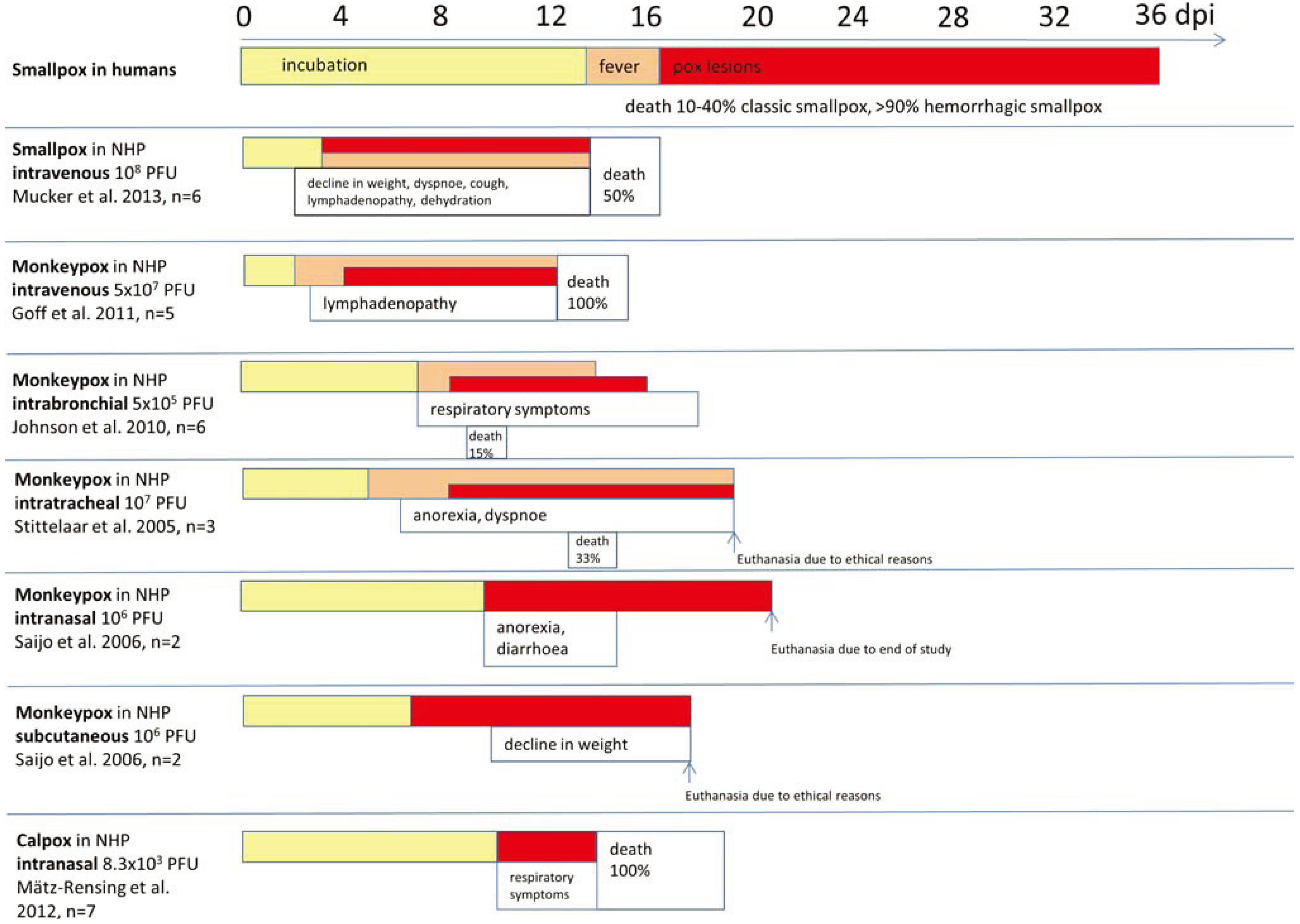
The main cause of NHP studies is to develop models to test new antiviral therapeutics and vaccines and to get a deeper insight into pathogenesis of OPXV infections. To sum up, there exists no NHP model that perfectly represents human disease (see Figure 5). Each model has its advantages and disadvantages. Concerning smallpox research, cynomolgus macaques infected with MPXV are, thus far, the best model for human smallpox [70]. Models, which use VARV, have the advantage that the original causative agent for human smallpox is used—but handling VARV is dangerous and can only be done in two laboratories worldwide.
In the last decades, promising new animal models have been developed, but none of them is good enough to safely predict a response to new therapeutics or vaccines in humans. Thus, there is a need for more research in this area. The next years will lead to interesting new findings concerning OPXV pathogenesis and development of new compounds driven by concerns of bioterrorism and increasing zoonotic potential of OPXV.
Figure 5. Comparison of disease courses in different orthopoxvirus models.
Vetsci 01 00040 g005
Table 1. Orthopoxvirus models in non-human primates.
SpeciesRoute of InfectionVirusDosePurpose of StudyReference
M.f. and M.m.i.m.MPXV105 PFUpathogenesis[33]
P.c.i.m.MPXV Copenhagen106.5–107.5 TCID50pathogenesis[34]
P.c.via skin scarificationMPXV107 TCID50pathogenesis[35]
M.f.i.v.MPXV Zaire 795 × 107 PFUTecovirimat efficacy study[20]
M.f.i.v.MPXV Zaire 795 × 107 PFUTecovirimat efficacy study[37]
M.f.i.v.MPXV-GFP Zaire 795 × 107 PFUpathogenesis[38]
M.m.i.v.MPXV Zaire 795 × 108 PFUDNA vaccine study[39]
M.m.i.v.MPXV Zaire 792 × 107 PFUMVA/gene based vaccine study[40]
M.m.i.v.MPXV Zaire 791.5–2.5 × 10⁶ PFUpathogenesis[41]
M.f.i.v. and i.b.MPXV Zaire 795 × 106–5 × 10⁷ PFUpathogenesis[42]
M.f.i.v.MPXV Zaire 795 × 107 PFUDryvax/MVA vaccine study[43]
M.m.i.v.MPXV Zaire 795 × 107 PFUVaccine study in SIV-infected macaques[44]
M.m.i.v.MPXV Zaire 795 × 107 PFUMVA/NYVAC/Dry-vax vaccine study[45]
M.m.i.v.MPXV Zaire 795 × 107 PFUSubunit recombinant vaccine study[46]
M.f.i.v.MPXV Zaire 795 × 107 PFUIl-15/Wyeth vaccine study[47]
M.f.i.t.MPXV MSF #6106–107 PFU in 5 mLMVA vaccine study[49]
M.f.i.t.MPXV MSF #6107 PFU in 5 mLComparison post-exposure vaccination with antiviral therapeutics[50]
M.f.via head-only exposure chamberMPXV Zaire 79104–105 PFUpathogenesis[51]
M.f.via head-only exposure chamberMPXV Zaire 794.3 × 10⁴–1.1 × 10⁶ PFUpathogenesis[21]
M.f.via head-only exposure systemMPXV Zaire 792.5 × 10⁴–9.3 × 10⁵ PFUpathogenesis[52]
M.f.via Henderson- apparatus and modified anesthesia maskMPXV Zaire 792.1 × 10⁵–3.1 × 10⁵ PFUImvamune, Acam2000 vaccine study[53]
M.f.bronchoscope and liquid MicroSprayer aerosolizerMPXV Zaire3.42 × 106–3.53 × 10⁷ PFUpathogenesis[36]
M.m.i.b.MPXV Zaire and D14L KO MPXV2 × 105 PFUpathogenesis[54]
M.f.i.v. and i.b.MPXV Zaire 79i.v.: 5 × 10⁷–5 × 10⁴ PFU
i.b.: 5 × 10⁶–5 × 10⁴ PFU
pathogenesis[55]
M.m.i.b.MPXV Zaire 792 × 105 PFUpathogenesis[56]
M.f.i.n. and subcutaneousMPXV Liberia and Zaire-5591 × 106 PFULC16m8 vaccine study[57]
M.f.i.n. and subcutaneousMPXV Liberia and Zaire-5591 × 106 PFUpathogenesis[58]
M.m.via Henderson apparatusVARV Higgins?pathogenesis[64,66]
M.f.aerosolVARV>108 PFUpathogenesis[67]
M.f.aerosol and/or i.v.VARV Harper and India 7124106–109 PFUpathogenesis[68]
M.f.aerosol and/or i.v.VARV Harper and India 7124Aerosol: 5 × 10⁸ PFU
i.v.: 109 PFU
pathogenesis[62]
M.f.i.v.VARV Harper108–109 PFUpathogenesis[69]
M.f.i.v.VARV Harper1 × 108 PFUST-246 efficacy study[20]
M.f.i.v.VARV Harper1 × 108 PFUST-246 efficyacy study[70]
C.j.i.v. and i.n.calpoxi.v.: 1.25 × 10⁷–1 × 10⁴ PFU
i.n.: 5 × 10²–3.5 × 10⁵ PFU
pathogenesis[93,94]
M.f.i.b.CPXV5 × 107–5 × 10⁴ PFUpathogenesis[98]
M.f.i.v.CPXV Brighton Red5 × 107–5 × 10⁵ PFUpathogenesis[99]
M.f.i.v.CPXV Brighton5 × 102–5 × 10⁴ PFUpathogenesis[100]
M.f.: Macaca fascicularis; M.m.: Macaca mulatta; P.c.: Papio cynocephalus; C.j.: Callithrix jacchus; i.v.: intravenous; i.n.: intranasal; i.b.: intrabronchial; i.t.: intratracheal; PFU: plaque-forming units.

Acknowledgments

We would like to thank Christiane Stahl-Hennig (German Primate Center) and Heinz Ellerbrok (Robert-Koch-Institute) for their collaboration.

Conflicts of Interest

The authors declare no conflict of interest.

References

  1. Fenner, F.; Henderson, D.A.; Arita, I.; Jezek, Z.; Ladnyi, I.D. Variola virus and other orthopoxviruses. In Smallpox and Its Eradication; WHO: Geneva, Switzerland, 1988. [Google Scholar]
  2. Shchelkunov, S.N. An increasing danger of zoonotic orthopoxvirus infections. PLoS Pathog. 2013, 9. [Google Scholar] [CrossRef]
  3. Nitsche, A. Untersuchungen zur Diagnostik und Risikobewertung von Emerging und Re-Emerging Orthopockenviren in Deutschland. Habilitation Thesis, Robert Koch-Institute, Berlin, Germany, 2010. [Google Scholar]
  4. Cardeti, G.; Brozzi, A.; Eleni, C.; Polici, N.; D’Alterio, G.; Carletti, F.; Scicluna, M.T.; Castilletti, C.; Capobianchi, M.R.; di Caro, A.; et al. Cowpox virus in llama, italy. Emerg. Infect. Dis. 2011, 17, 1513–1515. [Google Scholar]
  5. Bonnekoh, B.; Falk, K.; Reckling, K.F.; Kenklies, S.; Nitsche, A.; Ghebremedhin, B.; Pokrywka, A.; Franke, I.; Thriene, B.; König, W.; et al. Cowpox infection transmitted from a domestic cat. J. Dtsch. Dermatol. Ges. 2008, 6, 210–213. [Google Scholar]
  6. Baxby, D. Cowpox: Increased incidence or interest? Lancet 1994, 343, 543. [Google Scholar] [CrossRef]
  7. Dabrowski, P.W.; Radonic, A.; Kurth, A.; Nitsche, A. Genome-wide comparison of cowpox viruses reveals a new clade related to variola virus. PLoS ONE 2013, 8. [Google Scholar] [CrossRef]
  8. Rimoin, A.W.; Mulembakani, P.M.; Johnston, S.C.; Lloyd Smith, J.O.; Kisalu, N.K.; Kinkela, T.L.; Blumberg, S.; Thomassen, H.A.; Pike, B.L.; Fair, J.N.; et al. Major increase in human monkeypox incidence 30 years after smallpox vaccination campaigns cease in the democratic republic of congo. Proc. Natl. Acad. Sci. USA 2010, 107, 16262–16267. [Google Scholar] [CrossRef]
  9. Breman, J.G.; Henderson, D.A. Poxvirus dilemmas-monkeypox, smallpox, and biologic terrorism. N. Engl. J. Med. 1998, 339, 556–559. [Google Scholar] [CrossRef]
  10. Jahrling, P.B.; Fritz, E.A.; Hensley, L.E. Countermeasures to the bioterrorist threat of smallpox. Curr. Mol. Med. 2005, 5, 817–826. [Google Scholar] [CrossRef]
  11. Anderson, P.D.; Bokor, G. Bioterrorism: Pathogens as weapons. J. Pharm. Pract. 2012, 25, 521–529. [Google Scholar] [CrossRef]
  12. Hutson, C.L.; Damon, I.K. Monkeypox virus infections in small animal models for evaluation of anti-poxvirus agents. Viruses 2010, 2, 2763–2776. [Google Scholar] [CrossRef]
  13. Safronetz, D.; Geisbert, T.W.; Feldmann, H. Animal models for highly pathogenic emerging viruses. Curr. Opin. Virol. 2013, 3, 205–209. [Google Scholar] [CrossRef]
  14. Jordan, R.; Hruby, D. Smallpox antiviral drug development: Satisfying the animal efficacy rule. Expert Rev. Anti-Infect. Ther. 2006, 4, 277–289. [Google Scholar] [CrossRef]
  15. Parker, S.; Buller, R.M. A review of experimental and natural infections of animals with monkeypox virus between 1958 and 2012. Future Virol. 2013, 8, 129–157. [Google Scholar]
  16. Chapman, J.L.; Nichols, D.K.; Martinez, M.J.; Raymond, J.W. Animal models of orthopoxvirus infection. Vet. Pathol. 2010, 47, 852–870. [Google Scholar] [CrossRef]
  17. Smee, D.F.; Sidwell, R.W. A review of compounds exhibiting anti-orthopoxvirus activity in animal models. Antivir. Res. 2003, 57, 41–52. [Google Scholar] [CrossRef]
  18. Smee, D.F. Progress in the discovery of compounds inhibiting orthopoxviruses in animal models. Antivir. Chem. Chemother. 2008, 19, 115–124. [Google Scholar]
  19. Smee, D.F. Orthopoxvirus inhibitors that are active in animal models: An update from 2008 to 2012. Future Virol. 2013, 8, 891–901. [Google Scholar] [CrossRef]
  20. Huggins, J.; Goff, A.; Hensley, L.; Mucker, E.; Shamblin, J.; Wlazlowski, C.; Johnson, W.; Chapman, J.; Larsen, T.; Twenhafel, N.; et al. Nonhuman primates are protected from smallpox virus or monkeypox virus challenges by the antiviral drug st-246. Antimicrob. Agents Chemother. 2009, 53, 2620–2625. [Google Scholar] [CrossRef]
  21. Nalca, A.; Livingston, V.A.; Garza, N.L.; Zumbrun, E.E.; Frick, O.M.; Chapman, J.L.; Hartings, J.M. Experimental infection of cynomolgus macaques (Macaca fascicularis) with aerosolized monkeypox virus. PLoS ONE 2010, 5. [Google Scholar] [CrossRef]
  22. Jezek, Z.; Szczeniowski, M.; Paluku, K.M.; Mutombo, M. Human monkeypox: Clinical features of 282 patients. J. Infect. Dis. 1987, 156, 293–298. [Google Scholar] [CrossRef]
  23. Reynolds, M.G.; Carroll, D.S.; Karem, K.L. Factors affecting the likelihood of monkeypox’s emergence and spread in the post-smallpox era. Curr. Opin. Virol. 2012, 2, 335–343. [Google Scholar] [CrossRef]
  24. McCollum, A.M.; Damon, I.K. Human monkeypox. Clin. Infect. Dis. 2014, 58, 260–267. [Google Scholar] [CrossRef]
  25. Damon, I.K. Status of human monkeypox: Clinical disease, epidemiology and research. Vaccine 2011, 29 (Suppl. 4), D54–D59. [Google Scholar] [CrossRef]
  26. Von Magnus, P.; Anderson, E.; Petersen, K.; Birch-Anderson, A. A pox-like disease in cynomolgus monkeys. Acta Pathol. Microbiol. Scand. 1959, 46, 156–176. [Google Scholar]
  27. Prier, J.E.; Sauer, R.M. A pox disease of monkeys. Ann. N. Y. Acad. Sci. 1960, 85, 951–959. [Google Scholar] [CrossRef]
  28. Khodakevich, L.; Jezek, Z.; Kinzanzka, K. Isolation of monkeypox virus from wild squirrel infected in nature. Lancet 1986, 1, 98–99. [Google Scholar] [CrossRef]
  29. DiGiulio, D.B.; Eckburg, P.B. Monkeypox in the western hemisphere. N. Engl. J. Med. 2004, 350, 1790–1791, author reply 1790–1791. [Google Scholar] [CrossRef]
  30. Reed, K.D.; Melski, J.W.; Graham, M.B.; Regnery, R.L.; Sotir, M.J.; Wegner, M.V.; Kazmierczak, J.J.; Stratman, E.J.; Li, Y.; Fairley, J.A.; et al. The detection of monkeypox in humans in the western hemisphere. N. Engl. J. Med. 2004, 350, 342–350. [Google Scholar] [CrossRef]
  31. Centers for Disease, Control and Prevention. Update: Multistate outbreak of monkeypox—Illinois, Indiana, Kansas, Missouri, Ohio, and Wisconsin, 2003. MMWR. Morb. Mortal. Wkly. Rep. 2003, 52, 642–646. [Google Scholar]
  32. Parker, S.; Nuara, A.; Buller, R.M.; Schultz, D.A. Human monkeypox: An emerging zoonotic disease. Future Microbiol. 2007, 2, 17–34. [Google Scholar] [CrossRef]
  33. Wenner, H.A.; Bolano, C.R.; Cho, C.T.; Kamitsuka, P.S. Studies on the pathogenesis of monkey pox. 3. Histopathological lesions and sites of immunofluorescence. Arch. Gesamte Virusforsch. 1969, 27, 179–197. [Google Scholar] [CrossRef]
  34. Heberling, R.L.; Kalter, S.S. Induction, course, and transmissibility of monkeypox in the baboon (Papio cynocephalus). J. Infect. Dis. 1971, 124, 33–38. [Google Scholar] [CrossRef]
  35. Heberling, R.L.; Kalter, S.S.; Rodriguez, A.R. Poxvirus infection of the baboon (Papio cynocephalus). Bull. World Health Organ. 1976, 54, 285–294. [Google Scholar]
  36. Goff, A.J.; Chapman, J.; Foster, C.; Wlazlowski, C.; Shamblin, J.; Lin, K.; Kreiselmeier, N.; Mucker, E.; Paragas, J.; Lawler, J.; et al. A novel respiratory model of infection with monkeypox virus in cynomolgus macaques. J. Virol. 2011, 85, 4898–4909. [Google Scholar] [CrossRef]
  37. Jordan, R.; Goff, A.; Frimm, A.; Corrado, M.L.; Hensley, L.E.; Byrd, C.M.; Mucker, E.; Shamblin, J.; Bolken, T.C.; Wlazlowski, C.; et al. St-246 antiviral efficacy in a nonhuman primate monkeypox model: Determination of the minimal effective dose and human dose justification. Antimicrob. Agents Chemother. 2009, 53, 1817–1822. [Google Scholar] [CrossRef]
  38. Goff, A.; Mucker, E.; Raymond, J.; Fisher, R.; Bray, M.; Hensley, L.; Paragas, J. Infection of cynomolgus macaques with a recombinant monkeypox virus encoding green fluorescent protein. Arch. Virol. 2011, 156, 1877–1881. [Google Scholar] [CrossRef]
  39. Hooper, J.W.; Thompson, E.; Wilhelmsen, C.; Zimmerman, M.; Ichou, M.A.; Steffen, S.E.; Schmaljohn, C.S.; Schmaljohn, A.L.; Jahrling, P.B. Smallpox DNA vaccine protects nonhuman primates against lethal monkeypox. J. Virol. 2004, 78, 4433–4443. [Google Scholar] [CrossRef]
  40. Golden, J.W.; Josleyn, M.; Mucker, E.M.; Hung, C.F.; Loudon, P.T.; Wu, T.C.; Hooper, J.W. Side-by-side comparison of gene-based smallpox vaccine with MVA in nonhuman primates. PLoS ONE 2012, 7. [Google Scholar] [CrossRef]
  41. Song, H.; Josleyn, N.; Janosko, K.; Skinner, J.; Reeves, R.K.; Cohen, M.; Jett, C.; Johnson, R.; Blaney, J.E.; Bollinger, L.; et al. Monkeypox virus infection of rhesus macaques induces massive expansion of natural killer cells but suppresses natural killer cell functions. PLoS ONE 2013, 8. [Google Scholar] [CrossRef]
  42. Dyall, J.; Johnson, R.F.; Chen, D.Y.; Huzella, L.; Ragland, D.R.; Mollura, D.J.; Byrum, R.; Reba, R.C.; Jennings, G.; Jahrling, P.B.; et al. Evaluation of monkeypox disease progression by molecular imaging. J. Infect. Dis. 2011, 204, 1902–1911. [Google Scholar] [CrossRef]
  43. Earl, P.L.; Americo, J.L.; Wyatt, L.S.; Espenshade, O.; Bassler, J.; Gong, K.; Lin, S.; Peters, E.; Rhodes, L., Jr.; Spano, Y.E.; et al. Rapid protection in a monkeypox model by a single injection of a replication-deficient vaccinia virus. Proc. Natl. Acad. Sci. USA 2008, 105, 10889–10894. [Google Scholar] [CrossRef]
  44. Edghill-Smith, Y.; Bray, M.; Whitehouse, C.A.; Miller, D.; Mucker, E.; Manischewitz, J.; King, L.R.; Robert-Guroff, M.; Hryniewicz, A.; Venzon, D.; et al. Smallpox vaccine does not protect macaques with AIDS from a lethal monkeypox virus challenge. J. Infect. Dis. 2005, 191, 372–381. [Google Scholar] [CrossRef]
  45. Edghill-Smith, Y.; Golding, H.; Manischewitz, J.; King, L.R.; Scott, D.; Bray, M.; Nalca, A.; Hooper, J.W.; Whitehouse, C.A.; Schmitz, J.E.; et al. Smallpox vaccine-induced antibodies are necessary and sufficient for protection against monkeypox virus. Nat. Med. 2005, 11, 740–747. [Google Scholar] [CrossRef]
  46. Heraud, J.M.; Edghill-Smith, Y.; Ayala, V.; Kalisz, I.; Parrino, J.; Kalyanaraman, V.S.; Manischewitz, J.; King, L.R.; Hryniewicz, A.; Trindade, C.J.; et al. Subunit recombinant vaccine protects against monkeypox. J. Immunol. 2006, 177, 2552–2564. [Google Scholar] [CrossRef]
  47. Zielinski, R.J.; Smedley, J.V.; Perera, P.Y.; Silvera, P.M.; Waldmann, T.A.; Capala, J.; Perera, L.P. Smallpox vaccine with integrated il-15 demonstrates enhanced in vivo viral clearance in immunodeficient mice and confers long term protection against a lethal monkeypox challenge in cynomolgus monkeys. Vaccine 2010, 28, 7081–7091. [Google Scholar] [CrossRef]
  48. Arita, I.; Jezek, Z.; Khodakevich, L.; Ruti, K. Human monkeypox: A newly emerged orthopoxvirus zoonosis in the tropical rain forests of africa. Am. J. Trop. Med. Hyg. 1985, 34, 781–789. [Google Scholar]
  49. Stittelaar, K.J.; van Amerongen, G.; Kondova, I.; Kuiken, T.; van Lavieren, R.F.; Pistoor, F.H.; Niesters, H.G.; van Doornum, G.; van der Zeijst, B.A.; Mateo, L.; et al. Modified vaccinia virus ankara protects macaques against respiratory challenge with monkeypox virus. J. Virol. 2005, 79, 7845–7851. [Google Scholar] [CrossRef]
  50. Stittelaar, K.J.; Neyts, J.; Naesens, L.; van Amerongen, G.; van Lavieren, R.F.; Holy, A.; de Clercq, E.; Niesters, H.G.; Fries, E.; Maas, C.; et al. Antiviral treatment is more effective than smallpox vaccination upon lethal monkeypox virus infection. Nature 2006, 439, 745–748. [Google Scholar] [CrossRef]
  51. Zaucha, G.M.; Jahrling, P.B.; Geisbert, T.W.; Swearengen, J.R.; Hensley, L. The pathology of experimental aerosolized monkeypox virus infection in cynomolgus monkeys (Macaca fascicularis). Lab. Investig. 2001, 81, 1581–1600. [Google Scholar] [CrossRef]
  52. Barnewall, R.E.; Fisher, D.A.; Robertson, A.B.; Vales, P.A.; Knostman, K.A.; Bigger, J.E. Inhalational monkeypox virus infection in cynomolgus macaques. Front. Cell. Infect. Microbiol. 2012, 2, 117. [Google Scholar]
  53. Hatch, G.J.; Graham, V.A.; Bewley, K.R.; Tree, J.A.; Dennis, M.; Taylor, I.; Funnell, S.G.; Bate, S.R.; Steeds, K.; Tipton, T.; et al. Assessment of the protective effect of Imvamune and Acam2000 vaccines against aerosolized monkeypox virus in cynomolgus macaques. J. Virol. 2013, 87, 7805–7815. [Google Scholar] [CrossRef]
  54. Estep, R.D.; Messaoudi, I.; O’Connor, M.A.; Li, H.; Sprague, J.; Barron, A.; Engelmann, F.; Yen, B.; Powers, M.F.; Jones, J.M.; et al. Seletion of the monkeypox virus inhibitor of complement enzymes locus impacts the adaptive immune response to monkeypox virus in a nonhuman primate model of infection. J. Virol. 2011, 85, 9527–9542. [Google Scholar] [CrossRef]
  55. Johnson, R.F.; Dyall, J.; Ragland, D.R.; Huzella, L.; Byrum, R.; Jett, C.; St Claire, M.; Smith, A.L.; Paragas, J.; Blaney, J.E.; et al. Comparative analysis of monkeypox virus infection of cynomolgus macaques by the intravenous or intrabronchial inoculation route. J. Virol. 2011, 85, 2112–2125. [Google Scholar] [CrossRef]
  56. Brown, J.N.; Estep, R.D.; Lopez-Ferrer, D.; Brewer, H.M.; Clauss, T.R.; Manes, N.P.; O’Connor, M.; Li, H.; Adkins, J.N.; Wong, S.W.; et al. Characterization of macaque pulmonary fluid proteome during monkeypox infection: Dynamics of host response. Mol. Cell. Proteomics 2010, 9, 2760–2771. [Google Scholar] [CrossRef]
  57. Saijo, M.; Ami, Y.; Suzaki, Y.; Nagata, N.; Iwata, N.; Hasegawa, H.; Ogata, M.; Fukushi, S.; Mizutani, T.; Sata, T.; et al. Lc16m8, a highly attenuated vaccinia virus vaccine lacking expression of the membrane protein b5r, protects monkeys from monkeypox. J. Virol. 2006, 80, 5179–5188. [Google Scholar] [CrossRef]
  58. Saijo, M.; Ami, Y.; Suzaki, Y.; Nagata, N.; Iwata, N.; Hasegawa, H.; Iizuka, I.; Shiota, T.; Sakai, K.; Ogata, M.; et al. Virulence and pathophysiology of the congo basin and west african strains of monkeypox virus in non-human primates. J. Gen. Virol. 2009, 90, 2266–2271. [Google Scholar] [CrossRef]
  59. Olsen, R.G.; Blakeslee, J.R.; Mathes, L.; Nakano, J.H. Preparation and evaluation of a noninfectious monkey pox virus vaccine. J. Clin. Microbiol. 1977, 6, 50–54. [Google Scholar]
  60. Sauer, R.M.; Prier, J.E.; Buchanan, R.S.; Creamer, A.A.; Fegley, H.C. Studies on a pox disease of monkeys. I. Pathology. Am. J. Vet. Res. 1960, 21, 377–380. [Google Scholar]
  61. Cann, J.A.; Jahrling, P.B.; Hensley, L.E.; Wahl-Jensen, V. Comparative pathology of smallpox and monkeypox in man and macaques. J. Comp. Pathol. 2013, 148, 6–21. [Google Scholar] [CrossRef]
  62. Rubins, K.H.; Hensley, L.E.; Jahrling, P.B.; Whitney, A.R.; Geisbert, T.W.; Huggins, J.W.; Owen, A.; Leduc, J.W.; Brown, P.O.; Relman, D.A. The host response to smallpox: Analysis of the gene expression program in peripheral blood cells in a nonhuman primate model. Proc. Natl. Acad. Sci. USA 2004, 101, 15190–15195. [Google Scholar] [CrossRef]
  63. Brinckerhoff, W.R.; Tyzzer, E.E. Studies upon experimental variola in monkeys (Macacus cynomologus and Macaca nemestrinus) and in the orang utan (Simia satyrus): Part II. J. Med. Res. 1906, 14, 263–320. [Google Scholar]
  64. Hahon, N.; Wilson, B.J. Pathogenesis of variola in Macaca irus monkeys. Am. J. Hyg. 1960, 71, 69–80. [Google Scholar]
  65. Westwood, J.C.; Boulter, E.A.; Bowen, E.T.; Maber, H.B. Experimental respiratory infection with poxviruses. I. Clinical virological and epidemiological studies. Br. J. Exp. Pathol. 1966, 47, 453–465. [Google Scholar]
  66. Lancaster, M.C.; Boulter, E.A.; Westwood, J.C.; Randles, J. Experimental respiratory infection with poxviruses. Pathological studies. Br. J. Exp. Pathol. 1966, 47, 466–471. [Google Scholar]
  67. LeDuc, J.W.; Jahrling, P.B. Strengthening national preparedness for smallpox: An update. Emerg. Infect. Dis. 2001, 7, 155–157. [Google Scholar] [CrossRef]
  68. Jahrling, P.B.; Hensley, L.E.; Martinez, M.J.; Leduc, J.W.; Rubins, K.H.; Relman, D.A.; Huggins, J.W. Exploring the potential of variola virus infection of cynomolgus macaques as a model for human smallpox. Proc. Natl. Acad. Sci. USA 2004, 101, 15196–15200. [Google Scholar] [CrossRef]
  69. Wahl-Jensen, V.; Cann, J.A.; Rubins, K.H.; Huggins, J.W.; Fisher, R.W.; Johnson, A.J.; de Kok-Mercado, F.; Larsen, T.; Raymond, J.L.; Hensley, L.E.; et al. Progression of pathogenic events in cynomolgus macaques infected with variola virus. PLoS ONE 2011, 6. [Google Scholar] [CrossRef]
  70. Mucker, E.M.; Goff, A.J.; Shamblin, J.D.; Grosenbach, D.W.; Damon, I.K.; Mehal, J.M.; Holman, R.C.; Carroll, D.; Gallardo, N.; Olson, V.A.; et al. Efficacy of Tecovirimat (ST-246) in nonhuman primates infected with variola virus (smallpox). Antimicrob. Agents Chemother. 2013, 57, 6246–6253. [Google Scholar] [CrossRef]
  71. Alzhanova, D.; Fruh, K. Modulation of the host immune response by cowpox virus. Microbes Infect. 2010, 12, 900–909. [Google Scholar] [CrossRef]
  72. Wolfs, T.F.; Wagenaar, J.A.; Niesters, H.G.; Osterhaus, A.D. Rat-to-human transmission of cowpox infection. Emerg. Infect. Dis. 2002, 8, 1495–1496. [Google Scholar] [CrossRef]
  73. Postma, B.H.; Diepersloot, R.J.; Niessen, G.J.; Droog, R.P. Cowpox-virus-like infection associated with rat bite. Lancet 1991, 337, 733–734. [Google Scholar]
  74. Becker, C.; Kurth, A.; Hessler, F.; Kramp, H.; Gokel, M.; Hoffmann, R.; Kuczka, A.; Nitsche, A. Cowpox virus infection in pet rat owners: Not always immediately recognized. Dtsch. Ärztebl. Int. 2009, 106, 329–334. [Google Scholar]
  75. Campe, H.; Zimmermann, P.; Glos, K.; Bayer, M.; Bergemann, H.; Dreweck, C.; Graf, P.; Weber, B.K.; Meyer, H.; Büttner, M.; et al. Cowpox virus transmission from pet rats to humans, germany. Emerg. Infect. Dis. 2009, 15, 777–780. [Google Scholar] [CrossRef]
  76. Baxby, D.; Bennett, M.; Getty, B. Human cowpox 1969–93: A review based on 54 cases. Br. J. Dermatol. 1994, 131, 598–607. [Google Scholar] [CrossRef]
  77. Schwarzer, H.; Kurth, A.; Hermel, M.; Plange, N. Severe ulcerative keratitis in ocular cowpox infection. Graefe’s Arch. Clin. Exp. Ophthalmol. 2013, 251, 1451–1452. [Google Scholar] [CrossRef]
  78. Hall, C.J.; Stevens, J.D. Ocular cowpox. Lancet 1987, 1, 111. [Google Scholar] [CrossRef]
  79. Dugmore, W.N.; Dabir, Z.M. Cowpox virus. Br. J. Ophthalmol. 1992, 76, 510. [Google Scholar] [CrossRef]
  80. Blackford, S.; Roberts, D.L.; Thomas, P.D. Cowpox infection causing a generalized eruption in a patient with atopic dermatitis. Br. J. Ophthalmol. 1993, 129, 628–629. [Google Scholar]
  81. Eis-Hubinger, A.M.; Gerritzen, A.; Schneweis, K.E.; Pfeiff, B.; Pullmann, H.; Mayr, A.; Czerny, C.P. Fatal cowpox-like virus infection transmitted by cat. Lancet 1990, 336, 880. [Google Scholar]
  82. Pelkonen, P.M.; Tarvainen, K.; Hynninen, A.; Kallio, E.R.; Henttonen, K.; Palva, A.; Vaheri, A.; Vapalahti, O. Cowpox with severe generalized eruption, Finland. Emerg. Infect. Dis. 2003, 9, 1458–1461. [Google Scholar] [CrossRef]
  83. Czerny, C.P.; Eis-Hübinger, A.M.; Mayr, A.; Schneweis, K.E.; Pfeiff, B. Animal poxviruses transmitted from cat to man: Current event with lethal end. Zentralbl. Veterinärmed. B 1991, 38, 421–431. [Google Scholar]
  84. Klingebiel, T.; Vallbracht, A.; Doller, G.; Stierhof, Y.D.; Gerth, H.J.; Glashauser, E.; Herzau, V. A severe human cowpox infection in south germany. Pediatr. Infect. Dis. J. 1988, 7, 883–885. [Google Scholar] [CrossRef]
  85. Baxby, D. Is cowpox misnamed? A review of 10 human cases. Br. Med. J. 1977, 1, 1379–1381. [Google Scholar] [CrossRef]
  86. Essbauer, S.; Pfeffer, M.; Meyer, H. Zoonotic poxviruses. Vet. Microbiol. 2010, 140, 229–236. [Google Scholar] [CrossRef]
  87. Kinnunen, P.M.; Henttonen, H.; Hoffmann, B.; Kallio, E.R.; Korthase, C.; Laakkonen, J.; Niemimaa, J.; Palva, A.; Schlegel, M.; Ali, H.S.; et al. Orthopox virus infections in eurasian wild rodents. Vector Borne Zoonotic Dis. 2011, 11, 1133–1140. [Google Scholar] [CrossRef]
  88. Coras, B.; Essbauer, S.; Pfeffer, M.; Meyer, H.; Schroder, J.; Stolz, W.; Landthaler, M.; Vogt, T. Cowpox and a cat. Lancet 2005, 365, 446. [Google Scholar] [CrossRef]
  89. Kurth, A.; Wibbelt, G.; Gerber, H.P.; Petschaelis, A.; Pauli, G.; Nitsche, A. Rat-to-elephant-to-human transmission of cowpox virus. Emerg. Infect. Dis. 2008, 14, 670–671. [Google Scholar] [CrossRef]
  90. Wisser, J.; Pilaski, J.; Strauss, G.; Meyer, H.; Burck, G.; Truyen, U.; Rudolph, M.; Frölich, K. Cowpox virus infection causing stillbirth in an asian elephant (Elephas maximus). Vet. Rec. 2001, 149, 244–246. [Google Scholar] [CrossRef]
  91. Martina, B.E.; van Doornum, G.; Dorrestein, G.M.; Niesters, H.G.; Stittelaar, K.J.; Wolters, M.A.; van Bolhuis, H.G.; Osterhaus, A.D. Cowpox virus transmission from rats to monkeys, The Netherlands. Emerg. Infect. Dis. 2006, 12, 1005–1007. [Google Scholar] [CrossRef]
  92. Mätz-Rensing, K.; Ellerbrok, H.; Ehlers, B.; Pauli, G.; Floto, A.; Alex, M.; Czerny, C.P.; Kaup, F.J. Fatal poxvirus outbreak in a colony of new world monkeys. Vet. Pathol. 2006, 43, 212–218. [Google Scholar] [CrossRef]
  93. Kramski, M.; Mätz-Rensing, K.; Stahl-Hennig, C.; Kaup, F.J.; Nitsche, A.; Pauli, G.; Ellerbrok, H. A novel highly reproducible and lethal nonhuman primate model for orthopox virus infection. PLoS ONE 2010, 5. [Google Scholar] [CrossRef]
  94. Mätz-Rensing, K.; Stahl-Hennig, C.; Kramski, M.; Pauli, G.; Ellerbrok, H.; Kaup, F.J. The pathology of experimental poxvirus infection in common marmosets (Callithrix jacchus): Further characterization of a new primate model for orthopoxvirus infections. J. Comp. Pathol. 2012, 146, 230–242. [Google Scholar] [CrossRef]
  95. Abbott, D.H.; Barnett, D.K.; Colman, R.J.; Yamamoto, M.E.; Schultz-Darken, N.J. Aspects of common marmorset basic biology and life history important for biomedical research. Comp. Med. 2003, 53, 339–350. [Google Scholar]
  96. Vorou, R.M.; Papavassiliou, V.G.; Pierroutsakos, I.N. Cowpox virus infection: An emerging health threat. Curr. Opin. Infect. Dis. 2008, 21, 153–156. [Google Scholar] [CrossRef]
  97. Seet, B.T.; Johnston, J.B.; Brunetti, C.R.; Barrett, J.W.; Everett, H.; Cameron, C.; Sypula, J.; Nazarian, S.H.; Lucas, A.; McFadden, G. Poxviruses and immune evasion. Annu. Rev. Immunol. 2003, 21, 377–423. [Google Scholar] [CrossRef]
  98. Smith, A.L.; St Claire, M.; Yellayi, S.; Bollinger, L.; Jahrling, P.B.; Paragas, J.; Blaney, J.E.; Johnson, R.F. Intrabronchial inoculation of cynomolgus macaques with cowpox virus. J. Gen. Virol. 2012, 93, 159–164. [Google Scholar] [CrossRef]
  99. Johnson, R.F.; Yellayi, S.; Cann, J.A.; Johnson, A.; Smith, A.L.; Paragas, J.; Jahrling, P.B.; Blaney, J.E. Cowpox virus infection of cynomolgus macaques as a model of hemorrhagic smallpox. Virology 2011, 418, 102–112. [Google Scholar] [CrossRef]
  100. Song, H.; Janosko, K.; Johnson, R.F.; Qin, J.; Josleyn, N.; Jett, C.; Byrum, R.; St Claire, M.; Dyall, J.; Blaney, J.E.; et al. Poxvirus antigen staining of immune cells as a biomarker to predict disease outcome in monkeypox and cowpox virus infection in non-human primates. PLoS ONE 2013, 8. [Google Scholar] [CrossRef]