Article

L-Malic Acid Descaler for Drinking Water—Physicochemical Analysis and Biological Activity

by
Teodora Todorova
1,
Krassimir Boydzhiev
1,
Ignat Ignatov
2,*,
Teodora Petrova Popova
3,
Zhechko Dimitrov
4,
Irina Gotova
4,
Fabio Huether
5,
Alexander Ignat Ignatov
2 and
Yordan Georgiev Marinov
6
1
Institute of Biodiversity and Ecosystem Research, Bulgarian Academy of Sciences, 2 Gagarin Str., 1113 Sofia, Bulgaria
2
Scientific Research Center of Medical Biophysics (SRCMB), 1111 Sofia, Bulgaria
3
Faculty of Veterinary Medicine, University of Forestry, 10 Kl. Ohridski Blvd., 1756 Sofia, Bulgaria
4
LB-Bulgaricum PLC, R&D Center, 1000 Sofia, Bulgaria
5
EVODROP AG, 8306 Brüttisellen, Switzerland
6
Georgi Nadjakov Institute of Solid State Physics, Bulgarian Academy of Sciences, 72 Tzarigradsko Chaussee Blvd., 1784 Sofia, Bulgaria
*
Author to whom correspondence should be addressed.
Beverages 2025, 11(3), 62; https://doi.org/10.3390/beverages11030062
Submission received: 24 February 2025 / Revised: 24 April 2025 / Accepted: 25 April 2025 / Published: 29 April 2025
(This article belongs to the Special Issue Sports and Functional Drinks)

Abstract

The present study aimed to analyze the physicochemical properties and biological activity of an L-malic acid descaler. The treated water with L-malic acid descaler complies with EU Directive No. 2020/2184 for the quality of water intended for human consumption. The L-malic acid descaler contains L-malic acid as the active component, while polyethylene and activated charcoal function as structural and absorbent materials, respectively. The composition was analyzed in a licensed laboratory using Chemical Abstracts Service Number (CAS) and European List of Notified Chemical Substances (EINECS) standards. Fourier Transform Infrared (FT-IR) analysis confirmed the presence of hydroxyl (–OH), carbonyl (C=O), and carboxyl (–COOH) groups in L-malic acid descaler, which are connected with proton-donating ability, and redox activity. The biological activity was evaluated using Saccharomyces cerevisiae as a model system. The role of the YAP1 transcription factor, a key regulator of oxidative stress defense mechanisms, was also examined. The detrimental effects on a cellular level were induced by the well-known mutagen—methyl methanesulfonate (MMS). Our data revealed that yeast cells treated with such water decrease the MMS-induced superoxide anions (3.5-fold), total glutathione lipid peroxidation (1.5-fold), and total glutathione (3-fold) and increase cell survival (2-fold). In conclusion, water treated with L-malic acid descaler possesses antioxidant effects in yeast-cell-based tests, independent of YAP1 transcription factor activity. This study provides preliminary evidence that L-malic acid, when dissolved in water, significantly reduced MMS-induced superoxide anions, one of the biomarkers contributing to the genotoxic and carcinogenic effects of MMS.

Graphical Abstract

1. Introduction

Organic acids, such as malic acid, are widely present in fruits, vegetables, and spices. Although traditionally not considered highly biologically or pharmacologically active, recent research has indicated that some organic acids may possess notable biological properties.
Organic acids, including malic acid, citric acid, formic acid, acetic acid, and aconitic acid, are commonly used for their descaling properties due to their ability to dissolve mineral deposits [1]. These organic acids are biodegradable, non-toxic, and environmentally friendly, making them suitable for applications in water treatment and food industries.
Malic acid is particularly effective in dissolving calcium carbonate, making it a promising agent for descaling in water systems [2]. As a weak organic acid, it effectively dissolves calcium carbonate deposits, reducing scale formation in water pipes and ensuring compliance with drinking water standards. This property makes malic acid a promising descaler and a tool against health-related concerns.
Malic acid, with the formula C4H6O5 or HO2CCH(OH)CH2CO2H, is a carboxylic acid with four carbon atoms. It is an intermediate of the tricarboxylic acid (Krebs) cycle [3]. It contributes to cellular redox homeostasis by donating electrons, neutralizing reactive oxygen species (ROS), and preventing oxidative damage to cellular components, including lipids and proteins [4].
In aqueous solutions, malic acid dissociates in two steps, releasing protons and forming malate ions (C4H5O5 and C4H4O52−), which participate in metabolic and antioxidant reactions [5]. Malic acid and citric acid are the main compounds that determine the acidity of apple fruit. Malic acid is the leading organic acid accumulated in the fruits of cultivated apples [6]. It is used as a feed additive due to its digestive and health-promoting benefits of animals [7]. Malic acid has been found to have beneficial effects on the body. Malic acid and a mixture of its salts (sodium and calcium malate) are safe and effective as technological additives for domestic animals. They improve feed acidity and support digestion without harming animals, consumers, or the environment [8]. Its antioxidant effects are also helpful for the growth of chickens and broilers [9]. Its antioxidant potential has been studied in poultry and aquaculture, where it stimulates growth and improves oxidative balance [10,11]. However, most current data are based in vitro assays or enzyme-level evaluations, and more direct biological models are needed [9,11,12].
Through oligomerization and copolymerization of malic acid, significant effects are achieved in preventing limescale buildup [13].
In the application of the Fourier transform infrared (FT-IR) spectroscopy method, the peak of 1700–1725 cm−1 is characteristic of the stretching vibration of the carbonyl bond (C=O) in the carboxyl groups (COOH) of the carboxylic acids, including malic acid [14]. The effects on the carbonyl bond (C=O) stretching vibration in COOH were studied with deuterium enrichment [15]. The bands’ 3200–3600 cm−1 peaks are assigned to the hydroxyl (OH) stretching vibrations. They are associated with both hydroxyl groups in the hydroxyl part of the malic acid carboxyl groups [16]. The peak is around 1170 cm−1, characteristic of the C–O stretching vibration in the carboxyl group [17].
In the present research, L-malic acid (45%) was combined with active charcoal (30%) and polyethylene (25%). Polyethylene serves as inert structural matrix. Polyethylene has stretching vibrations at 2848 cm−1 and 2916 cm−1 for C-H bonds in CH2 groups. There are strain bending vibrations at 1465 cm−1 for C-H bonds in CH2 groups. Moreover, “rocking” vibrations at 720 cm−1 for C–H bonds in CH2 groups are characteristic. Polyethylene is an inert material, included as a structural component [18].
In the present work, we hypothesized that water treated with a descaler containing L-malic acid would have antigenotoxic and antioxidant properties on a cellular level.
The aim of this study was to investigate the antioxidant potential of an L-malic acid descaler by evaluating its effects on oxidative stress biomarkers—superoxide anions, lipid peroxidation, and glutathione levels in the model eukaryotic organism Saccharomyces cerevisiae. This study further explored the involvement of the YAP1 transcription factor in mediating the antioxidant response. To ensure structural–functional consistency, the spectral parameters of the L-malic acid descaler were analyzed via FT-IR spectroscopy, confirming the presence of key functional groups responsible for redox activity. The treated water was verified to meet the safety and quality requirements of EU Directive 2020/2184, thereby supporting the potential for safe biological applications [19].
One of the first steps in product safety research is performing experiments on unicellular organisms such as Saccharomyces cerevisiae. This approach helps to overcome some ethical obstacles and provides information for the mechanisms of action of various compounds [20,21,22,23]. Saccharomyces cerevisiae is a eukaryotic organism with a fully sequenced genome, and the response to oxidative stress is similar to that of higher eukaryotes including humans. Yeasts have similar defense mechanisms against pro-oxidative damage as all eukaryotic organisms [24,25]. The Yap1 transcription factor is the yeast functional homolog of the mammalian AP-1. This transcription factor is a specific regulator of oxidative stress defense mechanisms. Under oxidative stress, Yap1 is involved in expressing some antioxidant genes [24,25,26,27]. A mutation or disruption of the YAP1 gene blocks the synthesis of the Yap1 protein, increasing the intracellular H2O2 concentrations [28] and, consequently, oxidative stress.

2. Materials and Methods

2.1. L-Malic Acid Descaler

EVOdrop L-Malic Acid Descaler (EMAD) (Brüttisellen, Switzerland) diluted in water that we used in the research has a patent registration with number PCT/EP2023/069837. Table 1 illustrates the chemical composition of L-malic acid descaler.
Polyethylene is insoluble in water and functions as a structural component in the malic acid descaler [29]. Activated charcoal, also insoluble in water, is incorporated for its absorption properties, enhancing the descaler’s ability to remove impurities [30].
The certificate is from 9 August 2022 and is a test report issued by Shenzhen ZTS Testing Service Co., Ltd. (Shenzhen, China) for a product, Evodrop AG-the EVO-CMASI tablet (Brüttisellen, Switzerland). The document certifies that the product has passed FDA Food contact compliance to 21CFR 175.300, which defines the total extractable substances upon contact with various liquids. The certificate from 1 February 2023 is a laboratory report from SGS Taiwan Ltd. (New Taipei City, Taiwan) containing the results for in vitro cytotoxicity. The tested sample did not exhibit any cytotoxicity.

2.2. Standards for Drinking Water

For the European Union, Directive No. 2020/2184 validates the quality of water intended for human consumption [19,31]. The drinking water with L-malic acid descaler met this standard with certificate No. 19697/29.03.2024, pH 6.54 ± 0.11, electrical conductivity 79.0 ± 3.9 μS cm−1, and hardness 0.60 ± 0.06 mgeqv L−1.
The studies with physicochemical indicators were performed in licensed laboratories of Bulgarian and EU standards [19,31].
Each L-malic acid descaler granule weighed 0.055 g and was placed in a filter, designed for application in water systems as an EVOdrop L-malic acid descaler. Malic acid reduces calcium carbonate deposits in water pipes [32]. The water is purified from calcium carbonate by passing through a filter containing granules of L-malic acid. The acid reacts with the calcium carbonate, dissolving it and preventing the formation of limescale in water pipes.

2.3. Fourier Transform Infrared Spectroscopy (FT-IR)

The Fourier-IR spectrometer Bruker Vertex was used to analyze the IR spectra of L-malic acid descaler.
Thermo Nicolet Avatar 360 Fourier-transform IR (Bruker, Ettlingen, Germany) has the following parameters: average spectral range: 370–7800 cm−1; visible spectral range 2500–8000 cm−1; resolution: 0.5 cm−1; accuracy of wave number: 0.1 on 2000 cm−1.

2.4. Yeast-Cell-Based Analyses

2.4.1. Strains

For yeast-cell-based analyses, two strains of Saccharomyces cerevisiae were chosen. Strain 551 rho+ (genotype: MATα, ura3, his3Δ200:TymHis3AI, sec53, rho+) is haploid with a normal antioxidant defense system and intact mitochondrial DNA, and 551yap1Δ (genotype: MATα, ura3, his3Δ200:TymHis3AI, sec53, yap1Δ)—with an impaired antioxidant defense system—disrupts the yap1 gene. The genotype characterization of the strains is fully presented in [23,33]. These strains were selected because they may provide significant insights into the mode of action of antioxidants, based on the presence or absence of an antioxidant defense system. Yeasts have similar defense mechanisms against pro-oxidative damage as all eukaryotic organisms [24,25]. The Yap1 transcription factor is the yeast homolog of the mammalian AP-1. This transcription factor is a specific regulator of oxidative stress defense mechanisms. Under oxidative stress, Yap1 is involved in expressing some antioxidant genes [24,25,26,27]. A mutation or disruption of the YAP1 gene blocks the synthesis of the Yap1 protein, increasing the intracellular H2O2 concentrations [28] and, consequently, oxidative stress.

2.4.2. Treatment with Malic Acid Granules

In order to determine the beneficial effects of EVOdrop L-malic acid descaler (EMAD) added to drinking water, the following procedure was performed: EMAD granules (2.5 g) were added to 1 L of drinking water for 24 h. After that, the granules were removed and the water was used for further experiments.
Cell suspensions of the two strains were cultivated under standard conditions until the end of the exponential growth phase and a cell density of 5–7 × 10⁷ cells/mL. Then, the following treatments were performed: negative control—untreated cells; cell suspensions treated with 16 mM methyl methanesulfonate (MMS) (Thermo Fisher Scientific, Waltham, MA, USA) for 30 min; and combined treatment performed for 30 min at 30 °C, 200 rpm, and with the solution filtered after the incubation of EMAD in distilled water and MMS. MMS was chosen based on the current knowledge of the effects—induction of superoxide anions and malondialdehyde and reduction in cell survival [20,34,35,36]. After the treatment, all the samples were washed [34].

2.4.3. Cell Survival

After the treatment, cell suspensions of the two strains were diluted and plated on at least five Petri dishes containing a YEPD medium (1% yeast extract, 2% peptone, 2% dextrose) (Thermo Fisher Scientific, Waltham, MA, USA) for cell survival.

2.4.4. Quantitative Measurement of Physiologically Active Superoxide Anions in Living Cells

As described in [35,36,37], 2,3-bis-(2-methoxy-4-nitro-5-sulfophenyl)-2h-tetrazolium-5-carboxanilide (XTT) (Thermo Fisher Scientific, Waltham, MA, USA) enters only living cells and is reduced by O2•− to water-soluble, orange-colored formazans. This is a routine method for measuring levels of superoxide anions not only in Saccharomyces cerevisiae but also in plants, mold, and amoeba [37].
The assay based on reducing the tetrazolium dye XTT by superoxide anions (O2•−) to water-soluble, orange-colored formazans was performed as described in [36,37,38].
Briefly, after the treatments, cell suspensions were washed with Phosphate-Buffered Saline (PBS) buffer (Thermo Fisher Scientific, Waltham, MA, USA), and XTT was added at a final concentration of 125 µM. Cell suspensions were incubated at 30 °C on a rotary shaker for 6 h. After that, aliquots were centrifuged, and the absorbance of the supernatant was measured at a wavelength λ = 470 nm. A blank sample of PBS buffer with XTT was also measured and subtracted from that of the samples. Results were calculated based on the colony-forming units, considering the molar extinction coefficient of XTT (2.16 × 10⁴ M−1 S−1). Data are presented as pM O2•−/cell ± SEM.

2.4.5. Lipid Peroxidation Measured by the TBARS Method-Thiobarbituric Acid Reactive Species

After the treatment, cells were harvested by centrifugation and washed twice with MQ water. The pellets were resuspended in 500 μL of 10% Trichloroacetic acid (TCA) (w/v) (Merck KGaA, Darmstadt, Germany), and 500 μL of 1× PBS was added. The samples were lysed by adding 1.5 g of glass beads and performing 6 cycles—20 s vortex mixer agitation/20 s on ice. The extracts were used to detect lipid peroxidation, as described [39].

2.4.6. Total Glutathione (GSH)

Glutathione levels were measured following the Ellman procedure [40,41,42] with slight modifications. Glutathione levels were calculated using an extinction coefficient of 1.36 × 10⁴ M−1 cm−1. The results are expressed in mmol GSH/g sample.

2.5. Statistical Analysis

Each test was conducted with at least three independent experiments from independently grown Saccharomyces cerevisiae cultures. The significance of differences among the strains, tested substances, and controls was calculated by two-way ANOVA with Tukey’s multiple-comparisons test.

3. Results and Discussion

3.1. Fourier Transform Infrared (FT-IR) Method

FT-IR spectroscopy was used in our research with L-malic acid descaler [43].
Figure 1 shows Fourier transform infrared spectroscopy of L-malic acid descaler (EMAD).
Our data (Figure 1; Table 2) show the following medium and peaks for malic acid as a key component of malic acid descaler.
Fourier transform infrared (FT-IR) spectroscopy was performed to evaluate structural composition and functional integrity of the L-malic acid descaler (EMAD). It is a complex formulation consisting of L-malic acid, polyethylene, and activated charcoal. FT-IR analysis was applied to identify the characteristic functional groups associated with each ingredient and verify their presence in the final granulated form. This structural verification was essential to ensure the reproducibility and consistency of the EMAD formulation used in the biological experiments.
The FT-IR confirmed the hydroxyl (OH), carbonyl (C=O), and carboxyl (–COOH) groups in L-malic acid. The carboxyl groups are primarily involved in proton release, contributing to the compound’s acidic properties. Furthermore, the ionized form of malic acid (malate) participates in redox reactions, such as its oxidation to oxaloacetate in the Krebs cycle, illustrating its biochemical role in both proton donation and electron transfer. The presence of hydroxyl and carboxyl groups is insufficient to determine antioxidant activity. Their effectiveness depends on the overall molecular structure and specific physicochemical properties [44].
L-malic acid shows the following FT-IR data: 3432 and 3601 cm−1 are O–H stretching (hydroxyl groups), 1714 cm−1 is C=O stretching (carboxyl group), 1166–1289 cm−1 is C–O stretching (in carboxyl and hydroxyl groups), 1362 and 1397 cm−1 are C–H deformation/bending, and 1058 cm−1 is C–O stretching (secondary alcohol, COO groups) [14,15,16].
Polyethylene exhibits the following absorption bands: 2849, 2919, and 2952 cm−1, which are C–H stretching in CH2, while they are C–H bending in CH2, and 719 cm−1, which is CH2, a typical polyethylene peak.
At 640 and 1034 cm−1 for activated charcoal, there are deformation vibrations and residual surface groups (C–O, and C–H vibrations), typically weak due to its amorphous structure.
These FT-IR spectral data for L-malic acid confirm the presence of functional groups, such as (–OH) hydroxyl, carbonyl (C=O), and carboxyl (–COOH), in L-malic acid, for biochemical ability to release protons and participate in redox reactions [45,46].

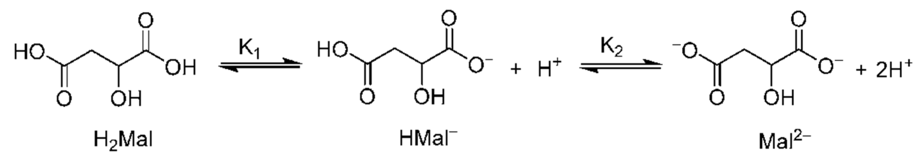
3.2. Reactions of Malic Acid in Water

The following chemical reaction is valid for malic acid in water:
C4H6O5 + H2O → C4H5O5 + H3O+
As a result of the reaction, there are malate ions (C4H5O5) and hydronium ions (H3O+).
Malic acid is a dibasic acid, and a second reaction in water is also valid:
C4H5O5 + H2O → C4H4O52− + H3O+
The malate ion participates in the Krebs cycle, redox reactions, and electron transfer for energy transfer [40].
The protons from the hydronium ions (H3O+) move from one water molecule to another by the Grotthuss shutting process. The clusters have a positive charge [47,48]. Water clusters with different numbers of water molecules and a hydronium ion are indicated by [49].
The proton (H+) from the hydronium ion (H3O+) is attached to biomolecules such as proteins and enzymes. Bioreactions of malic acid are shown in Figure 2.
Proton migration between the clusters [50] is related to electric conductivity. The malate ions donate electrons with antioxidant activity and participate in reactions where electrons bind with free radicals.

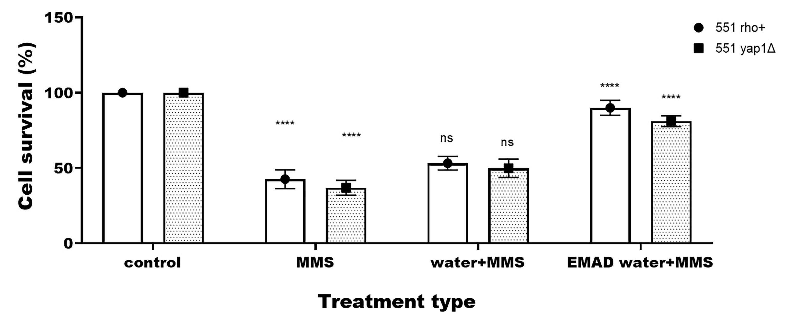
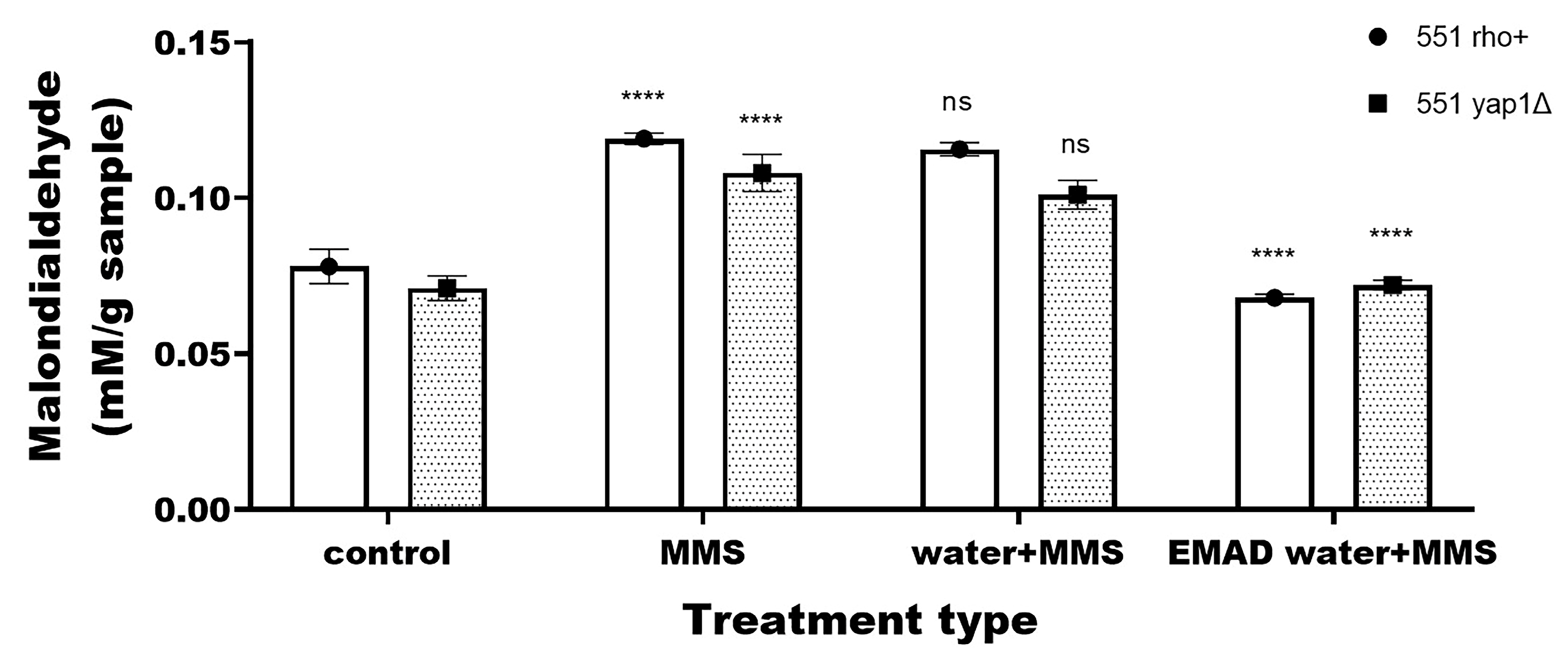
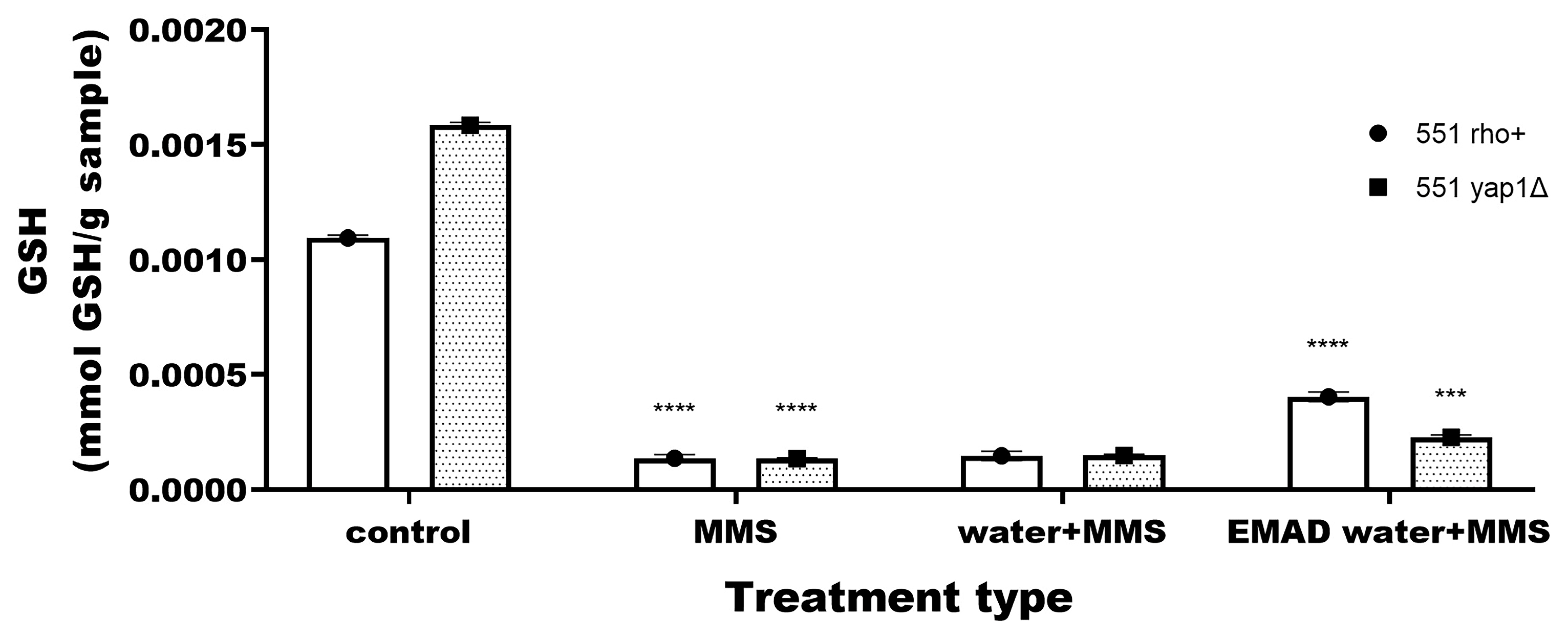
3.3. Yeast-Cell-Based Analyses

Preliminary evaluation of the effect of single EMAD water treatment
As a first step, the cell survival, levels of superoxide anions, MDA, and GSH were evaluated (Table 3). Our preliminary data (Table 3) revealed that the studied effects do not differ among treatments with descaled water, non-descaled water, and untreated cells. No statistically significant difference was observed.
Based on this, further experiments were compared to the control—untreated cells.
The first endpoint analyzed was the cell survival of both strains (Figure 3). A lack of positive effect was obtained for both strains when treatment was performed with drinking water and MMS. Such a result is expected as the water itself does not possess antigenotoxic properties.
Further, combined treatment with EMAD water and MMS resulted in around 2-fold higher cell survival compared to the single MMS treatment. The effect was similar for both strains regardless of the presence/absence of the strain’s antioxidant defense system.
Based on this, it could be suggested that the antigenotoxic effect obtained is probably due to the presence of malate ions in the water.
Previous research with malic acid descaler (EMAD) presented beneficial properties based on in vitro results [51]. The study demonstrated that tap water treated with a malic acid granulated filter significantly improves intestinal epithelial cell regeneration and barrier integrity and reduces inflammation compared to untreated tap water. The treated water solution reduced oxidative stress and endogenous superoxide radical formation by neutrophils more effectively than untreated water.
Based on this observation, in the present study, we employed another approach in order to gain more detailed information concerning the antioxidant potential in superoxide anions. The levels of physiologically active superoxide anions in live cells were measured (Figure 4). Data revealed that EMAD water results in a 3.5-fold decrease in the MMS-induced superoxide anions. No statistically significant difference was calculated between the strains, suggesting that the presence/absence of the antioxidant defense system in the strain does not play a role.
Next, the levels of lipid peroxidation were measured. Our data revealed that MMS induces around 1.5-fold malondialdehyde (MDA) in yeast cells (Figure 5).
Adding EMAD water to MMS reduced the MDA levels to values comparable to control, untreated cells (Figure 6). Again, a positive effect of EMAD was obtained regardless of the presence/absence of the antioxidant defense system of the strain. The well-expressed reduction in MDA levels (p < 0.05) suggests very pronounced antioxidant activity in lipid peroxidation. This observation follows the beneficial effect of EMAD on the intestinal epithelial barrier integrity [51].
Finally, our research focused on EMAD water’s effect on the antioxidant molecule glutathione. Genotype differences were obtained concerning this biomarker.
The total glutathione (GSH) quantity in the impaired strain was higher than that measured in the strain with a regular antioxidant system (Figure 6). Treatment with MMS resulted in an 8-fold decrease in GSH levels for strain 551 and more than 11-fold for 551yap1Δ, suggesting a more detrimental effect on the impaired strain. Further, when EMAD was added to the solution, the GSH levels in the impaired strain increased to some extent (1.7-fold) but less than the normal strain (3-fold). Around a 1.8-fold difference in the levels of GSH was measured between the strains in favor of the strain with a normal antioxidant system.
Such an observation is not surprising, as it is already known that not only YAP1 but also its mammalian functional homolog AP-1 controls the expression of antioxidant enzymes, such as glutathione-S-transferase, glutathione reductase, thioredoxin, and thioredoxin reductase [52]. Data concerning the significant reduction in the physiologically active superoxide anions and lipid peroxidation regardless of the strain specificity are presented. This is the first report to our knowledge that water treated with malic acid descaler can reduce MMS-induced superoxide anions. That observation is essential since these anions correlate with MMS’s carcinogenic potential [53,54].
Nevertheless, to determine at least partially the mode of action of the L-malic acid descaler, two isogenic strains were used: 551 rho+ and 551yap1Δ. It was expected that strain 551yap1Δ’s response to MMS treatment combined with EMAD water would differ. Surprisingly, cell survival, physiologically active superoxide anions, and MDA were similar for both strains. The only difference observed was in the total glutathione. Based on this, it could be suggested that the main mechanism of action of L-malic acid ions may be related to direct action on the mutagen, regardless of the presence of antioxidant defense systems. The contribution of L-malic acid to the antioxidant enzymatic and non-enzymatic systems should also be mentioned as the glutathione levels differed significantly in both strains.

4. Conclusions

The water treated with L-malic acid descaler complies with the European Union’s Directive No. 2020/2184 on the quality of water intended for human consumption, ensuring its safety for potential biological applications.
The present work provides new information concerning the in vivo biological activity of L-malic acid-descaled drinking water. This is the first evidence to our knowledge that EMAD water can reduce the genotoxic and pro-oxidant potential of the standard mutagen methyl methanesulfonate either by direct action on the mutagen or via alternative metabolic or redox-modulating mechanisms. The partial contribution to the activation of some of the antioxidant defense systems should not be excluded.
This study provides novel in vivo evidence that water treated with L-malic acid descaler can mitigate oxidative stress markers at the cellular level. These findings highlight its dual role as an effective water treatment agent and a biologically active compound with antioxidant potential.

Author Contributions

Conceptualization, F.H. and Y.G.M.; methodology, T.T., K.B. and I.I.; software, A.I.I.; validation, Z.D. and I.G.; formal analysis, I.I.; investigation, F.H.; resources, T.T.; data curation, I.I.; writing—original draft preparation, T.T.; writing—review and editing, I.I. and T.P.P.; visualization, F.H.; supervision, A.I.I.; project administration, A.I.I.; funding acquisition, I.I. All authors have read and agreed to the published version of the manuscript.

Funding

This research received no external funding.

Institutional Review Board Statement

Not applicable.

Informed Consent Statement

Not applicable.

Data Availability Statement

The original contributions presented in this study are included in the article. Further inquiries can be directed to the corresponding author.

Conflicts of Interest

Co-author Fabio Huether has a patent registration with number PCT/EP2023/069837. The remaining authors declare that the research was conducted in the absence of any commercial or financial relationships that could be construed as potential conflicts of interest.

References

  1. El-Nahas, S.; Khodari, M.; Hamam, A.A.; Gad El Rab, A.N.; Toghan, A. Descaling of Evaporator Tubes in Sugarcane Factories Using Molasses as a Green and Effective Technology. Sugar Tech. 2024, 26, 1157–1170. [Google Scholar] [CrossRef]
  2. Nayunigari, M.K.; Maity, A.; Agarwal, S.; Vinod Kumar Gupta, V.K. Curcumin-malic acid based green copolymers for control of scale and microbiological growth applications in industrial cooling water treatment. J. Mol. Liq. 2016, 214, 400–410. [Google Scholar] [CrossRef]
  3. Chi, Z.; Wang, Z.P.; Wang, G.Y.; Khan, I.; Chi, Z.M. Microbial biosynthesis and secretion of l-malic acid and its applications. Spectrochim. Acta A Mol. Biomol. Spectrosc. 2016, 36, 99–107. [Google Scholar] [CrossRef] [PubMed]
  4. Wu, N.; Wu, X.; Zhang, M.; Zhang, C.; Xu, Q. Metabolic engineering of Aspergillus niger for accelerated malic acid biosynthesis by improving NADPH availability. Biotechnol. J. 2024, 19, 2400014. [Google Scholar] [CrossRef]
  5. Halevas, E.; Karamelidou, A.; Hatzidimitriou, A.; Mateescu, C.; Salifoglou, A. pH-specific halide-dependent materials from ZrIV/hydroxycarboxylic acid/aromatic chelator reactivity: Architecture–lattice dimensionality and spectroscopic fingerprint relations. Eur. J. Inorg. Chem. 2015, 2015, 664–679. [Google Scholar] [CrossRef]
  6. Ma, B.; Yuan, Y.; Gao, M.; Li, C.; Ogutu, C.; Li, M.; Ma, F. Determination of Predominant Organic Acid Components in Malus Species: Correlation with Apple Domestication. Metabolites 2018, 8, 74. [Google Scholar] [CrossRef]
  7. Kovilein, A.; Kubisch, C.; Cai, L.; Ochsenreither, K. Malic acid production from renewables: A review. J. Chem. Technol. Biotechnol. 2020, 95, 513–526. [Google Scholar] [CrossRef]
  8. EFSA Panel on Additives and Products or Substances used in Animal Feed (FEEDAP). Scientific Opinion on the safety and efficacy of malic acid and a mixture of sodium and calcium malate when used as technological additives for all animal species. EFSA Panel on Additive and Products or Substances Used in Animal Feed. EFSA J. 2014, 12, 3563. [Google Scholar]
  9. Qiu, K.; He, W.; Zhang, H.; Wang, J.; Qi, G.; Guo, N.; Zhang, X.; Wu, S. Bio-Fermented Malic Acid Facilitates the Production of High-Quality Chicken via Enhancing Muscle Antioxidant Capacity of Broilers. Antioxidants 2022, 11, 2309. [Google Scholar] [CrossRef]
  10. Zhang, L.; Zhang, P.; Xia, C.; Cheng, Y.; Guo, X.; Li, Y. Effects of malic acid and citric acid on growth performance, antioxidant capacity, haematology, and immune response of Carassius auratus gibelio. Aquac. Res. 2020, 51, 2766–2776. [Google Scholar] [CrossRef]
  11. Yousefi, M.; Ghafarifarsani, H.; Raissy, M.; Yilmaz, S.; Vatnikov, Y.A.; Kulikov, E.V. Effects of dietary malic acid supplementation on growth performance, antioxidant and immunological parameters, and intestinal gene expressions in rainbow trout, Oncorhynchus mykiss. Aquaculture 2023, 563, 738864. [Google Scholar] [CrossRef]
  12. Chen, M.; Zhao, Y.; Li, S.; Chang, Z.; Liu, H.; Zhang, D.; Wang, S.; Zhang, X.; Wang, J. Maternal malic acid may ameliorate oxidative stress and inflammation in sows through modulating gut microbiota and host metabolic profiles during late pregnancy. Antioxidants 2024, 13, 253. [Google Scholar] [CrossRef] [PubMed]
  13. Li, N.; Yan, M.; Guo, R.; Liu, Z. The research progress of water treatment technology on recirculated cooling water. IOP Conf. Ser. Earth Environ. Sci. 2020, 508, 012041. [Google Scholar] [CrossRef]
  14. Cabansis, S.E.; Leenheer, J.A.; McVey, I.F. Aqueous infrared carboxylate absorbances: Aliphatic di-acids. Spectrochim. Acta A Mol Biomol. Spectrosc. 1998, 54, 449–458. [Google Scholar] [CrossRef]
  15. Mosin, O.V.; Shvets, V.I.; Skladnev, D.A.; Ignatov, I. Microbial synthesis of 2H-labelled L-phenylalanine with different levels in isotopic enrichment by a facultative methylotrophic Brevibacterium Methylicum with RuMP assimilation of Carbon. Biochem. Suppl. B Biomed. Chem. 2013, 7, 247–258. [Google Scholar] [CrossRef]
  16. Le Tran, H.; Van, L.T.M.; Vuu, M.N.; Mai, P.T. Separation performance of polyvinyl alcohol) based nanofiltration membranes crosslinked by malic acid for salt solutions. Sci. Technol. Dev. J. 2016, 19, 70–78. [Google Scholar] [CrossRef]
  17. Telegdi, J.; Trif, L.; Mihaly, J.; Nagy, E.; Nyikos, L. Controlled synthesis and characterization of biodegradable, stereomer co-polycondensates of l-malic acid. J. Therm. Anal. Calorim. 2015, 121, 663–673. [Google Scholar] [CrossRef]
  18. Eck, M.; Schwab, S.T.; Nelson, T.F.; Wurst, K.; Iberl, S.; Schleheck, D.; Link, C.; Battagliarin, G.; Mecking, S. Biodegradable high-density polyethylene-like material. Angew. Chem. 2022, 135, e202213438. [Google Scholar]
  19. Directive (EU) 2020/2184 of the European Parliament and of the Council of 16 December 2020 on the Quality of Water Intended for Human Consumption. Available online: https://www.legislation.gov.uk/eudr/2020/2184 (accessed on 23 February 2025).
  20. Todorova, T.; Pesheva, M.; Gregan, F.; Chankova, S. Antioxidant, antimutagenic, and anticarcinogenic effects of Papaver rhoeas L. extract on Saccharomyces cerevisiae. J. Med. Food 2015, 18, 460–467. [Google Scholar] [CrossRef]
  21. Ikner, A.; Shiozaki, K. Yeast signaling pathways in the oxidative stress response. Mutat. Res. 2005, 569, 13–27. [Google Scholar] [CrossRef]
  22. Temple, M.D.; Perrone, G.G.; Dawes, I.W. Complex cellular responses to reactive oxygen species. Trends Cell Biol. 2005, 15, 319–326. [Google Scholar] [CrossRef] [PubMed]
  23. Todorova, T.; Boyadzhiev, K.; Dimitrov, M.; Parvanova, P. Bee venom genotoxicity on Saccharomyces cerevisiae cells–The role of mitochondria and YAP1 transcription factor. Toxicology 2024, 503, 153768. [Google Scholar] [CrossRef] [PubMed]
  24. Daskalova, A.V.; Tomova, A.A.; Kujumdzieva, A.V.; Velkova, L.G.; Dolashka, P.A.; Petrova, V.Y. Menadione and hydrogen peroxide trigger specific alterations in RNA polymerases profiles in quiescent Saccharomyces cerevisiae cells. Biotechnol. Biotechnol. Equip. 2021, 35, 1190–1199. [Google Scholar] [CrossRef]
  25. Shirvanyan, A.; Trchounian, K. Sodium transport and redox regulation in Saccharomyces cerevisiae under osmotic stress depending on oxygen availability. Sci. Rep. 2024, 14, 23982. [Google Scholar] [CrossRef]
  26. Chen, X.; Li, C.; Liu, H. Enhanced recombinant protein production under special environmental stress. Front. Microbiol. 2021, 12, 630814. [Google Scholar] [CrossRef]
  27. Zbieralski, K.; Staszewski, J.; Konczak, J.; Lazarewicz, N.; Nowicka-Kazmierczak, M.; Wawrzycka, D.; Maciaszczyk-Dziubinska, E. Multilevel regulation of membrane proteins in response to metal and metalloid stress: A Lesson from Yeast. Int. J. Mol. Sci. 2024, 25, 4450. [Google Scholar] [CrossRef]
  28. Stephen, D.W.; Rivers, S.L.; Jamieson, D.J. The role of the YAP1 and YAP2 genes in the regulation of the adaptive oxidative stress responses of Saccharomyces cerevisiae. Mol. Microbiol. 1995, 16, 415–423. [Google Scholar] [CrossRef]
  29. Aboughaly, M.; Babaei-Ghazvini, A.; Dhar, P.; Patel, R.; Acharya, B. Enhancing the potential of polymer composites using biochar as a filler: A Review. Polymers 2023, 15, 3981. [Google Scholar] [CrossRef]
  30. Jamaković, N.; Karahmet, E.; Varešić, B. Assessment of Activated Charcoal Efficiency with Filter Paper in the Water Purification Process. In New Technologies, Development and Application II. NT 2019. Lecture Notes in Networks and Systems; Karabegović, I., Ed.; Springer: Cham, Switzerland, 2020; Volume 76. [Google Scholar]
  31. Ignatov, I. Review of different types of mountain springs and mineral waters from Bulgaria based on their natural origin and health benefits. Med. Perspekt. 2023, 51, 199–206. [Google Scholar] [CrossRef]
  32. Gamal, H.; Elkatatny, S.; Al-Afnan, S.; Bahdat, M. Development of a unique organic acid solution for removing composite field Scales. ACS Omega 2021, 6, 1205–1215, Erratum in Sustain. Chem. Pharm. 2023, 33, 101131. [Google Scholar] [CrossRef]
  33. Enchev, P.; Zarev, Y.; Dakovska, A.; Todorova, T.; Dimitrov, M.; Rivera-Mondragón, A.; Ionkova, I. Pentacyclic triterpenoids from the leaves of Cecropia longipes. Pharmacia 2024, 71, 1–8. [Google Scholar] [CrossRef]
  34. Pesheva, M.; Krastanova, O.; Staleva, L.; Dentcheva, V.; Hadzhitodorov, M.; Venkov, P. The Ty1 transposition assay: A new short-term test for detection of carcinogens. J. Microbiol. Methods 2005, 61, 1–8. [Google Scholar] [CrossRef] [PubMed]
  35. Pesheva, M.; Krastanova, O.; Stamenova, R.; Todorova, I.; Shoevska, R.; Venkov, P.; Tsvetkov, T. The Ty1 transposition assay: A short–term test for selective detection of carcinogenic pollutants in environmental samples. Bulg. J. Agric. Sci. 2008, 14, 271–282. [Google Scholar]
  36. Stoycheva, T.; Pesheva, M.; Venkov, P. The role of reactive oxygen species in the induction of Ty1 retrotransposition in Saccharomyces cerevisiae. Yeast 2010, 27, 259–267. [Google Scholar] [CrossRef]
  37. Stamenova, R.; Dimitrov, M.; Stoycheva, T.; Pesheva, M.; Venkov, P.; Tsvetkov, T.S. Transposition of Saccharomyces cerevisiae Ty1 retrotransposon is activated by improper cryopreservation. Cryobiology 2008, 56, 241–247. [Google Scholar] [CrossRef]
  38. Todorova, T.; Pesheva, M.; Stamenova, R.; Dimitrov, M.; Venkov, P. Mutagenic effect of freezing on nuclear DNA of Saccharomyces cerevisiae. Yeast 2012, 29, 191–199. [Google Scholar] [CrossRef]
  39. Steels, E.L.; Learmonth, R.P.; Watson, K. Stress tolerance and membrane lipid unsaturation in Saccharomyces cerevisiae grown aerobically or anaerobically. Microbiology 1994, 140, 569–576. [Google Scholar] [CrossRef]
  40. Jamnik, P.; Medved, P.; Raspor, P. Increased glutathione content in yeast Saccharomyces cerevisiae exposed to NaCl. Ann. Microbiol. 2006, 56, 175–178. [Google Scholar] [CrossRef]
  41. Dao, O.; Kuhnert, F.; Weber, A.P.N.; Peltier, G.; Li-Besson, Y. Physiological functions in malate shuttles in plants and algae. Trends Plant Sci. 2022, 27, 488–504. [Google Scholar] [CrossRef]
  42. Zheng, Q.; Zhu, H.; Lv, C.; Zhu, Z.; Cui, H.; Fan, Z.; Sun, J.; Huang, Z.; Shi, P. Clioquinol rescues yeast cells from Aβ42 toxicity via the inhibition of oxidative damage. Biotechnol. J. 2024, 19, 2300662. [Google Scholar] [CrossRef]
  43. Smith, B.C. Fundamentals of Fourier Transform Infrared Spectroscopy, 2nd ed.; CRC Press, Taylor & Francis Group, LLC: New York, NY, USA, 2011; ISBN 978-1-4200-6930-3. [Google Scholar]
  44. Charlton, N.C.; Mastyugin, M.; Török, B.; Török, M. Structural Features of Small Molecule Antioxidants and Strategic Modifications to Improve Potential Bioactivity. Molecules 2023, 28, 1057. [Google Scholar] [CrossRef] [PubMed]
  45. Max, J.-J.; Chapados, C. Infrared scpctroscopy of aquous carboxylic acids: Malic acid. J. Phys. Chem. A 2002, 27, 6452–6461. [Google Scholar] [CrossRef]
  46. Rochette, L.; Zeller, M.; Cottin, Y.; Vergely, C. Antitumor activity of protons and molecular hydrogen: Underlying mechanisms. Cancers 2021, 13, 893. [Google Scholar] [CrossRef]
  47. Igamberdiev, A.V.; Bykova, N.V. Role of organic acids in the integration of cellular redox metabolism and mediation of redox signalling in photosynthetic tissues of higher plants. Free Radic. Biol. Med. 2018, 122, 74–85. [Google Scholar] [CrossRef]
  48. Benjamin, I. Hydronium ion transport across the liquid/liquid interface assisted by a phase-transfer catalyst: Structure and thermodynamics using molecular dynamics simulation. J. Phys. Chem. B 2024, 128, 9613–9618. [Google Scholar] [CrossRef]
  49. Vassilev, N.; Ignatov, I.; Popova, T.P.; Huether, F.; Ignatov, A.I.; Iliev, M.T.; Marinov, Y. Nuclear Magnetic Resonance (NMR) and Density Functional Theory (DFT) Study of Water Clusters of Hydrogen-Rich Water (HRW). Water 2024, 16, 3261. [Google Scholar] [CrossRef]
  50. Kolasinski, K.W.; Salkowski, A.N. Density functional theory (DFT) study of water autoionization in solvated clusters. J. Chem. Phys. 2024, 161, 1143. [Google Scholar] [CrossRef]
  51. Dartsch, P.C.; Huether, F. Beneficial health effects of malic acid water treatment on cultured intestinal epithelial cells and functional neutrophils. Appl. Cell. Biol. 2024, 12, 18–22. [Google Scholar] [CrossRef]
  52. Krysan, K.; Lou, M.F. Regulation of human thioltransferase (hTTase) gene by AP-1 transcription factor under oxidative stress. Investig. Ophthalmol. Vis. Sci. 2002, 43, 1876–1883. [Google Scholar]
  53. Stoycheva, T.; Pesheva, M.; Dimitrov, M.; Venkov, P. The Ty1 Retrotransposition Short-Term Test for Selective Detection of Carcinogenic Genotoxins; InTech: Rijeka, Croatia, 2012; pp. 83–610. [Google Scholar] [CrossRef]
  54. Stoycheva, T. Methylmethane sulfonate increases the level of superoxide anions in yeast cells. Biotechnol. Biotechnol. Equip. 2009, 23, 688–692. [Google Scholar] [CrossRef]
Figure 1. Fourier transform infrared spectroscopy of malic acid.
Beverages 11 00062 g001
Figure 2. Bioreactions of malic acid.
Beverages 11 00062 g002
Figure 3. Cell survival of Saccharomyces cerevisiae strains 551ρ+ (●) and 551 yap1Δ (■) after single treatment with 16 mM MMS as well as the combined treatments with EMAD water and MMS, and drinking water and MMS. Results are presented as mean ± SEM from at least three independent experiments. Where no error bars are evident, errors were equal to or smaller than the symbols (ns p > 0.05; **** p < 0.0001).
Beverages 11 00062 g003
Figure 4. Levels of superoxide anions measured in Saccharomyces cerevisiae strains 551ρ+ (●) and 551 yap1Δ (■) after a single treatment with 16 mM MMS as well as the combined treatments with EMAD water and MMS, and drinking water and MMS. Results are presented as mean ± SEM from at least three independent experiments. Where no error bars are evident, errors were equal to or smaller than the symbols (ns p ≥ 0.05; **** p < 0.0001).
Beverages 11 00062 g004
Figure 5. Levels of malondialdehyde (MDA) measured in Saccharomyces cerevisiae strains 551ρ+ (●) and 551 yap1Δ (■) after a single treatment with 16 mM MMS as well as the combined treatments with EMAD water and MMS, and drinking water and MMS. Where no error bars are evident, errors were equal to or smaller than the symbols (ns—p ≥ 0.05; **** p < 0.0001).
Beverages 11 00062 g005
Figure 6. Levels of total glutathione (GSH) measured in Saccharomyces cerevisiae strains 551ρ+ (●) and 551 yap1Δ (■) after single treatment with 16 mM MMS as well as combined treatments with EMAD water and MMS, and drinking water and MMS. Where no error bars are evident, errors were equal to or smaller than the symbols (*** p < 0.001; **** p < 0.0001).
Beverages 11 00062 g006
Table 1. Chemical composition of L-malic acid descaler.
Composition of L-Malic Acid Descaler
CAS: 617-48-1
EINECS: 210-514-9
L-malic acid45%
CAS: 9002-88-4Polyethylene30%
CAS: 64365-11-3
EINECS: 264-846-4
Charcoal, activated25%
CAS—Chemical Abstracts Service Number; EINECS—European List of Notified Chemical Substances.
Table 2. L-malic acid descaler absorption bands tested by FT-IR method.
Absorption Bands
Wave Number
(cm−1)
Wavelength
(µm)
Type
36012.78medium
34322.91medium
29523.39weak
29193.43medium
28493.51weak
17925.58weak
17145.83strong
15316.53weak
13977.16medium
13627.34medium
12897.76weak
12587.95weak
12178.22weak
11668.58medium
10589.45weak
10349.67weak
71913.91weak
64015.63weak
Table 3. Comparative analysis of the endpoints—cell survival, levels of superoxide anions, malondialdehyde (MDA), and total glutathione (GSH) among untreated cells and cells treated either with drinking water or EMAD water.
EndpointType of Treatment551 rho+ 1551 yap1Δ
Cell survival (%)Untreated100100
Treated with water101 ± 2.65 ns99.67 ± 1.27 ns
Treated with EMAD water98.24 ± 3.29 ns97.23 ± 4.21 ns
Superoxide anionsUntreated0.53 ± 0.060.82 ± 0.07
Treated with water0.57 ± 0.09 ns0.85 ± 0.1 ns
Treated with EMAD water0.52 ± 0.05 ns0.80 ± 0.06 ns
MDAUntreated0.08 ± 0.0050.07 ± 0.004
Treated with water0.1 ± 0.002 ns0.09 ± 0.005 ns
Treated with EMAD water0.06 ± 0.005 ns0.06 ± 0.006 ns
GSHUntreated0.0011 ± 0.000010.0016 ± 0.00001
Treated with water0.0013 ± 0.00001 ns0.0015 ± 0.00001 ns
Treated with EMAD water0.0011 ± 0.00001 ns0.0014 ± 0.00001 ns
1 Results are presented as mean ± SEM from at least three independent experiments (ns—non-significant statistical difference). Untreated: Cells that were not subjected to any treatment served as control group. Treated with water: Cells were treated with drinking water under the same experimental conditions. Treated with EMAD water to asses with potential biological effects.
Disclaimer/Publisher’s Note: The statements, opinions and data contained in all publications are solely those of the individual author(s) and contributor(s) and not of MDPI and/or the editor(s). MDPI and/or the editor(s) disclaim responsibility for any injury to people or property resulting from any ideas, methods, instructions or products referred to in the content.

