
Physiological Ventricular Simulator for Valve Surgery Training


 ,
, 
Abstract
1. Introduction
2. Materials and Methods
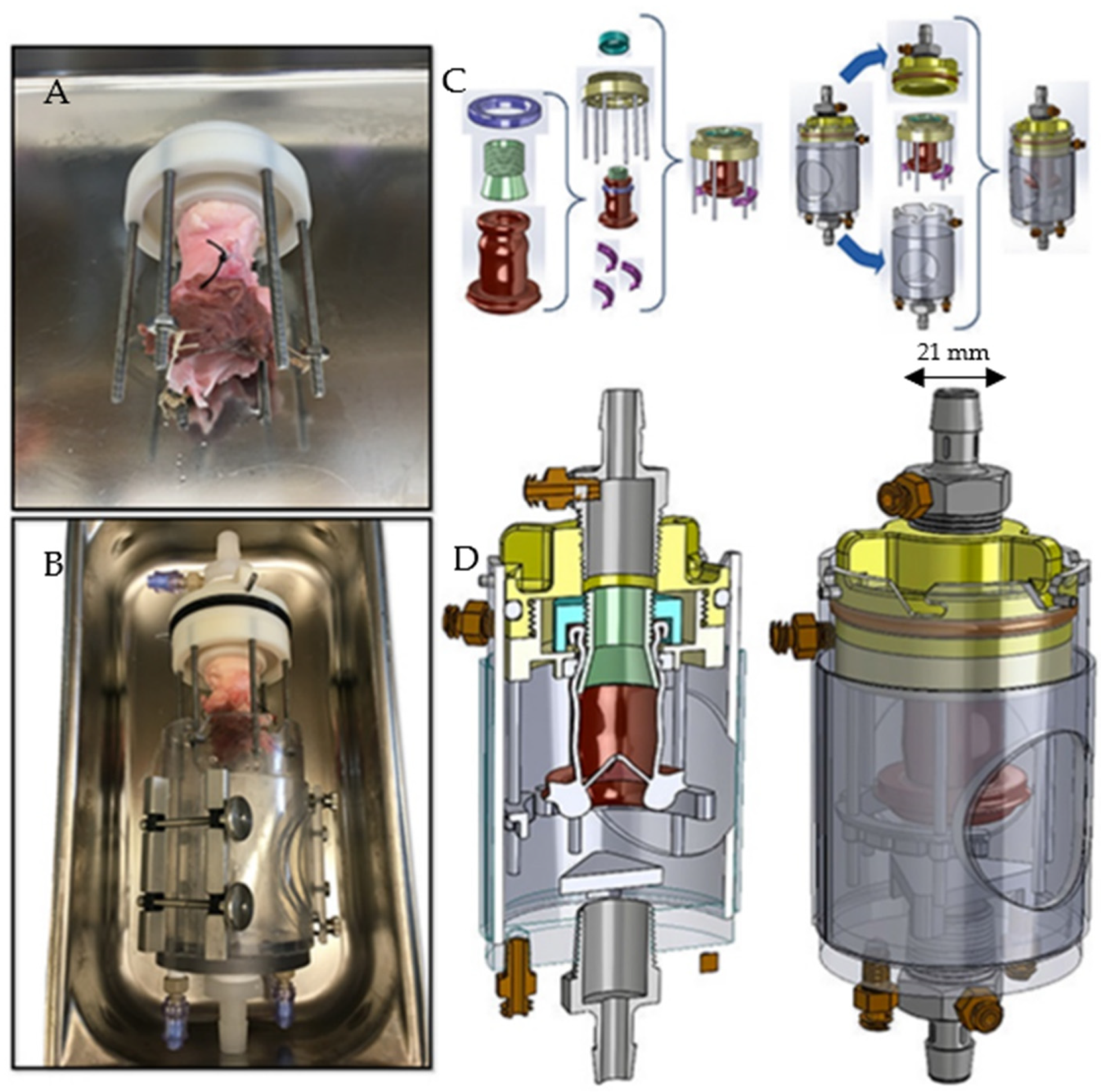
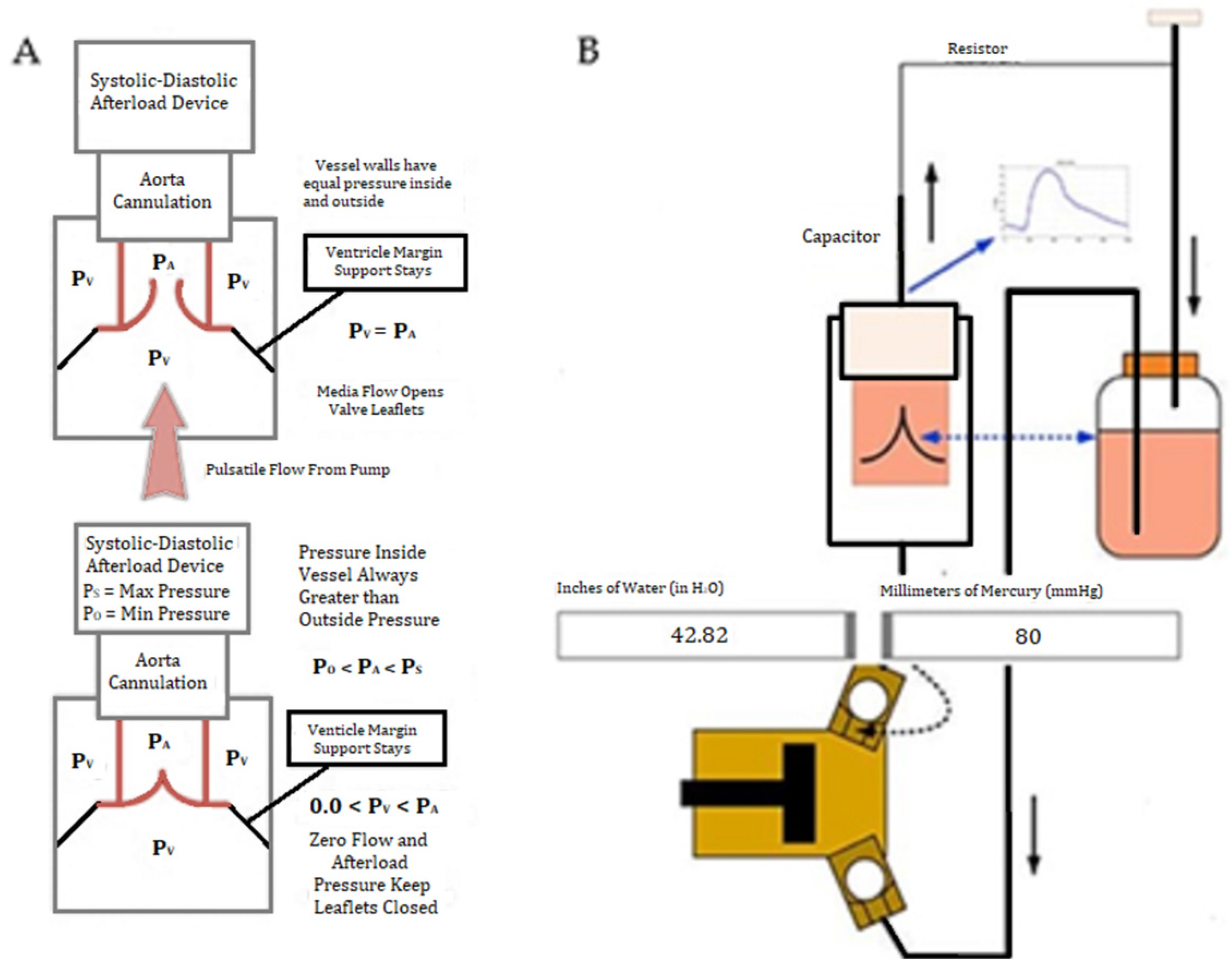
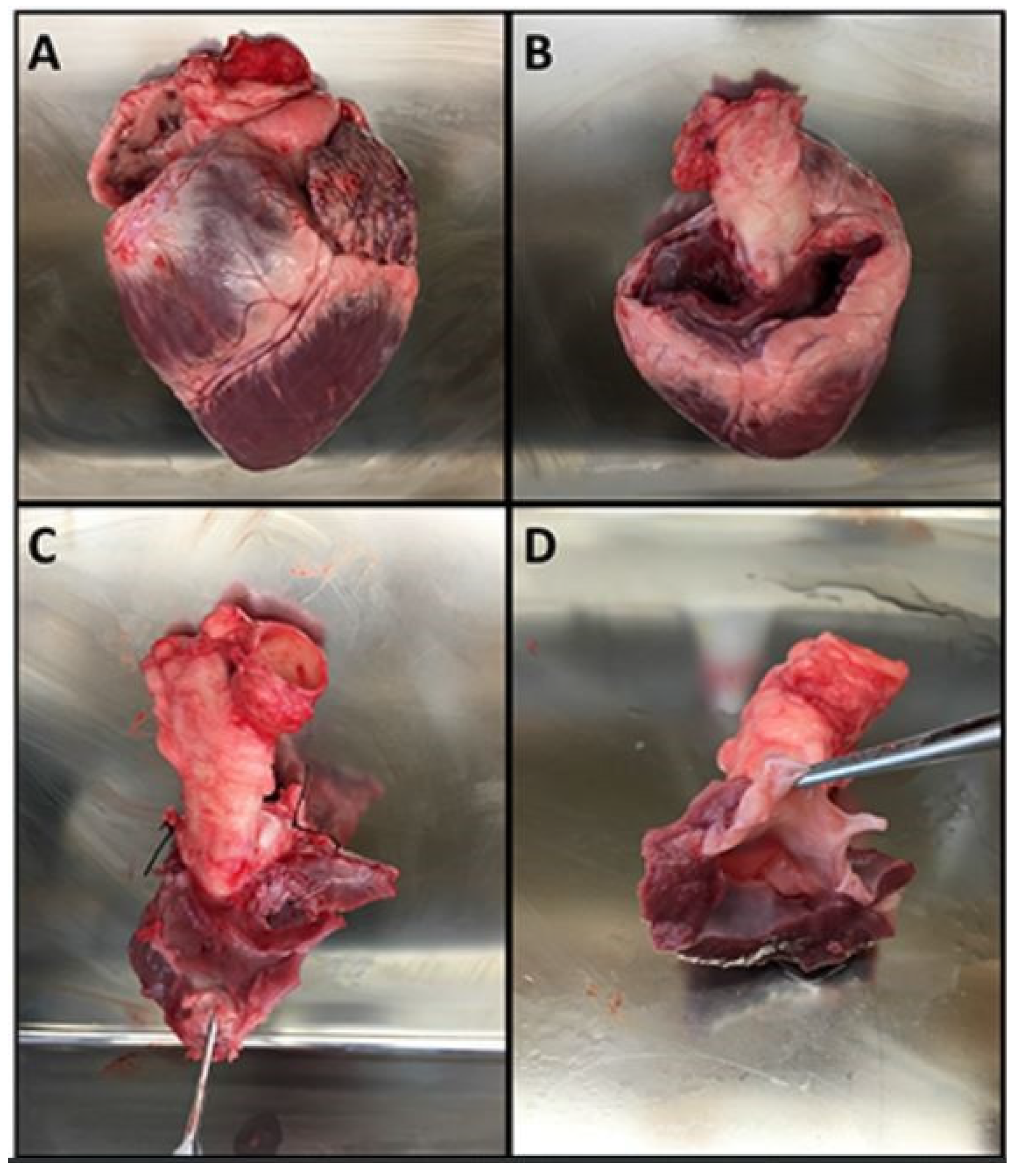
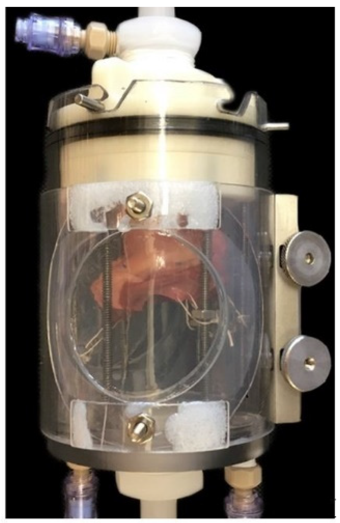
2.1. Part 1
2.2. Part 2
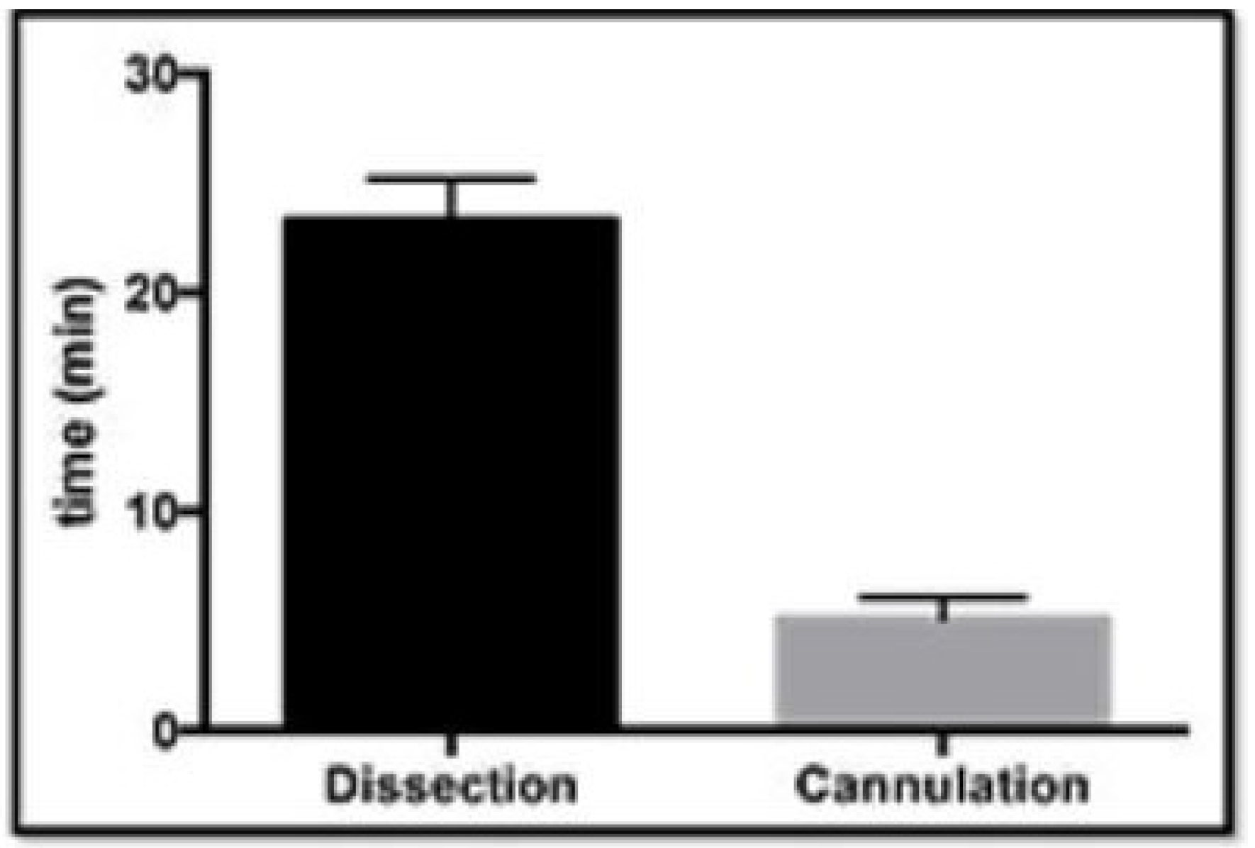
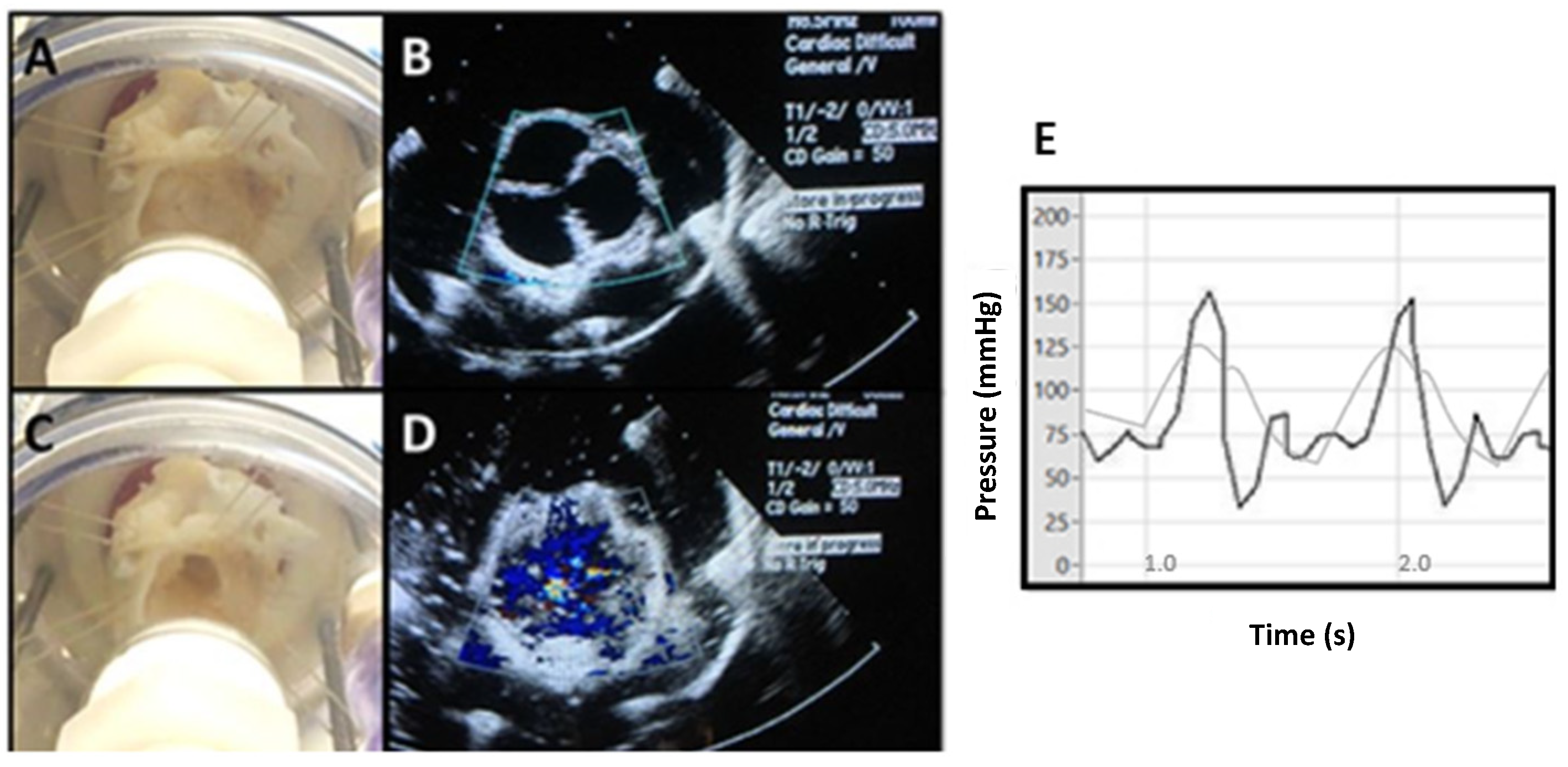
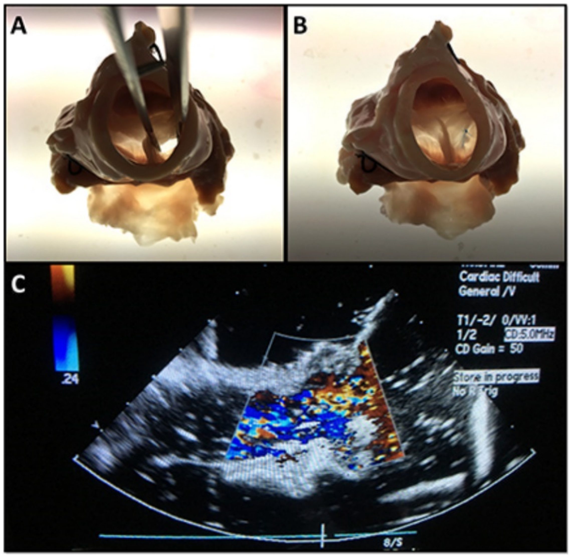
3. Results
4. Discussion
Author Contributions
Funding
Institutional Review Board Statement
Informed Consent Statement
Data Availability Statement
Conflicts of Interest
References
- Fann, J.I.; Calhoon, J.H.; Carpenter, A.J.; Merrill, W.H.; Brown, J.W.; Poston, R.S.; Kalani, M.; Murray, G.F.; Hicks, G.L., Jr.; Feins, R.H. Simulation in coronary artery anastomosis early in cardiothoracic surgical residency training: The Boot Camp experience. J. Thorac. Cardiovasc. Surg. 2010, 139, 1275–1281. [Google Scholar] [CrossRef] [PubMed]
- Fann, J.I.; Feins, R.H.; Hicks, G.L.; Nesbitt, J.C.; Hammon, J.W.; Crawford, F.A., Jr. Evaluation of simulation training in cardiothoracic surgery: The Senior Tour perspective. J. Thorac. Cardiovasc. Surg. 2012, 143, 264–272. [Google Scholar] [CrossRef]
- Price, J.; Naik, V.; Boodhwani, M.; Brandys, T.; Hendry, P.; Lam, B.K. A randomized evaluation of simulation training on performance of vascular anastomosis on a high-fidelity in vivo model: The role of deliberate practice. J. Thorac. Cardiovasc. Surg. 2011, 142, 496–503. [Google Scholar] [CrossRef] [PubMed]
- Feins, R.H.; Burkhart, H.M.; Conte, J.V.; Coore, D.N.; Fann, J.I.; Hicks, G.L., Jr.; Nesbitt, J.C.; Ramphal, P.S.; Schiro, S.E.; Shen, K.R.; et al. Simulation-based training in cardiac surgery. Ann. Thorac. Surg. 2017, 103, 312–321. [Google Scholar] [CrossRef] [PubMed]
- Fann, J.I.; Caffarelli, A.D.; Georgette, G.; Howard, S.K.; Gaba, D.M.; Youngblood, P.; Mitchell, R.S.; Burdon, T.A. Improvement in coronary anastomosis with cardiac surgery simulation. J. Thorac. Cardiovasc. Surg. 2008, 136, 1486–1491. [Google Scholar] [CrossRef]
- Wang, H.; Song, H.; Yang, Y.; Cao, Q.; Hu, Y.; Chen, J.; Guo, J.; Wang, Y.; Jia, D.; Cao, S.; et al. Three-dimensional printing for cardiovascular diseases: From anatomical modeling to dynamic functionality. BioMedical Eng. Online 2020, 19, 76. [Google Scholar] [CrossRef]
- Olin, C. Pulsatile flow Studies of Prosthetic Aortic Valves. Scand. J. Thorac. Cardiovasc. Surg. 2009, 5, 1–12. [Google Scholar] [CrossRef]
- Harvard Apparatus. Series 1400 Pulsatile Blood Pumps User’s Manual; Harvard Apparatus Inc.: Natick, MA, USA, 2005. [Google Scholar]
- Taylor, C.E.; Dziczkowski, Z.W.; Miller, G.E. Automation of the Harvard Apparatus Pulsatile Blood Pump. ASME J. Med. Devices 2012, 6, 45–55. [Google Scholar] [CrossRef]
- Chikwe, J.; Cooke, D.; Weiss, A. Cardiothoracic Surgery, 2nd ed.; Oxford University Press: Oxford, UK, 2013; p. 26. [Google Scholar]
- Pfeiffer, S.; Sirch, J.; Vogt, F.; Fischlein, T.; Santarpino, G. Implantation of the Sorin Perceval® sutureless aortic valve: A step by step approach. Minerva Cardioangiol. 2017, 65, 184–192. [Google Scholar] [CrossRef]
- Mokadam, N.A.; Fann, J.I.; Hicks, G.L.; Nesbitt, J.C.; Burkhart, H.M.; Conte, J.V.; Coore, D.N.; Ramphal, P.S.; Shen, K.R.; Walker, J.D.; et al. Experience with the Cardiac Surgery Simulation Curriculum: Results of the Resident and Faculty Survey. Ann. Thorac. Surg. 2017, 103, 322–328. [Google Scholar] [CrossRef][Green Version]
- Ribeiro, I.B.; Ngu, J.M.C.; Lam, B.; Edwards, R.A. Simulation-Based Skill Training for Trainees in Cardiac Surgery: A Systematic Review. Ann. Thorac. Surg. 2018, 105, 972–982. [Google Scholar] [CrossRef] [PubMed]
- Loor, G.; Doud, A.; Nguyen, T.C.; Antonoff, M.B.; Morancy, J.D.; Robich, M.P.; Odell, D.D.; Yarboro, L.T.; Vaporciyan, A.A.; Roselli, E. Development and Evaluation of a Three-Dimensional Multistation Cardiovascular Simulator. Ann. Thorac. Surg. 2016, 102, 62–68. [Google Scholar] [CrossRef] [PubMed]
- Wang, X. Bioartificial Organ Manufacturing Technologies. Cell Transplant. 2019, 28, 5–17. [Google Scholar] [CrossRef] [PubMed]
- Johnson, A.; Cupp, G.; Armour, N.; Warren, K.; Stone, C.; Lee, D.; Gilbert, N.; Hammond, C.; Moore, J.; Kang, Y. An Inexpensive Cardiovascular Flow Simulator for Cardiac Catheterization Procedure Using a Pulmonary Artery Catheter. Front. Med. Technol. 2021, 3, 764007. [Google Scholar] [CrossRef]
- Zhu, Y.; Imbrie-Moore, A.M.; Paulsen, M.J.; Priromprintr, B.; Park, M.H.; Wang, H.; Lucian, H.J.; Farry, J.M.; Woo, Y.J. A novel aortic regurgitation model from cusp prolapse with hemodynamic validation using an Ex Vivo left heart simulator. J. Cardiovasc. Transl. Res. 2021, 14, 283–289. [Google Scholar] [CrossRef]
- Sharma, V.J.; Barton, C.; Page, S.; Ganesh, J.S.; Patel, N.; Pirone, F.; Lin, Z.; Kejriwal, N.K.; El Gamel, A.; McCormack, D.J.; et al. Cardiac surgery simulation: A low-cost feasible option in an Australasian setting. ANZ J. Surg. 2021, 91, 2042–2046. [Google Scholar] [CrossRef]
- Valdis, M.; Chu, M.; Schlachta, C.; Kiaii, B. Validation and comparison of novel robotic cardiac surgery simulation-based training programs. Can. J. Cardiol. 2015, 31, S268. [Google Scholar] [CrossRef]
- Ozaki, S.; Kawase, I.; Yamashita, H.; Uchida, S.; Nozawa, Y.; Matsuyama, T.; Takatoh, M.; Hagiwara, S. Aortic valve reconstruction using self-developed aortic valve plasty system in aortic valve disease. Interact. Cardiovasc. Thorac. Surg. 2011, 12, 550–553. [Google Scholar] [CrossRef]
- Ozaki, S.; Kawase, I.; Yamashita, H.; Uchida, S.; Nozawa, Y.; Takatoh, M.; Hagiwara, S. A total of 404 cases of aortic valve reconstruction with glutaraldehyde-treated autologous pericardium. J. Thorac. Cardiovasc. Surg. 2014, 147, 301–306. [Google Scholar] [CrossRef]
- Marathe, S.P.; Chávez, M.; Sleeper, S.A.; Marx, G.R.; Friedman, K.; Feins, E.N.; Nido, P.J.; Baird, C.W. Single-Leaflet Aortic Valve Reconstruction Utilizing the Ozaki Technique in Patients with Congenital Aortic Valve Disease. Semin. Thorac. Cardiovasc. Surg. 2021; in press. [Google Scholar] [CrossRef]
- Baird, C.W.; Marathe, S.P.; Nido, P.J. Aortic valve neo-cuspidation using the Ozaki technique for acquired and congenital disease: Where does this procedure currently stand? Indian J. Thorac. Cardiovasc. Surg. 2020, 36, S113–S122. [Google Scholar] [CrossRef] [PubMed]
- Baird, C.W.; Cooney, B.; Chávez, M.; Sleeper, L.A.; Marx, G.R.; Nido, P.J. Congenital aortic and truncal valve reconstruction using the Ozaki technique: Short-term clinical results. J. Thorac. Cardiovasc. Surg. 2021, 161, 1567–1577. [Google Scholar] [CrossRef] [PubMed]
- Baird, C.W.; Myers, P.O.; Nido, P.J. Aortic Valve Reconstruction in the Young Infants and Children. Semin. Thorac. Cardiovasc. Surg. Pediatric Card. Surg. Annu. 2012, 15, 9–19. [Google Scholar] [CrossRef] [PubMed]


Publisher’s Note: MDPI stays neutral with regard to jurisdictional claims in published maps and institutional affiliations. |
© 2022 by the authors. Licensee MDPI, Basel, Switzerland. This article is an open access article distributed under the terms and conditions of the Creative Commons Attribution (CC BY) license (https://creativecommons.org/licenses/by/4.0/).
Share and Cite
Zilinskas, K.; Kwon, J.H.; Bishara, K.; Hayden, K.; Quintao, R.; Rajab, T.K. Physiological Ventricular Simulator for Valve Surgery Training. Bioengineering 2022, 9, 264. https://doi.org/10.3390/bioengineering9060264
Zilinskas K, Kwon JH, Bishara K, Hayden K, Quintao R, Rajab TK. Physiological Ventricular Simulator for Valve Surgery Training. Bioengineering. 2022; 9(6):264. https://doi.org/10.3390/bioengineering9060264
Chicago/Turabian StyleZilinskas, Kasparas, Jennie H. Kwon, Katherine Bishara, Kaila Hayden, Ritchelli Quintao, and Taufiek Konrad Rajab. 2022. "Physiological Ventricular Simulator for Valve Surgery Training" Bioengineering 9, no. 6: 264. https://doi.org/10.3390/bioengineering9060264
APA StyleZilinskas, K., Kwon, J. H., Bishara, K., Hayden, K., Quintao, R., & Rajab, T. K. (2022). Physiological Ventricular Simulator for Valve Surgery Training. Bioengineering, 9(6), 264. https://doi.org/10.3390/bioengineering9060264