
What Is the Most Effective Technique for Bonding Brackets on Ceramic—A Systematic Review and Meta-Analysis

 ,
,  ,
,  , ,
, ,  , ,
, ,  and
and 
Abstract
1. Introduction
2. Materials and Methods
Statistical Analysis
3. Results
3.1. Risk of Bias
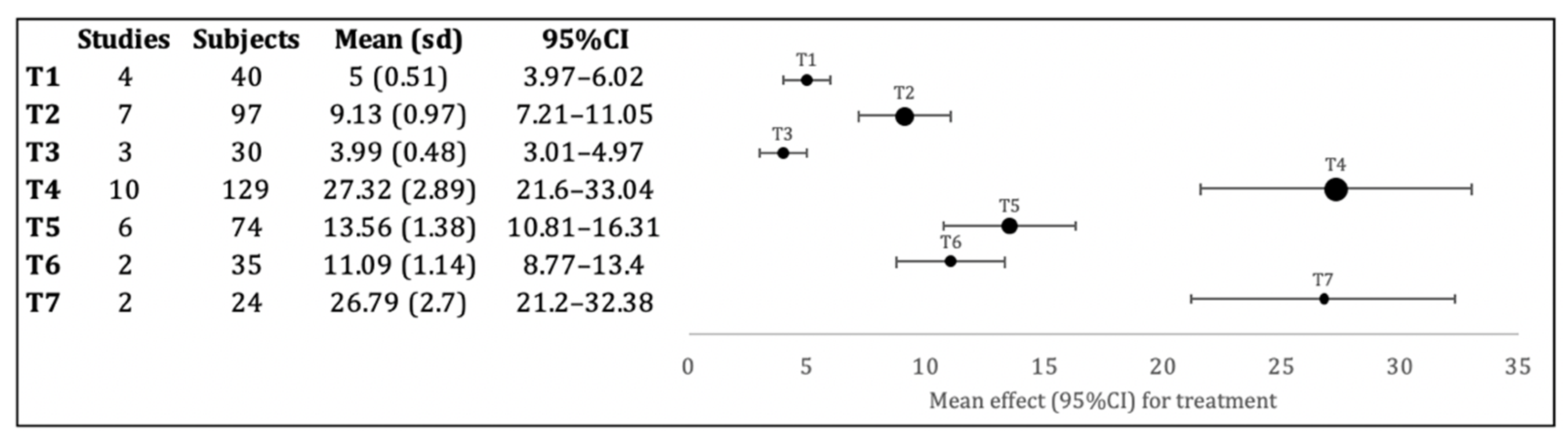
3.2. Meta-Analysis
4. Discussion
4.1. Design and Bracket Material
4.2. Orthophosphoric Acid, Fine Burr and Sandblasting
4.3. Hydrofluoric Acid
4.4. Silane
4.5. Adhesive System
4.6. LASER
5. Conclusions
Author Contributions
Funding
Institutional Review Board Statement
Informed Consent Statement
Data Availability Statement
Conflicts of Interest
References
- Alzainal, A.H.; Majud, A.S.; Al-Ani, A.M.; Mageet, A.O. Orthodontic Bonding: Review of the Literature. Int. J. Dent. 2020, 2020, 8874909. [Google Scholar] [CrossRef] [PubMed]
- González-Serrano, C.; Phark, J.-H.; Fuentes, M.V.; Albaladejo, A.; Sánchez-Monescillo, A.; Duarte, S.; Ceballos, L. Effect of a single-component ceramic conditioner on shear bond strength of precoated brackets to different CAD/CAM materials. Clin. Oral Investig. 2021, 25, 1953–1965. [Google Scholar] [CrossRef] [PubMed]
- Lopes, G.V.; Correr-Sobrinho, L.; Correr, A.B.; de Godoi, A.P.T.; Vedovello, S.A.S.; de Menezes, C.C. Light Activation and Thermocycling Methods on the Shear Bond Strength of Brackets Bonded to Porcelain Surfaces. Braz. Dent. J. 2020, 31, 52–56. [Google Scholar] [CrossRef]
- Golshah, A.; Mohamadi, N.; Rahimi, F.; Pouyanfar, H.; Tabaii, E.; Imani, M. Shear Bond Strength of Metal Brackets to Porcelain Using a Universal Adhesive. Med. Arch. 2018, 72, 425. [Google Scholar] [CrossRef] [PubMed]
- Gardiner, R.; Ballard, R.; Yu, Q.; Kee, E.; Xu, X.; Armbruster, P. Shear bond strength of orthodontic brackets bonded to a new all-ceramic crown composed of lithium silicate infused with zirconia: An in vitro comparative study. Int. Orthod. 2019, 17, 726–732. [Google Scholar] [CrossRef]
- Durgesh, B.H.; Alhijji, S.; Hashem, M.I.; Al Kheraif, A.A.; Durgesh, P.; Elsharawy, M.; Vallittu, P.K. Influence of tooth brushing on adhesion strength of orthodontic brackets bonded to porcelain. Biomed. Mater. Eng. 2016, 27, 365–374. [Google Scholar] [CrossRef]
- Juntavee, N.; Juntavee, A.; Wongnara, K.; Klomklorm, P.; Khechonnan, R. Shear bond strength of ceramic bracket bonded to different surface-treated ceramic materials. J. Clin. Exp. Dent. 2018, 10, e1167–e1176. [Google Scholar] [CrossRef]
- Mehmeti, B.; Kelmendi, J.; Iiljazi-Shahiqi, D.; Azizi, B.; Jakovljevic, S.; Haliti, F.; Anić-Milošević, S. Comparison of Shear Bond Strength Orthodontic Brackets Bonded to Zirconia and Lithium Disilicate Crowns. Acta Stomatol. Croat. 2019, 53, 17–27. [Google Scholar] [CrossRef]
- Tahmasbi, S.; Shiri, A.; Badiee, M. Shear bond strength of orthodontic brackets to porcelain surface using universal adhesive compared to conventional method. Dent. Res. J. (Isfahan.) 2020, 17, 19. [Google Scholar] [CrossRef]
- Naseh, R.; Afshari, M.; Shafiei, F.; Rahnamoon, N. Shear bond strength of metal brackets to ceramic surfaces using a universal bonding resin. J. Clin. Exp. Dent. 2018, 10, e739-45. [Google Scholar] [CrossRef]
- Pinho, M.; Manso, M.C.; Almeida, R.F.; Martin, C.; Carvalho, Ó.; Henriques, B.; Silva, F.; Pinhão Ferreira, A.; Souza, J.C.M. Bond Strength of Metallic or Ceramic Orthodontic Brackets to Enamel, Acrylic, or Porcelain Surfaces. Materials 2020, 13, 5197. [Google Scholar] [CrossRef] [PubMed]
- Kaya, Y.; Unalan Degirmenci, B.; Degirmenci, A. Comparison of the Shear Bond Strength of Metal Orthodontic Brackets Bonded to Long-term Water-aged and Fresh Porcelain and Composite Surfaces. Turkish J. Orthod. 2019, 32, 28–33. [Google Scholar] [CrossRef] [PubMed]
- Ghozy, E.A.; Shamaa, M.S.; El-Bialy, A.A. In vitro testing of shear bond strength of orthodontic brackets bonded to different novel CAD/CAM ceramics. J. Dent. Res. Dent. Clin. Dent. Prospects 2020, 14, 239–243. [Google Scholar] [CrossRef] [PubMed]
- Kara, M.; Demir, O.; Dogru, M. Bond Strength of Metal and Ceramic Brackets on Resin Nanoceramic Material With Different Surface Treatments. Turkish J. Orthod. 2020, 33, 115–122. [Google Scholar] [CrossRef] [PubMed]
- Juntavee, P.; Kumchai, H.; Juntavee, N.; Nathanson, D. Effect of Ceramic Surface Treatment and Adhesive Systems on Bond Strength of Metallic Brackets. Int. J. Dent. 2020, 2020, 7286528. [Google Scholar] [CrossRef] [PubMed]
- Alavi, S.; Samie, S.; Raji, S.A.H. Comparison of lithium disilicate–reinforced glass ceramic surface treatment with hydrofluoric acid, Nd:YAG, and CO2 lasers on shear bond strength of metal brackets. Clin. Oral Investig. 2021, 25, 2659–2666. [Google Scholar] [CrossRef]
- Cevik, P.; Eraslan, O.; Eser, K.; Tekeli, S. Shear bond strength of ceramic brackets bonded to surface-treated feldspathic porcelain after thermocycling. Int. J. Artif. Organs 2018, 41, 160–167. [Google Scholar] [CrossRef]
- Faggion, C.M. Guidelines for Reporting Pre-clinical In Vitro Studies on Dental Materials. J. Evid. Based Dent. Pract. 2012, 12, 182–189. [Google Scholar] [CrossRef]
- Al-Hity, R.; Gustin, M.-P.; Bridel, N.; Morgon, L.; Grosgogeat, B. In vitro orthodontic bracket bonding to porcelain. Eur. J. Orthod. 2012, 34, 505–511. [Google Scholar] [CrossRef]
- Durgesh, B.H. Experimental silane primer and grit-blasting distance in orthodontic bonding of zirconia surfaces. Ceram.-Silikaty 2020, 64, 469–477. [Google Scholar] [CrossRef]
- Mohammed, M.A.; Bhuminathan, S. A Comparative Evaluation of Shear Bond Strength of Orthodontic Brackets Bonded to Porcelain Fused Metal Crowns Treated with Different Surface Conditioning Techniques―An Invitro Study. Indian J. Public Health Res. Dev. 2019, 10, 2416. [Google Scholar] [CrossRef]
- Dilber, E.; Aglarcı, C.; Akın, M.; Özcan, M. Adhesion of metal brackets to glassy matrix and hybrid CAD/CAM materials after different physico-chemical surface conditioning. J. Adhes. Sci. Technol. 2016, 30, 1700–1709. [Google Scholar] [CrossRef][Green Version]
- Miersch, S.; König, A.; Mehlhorn, S.; Fuchs, F.; Hahnel, S.; Rauch, A. Adhesive luting of orthodontic devices to silica-based ceramic crowns—Comparison of shear bond strength and surface properties. Clin. Oral Investig. 2020, 24, 3009–3016. [Google Scholar] [CrossRef] [PubMed]
- Kurt, İ.; Çehreli, Z.C.; Özçırpıcı, A.A.; Şar, Ç. Biomechanical evaluation between orthodontic attachment and three different materials after various surface treatments: A three-dimensional optical profilometry analysis. Angle Orthod. 2019, 89, 742–750. [Google Scholar] [CrossRef] [PubMed]
- Zhang, Z.; Qian, Y.; Yang, Y.; Feng, Q.; Shen, G. Bond strength of metal brackets bonded to a silica-based ceramic with light-cured adhesive. J. Orofac. Orthop./Fortschritte der Kieferorthopädie 2016, 77, 366–372. [Google Scholar] [CrossRef]
- Recen, D.; Yıldırım, B.; Othman, E.; Çömlekoğlu, M.E.; Aras, I. Bond strength of metal brackets to feldspathic ceramic treated with different surface conditioning methods: An in vitro study. Eur. Oral Res. 2021, 55, 1–7. [Google Scholar] [CrossRef] [PubMed]
- Mehta, A.S.; Evans, C.A.; Viana, G.; Bedran-Russo, A.; Galang-Boquiren, M.T.S. Bonding of Metal Orthodontic Attachments to Sandblasted Porcelain and Zirconia Surfaces. Biomed Res. Int. 2016, 2016, 5762785. [Google Scholar] [CrossRef]
- Xu, Z.; Li, J.; Fan, X.; Huang, X. Bonding Strength of Orthodontic Brackets on Porcelain Surfaces Etched by Er:YAG Laser. Photomed. Laser Surg. 2018, 36, 601–607. [Google Scholar] [CrossRef]
- Ahrari, F.; Heravi, F.; Hosseini, M. CO2 laser conditioning of porcelain surfaces for bonding metal orthodontic brackets. Lasers Med. Sci. 2013, 28, 1091–1097. [Google Scholar] [CrossRef]
- Mirhashemi, A.; Chiniforush, N.; Jadidi, H.; Sharifi, N. Comparative study of the effect of Er:YAG and Er:Cr;YSGG lasers on porcelain: Etching for the bonding of orthodontic brackets. Lasers Med. Sci. 2018, 33, 1997–2005. [Google Scholar] [CrossRef]
- Girish, P.; Dinesh, U.; Bhat, C.R.; Shetty, P.C. Comparison of Shear Bond Strength of Metal Brackets Bonded to Porcelain Surface using Different Surface Conditioning Methods: An in vitro Study. J. Contemp. Dent. Pract. 2012, 13, 487–493. [Google Scholar] [CrossRef]
- Lee, J.-Y.; Kim, J.-S.; Hwang, C.-J. Comparison of shear bond strength of orthodontic brackets using various zirconia primers. Korean J. Orthod. 2015, 45, 164. [Google Scholar] [CrossRef]
- Ihsan, S.S.A.; Mohammed, S.A. Comparison of Shear Bond Strength of Orthodontic Buccal Tube Bonded to Zirconia Crown after Using Two Different (10-MDP)- Containing Adhesive Systems. Int. J. Med. Res. Health Sci. 2019, 8, 69–78. [Google Scholar]
- Costa, A.R.; Correr, A.B.; Puppin-Rontani, R.M.; Vedovello, S.A.; Valdrighi, H.C.; Correr-Sobrinho, L.; Vedovello Filho, M. Effect of bonding material, etching time and silane on the bond strength of metallic orthodontic brackets to ceramic. Braz. Dent. J. 2012, 23, 223–227. [Google Scholar] [CrossRef]
- Dalaie, K.; Mirfasihi, A.; Eskandarion, S.; Kabiri, S. Effect of bracket base design on shear bond strength to feldspathic porcelain. Eur. J. Dent. 2016, 10, 351–355. [Google Scholar] [CrossRef]
- Kaygisiz, E.; Egilmez, F.; Ergun, G.; Yuksel, S.; Cekic-Nagas, I. Effect of different surface treatments on bond strength of recycled brackets to feldspathic porcelain. J. Adhes. Sci. Technol. 2016, 30, 45–55. [Google Scholar] [CrossRef]
- Topcuoglu, T.; Oksayan, R.; Topcuoglu, S.; Coskun, M.E.; Isman, N.E. Effect of Er:YAG Laser Pulse Duration on Shear Bond Strength of Metal Brackets Bonded to a Porcelain Surface. Photomed. Laser Surg. 2013, 31, 240–246. [Google Scholar] [CrossRef]
- Gonçalves, P.R.A.; de Moraes, R.R.; Costa, A.R.; Correr, A.B.; Nouer, P.R.A.; Sinhoreti, M.A.C.; Correr-Sobrinho, L. Effect of etching time and light source on the bond strength of metallic brackets to ceramic. Braz. Dent. J. 2011, 22, 245–248. [Google Scholar] [CrossRef] [PubMed]
- Akpinar, Y.Z.; Irgin, C.; Yavuz, T.; Aslan, M.A.; Kilic, H.S.; Usumez, A. Effect of Femtosecond Laser Treatment on the Shear Bond Strength of a Metal Bracket to Prepared Porcelain Surface. Photomed. Laser Surg. 2015, 33, 206–212. [Google Scholar] [CrossRef] [PubMed]
- Asiry, M.A.; Alshahrani, I.; Hameed, M.S.; Durgesh, B.H. Effect of Surface Conditioning Protocols and Finishing on Bonding Orthodontic Brackets to Ceramics. J. Biomater. Tissue Eng. 2018, 8, 591–596. [Google Scholar] [CrossRef]
- Erdur, E.A.; Basciftci, F.A. Effect of Ti:sapphire laser on shear bond strength of orthodontic brackets to ceramic surfaces. Lasers Surg. Med. 2015, 47, 512–519. [Google Scholar] [CrossRef]
- Asiry, M.A.; AlShahrani, I.; Alaqeel, S.M.; Durgesh, B.H.; Ramakrishnaiah, R. Effect of two-step and one-step surface conditioning of glass ceramic on adhesion strength of orthodontic bracket and effect of thermo-cycling on adhesion strength. J. Mech. Behav. Biomed. Mater. 2018, 84, 22–27. [Google Scholar] [CrossRef]
- Franz, A.; Raabe, M.; Lilaj, B.; Dauti, R.; Moritz, A.; Müßig, D.; Cvikl, B. Effect of two different primers on the shear bond strength of metallic brackets to zirconia ceramic. BMC Oral Health 2019, 19, 51. [Google Scholar] [CrossRef]
- Yu, J.; Zhang, Z.; Yang, H.; Wang, Y.; Muhetaer, A.; Lei, J.; Huang, C. Effect of universal adhesive and silane pretreatment on bond durability of metal brackets to dental glass ceramics. Eur. J. Oral Sci. 2021, 129, e12772. [Google Scholar] [CrossRef]
- Abdelnaby, Y.L. Effects of cyclic loading on the bond strength of metal orthodontic brackets bonded to a porcelain surface using different conditioning protocols. Angle Orthod. 2011, 81, 1064–1069. [Google Scholar] [CrossRef]
- Cevik, P.; Karacam, N.; Eraslan, O.; Sari, Z. Effects of different surface treatments on shear bond strength between ceramic systems and metal brackets. J. Adhes. Sci. Technol. 2017, 31, 1105–1115. [Google Scholar] [CrossRef]
- Sarac, Y.S.; Kulunk, T.; Elekdag-Turk, S.; Sarac, D.; Turk, T. Effects of surface-conditioning methods on shear bond strength of brackets bonded to different all-ceramic materials. Eur. J. Orthod. 2011, 33, 667–672. [Google Scholar] [CrossRef] [PubMed]
- Hsu, H.-M.; Chang, C.-J.; Liu, J.-K. Effects of Various Acid-Silane Surface Treatments on Bond Strength of Metal Brackets to Porcelain Crowns. J. Med. Biol. Eng. 2015, 35, 375–380. [Google Scholar] [CrossRef]
- Durgesh, B.H.; Alhijji, S.; Hashem, M.I.; Kheraif, A.A.; Malash, A.M.; Divakar, D.D.; Shahrani, O.A.; Asmari, M.A.; Matinlinna, J.P. Evaluation of an Experimental Silane Primer System in Promoting Adhesion Between Orthodontic Bracket and Ceramic. J. Biomater. Tissue Eng. 2016, 6, 239–245. [Google Scholar] [CrossRef]
- Bavbek, N.C.; Roulet, J.-F.; Ozcan, M. Evaluation of microshear bond strength of orthodontic resin cement to monolithic zirconium oxide as a function of surface conditioning method. J. Adhes. Dent. 2014, 16, 473–480. [Google Scholar] [CrossRef]
- Lima Sandoval, P.C.; Ratto Tempestini Horliana, A.C.; Calabró Calheiros, F.; de Moura-Netto, C.; Gonçalves, F.; Antunes Santos, A.M.; Volpi Mello-Moura, A.C. Evaluation of shear strength in metallic brackets bonded to ceramic surface. Chirurgia (Bucur) 2020, 33, 18–23. [Google Scholar] [CrossRef]
- Zarif Najafi, H.; Oshagh, M.; Torkan, S.; Yousefipour, B.; Salehi, R. Evaluation of the Effect of Four Surface Conditioning Methods on the Shear Bond Strength of Metal Bracket to Porcelain Surface. Photomed. Laser Surg. 2014, 32, 694–699. [Google Scholar] [CrossRef]
- García-Sanz, V.; Paredes-Gallardo, V.; Bellot-Arcís, C.; Martínez-León, L.; Torres-Mendieta, R.; Montero, J.; Albaladejo, A. Femtosecond laser settings for optimal bracket bonding to zirconia. Lasers Med. Sci. 2019, 34, 297–304. [Google Scholar] [CrossRef]
- Stella, J.P.F.; Oliveira, A.B.; Nojima, L.I.; Marquezan, M. Four chemical methods of porcelain conditioning and their influence over bond strength and surface integrity. Dental Press J. Orthod. 2015, 20, 51–56. [Google Scholar] [CrossRef]
- Epperson, M.; Vazirani, N.; Armbruster, P.C.; Kee, E.L.; Xu, X.; Ballard, R.W. Hybrid crowns—Bonding protocols and shear bond strength. Australas. Orthod. J. 2021, 33, 40–47. [Google Scholar] [CrossRef]
- Ramos, T.; Lenza, M.; Reges, R.; Freitas, G. Influence of ceramic surface treatment on shear bond strength of ceramic brackets. Indian, J. Dent. Res. 2012, 23, 789. [Google Scholar] [CrossRef]
- Mehmeti, B.; Haliti, F.; Azizi, B.; Kelmendi, J.; Shahiqi, D.I.; Jakovljevic, S.; Milosevic, S.A. Influence of different orthodontic brackets and chemical preparations of ceramic crowns on shear bond strength. Australas. Med. J. 2018, 11, 107–112. [Google Scholar] [CrossRef]
- Baeshen, H.A. Influence of photodynamic therapy and different conventional methods on conditioning of lithium di silicate ceramics bonded to metallic brackets: An assessment of bond strength. Photodiagnosis Photodyn. Ther. 2021, 34, 102210. [Google Scholar] [CrossRef] [PubMed]
- Blerim, M.; Azizi, B.; Kelmendi, J.; Iljazi-Shahiqi, D.; Alar, Ž.; Anić-Milošević, S. Shear Bond Strength of Orthodontic Brackets Bonded to Zirconium Crowns. Acta Stomatol. Croat. 2017, 51, 99–105. [Google Scholar] [CrossRef]
- Yassaei, S.; Moradi, F.; Aghili, H.; Lotfi Kamran, M.H. Shear bond strength of orthodontic brackets bonded to porcelain following etching with Er:YAG laser versus hydrofluoric acid. Orthod. Art Pract. Dentofac. Enhanc. 2013, 14, e82–e87. [Google Scholar] [CrossRef] [PubMed]
- Hosseini, M.H.; Sobouti, F.; Etemadi, A.; Chiniforush, N.; Ayoub Bouraima, S. Scanning Electron Microscope Comparative Evaluation of Feldspathic Porcelain Surfaces under Irradiation by Different Powers of Neodymium-Doped Yttrium Aluminium Garnet (Nd:YAG) Laser. J. lasers. Med. Sci. 2013, 4, 75–78. [Google Scholar]
- Alakuş Sabuncuoğlu, F.; Ertürk, E. Shear bond strength of brackets bonded to porcelain surface: In vitro study. J. Istanbul. Univ. Fac. Dent. 2016, 50, 9–18. [Google Scholar] [CrossRef] [PubMed]
- Aksakalli, S.; Ileri, Z.; Yavuz, T.; Malkoc, M.A.; Ozturk, N. Porcelain laminate veneer conditioning for orthodontic bonding: SEM-EDX analysis. Lasers Med. Sci. 2015, 30, 1829–1834. [Google Scholar] [CrossRef]
- Lestrade, A.M.; Ballard, R.W.; Xu, X.; Yu, Q.; Kee, E.L.; Armbruster, P.C. Porcelain surface conditioning protocols and shear bond strength of orthodontic brackets. Australas. Orthod. J. 2021, 32, 18–22. [Google Scholar] [CrossRef]
- Poosti, M.; Jahanbin, A.; Mahdavi, P.; Mehrnoush, S. Porcelain conditioning with Nd:YAG and Er:YAG laser for bracket bonding in orthodontics. Lasers Med. Sci. 2012, 27, 321–324. [Google Scholar] [CrossRef]
- AlShahrani, I.; Kamran, M.A.; Almoammar, S.; Alhaizaey, A. Photosensitization of lithium di-silicate ceramic by Er, Cr: YSGG and fractional carbon dioxide laser bonded to orthodontic bracket. Photodiagnosis Photodyn. Ther. 2019, 28, 273–276. [Google Scholar] [CrossRef]
- May, N.B.; Araújo, É.; Vieira, L.C.C. Shear bond strength of ceramic brackets after different pre-treatments in porcelain surface. Brazilian J. Oral Sci. 2015, 14, 31–35. [Google Scholar] [CrossRef][Green Version]
- Alqerban, A. Lithium di silicate ceramic surface treated with Er,Cr:YSGG and other conditioning regimes bonded to orthodontic bracket. Saudi Dent. J. 2021, 33, 188–193. [Google Scholar] [CrossRef]
- Elsaka, S.E. Influence of surface treatments on bond strength of metal and ceramic brackets to a novel CAD/CAM hybrid ceramic material. Odontology 2016, 104, 68–76. [Google Scholar] [CrossRef]
- Falkensammer, F.; Freudenthaler, J.; Pseiner, B.; Bantleon, H.P. Influence of surface conditioning on ceramic microstructure and bracket adhesion. Eur. J. Orthod. 2012, 34, 498–504. [Google Scholar] [CrossRef]
- Kim, J.; Park, C.; Lee, J.-S.; Ahn, J.; Lee, Y. The Effect of Various Types of Mechanical and Chemical Preconditioning on the Shear Bond Strength of Orthodontic Brackets on Zirconia Restorations. Scanning 2017, 2017, 6243179. [Google Scholar] [CrossRef]
- Alaqeel, S.M. The effect of heat treatment and surface neutralization on bond strength of orthodontic brackets to lithium disilicate glass-ceramic. J. Mech. Behav. Biomed. Mater. 2020, 103, 103605. [Google Scholar] [CrossRef]
- Di Guida, L.A.; Benetti, P.; Corazza, P.H.; Della Bona, A. The critical bond strength of orthodontic brackets bonded to dental glass–ceramics. Clin. Oral Investig. 2019, 23, 4345–4353. [Google Scholar] [CrossRef] [PubMed]
- Martalia, C.; Anggitia, C.; Hamid, T.; Sjamsudin, J. The comparison of shear bond strength of metal orthodontics bracket to porcelain surface using silane and single bond: An in vitro study. J. Int. Oral Health 2020, 12, 470. [Google Scholar] [CrossRef]
- Jivanescu, A.; Bratu, D.C.; Zaharia, R.; Marsavina, L. Tensile Bond Strength Evaluation of Two Adhesive Cements Used for Bonding Orthodontic Metal Brackets to Porcelain Fused-to-metal Crowns. Mater. Plast. 2014, 51, 465–467. [Google Scholar]
- Park, K.-S.; Kim, N.-J.; Lee, C.-J.; Park, J.-H. Study on the Shear Bond Strength of Orthodontic Bracket according to the Type of Artificial Tooth Crown and Etching Method. Int. J. Clin. Prev. Dent. 2013, 9, 71–78. [Google Scholar]
- Hellak, A.; Ebeling, J.; Schauseil, M.; Stein, S.; Roggendorf, M.; Korbmacher-Steiner, H. Shear Bond Strength of Three Orthodontic Bonding Systems on Enamel and Restorative Materials. Biomed Res. Int. 2016, 2016, 6307107. [Google Scholar] [CrossRef]
- Grewal Bach, G.K.; Torrealba, Y.; Lagravère, M.O. Orthodontic bonding to porcelain: A systematic review. Angle Orthod. 2014, 84, 555–560. [Google Scholar] [CrossRef]
- Garboza, C.S.; Berger, S.B.; Guiraldo, R.D.; Fugolin, A.P.P.; Gonini-Júnior, A.; Moura, S.K.; Lopes, M.B. Influence of Surface Treatments and Adhesive Systems on Lithium Disilicate Microshear Bond Strength. Braz. Dent. J. 2016, 27, 458–462. [Google Scholar] [CrossRef] [PubMed]
- da Cunha, P.F.J.S.; Tavares, J.G.; Spohr, A.M.; Bellan, M.C.; Bueno, C.H.; Cardoso, L.I. Examining the effects of acid etching duration on the bond strength between two CAD/CAM materials and one composite resin. Odontology 2021. [Google Scholar] [CrossRef]
- May, M.M.; Fraga, S.; May, L.G. Effect of milling, fitting adjustments, and hydrofluoric acid etching on the strength and roughness of CAD-CAM glass-ceramics: A systematic review and meta-analysis. J. Prosthet. Dent. 2021, in press. [CrossRef] [PubMed]
- Caparroso Pérez, C.; Latorre Correa, F.; Arroyave Hoyos, L.J.; Grajales Gaviria, C.A.; Medina Piedrahita, V.M. In vitro evaluation of the effect of hydrofluoric acid concentration and application time on adhesion to lithium disilicate. Rev. Fac. Odontol. Univ. Antioq. 2014, 26, 62–75. [Google Scholar]
- dos Santos, D.; Bitencourt, S.; da Silva, E.; Matos, A.; Benez, G.; Rangel, E.; Pesqueira, A.; Barão, V.; Goiato, M. Bond strength of lithium disilicate after cleaning methods of the remaining hydrofluoric acid. J. Clin. Exp. Dent. 2020, 12, e103–e107. [Google Scholar] [CrossRef]
- Zeidan, L.C.; Esteves, C.M.; Oliveira, J.A.; Brugnera, A.; Cassoni, A.; Rodrigues, J.A. Effect of different power settings of Er,Cr:YSGG laser before or after tribosilicatization on the microshear bond strength between zirconia and two types of cements. Lasers Med. Sci. 2018, 33, 233–240. [Google Scholar] [CrossRef] [PubMed]
- Feitosa, F.A.; de Araújo, R.M.; Tay, F.R.; Niu, L.; Pucci, C.R. Effect of high-power-laser with and without graphite coating on bonding of resin cement to lithium disilicate ceramic. Sci. Rep. 2017, 7, 17422. [Google Scholar] [CrossRef] [PubMed]


| Population | Ceramic subtracts (crowns, veneers) … |
| Intervention | Adhesion Techniques … |
| Comparison | Diverse techniques (fluoride acid, sand blasting, adhesive, silane) …. |
| Outcome | Which is the most effective …. |
| Authors, Year | Study Design | Type of Adhesion Technique (Type, Time, Clinical Application) | Type of Porcelain | Sample Size (n) | Test Group | Control Group | Bracket Type | Intervention Test | Results | Conclusions |
|---|---|---|---|---|---|---|---|---|---|---|
| Mohammed et al., 2019 [21] | Ex vivo | Five different surface conditioning methods: G1: 37% H3PO4 acid gel (30 s) + washed + air dried + primer & bonding agent; G2: 9% HF acid (90s) G3: sandblasting for 2–3 s + 9% HF acid; G4: Sandblasting (2–3 s) + Silane, G5: Fine diamond bur roughening + silane | Porcelain | 60 | 50 ceramic crowns fabricated onto the premolar teeth following crown preparation | Natural teeth were acid etched in conventional manner using 37% H3PO4 acid (n = 10) | Metallic | SBST | G4 produced maximum bond strength of 12.34 ± 0.95 MPa comparable or even better than the control group 11.03 ± 1.63 MPa; G2 and G3 9% HF acid 11.48 ± 0.98 MPa; G5 F 9.28 ± 1.11 MPa. Ceramic surfaces conditioned with 37% H3PO4 acid produced least SBST of 5.51 ± 0.88 MPa and hence not suitable for bonding Orthodontic brackets in a clinical scenario. | G4 produced maximum bond strength comparable or even better than the control group followed by G3 and G5. G2 produced least SBST and hence not suitable for bonding Orthodontic brackets in a clinical scenario. |
| Dilber et al., 2016 [22] | In vitro | Three surface conditioning methods: G1: fine diamond burr; G2: fine diamond burr + air abrasion with 30 μm SiO2 + silane G3: fine diamond burr +9.5% HF acid + silane | Feldspathic ceramic; Lithium disilicate glass ceramic; Nanocomposite; Polymer infiltrated ceramic network | 204 | CAD/CAM blocks (n = 204, n = 17 per group) of (a) VITA Mark II (VM), (b) IPS e.max CAD (IP), (c) Lava Ultimate (LU), (d)VITA ENAMIC (VE): C-Control: (fine diamond bur); CJ: (fine diamond bur + air abrasion with 30 μm SiO2 + silane) HF: (fine diamond bur +9.5% HF acid + silane) | Specimens were mechanically roughened with fine diamond burrs placed with their shafts parallel to the specimen axes. Then, they were washed and rinsed thoroughly to remove the debris, and air-dried | Metallic | SBST | Mean bond strength (MPa) values were significantly affected by the surface conditioning method (p < 0.001) but not the CAD/CAM material type (p = 0.052); Bond strengths for all CJ and HF-conditioned specimens were two-fold higher (11.83 ± 1.95 − 9.44 ± 1.63) than those for control specimens with all materials (4.73 ± 0.93 − 6.02 ± 0.69). Significantly lower mean values were obtained in LU-CJ (9.78 ± 1.61) and LU-HF (9.44 ± 1.63) than those for other groups (11.83 ± 1.95 − 10.93 ± 1.33) groups (p < 0.05). | All CAD/CAM materials tested benefitted from additional surface conditioning either with HF acid or silica coating and silanization; Weibull parameters indicated more reliable adhesion of metal brackets to feldspathic ceramic when their graze was removed with fine diamond bur and then conditioned with either hydrofluoric acid or silica coating followed by silanization compared to those of other material conditioning combinations; |
| Miersch et al., 2019 [23] | In vitro | (1) Roughening, etching with 9% buffered HF acid; (2) Sandblasting and silane; (3) Roughening, and an experimental single component ceramic primer containing ammonium polyfluoride and trimethoxysilylpropyl methacrylate; (4) Applying the experimental single-component ceramic primer without prior roughening; (5) Only roughening; | Leucite reinforced glass ceramic | 60 | 60 identical molar crowns with the morphology of tooth 36 were computer-aided designed and computer-aided manufactured (CAD/CAM) from a leucite-reinforced glass ceramic. G1: roughening, hydrofluoric acid, silane; G2: roughening, silane; G3: roughening, experimental coupling agent; G4: experimental coupling agent; G5: roughening; | In group 6 (control), the buccal tube was positioned directly on the untreated ceramic surface only using the luting composite, which was polymerized by light curing (n = 10) | Metallic | SBST | The highest mean value of SBST was examined in group 1 (61.56 MPa), followed by group iii (45.53 MPa), group 2 (41.65 MPa), and group 4 (23.14 MPa). The comparison between groups 1–4 (with coupling agent) and group 5 (without coupling agent) revealed statistically significant differences (p ≤ 0.002), with the exception of the comparison between groups 4 and 5. Within groups 1–4, statistically significant results were determined between groups 1 and 4 as well as between groups 3 and 4 (p < 0.001). The SBST of group 6 was not calculated as the buccal tubes debonded after the incubation period. | A suitable coupling agent system produced clinically acceptable shear bond strengths capable of withstanding orthodontic forces. |
| Kurt et al., 2019 [24] | In vitro | G1: HF acid 9,6% for 2 min + silane; G2: Sandblasting with Al2O3 applied from a distance of 10 mm for 10 s in circling motions at 2.5 bar pressure + silane; G3: Silica coating with cojet under 2.5 bar pressure, at a 10-mm distance for 10 s + silane; G4: Roughening with diamond burr at 40,000 rpm for 10 s+ silane | Feldspathic porcelain monolithic zirconia hybrid porcelain | 168 | 56 feldspathic porcelain, 56 monolithic zirconia, and 56 hybrid porcelain samples were divided into 4 surface treatment subgroups. | NR | Metallic | SBST | Of the materials conditioned with HF acid, the feldspathic porcelain group had the significantly highest bonding resistance (8.84). The surface-conditioning method did affect the SBST on different surfaces. | Variations of surface types of the materials affected the bonding resistance of orthodontic attachments. Comparisons of the materials with each other showed the highest bonding resistance to be for the feldspathic porcelain in HF acid group. |
| Zhang et al., 2016 [25] | In vitro | G1: 9.6% HF acid for 2 min (HF); G2: HF acid for 2 min and silane (HFS); G3: Sandblasting from a distance of 10 mm at a pressure of 3 bar for 10 s, then washed and dried for 1 min and silane (sas); G4: Silica-coating by using the intraoral sandblaster filled with 30 mL silica-modified aluminum trioxide at 3 bar pressure, from a distance of 10 mm for 10 s and silane was applied afterward (sis). | Silica based ceramic | 80 | G1 (HF); G2 (HFS); G3 (sas); G4 (sis). | NR | Metallic | SBST | The HF-acid-treated group revealed the lowest bond strength value (3.1 MPa), which was significantly lower than those of the other three groups (p = 5.82 9 10–13). Silica-coating with silane (12.3 MPa) and sandblasting with silane (11.6 MPa) groups yielded similar bond strengths (p = 0.14), and both showed significantly higher shear bond strength than that of the HF acid with silane group. | Shear bond strengths exceeded the optimal range of ideal bond strength for clinical practice, except for the isolated HF group. HF acid etching followed by silane was the best suited method for bonding on IPS Classic. |
| Recen et al., 2021 [26] | In vitro | Four surface conditioning methods: G1: cojet sand from a 10 mm distance at a pressure of 0.25 MPa for 15 s.; G2: MEP was applied and agitated into the FC surface for 20 s; G3: 9% HF acid etching for 90 s. Followed by silane coupling agent for 60 s; G4: Diamond burr for 3 s followed by silane coupling agent for 60 s. | Feldspathic porcelain | 40 | G1: Sandblasting; G2: Monobond® Etch & Prime (MEP); G3: 9% HF and Silane coupling agent; G4: Roughening and silane. | NR | Metallic | SBST | No statistically significant difference (p > 0.05) was found in SBST between the groups | Considering the mean SBST values, all treatment methods except use of a diamond bur followed by a silane coupling agent can all be used for the bonding of metal brackets to the FC restorations with sufficient SBST for clinical performance. The clinical application of MEP has been found promising since it presented with comparably high SBST values to cojet and HF with safe ARI scores. Also, it eliminates the need for extra steps, minimizing the probability of contamination or the necessity to purchase additional instruments but also excludes potential detrimental effects of HF or sandblasting. |
| Mehta et al., 2016 [27] | In vitro | Hydrofluoric acid 4% (HF), porcelain conditioner silane primer, reliance assure primer, reliance assures plus primer, and z prime plus zirconia primer | Feldspathic porcelain and zirconia | 72 | 36 zirconia specimens divided into 2 groups: G 1- sandblasting + HF + silane + ra primer; G 2- sandblasting + silane + ra plus primer. 36 glazed feldspathic porcelain specimens divided into two groups: G1- sandblasting + z prime plus primer; G2- sandblasting + ra plus primer. | One control group for zirconia porcelain group (sandblasting + porcelain conditioner (silane)) and one control group for feldspathic porcelain group (sandblasting + porcelain conditioner (silane)) | Metallic | TBST | No statistically significant mean differences were found in tbs among the different bonding protocols for feldspathic and zirconia, 𝑝 values = 0.369 and 0.944, respectively. | Silanization following sandblasting resulted in tensile bond strengths comparable to other bonding protocols for feldspathic and zirconia surface. |
| Xu et al., 2018 [28] | In vitro | G1 9% HF acid for 2 min; G2 and G3 Er:YAG laser with two energy parameters: 250 mJ, 20 Hz and 300 mJ, 20 Hz; G4 and G5 Er:YAG laser with two energy parameters: 250 mJ, 20 Hz and 300 mJ, 20 Hz + 9% HF acid for 2 min | NR | 90 | 90 ceramic chips were divided into five groups (n = 18 each): | NR | NR | SBST | The SBST in G2 and G3 (treated by laser only) were low, only 2.97 and 3.11 MPa respectively; it was 5.28 MPa in G1 (HF). The SBST of G4 and G5, treated by both laser and HF, were 6.73 and 7.09 MPa respectively, much more than G1, G2, and G3. Based on the comparison between G1 and G2, there is a statistical difference in SBST (p < 0.05). By comparing G1 and G3, the SBST has statistical difference (p < 0.05). The comparison between G2 and G4 indicates the statistical difference in SBST (p < 0.05). Moreover, the statistical difference in SBST exists between G2 and G5 (p < 0.05), G3 and G5 ( p < 0.05). | The exclusive use of HF acid, or Er:YAG laser could not achieve sufficient bracketing bonding strength. The bonding strength of combination strategy of 250 mJ, 20 Hz Er:YAG laser and HF acid on porcelain restoration surface can be satisfied for orthodontic bracket bonding. |
| Ahrari et al., 2013 [29] | In vitro | G1, G2, G3: CO2 laser for 10 s a silane coupling agent was applied before bracket bonding; G4: 9.6% hydrofluoric HF acid gel was used for 2 min. | Feldspathic porcelain | 80 | Four groups of 20: the specimens in G 1 to G3 were treated with a fractional CO2 laser for 10 s using 10 mJ of energy, frequency of 200 Hhz and powers of 10 W (G1), 15 W (G2) and 20 W (G3). In G4: a 9.6% hydrofluoric HF acid gel was used for 2 min. | NR | NR | SBST | Deglazing caused significant increase in SBST of laser treated porcelain surfaces (p < 0.05 but had no significant effect on SBST when HF acid was used for etching (p < 0.137). ANOVA revealed no significant difference in SBST values of the study groups when glazed surfaces were compared (p < 0.269). However, a significant between group difference was found among the deglazed specimens (p < 0.001). Tukey test revealed that the bond strengths of 10 W and 15 W laser groups were significantly higher than that of the HF acid group (p < 0.05). | Application of 9.6% hydrofluoric acid produced bond strength values that surpassed the minimum strength required in clinical conditions, either used on glazed or deglazed porcelain; due to the significantly higher bond strength, porcelain treatment with a fractional CO2 laser could be recommended as a suitable alternative technique to HF acid for bonding orthodontic brackets to deglazed feldspathic porcelain. |
| Mirhashemi et al., 2018 [30] | In vitro | G1: 9% HF for 2 min; G2: etching with the 9% HF for 2 min followed by irradiation with the Er:CrYSGG laser for 10 s; G3: etching with the 9% HF for 2 min followed by irradiation with the Er:YAG laser for 10 s; G4: irradiation with the Er:CrYSGG laser for 10 s without acid etching; G5: irradiation with the Er:YAG laser | Feldspathic porcelain | 60 | 60 specimens of maxillary incisor crown were prepared and randomly assigned to five groups: G 1: etching with the 9% HF. G2: etching with the 9% HF + Er:CrYSGG laser; G3: etching with the 9% HF + Er:YAG laser; G4: Er:CrYSGG laser G5: Er:YAG laser | NR | Metallic | SBST | The average SBST [mean ± SD)] values in the five groups were as follows: HF (32.58 ± 9.21 MPa), Er:CrYSGG + HF (27.81 ± 7.66 MPa), Er:YAG + HF (23.08 ± 9.55 MPa), Er:CrYSGG (14.11 ± 9.35 MPa), and Er:YAG (6.30 ± 3.09 MPa). A statistically significant difference in SBST existed between the first three groups and the two laser groups (df = 4, F = 18.555, p < 0.001). | The Er:YAG laser with the stated specifications is not a suitable alternative to HF etching. In the case of Er:CrYSGG laser, although the conditioning outcome met the bond strength requirement for orthodontic brackets (that is, 6–8 MPa). Therefore, the bond strength must be further improved by fine-tuning the irradiation details. |
| Alavi et al., 2021 [16] | In vitro | G1: 9.6% hydrofluoric acid HF; G2: neodymium-doped yttrium aluminium garnet (Nd:YAG) laser; G3: carbon dioxide (CO2) laser; The glass ceramic surfaces were primed with a silane, and the brackets were bonded using a light-cured composite resin. | lithium disilicate–reinforced ceramic | 36 | 36 lithium disilicate ceramic blocks were assigned to three groups (n = 12): G1: 9.6% HF; G2: neodymium-doped yttrium aluminium garnet (Nd:YAG) laser; G3: carbon dioxide (CO2) laser | NR | Metallic | SBST | The median and interquartile range of SBST values in three groups were 6.48 (1.56–15.18), 1.26 (0.83–1.67), and 0.99 MPa (0.70–2.10), respectively. | Neither CO2 nor Nd:YAG lasers resulted in adequate surface changes for bonding of brackets on ceramics compared with the samples conditioned with HF. |
| Girish et al., 2012 [31] | G2: Bur for 10 s; G3: hydrofluoric acid HF; G4: sandblasting for 10 s; G5: bur for 10 s + silane; G6: Hydrofluoric acid + silane; G7: sandblasting+ silane | NR | 70 | G2: bur; G3: hydrofluoric acid HF; G4: sandblasting; G5: burr+silane; G6: hydrofluoric acid HF + silane; G7: sandblasting+ silane. | G1- untreated surface (n = 10) | Metallic | SBST | Sandblasting with silane produced the highest SBST among all the groups and showed a mean value of 15.18 MPa. The weakest SBST was seen in the control group with a mean of 1.57 MPa. The statistical results showed that there was a significant difference between all the groups. | Sandblasting with silane combination produced the highest SBST, so it is a clinically suitable method for bonding orthodontic metal brackets onto ceramic surface. | |
| Ji-Yeon Lee et al., 2015 [32] | In vitro | G0: No-primer (np); G1:porcelain conditioner (pc); G2: z-prime plus (zp); G3: monobond plus (mp); G4: zirconia liner premium (zl) | Zirconia | 100 | Four primer groups (n = 20 per group), and each primer was divided into two subgroups (n = 10 each) to examine by thermocycling protocols. | 1 control group (np) (n = 20) | Metallic | SBST | The SBST of all experimental groups decreased after thermocycling. Before thermocycling, the SBST was G4, G2 ≥ G3 ≥ G1 > G0 but after thermocycling, the SBST was G4 ≥ G3 ≥ G2 > G1 = G0 (p > 0.05). | Surface treatment with a zirconia primer increases the SBST relative to no-primer or silane primer application between orthodontic brackets and zirconia prostheses. |
| Ihsan et al., 2019 [33] | In vitro | G1: transbondtm XT primer; G2: single bond universal adhesive for 20 s, and also air dried for 5 s, and then light cured for 10 s; G3: theracem, was done in the same way as described with the previous groups except that no priming or bonding agent to the zirconia surfaces was needed according to manufacturer instructions. | Zirconia | 30 | Single bond universal adhesive group (n = 10); Theracem group (n = 10). | G1: control group (n = 10) | Metallic | SBST | The highest value of the mean shear bond strength was in G2 (16.299 ± 2.201 MPa), followed by that of G3 (15.373 ± 1.575 MPa), while the G1 had the lowest value (5.337 ± 1.274 MPa). ANOVA showed that there was a statistically highly significant difference (p ≤ 0.01) among the mean values of the shear bond strength of all groups. | The two types of 10-mdp-containing adhesive systems provide good value of shear bond strength for buccal tubes bonded to zirconia surface, however, single bond universal adhesive/composite resin is the best. |
| Mehmeti et al., 2019 [8] | In vitro | Two different etching materials were used for conditioning of the surface of ceramic crowns: 5% HF and 37% H3PO4 for 120 s, and subsequently silane. | Zirconia and lithium-disilicate ceramics | 96 (all-ceramic crowns, of which 48 full contour zirconia and 48 lithium disilicate) | Eight groups: G1: Metallic bracket bonded to zirconia surface etched with H3PO4; G2: Metallic bracket bonded to zirconia surface etched with HF; G3: Ceramic bracket bonded to zirconia surface etched with H3PO4; G4: Ceramic bracket bonded to zirconia surface etched with HF; G5: Metallic bracket bonded to lithium disilicate surface etched with H3PO4; G6: Metallic bracket bonded to lithium disilicate surface etched with HF; G7: Ceramic bracket bonded to lithium disilicate surface etched with H3PO4; G8: Ceramic bracket bonded to lithium disilicate surface etched with HF. | NR | Ceramic and metallic orthodontic brackets | SBST | Lithium-disilicate showed better bond strength in almost all groups. However, no significant difference between the groups was noticed and none of the factors had a significant influence on the mean values of SBST (p > 0.05). | The use of HF for surface etching of zirconia and lithium-disilicate, does not cause a significant increase in the SBST values as compared to etching with H3PO4 and silane application. |
| Costa et al., 2012 [34] | In vitro | G1 and G2: 10% hydrofluoric acid gel for 20s with or without silane; G3 and G4: 10% hydrofluoric acid gel for 60s with or without silane. | Feldspathic porcelain | 8 | G1 and G2: cylinders were etched using 10% hydrofluoric acid gel for 20 s only (n = 2) and 10% hydrofluoric acid gel for 20 s and silane (n = 2); G3 and G4: cylinders were etched using 10% hydrofluoric acid gel for 60s only (n = 2) and 10% hydrofluoric acid gel for 60s and silane (n = 2). | NR | Metallic | SBST | Silane application increased bond strength significantly (p < 0.05) compared with no silane application; the bonding material transbond XT promoted a significantly higher (p < 0.05) shear bond strength than fuji ortho lc, with or without silane application and for both etching times. The specimens etched for 20 s showed significantly lower (p < 0.05) shear bond strengths than those etched for 60s, for both bonding materials. | Etching time of 60 s, application of silane and transbond XT resin significantly improved the shear bond strength of brackets to feldspathic ceramics. |
| Dalaie et al., 2016 [35] | In vitro | 9% hydrofluoric acid for 2 min and silane | Feldspathic porcelain | 40 | 40 porcelain-fused-to-metal restorations and four different bracket base designs were bonded to these specimens | NR | Metallic and ceramic | SBST | One-way ANOVA showed that the SBST values were significantly different among the four groups (p < 0.001). Groups 1, 2, and 4 were not significantly different, but group 3 had significantly lower SBST (p < 0.001). | The bracket base design significantly affects the SBST of brackets to feldspathic porcelain. |
| Juntavee et al., 2020 [15] | In vitro | 9.6% HF for 15 s | Feldspathic based ceramic; lithium disilicate glass-ceramic; fluoroapatite-leucite glass-ceramic; BIS-GMA, BIS-EMA, TEGDMA 73–77% silanated quartz and silica; UDMA, TEGDMA, sodium fluoride, 85% fused silica; uncured methacrylate monomer, inert material fillers, fused silica. | 60 | Machined ceramic specimens (10 × 10 × 2 mm) were prepared from vitablocs mark II (vita) and IPS e.max® CAD (ivoclar). Layered porcelain fused to metal was used to fabricate PFM specimens. Half of specimens (n = 30) were etched. Three resin bonding systems were used for attaching metal brackets to each group (n = 10): transbond™ XT (3 m), light bond™ (reliance), or blugloo™ (Ormco), all cured with LED curing unit. | Control group (n = 30) specimens nonetched | Metallic | SBST | There were significant effects on SBST of metal bracket to the ceramic veneering materials due to the factor of different types of ceramic materials, surface treatment, resin bonding materials, interaction between types of ceramic materials, and types of adhesive resin cement (p < 0.05). The mean SBST of metal bracket bonded to vitablocs™ mark II was higher than bonded to IPS e.max® CAD and bonded to IPS d.SIGN® porcelain (p < 0.05). The mean SBST of metal bracket bonded to IPS d.SIGN® porcelain for PFM was significant lower than the mean SBST of metal bracket bonded to vitablocs™ mark II ceramic materials (p < 0.05). Also, the mean SBST of metal bracket bonded to IPS e.max® CAD ceramic reveals significantly lower than the mean SBST of metal bracket bonded to vitablocs™ mark II ceramic materials (p < 0.05). | Etching ceramic surface enhanced ceramic-bracket bond strength. However, bond strengths in nontreated ceramic surface groups were still higher than bond strength required for bonding in orthodontic treatment. |
| Kaygisiz et al., 2015 [36] | In vitro | G1: Sandblasting with AL2O3 for 4 s; G2: Er:CrYSGG laser; G3: sandblasting + etching with HF + silane; G4: etching with HF + silane | Three groups: metal, sapphire and zirconia (n = 28/group). | 84 | The mounted specimens were randomly divided into four groups: G1: sandblasting with Al2O3; G2: Er:CrYSGG laser; G3: sandblasting, etching with HF and silane, G4: etching with HF and silane application) (n = 7/group) | NR | Metallic, saphire and zirconia | SBST | Statistical analysis indicated significant differences among surface treatment procedures (p < 0.0001). In addition, the effect of the first and second bonding factors on SBST behaviors was shown to be significant for the brackets (p < 0.001). | The use of sandblasting, HF treatment and silanization procedure could be used for improving the rebond shear bond strength of zirconia brackets to porcelain surface. |
| Topcuoglu et al., 2013 [37] | Ex vivo | Sandblasted + 9.6% HF gel for 2 min; Er:YAG laser short pulse (sp); Er:YAG laser super short pulse (ssp); sandblasted+ sp, or sandblasted + ssp | Porcelain-fused-to-metal | 150 | Nine groups differing in adhesive system and surface treatment. In five groups, the adhesive system was Relyx u 200 and in the other four, Transbond XT was used. For each adhesive system, the porcelain surfaces were treated in one of five different ways: sandblasted + HF, Er:YAG laser sp, Er:YAG ssp, sandblasted + sp, or sandblasted + ssp. | Sandblasted group with transbond XT (n = 15) | Metallic | SBST | There were statistically significant differences among groups (p = 0.002). The highest SBST were observed in G2 (8.83–3.3 MPa), followed by groups 1, 8, 10, and 9 (in that order) with values of 8.25–3.2, 3.48–1.7, 3.11–0.93, and 1.56–0.86 MPa, respectively. The results of the independent samples t-test indicated that there were no statistically significant differences between G1 and the control group (p = 0.635). There were no statistically significant differences between G8 and G10 (p = 0.502). | Er:YAG laser application did not allow for elimination of the hydrofluoric acid step. |
| Gonçalves et al., 2011 [38] | In vitro | G1: 10% hydrofluoric acid for 20 s + silane; G2: 10% hydrofluoric acid for 60 s + silane (after application of silane on the ceramic surface, metallic brackets were bonded to the cylinders using Transbond XT). | Feldspathic ceramic | 60 | The specimens for each etching time were assigned to four groups (n = 15), according to the light source: xl2500 halogen light, Ultralume 5 LED, Acucure 3000 argon laser, and Apollo 95e plasma arc. Light-activation was carried out with total exposure times of 40, 40, 20 and 12 s, respectively. | NR | Metallic | SBST | Specimens etched for 20 s presented significantly lower bond strength (p < 0.05) compared with those etched for 60 s. | Only the etching time had significant influence on the bond strength of brackets to ceramic. |
| Akpinar et al., 2015 [39] | In vitro | G1: SB for 3 s with Al2O3; G2: 9.6% HF gel for 4 min (hf); G3: Nd:YAG laser irradiation (ny) for 20 s; G4: performance of femtosecond laser pulses; after surface conditioning, all specimens were cleaned for 380 sec in an ultrasonic cleaner and were air dried in air stream before bonding. | Feldspathic porcelain | 80 | G1: Group sandblasting; G2: group HF; G3: group any LASER; G4: performance of femtosecond laser pulses (each group, n = 20) | NR | Metallic | SBST | The bond strength in G3 (5.11–1.53) was significantly lower than the other groups (p < 0.05). There were no statistically significant differences among G1 (9.07–3.76), G2 (9.09–3.51), and G4 (11.58–4.16) (p = 0.28). | G4 treatment produced high SBST of the processes assessed; therefore, it appears to be an effective method for bonding orthodontic metal brackets to prepared porcelain surfaces. |
| Asiry, et al., 2018 [40] | In vitro | G1: HF; G2:deglazing using diamond burr (DB); G3: sand blasting (SB) with 25 μm aluminum trioxide (Al2O3); G4: tribochemical silica coating (TS) with 30 μm silica coated aluminum trioxide (Al2O3) | NR | 120 | Four groups of 30 specimens: G1: group HF; G2: group DB; G3: group SB; G4 group TS; 15 specimens from each group were subjected to thermocycling and the remaining 15 specimens served as the baseline (n = 15). | 60 | NR | SBST | Group 4 exhibited highest SBST at baseline (14.68 ± 0.28) and after thermo-cycling (12.67 ± 0.22) while G1 specimens exhibited lowest SBST at baseline (6.32 ± 0.15) and after thermo-cycling (4.32 ± 0.26). G1 specimens demonstrated lowest SBST; and G4 specimens showed the highest SBST. | Increased surface roughness enhanced SBST of the specimens. |
| Erdur et al., 2015 [41] | In vitro | G1: Sandblasting for 20 s; G2: etching with 5% HF acid for 20 s;G3: Nd:YAG laser for 20 s; G4: Er:YAG laser for 20 s; G5: Ti:sapphire laser; After conditioning all ceramic surfaces, silane was applied to the ceramic surfaces. | Feldspathic and IPS Empress e-Max | 150 | 150 ceramic discs were prepared and divided into two groups. In each group, the following five subgroups (n = 15) were set up: G5 Ti:sapphire laser, G3: Nd:YAG laser, G4: Er:YAG laser, G1: sandblasting, and G2: HF acid. | NR | NR | SBST | Feldspathic and IPS Empress e-Max ceramics had similar SBST values. The Ti:sapphire femtosecond laser (16.76–1.37 MPa) produced the highest mean bond strength, followed by sandblasting (12.79–1.42 MPa) and HF acid (11.28–1.26 MPa). The Er:YAG (5.43–1.21 MPa) and Nd:YAG laser (5.36–1.04 MPa) groups were similar and had the lowest SBST values. | Ti:sapphire laser- treated surfaces had the highest SBST values. Therefore, this technique may be useful for the pretreatment of ceramic surfaces as an alternative to conventional’ techniques. |
| Asiry et al., 2018 [42] | In vitro | G1: IPS ceramic etching gel™ and Monobond plus™; G2: Monobond etch and prime™. | Lithium disilicate | 40 | The specimens were randomly assigned to two experimental groups (n = 20), G1 specimens were treated with two-step surface conditioning system (IPS ceramic etching gel™ and Monobond plus™) and G2 specimens were treated with one-step surface conditioning system (Monobond etch and prime™). Ten randomly selected specimens from each group were subjected to thermo-cycling and the remaining ten served as baseline. | N = 20 | NR | SBST | The specimens treated with two-step conditioning system had higher bond strength than one-step conditioning system. | Traditional two-step conditioning provides better bond strength. The clinical importance of the study is that, the silane promoted adhesion significantly reduces on exposure to thermo-cycling. |
| Franz et al., 2019 [43] | In vitro | The bonding agent G1: Monobond S (Ivoclar Vivadent) or G2: Monobond Etch & Prime | Zirconia | 20 | The ceramic blocks (n = 20) were randomized and divided into two groups and fixation of brackets was done either by using the bonding agent Monobond S (Ivoclar Vivadent) or Monobond Etch & Prime | NR | Metallic | SBST | SBST resulted in significantly higher shear bond values when Monobond Etch & Prime was used compared to the use of Monobond S. | The use of Monobond Etch & Prime has great potential for the bonding of brackets on dental zirconia ceramics. |
| Yu et al., 2021 [44] | In vitro | All specimens were etched with 9.5% hydrofluoric acid for 20 s. The etched specimens were randomly assigned to one of four groups according to the adhesive used and the use (or not) of additional silane pretreatment for 20 s. | Lithium disilicate glass ceramic | 80 | Four groups (n = 20) defined by the pretreatment and adhesive used: G1 Adper Single Bond 2 (SB2); G2 silane + Adper Single Bond 2 (S@SB2); G3 Single Bond Universal (SBU); and G4 silane + Single Bond Universal (S@SBU). | NR | Metallic | SBST | In all groups, the mean SBST values were statistically significantly lower (p < 0.001) after thermocycling than before. Furthermore, specimens in groups S@SB2 and S@SBU, both of which had silane pretreatment, showed statistically significantly higher mean SBST values than did the corresponding groups without silane pretreatment (p < 0.05). | The application of a silane-containing universal adhesive without silane pretreatment achieves adequate durability of the bond of metal brackets to dental glass ceramics |
| Abdelnaby, 2011 [45] | In vitro | G1: 9.6% HF for 2 min; G2: 9.6% HF for 2 min + silane; G3: sandblasting for 10 s+ 9.6% HF for 2 min; G4: sandblasting for 10 s+ 9.6% HF for 2 min+ silane. | Feldspathic porcelain | 100 | The specimens were divided into four equal groups (n = 25). Porcelain surfaces were conditioned with different protocols. In G1, hydrofluoric acid and Embrace First-Coat primer were used. G2, hydrofluoric acid and silane were utilized. G3 and G4, sandblasting with aluminum oxide powder was done instead of etching. | NR | Metallic | SBST | Embrace First-Coat and silane exhibited a comparable SBST. The sandblasting process significantly increased SBST. No significant difference was found in bond SBST utilizing either hydrofluoric acid and Embrace First-Coat or sandblasting and silane. With regard to CSBS, the use of sandblasting and Embrace First-Coat revealed the highest significant CSBS value, followed by sandblasting and silane. Etching with hydrofluoric acid prior to application of either primer exhibited the least CSBS values; however, no significant difference was found between them. The SBST was significantly higher than CSBS. | Embrace First-Coat primer could be used successfully as an alternative to silane. Sandblasting provides higher bond strength than did hydrofluoric acid. Cyclic loading significantly decreased bond strength. |
| Cevik et al., 2017 [46] | In vitro | G1: 37.5% orthophosphoric acid for 4 min; G2: 9.6% hydrofluoric acid for 3 min; G3: Nd:YAG laser irradiation for 30 s.; G4: sandblasting with 50 μm Al2O3 particles for 10 s; G5: grinding with a diamond bur for 30 s. All samples were primed with silane before the bracket bonding, including the control group. | Feldspathic and lithium disilicate | 120 | Five subgroups depending on surface treatment (n = 10) G1: 37.5% orthophosphoric acid; G2: 9.6% hydrofluoric acid; G3: Nd-YAG laser irradiation; G4: sandblasting with 50 μm Al2O3 particles; G5: grinding with a diamond bur. | Control group (n = 10) | Metallic | SBST | G4 demonstrated significantly higher shear bond strengths than other groups | Surface conditioning methods, except for sandblasting and grinding, were associated with lower shear bond strengths; however, thermocycling may have had negative effects on bond strengths of specimens. Furthermore, in each ceramic system, there was a significant difference between surface-conditioning methods and surface roughness with regard to shear bond strength. |
| Saraç et al., 2011 [47] | In vitro | G1: Air-particle abrasion (APA) with 25 mm for 4 s Al2O3; G2: silica coating with 30 mm Al2O3 particles modified by silica for 4 s. Then silane application. | Feldspathic, fluoro-apatite, and leucite-reinforced ceramic. | 60 | 20 feldspathic, 20 fluoro-apatite, and 20 leucite-reinforced ceramic specimens were examined following two surface-conditioning methods: G1: APA with 25 mm Al2O3 and G2: silica coating with 30 mm Al2O3 particles modified by silica. | NR | Metallic | SBST | The lowest SBST was with APA for the fluoro-apatite ceramic (11.82 MPa), which was not significantly different from APA for the feldspathic ceramic (13.58 MPa). The SBST for the fluoro-apatite ceramic was significantly lower than that of leucite-reinforced ceramic with APA (14.82 MPa). The highest SBST value was obtained with silica coating of the leucite-reinforced ceramic (24.17 MPa), but this was not significantly different from the SBST for feldspathic and fluoroapatite ceramic (23.51 and 22.18 MPa, respectively). The SBST values with silica coating showed significant differences from those of APA. | Chairside tribochemical silica coating significantly increased mean bond strength values; With all surface-conditioning methods, leucite-reinforced ceramic, in general, showed a higher SBST than feldspathic and fluoro-apatite ceramics. |
| Hsu et al., 2015 [48] | In vitro | G1: 37% phosphoric acid solution for 60 s, and porcelain primer (H3PO4) was applied to the etched porcelain crown surface for another 60 s; G2: 9% HF acid solution for 60 s and silane for 60 s.; G3: 9% HF solution for 60 s and generic/pentron silane for 60 s; G4: 37% phosphoric acid etching solution for 60 s and ultradent silane for 60 s; G5: 37% phosphoric acid etching solution for 60 s and generic/pentron silane for 60 s. | Glazed ceramic porcelain fused to metal (PFM) | 50 | Five groups for bonding, each group n = 10; G1 (H3PO4-Porcelain Primer group); G2 (HF-Ultradent Silane); G 3 (HF-Jeneric/Pentron Silane); G 4 (H3PO4-Ultradent Silane)Ultradent; G 5 (H3PO4-Jeneric/Pentron Silane). | NR | Metallic | SBST | The Porcelain Primer group had the lowest bond strengths and the H3PO4-Jeneric/Pentron silane group had the highest bond strengths (p < 0.0005). Cross-matching of acid and silane showed that acid had a statistically significant effect on bond strength. The H3PO4-Jeneric/Pentron silane group had the highest bond strength among all acid silane groups. | The Porcelain Primer group had the lowest bond strength, showing statistically significant differences to those of the Jeneric/Pentron groups (either phosphoric acid or HF acid etching) (p < 0.0005). Although acid might be more important than silane (p = 0.005) for bond strength, there were no statistically significant differences in bond strength among the other four etching-silane groups (phosphoric acid vs. HF acid; Ultradent vs. Jeneric/Pentron). |
| Durgesh et al., 2016 [49] | In vitro | A silane-based primer consisting of 1.0 vol-% of 3 acryloxypropyltrimethoxysilane (ACPS) in 95.0 vol-%/5.0 vol-% ethanol/water, with a ph of 4.5; experimental primer, a novel silane system consisting of 0.5 vol-% of a cross-linker silane monomer bis-1,2-(triethoxysilyl) ethane (BTSE) which was added to 1.0 vol-% of acps, corresponding to a final 1.5 vol-% of silanes. | Glazed ceramic porcelain fused to metal (PFM) | 180 | Two groups of 90 specimens, according to the primer used. Each group was further divided into three subgroups according to the surface treatment to be received, thus there were 6 study groups; three with 3-acryloxypropyltrimethoxysilane (ACPS) silane primer, namely 1a (pretreatment with hydrofluoric acid, HF), 1b (pretreatment with grit-blasting) and 1c (pretreatment with tribochemical silica-coating) and 3 with a novel silane system (ACPS + bis-1,2(triethoxysilyl) ethane (BTSE)) assigned as 2a (HF), 2b (grit-blast), and 2c (tribochemical silica coating). | NR | NR | SBST | The highest SBST at baseline (26.8 + 1.7 MPa) and after thermocycling (24.6 + 1.7 MPa) was observed in group 2c, and the lowest (9.6 + 1.5 MPa and 4.5 + 1.1 MPa) was found in G1a. | The application of experimental silane primer system on specimens pretreated with tribochemical silica-coating demonstrated increased adhesion of orthodontic brackets making it an excellent choice in orthodontic bonding for a relatively long term use. |
| Bavbek et al., 2014 [50] | In vitro | Air abrasion with 30-μm silica coated aluminum oxide (Al2O3) particles (cojet) for 20 s; air abrasion with 50-μm Al2O3 particles. | Monolithic zirconium oxide ceramic (mz). | 120 | Two types of MZ (BruxZir Solid Zirconia, n = 60; Prettau-Zirkon, n = 60) with two types of surface finish (glazed, n = 30 per group; polished, n = 30 per group) were tested after two surface conditioning methods: 1. air abrasion with 30-μm silica coated aluminum oxide (Al2O3) particles (CoJet), or 2. air abrasion with 50-μm Al2O3 particles. | The non-conditioned group acted as the control. | NR | SBST | Mean μSBST values (MPa) did not show a significant difference between the two brands of MZ (p > 0.05). In both glazed (44 ± 6.4) and polished (45.9 ± 4.8) groups, CoJet application showed the highest μSBST values (p < 0.001). The control group (34.4 ± 6) presented significantly better results compared to that of Al2O3 (30 ± 3.8) (p < 0.05) on glazed surfaces, but it was the opposite in the polished groups (control: 20.3 ± 4.7; Al2O3: 33.8 ± 4.7; p < 0.001). | Air abrasion with CoJet followed by the application of universal primer improved the μSBST (microshear bond strength) of orthodontic resin to both the polished and glazed monolithic zirconium oxide materials tested |
| Sandoval et al., 2020 [51] | In vitro | Hydrofluoric acid 10% + silane; sandblasting with aluminum oxide + silane; hydrofluoric acid 10% + Single Bond Universal; blasting with aluminum oxide + Single Bond Universal. | NR | 60 | G1: hydrofluoric acid + 10% silane; G2: blasting with aluminum oxide + silane; G3: hydrofluoric acid 10% + Single Bond Universal and G4: blasting with aluminum oxide + Single Bond Universal. | NR | Metallic | SBST | The average shear strengths were: G1 = 24.2 MPa; G2 = 21.3 MPa; G3 = G4 = 19.1 MPa to 14.2 MPa. There were differences between all groups (p < 0.05) except for G3 (p > 0.05). | Single Bond Universal treated with blasting aluminum oxide had the best performance, and promoted good shear strength, it caused less cohesive damage to the ceramic. |
| Najafi et al., 2014 [52] | In vitro | Roughened with a diamond bur and etched with hydrofluoric acid (HF) gel for 4 min; roughened with a bur and irradiated by a CO2 laser with a 2W power setting for 20 s; CO2 laser; sandblasted with 50 μm aluminum oxide for 20 s. Before bonding, the bracket silane was applied on the porcelain surfaces. | Feldspathic porcelain fused to metal. | 48 | Four groups: G1: Deglazed +HF group; G2: Deglazed +CO2 group; G3: CO2 group; G4: Sandblasted group. In the four groups, a silane coupling agent) was applied. | NR | Metallic | SBST | ANOVA revealed significant differences in SBS among the four groups (p < 0.001). G1 demonstrated significantly higher bond strength (13.13–2.47) when compared with the other groups. G2 showed higher bond strength (9.60–1.91) when compared with G4 (6.40–1.67) (p = 0.016). | Deglazing combined with HF etching produced the highest bond strength, but CO2 laser irradiation provided adequate bond strength and allowed for elimination of the HF step. Deglazing is not recommended as a preliminary step before CO2 laser conditioning. |
| Durgesh, 2020 [20] | In vitro | Grit-blasted with various distance (5 mm, 10 mm and 15 mm) with 1.0 vol. % 3 methacryloyloxypropyltrimethoxy-silane (ep1) or their blends with 0.5% (ep2), and 1.0 vol. % (ep3) 1, 2-bis-(triethoxysilyl) ethane (all in ethanol/water). | Zirconia | 180 | A total of 180 zirconia specimens were used for three test groups (n = 60), and then grit-blasted with various distance (5 mm, 10 mm and 15 mm). The grit-blasted specimens were allocated to three silanizations (n = 30): with 1.0 vol. % 3 methacryloyloxypropyltrimethoxysilane (EP1) or their blends with 0.5% (EP2), and 1.0 vol. % (EP3) 1, 2-bis-(triethoxysilyl) ethane (all in ethanol/water). | NR | NR | AST | ANOVA showed a significant influence of the grit-blasting distance, silane blend and artificial aging on the shear bond strength values (p < 0.05). The highest adhesion strengths were obtained for baseline specimens irrespective of the grit-blasting distance or the silane primer blend system used. | Grit-blasting at 10 mm and silane primer blend of 1.0 vol. % 3-MPS and 0.5 vol. % BTSE provided acceptable orthodontic bonding with least surface damage to zirconia surface. Adhesion strength values significantly decreased following thermo-cycling, irrespective of the grit-blasting distance and the silane primer blend system used. |
| García-Sanz et al., 2019 [53] | In vitro | Air particle abrasion (APA) with alumina particles (Al2O3) for 20 s; femtosecond Ti:sapphire laser for 12 min. | Zirconia | 180 | Five groups (n = 30) according to surface treatment: G1: air-particle abrasion (APA); G2: FS laser irradiation (300 mW output power, 60 μm inter-groove distance); G3: FS laser irradiation (200 mW, 100 μm); G4: FS laser irradiation (40 mW, 60 μm); G5: FS laser irradiation (200 mW, 60 μm). | G1- control group: No treatment applied (n = 30). | NR | SBST | SBST in groups 3 and 6 was significantly higher than the other groups (5.92 ± 1.12 MPa and 5.68 ± 0.94 MPa). No significant differences were found between groups 1, 2, 4, and 5 (3.87 ± 0.77 MPa, 4.25 ± 0.51 MPa, 3.74 ± 0.10 MPa, and 3.91 ± 0.53 MPa). | FS laser at 200 mW, 60 μm can be recommended as the ideal settings for treating zirconia surfaces, producing good SBST and more economical energy use. |
| Stella et al., 2015 [54] | In vitro | G1: 37% gel phosphoric acid etching for one minute + Silane application for one minute; G2: 37% liquid phosphoric acid etching for one minute+ Silane application for one minute; G3: 10% hydrofluoric acid etching for one minute; G4: 10% hydrofluoric acid etching for one minute + Silane application for one minute. | NR | 52 | Four experimental groups (n = 13) were set up according to the ceramic conditioning method: G1: 37% phosphoric acid etching followed by silane application; G2: 37% liquid phosphoric acid etching, no rinsing, followed by silane application; G3: 10% hydrofluoric acid etching alone. G4: 10% hydrofluoric acid etching followed by silane application. | NR | Metallic | SBST | The highest shear bond strength values were found in groups G3 and G4 (22.01 ± 2.15 MPa and 22.83 ± 3.32 Mpa, respectively), followed by G1 (16.42 ± 3.61 MPa) and G2 (9.29 ± 1.95 MPa). | Acceptable levels of bond strength for clinical use were reached by all methods tested; however, liquid phosphoric acid etching followed by silane application (G2) resulted in the least damage to the ceramic surface. |
| Epperson et al., 2021 [55] | Ex vivo | 9.6% HF was for 4 min; 35% phosphoric acid (PA) with subsequent silanation; 50 μ aluminum oxide microetching (MIC) | Hybrid ceramics | 60 | G1: Lava (HF); G2: Lava (PA); G3 Lava (MIC); G1 Enamic (HF); G2 Enamic (PA); G3 Enamic (MIC). | Enamel control group (n = 10) (35% phosphoric acid for 30s and rinsed for 10 s. An adhesive primer, Transbond™ XT Primer was applied for 5 s and lightly air-thinned for 1 s. | Metallic | SBST | The SBST of all groups, except the HF Enamic® group, were significantly lower than the mean SBS of the enamel control group (8.8 MPa). The mean shear bond strength values of Enamic® were significantly higher than those of Lava™ Ultimate (p-values < 0.05). | Statistically, only Enamic® treated with HF exhibited sufficient SBST when compared with the enamel control. |
| Al-Hity et al., 2012 [19] | In vitro | The influence of using different combinations of bracket, adhesive, and light- curing source on the tensile bond strength to porcelain Tensile tests were performed using: one ceramic bracket versus one metal bracket, two orthodontic composites; type bisphenol A-glycidyldimethacrylate and urethane dimethacrylate (UDMA), and four light- curing units with the same range of emission spectrum but various light intensities: three light- emitting diode (LED) units and one halogen-based unit. | Fluorapatite glass-ceramic- | 160 | 160 porcelain samples were randomly divided into 16 equal groups. The porcelain surface was conditioned with 9% hydrofluoric acid before silane application. The composite was photo- polymerized for 40 s. | NR | Metallic and ceramic | TBST | The bond strength in all groups was sufficient to withstand orthodontic treatment (>6 MPa). There was no statistical difference between the adhesives, but comparing bracket × light interaction, it was significantly higher with the ceramic bracket. No significant differences were seen between the metal bracket groups, but for the ceramic bracket, the results were significantly higher with the LED light | No significant difference between adhesives’ composition related to the bonding strength on porcelain. Bonding strength of ceramic brackets on porcelain is significantly higher than metal bracket. Bonding strength of ceramic bracket is significantly higher when an LED LCU of high light intensity is used compared to halogen-based or LED LCU with low intensity. |
| Ghozy et al., 2020 [13] | In vitro | 9.5% HF for 1 min; 37% PA gel for 1 min. | VITABLOCS Mark II, VITAENAMIC, and IPS e.max CAD. | 120 | 120 CAD/CAM ceramic blocks in 12 groups were fabricated from three different CAD/CAM ceramic materials: VITABLOCS Mark II, VITAENAMIC, and IPS e.max CAD. Each ceramic material group was divided into two etching groups: 60 metal (BM) and ceramic brackets(CB) of the upper right central incisor were bonded to the HF-treated blocks. Another 60 metal and CBs were bonded to the PA treated blocks. | NR | Metallic and ceramic | SBST | There were no significant differences in SBS values between the three CAD/CAM ceramic materials. The HF-treated specimens exhibited significantly higher SBS values than the PA-treated specimens. Also, the SBS values of CBs were significantly higher than the BM. | The CAD/CAM ceramic type did not influence SBST; however, HF exhibited significantly higher SBST compared to PA. |
| Ramos et al., 2012 [56] | Ex vivo | G2: Diamond bur and processed with phosphoric acid 37% for 30 s; G3: Etching with HF 10% for 1 min; G4: etching with HF 10% for 1 min and application of 2 layers of silanization agent. | NR | 40 | n = 10 for each group. G2: fine diamond bur + orthophosphoric acid gel 37%; G3: HF 10%; G4: HF 10% + silane. | G1-control group: No surface treatment (n = 10). | Ceramic | SBST | There was a significant difference (p < 0.05) between the control group and all other groups. There was no significant difference (p < 0.05) between treated porcelain surface with diamond bur + orthophosphoric acid gel 37% (4.8 MPa) and HF 10% (6.1 MPa), but the group treated with HF 10% had clinically acceptable bond strength values. The group treated with HF 10% + silane (17.5 MPa) resulted in a statistically significant higher tensile bond strength (p < 0.05). In G4, 20% of the porcelain facets displayed damage. | Etching of the surface with HF 10% increased the bond strength values. Silane application was recommended to bond a ceramic bracket to the porcelain surface to achieve bond strengths that are clinically acceptable. |
| Mehmeti et al., 2018 [57] | In vitro | G1: 5% HF for 2 min; G2: 37% phosphoric acid for 2 min, and subsequently, silane. | Feldspar-based porcelain PFM. | 48 | Four groups (n = 12): G1: Metal bracket bonded after surface conditioning with 37 per cent phosphoric acid and silane; G2: Metal bracket bonded after surface conditioning with 5% HF and silane; G3: Ceramic bracket bonded after surface conditioning with 37% phosphoric acid and silane; G4: Ceramic bracket bonded after surface conditioning with 5% HF and silane. | NR | Metallic and ceramic | SBST | SBST values of the groups etched with HF and silane, compared to the groups etched with phosphoric acid and silane, are not significantly increased. However, ceramic brackets show significantly higher SBST values than metallic brackets. | Both types of ceramic surface conditioning procedures have similar features and provide strong enough SBST values to realize the orthodontic treatment. Also, the assumption that only the type of bracket significantly affects the SBST value can be accepted. |
| Baeshen, 2021 [58] | In vitro | G1: Er-YAG laser for about 30 s + S coupling agent for 30 s; G2: Photodynamic therapy (PDT) using methylene blue (MB) photosensitizer at a concentration of 100 mg/L; G3: 9.5% of HF acid for 60 s +S coupling agent was applied and air dried for 60 s; G4: 9.5% HF acid for 60 s + ultrasonic bath UB along with distilled water and air dried for 120 s + S; G5: Sandblasting with Al2O3 particles for 15 s; G6: SECP (Monobond etch and prime) for 60s followed by rinse for 20 s; G7: Er,Cr:YSGG for 60 s + S. | Lithium di silicate (LDC) | 70 | Seven groups according to ceramic surface conditioning. G1: surface treated with Er-YAG laser and saline (S); G2: PDT using MBP + S; G3: HF (Hydrofluoric acid) +saline, G4: HF (Hydrofluoric acid) +ultrasonic bath (UB) + S; G5: sand blasting the glass ceramic surface with 120 um Al2O3; G6: LDC surface conditioned with SECP (Etch and Prime); G7: Er,Cr:YSGG + S on was irradiated on LDC. | G3 HF + S (control). | Metallic | SBST | SBST values of G2 HF acid + S displayed highest bond durability (22.28 ± 1.09 MPa). Whereas, specimens in G4 surface treated with 120 μm Al2O3 displayed lowest SBST scores (11.81 ± 0.55 MPa) and these bond scores were comparable to PDT using MBP + S (12.54 ± 1.09 MPa) (p > 0.05). LDC surface treated by Er,Cr:YSGG + S (21.11 ± 3.85 MPa), HF + UB + S (19.28 ± 0.52 MPa) exhibited results comparable to HF acid + S (p > 0.05). | LDC conditioned with HF–S still remains as gold standard. Use of PDT for surface treatment of LDC and bonded to metallic bracket is not recommended as it results in decreased bond durability. Use of Er,Cr:YSGG-S and HF + UB + S has a potential to be used alternatively to HF–S for LDC conditioning. |
| Tahmasbi et al., 2020 [9] | In vitro | G1: universal adhesive (ScotchbondTM Universal adhesive) 20 s, air spray 5 s, light cured 10 s 650 mW/cm3; G2: universal adhesive/silane 1min, air spray 30 s, Scotchbond Universal adhesive 20 s, air spray 5 s, light curinf 10 s; G3: conventional adhesive–two layers of Single Bond 2 conventional adhesive 20 s, air sprayed 5 s, light cured 10 s; G4: conventional adhesive/silane–silane 1 min, air spray 30 s, two layers of Single Bond 2 conventional adhesive 20 s, air spray 5 s, light cured 10 s. | Feldspathic porcelain | 56 | n = 14–universal adhesive; n = 14–universal adhesive/silane; n = 14–conventional adhesive; n = 14–conventional adhesive/silane. | NR | NR | SBST | The highest SBST was noted in the universal adhesive/silane group (12.7 MPa) followed by conventional adhesive/silane (11.9 MPa), conventional adhesive without silane (7.6 MPa), and universal adhesive without silane (4.4 MPa). | SBST of bracket to porcelain mainly depends on the use of silane rather than the type of adhesive. Both universal and conventional adhesives yield significantly higher SBST in the presence of silane compared to that in the absence of silane |
| Mehmeti et al., 2017 [59] | In vitro | Phosphoric acid 120 s, composite resin-based bonding system, T light cured 40 s using light-emitting diode. | All-zirconium ceramic | 20 | n = 10–metallic bracket; n = 10–ceramic polycrystalline bracket. | NR | Metallic and ceramic polycrystalline | SBST | Force necessary to debond metallic brackets (sum of 10 tests = 70,797 N) of the zirconium crowns were higher than those of ceramic brackets (sum of 10 tests = 59,770 N), with a significant difference. | Metallic brackets compared with ceramic polycrystalline brackets, seem to create stronger adhesion with all- zirconium surfaces due to their better base surface design or retention mode. Also, ceramic brackets show higher fragility during debonding. |
| Yassaei et al., 2013 [60] | In vitro | G1: 9.6% HF; G2, 3 and 4: Er:YAG lasers of 1.6, 2, and 3.2 W. | Porcelain | 100 | Four groups: G1: n = 25–HF; G2: n = 25–Er:YAG lasers of 1.6; G3: n = 25–Er:YAG lasers of 2; G4: n = 25–Er:YAG lasers of 3.2 | NR | Metallic | SBST | The mean shear bond strength in the laser group with power of 1.6 W (7.88 MPa) was more than that of the HF (7.4 MPa), 2-W power (7.52 MPa), and 3.2-W power (7.45 MPa) groups, but this difference was not statistically significant. | Er:YAG laser can be a suitable method for bonding of orthodontic brackets to porcelain surfaces. |
| Gardiner et al., 2019 [5] | In vitro | Hydrofluoric acid etch | Zirconia; lithium disilicate (IPS e.max); lithium silicate infused with zirconia (CELTRA DUO) | 60 | Zirconia (n = 20): 9.6%HF+silane (n = 10), silane (n = 10); IPS e.max (n = 20): 9.6% HF+silane (n = 10), silane (n = 10); CELTRA DUO (n = 20): 9.6%HF + silane (n = 10), silane (n = 10). | Enamel (n = 10): 35% PA etch | Metallic | SBST | SBST of the lithium silicate infused with zirconia groups were significantly less than the chemically pre-treated lithium disilicate group, however both materials, when chemical pre- treatment protocol was used, were not statistically different than the enamel control. | Orthodontic bonding to lithium silicate infused with zirconia yielded a weaker shear bond strength than bonding to traditional lithium disilicate, however, when the surface was pre- treated with hydrofluoric acid etch it provides a bond strength that is within an acceptable clinical range. |
| Golshah et al., 2018 [4] | In vitro | 10% HF acid 2 min and the following bonding protocols: G1: Transbond XT bonding agent cured 10 s; G2: silane plus Transbond XT bonding agent cured 10 s; G3: silane plus universal adhesive (G-Premio bond) cured 10 s; G4: universal adhesive cured 10 s. | Glazed feldspathic porcelain | 40 | G1: Transbond XT bonding agent (n = 10); G2: silane plus Transbond XT bonding agent (n = 10); G3: silane plus universal adhesive (G-Premio bond) (n = 10); G4: universal adhesive (n = 10). | NR | Metallic | SBST | The highest and the lowest SBST values were noted in groups silane plus universal adhesive (17.06 ± 2.58 MPa) and universal adhesive (9.85 ± 4.76 MPa), respectively. Type of adhesive had no significant effect on SBST (p = 0.611). However, the effect of application of silane on SBST was significant (p = 0.000). Groups subjected to the application of silane showed higher SBST values than others. | Universal adhesive and Transbond XT were not significantly different in SBST. However, application of silane significantly increased the bond strength. |
| Hosseini et al., 2013 [61] | In vitro | 0.75-, 1-, 1.25-, 1.5- and 2-W neodymium-doped yttrium aluminum garnet (Nd:YAG) laser 10 s; hydrofluoric acid 9,6% 4 min | Glazed porcelain | 72 | n = 12–HF; n = 12–0.75-Nd:YAG laser; n = 12–1-Nd:YAG laser; n = 12–1.25-Nd:YAG laser; n = 12–1.5-Nd:YAG laser; n = 12–2-Nd:YAG laser. | NR | Metallic | SBST | The mean ± SD of the shear bond strength in the laser group 0.75, 1, 1.25, 1.5, and 2 W and HF group was 2.2 ± 0.9, 4.2 ± 1.1, 4.9 ± 2.4, 7 ± 1.7, 9.6 ± 2.7, and 9.4 ± 2.5, respectively. Together with the increased power of laser, the mean shear bond strength was increased continuously and no significant differences were found between the HF group and the laser groups with power of 1.5 or 2 W. | 1.5 and 2 W powers of Nd:YAG laser can be used as an alternative method for porcelain etching. |
| Naseh et al., 2018 [10] | In vitro | 9.6% hydrofluoric acid and divided into two groups: silane, air-dried, Transbond XT primer light-cured; Assure Plus, air-dried. | Feldspathic; lithium disilicate | 40 | n = 10–feldspathic with Assure Plus; n = 10–IPS E-max with Assure Plus | n = 10–feldspathic with silane+Transbon; n = 10–IPS E-max with silane+Transbond. | Metallic | SBST | Bracket bond to lithium disilicate by Assure Plus was significantly stronger than that to Feldspathic porcelain (p = 0.041). | Assure Plus provided high bond strength between ceramic and brackets and minimized damage to lithium disilicate ceramic during debonding. Assure Plus is recommended for use in orthodontic treatment of adults with ceramic restorations. |
| Cevik et al., 2018 [17] | In vitro | G1: 37% phosphoric acid 4 min; G2: 9.6% hydrofluoric acid 3 min; G3: grinding with diamond burs 20 μm with a high-speed handpiece 30s in wet conditions; G4: Nd:YAG laser 15 Hz 1 W 30 s with pulse duration range was 300 μs; G5: Airborne-particle abrasion 50 μm alumina (Al2O3) particles 2.5 bars of pressure 10 s at a direction perpendicular to the surface with the distance of 10 mm. | Feldspathic porcelain | 60 | G1: 37% phosphoric acid (n = 10); G2: 9.4% hydrofluoric acid (n = 10); G3: grinding with diamond burs (n = 10); G4: Nd:YAG laser (n = 10); G5: Airborne-particle abrasion (n = 10). | Without surface treatment (n = 10). | Ceramic | SBST | Using G5 specimens resulted in the highest shear bond strength value of 8.58 MPa for feldspathic porcelain. However, the other specimens showed lower values: G3 (6.51 MPa), G4 (3.37 MPa), G2 (2.71 MPa), G1 (1.17 MPa), and control group (0.93 MPa). | Airborne-particle abrasion and grinding can be used as surface treatment techniques on the porcelain surface for a durable bond strength. Hydrofluoric acid and phosphoric acid etching methods were not convenient as surface treatment methods for the feldspathic porcelain. |
| Juntavee et al., 2018 [7] | In vitro | Er-YAG laser power 200 mJ, 10 W, 20 Hz, 10 s-pulse length for 20 s; Etching with 9.5% HF acid gel 5 s (E5) or 15 s (E15). | Machined ceramic specimens: Empress® CAD, and e.max® CAD; Ceramic veneering metal (d.Sign® porcelain (1.27 mm thickness) over d.Sign® 10 metal (0.23 mm thickness)). | 45 | n = 15–Er-YAG laser; n = 15–HF 5 s; n = 15–HF 15 s. | NR | Ceramic | SBST | Significant differences in bond strength among groups were found related to surface treatment (p < 0.05), but not significant difference upon type of ceramics (p > 0.05). E15 provided higher bond strength than Er-YAG laser and E5 (p < 0.05). | Bond strength was affected by surface treatment. Both Er-YAG laser and E15 treated surface provided higher bond strength than E5. Considering possibly inducing defect on ceramic surface, Er-YAG laser seems to provide better favorable surface preparation than others. Treated ceramic surface with Er-YAG prior to bracket bonding is recommended. |
| Sabuncuoglu et al., 2016 [62] | In vitro | G1: Cylindrical diamond bur rotate at 40,000 rpm 3 s; G2: 37% Orthosphoric acid 2 min; G3: 9,6% HF 2 min; G4: SB with 50 μm aluminum oxide at 60 psi for 3 s at a distance of 10 mm; G5: SB+HF; G6: Nd:YAG laser wavelength 1064nm (300 μm fiber), 2 W power and frequency of 10 Hz for 10 s in a pulse mode (100 μs) using a sweeping motion at approximately 2 mm distance; G7: Er:YAG laser 2 W, 10 Hz, 10 s, 2 mm. | Feldspathic | 70 | G1: Diamond bur (n = 10); G2: Orthosphoric acid (n = 10); G3: Hydrofluoric acid (n = 10); G4: Sandblasted with aluminum oxide (n = 10); G5: SB+HF (n = 10); G6: Nd:YAG laser (n = 10); G7: Er:YAG laser (n = 10). | NR | NR | SBST | The highest SBST values were observed for SB + HF, with no significant difference between SB+HF and HF. SBST values for Diamond bur were significantly lower than those of all other groups tested. | Diamond bur alone is unable to sufficiently etch porcelain surfaces for bracket bonding. SB+HF results in a significantly higher shear-bond strength than HF or SB alone. Nd:YAG or Er:YAG laser was found to be more effective and less time-consuming than both HF acid and SB. |
| Aksakalli et al., 2015 [63] | Ex vivo | G1: SB with alumina particles 50 μm, at 65–70 psi, 10 s, 10 mm (SB); G2: 9.6% HF 4 min; G3: Er: YAG irradiation from 1 mm distance, 2 W, 10 Hz, 200 mJ, 100-μs pulse length, energy density of 25.31 J/cm2 for 10 s. | Porcelain laminate veneer | 39 | G1: SB (n = 13); G2: HF (n = 13); G3: Er:YAG (n = 13). | NR | NR | SBST | The highest shear bond strength values were obtained with group HF (10.8 ± 3.8 MPa) and group ER (9.3 ±1.5 MPa), whereas group SB revealed the lowest values. The sandblasting method did not demonstrate any ideal bond strength values; however, the 9.6% hydrofluoric acid etching and Er: YAG laser did. | The Er: YAG laser can be selected for bonding brackets to porcelain surfaces with acceptable bond strength and minimal surface damage as compared to the other methods. |
| Lestrade et al., 2021 [64] | In vitro/Ex vivo | G1: Bond enhancer (Assure, Reliance, IL, USA); G2: Green stone at 25.000 rpm; G3: Diamond bur at 25.000 rpm; G4: SB 25 μm aluminum oxide particles, distance 10 mm,10 s; SB with Rocatec (3 M ESPE, MN, USA) 100 μm aluminum oxide particles treated with silicon dioxide. | Lithium disilicate | 70 | G1: Porc-Etch, 9.6% HF, Porcelain Conditioner, silane, bond enhancer (Assure, Reliance, IL, USA) (n = 10); G2: green stone (n = 10); G3: diamond burr (n = 10); G4: SB (n = 10); G5: SB with Rocatec (n = 10). | n = 10–HF + silane; n = 10–teeth with 37% phosphoric acid + self-etching + primer + adhesive. | Metallic | SBST | No significant differences were found in SBST values, with the exception of surface roughening with a green stone prior to HF and silane treatment. This protocol yielded slightly higher bond strength which was statistically significant | The present in-vitro study found that SBST values for ceramic pretreatment all fell within an acceptable clinical range and similar to the bond strength of enamel. No significant differences were found in the SBST values, with the exception of roughening with a green stone prior to HF and silane treatment, which yielded slightly higher bond strength. |
| Poosti et al., 2012 [65] | In vitro | Tungstem carbide burrs; 9.6% Hydrofluoric acid 4 min; Er:YAG; Neodymium-doped yttrium aluminum garnet laser (Nd: YAG) 10 s. | Glazed porcelain | 100 | G1: tungsten carbide burs (n = 20); G2: tungsten carbide burs + 9.6% hydrofluoric acid (n = 20); G3: 0.8-W Nd:YAG laser (n = 20); G4: 2W Er:YAG laser (n = 20); G5: 3-W Er:YAG laser (n = 20). | NR | Metallic | SBST | Although Tukey’s test showed SBST in tungsten carbide burs+ 9.6% hydrofluoric acid and Nd:YAG laser were significantly higher than the other groups, they did not differ with each other significantly (p > 0.05). The results revealed that SBST of 9.6% hydroflouric acid and Nd:YAG Laser was in an acceptable range for orthodontic treatment. | Nd:YAG laser was shown to be an acceptable substitute for hydrofluoric acid while Er:YAG laser with the mentioned power and duration was not a suitable option. |
| AlShahrani et al., 2019 [66] | In vitro | G1: 9.6% Hydrofluoric acid+ S 60 s; G2: Er,Cr:YSGG laser 2 mm, 4.5 W, 30 Hz, 60 s; G3: Fractional carbon dioxide (CO2) laser 10 W, 200 Hz, 3 mm, 60s, pulse duration 1.75 ms; G4: SB Al2O3 10 mm, 2.8 MPa, 20 s; G5: Monobond etch & prime, Ivoclar Vivadent, Schaan, Liechtenstein. | Lithium disilicate | 50 | G2: Er,Cr:YSGG laser+ S (n = 10); G3: CO2 laser + S (n = 10); G4: SB (n = 10); G5: Self-Etch Glass Ceramic Primer (n = 10). | G1: HF+ S (n = 10). | Metallic and ceramic polycrystalline | SBST | The highest SBST values were presented by HF + S (21.08 ± 1.06). The lowest SBST values were displayed by Al2O3 (12.61 ± 0.45). SBST of samples conditioned with self-etch glass ceramic primer showed significant difference amongst all experimental groups (16.76 ± 0.81). | Lithium disilicate ceramics photosensitized with CO2 and Er,Cr:YSGG has a potential to be recommended in clinical settings alternate to HF+S when bonded to metallic bracket. |
| May et al., 2015 [67] | In vitro | 10% HF 20 s; aluminum oxide blasting 15 s, pressure at 80 psi, 5 mm; 35% phosphoric acid 30 s; CoJet blasting; S. | Eris ceramic; d. Sign ceramic | 120 | n = 60–Eris Ceramic; n = 15 -10% HF; n = 15–10% HF + S; n = 15 -aluminum oxide blasting + 35% phosphoric acid + S; n = 15 -CoJet blasting + 35% phosphoric acid + S); n = 60–d.Sign Ceramic; n = 15 -10% HF; n = 15–10% HF + S; n = 15 -aluminum oxide blasting + 35% phosphoric acid + S; n = 15 -CoJet blasting + 35% phosphoric acid + S). | NR | Metallic | SBST | There were statistically significant differences among the ceramics (p = 0.01) and surface treatments (p = 0.0001), but it did not show interaction among them (p = 0.14). | The tested ceramics performed similarly in terms of bond strength; the use of S after HF was responsible for the increase of bond strength values; HF+ S, as well as aluminum oxide +phosphoric acid+S provided significantly higher bond strength values to metallic brackets; the CoJet system did not result in significantly higher values than those observed for aluminum oxide blasting, becoming similar to the groups treated with HF without S; aluminum oxide blasting followed by phosphoric acid etching and S presented results similar to the treatment with HF + S. |
| Alqerban, 2021 [68] | In vitro | G2: S; G3: HF 9.5% 60 s; G4: SB 50 um Al2O3 1 mm, 2.8 atm, 15 s; G5: Self-etch ceramic primer (Monobond etch & prime, Ivoclar Vivadent, Schaan, Liechtenstein) (SECP) 60 s; G6: Er,Cr:YSGG laser 4.5 W, 30 Hz, 1 mm. | Lithium disilicate | 90 | G2: S 30 s (n = 15); G3: HF + UB + S 20 s (n = 15); G4: SB (n = 15); G5: SECP (n = 15); G6: Er,Cr:YSGG laser + S 20s (n = 15). | G1: HF + S 20s (n = 15). | Metallic | SBST | The highest SBST values were observed in HF+ UB + S (18.21 ± 1.241) and the lowest SBST values IN S only (5.21 ± 0.23). Specimens surface conditioned with HF+ S (17.85 ± 1.25), HF+ UB + S (18.21 ± 1.241) and Er,Cr:YSGG laser+ S (17.09 ± 1.114) unveiled comparable SBST values (p > 0.05). | Lithium disilicate conditioned with Er,Cr:YSGG laser has a potential to be used in clinical settings alternate to HF. |
| Elsaka, 2016 [69] | In vitro | 9% HF 1 min; 37% H3PO4 1 min; Diamond ceramic grinding bur (VOCO, Cuxhaven, Germany) 6000–10,000 rpm; Silica coating using CoJet system (CJ). | Vita Enamic (VE) CAD/CAM hybrid ceramic | 240 | n = 120–ceramic bracket (n = 30-HF; n = 30-H3PO4; n = 30–diamond ceramic grinding bur; n = 30-CJ). n = 120–metal bracket (n = 30-HF; n = 30-H3PO4; n = 30-diamond ceramic grinding bur; n = 30–CJ). | NR | Ceramic and metal brackets | SBST | SBST was significantly affected by the type of bracket and by type of treatment (p < 0.001). Specimens treated with CJ presented with significantly higher SBST compared to other groups (p < 0.05). Improvements in SBST values (MPa) were found in the following order: CJ [ HF [ Bur [H3PO4. Ceramic bracket showed higher SBST compared to metal bracket. | Surface treatment of VE CAD/CAM hybrid ceramic with silica coating enhanced the adhesion with ceramic and metal brackets. Ceramic bracket provided higher bond strength com- pared to metal bracket. |
| Falkensammer et al., 2012 [70] | In vitro | 5% HF 60/30 s, 9.6% buffered HF 9.6%, 60/30 s; SB Al2O3/SiO2 particles. | Metal- and all-ceramic veneering: feldspathic; leucite; leucite-free; fluorapatite. | 960 | The four types of ceramic were allocated to each of the six conditioning groups, resulting in 24 subgroups of 40 brackets each. | NR | NR | SBST | HF 5% or SB resulted in significantly (p < 0.001) higher bond strengths (mean values: 34.11 and 32.86 MPa, respectively) than with HF 9.6% (mean value: 12.49 MPa). Etching time or SB particles had no statistical (p > 0.001) influence on bond strength. | Different conditioning procedures have an effect on ceramic microstructures and bracket adhesion. High SBST (29.74–36.80 MPa) were found for all ceramic surfaces when HF 5% or SB, indicating a higher risk of ceramic fracture. The HF 9.6% appeared to have a minor conditioning effect, resulting in a lower SBST (9.34–15.92 MPa), but fewer ceramic fractures. A short etching time (30 s) was as effective as standard etching (60 s). SB SiO2 showed no advantage as compared with SB AL2O3. |
| Kim et al., 2017 [71] | In vitro | SB Al2O3 and CO (Colet TM); S; Zirconia Prime Plus (ZPP) and SBU. | Zirconia | 120 | n = 10–Al2O3 + S-T; n = 10–Al2O3 + S-N; n = 10–Al2O3 + ZPP-T; n = 10–Al2O3 + ZPP-N; n = 10–Al2O3 + SBU-T; n = 10–Al2O3 + SBU-N; n = 10–CO + S-T; n = 10–CO + S-N; n = 10–CO + ZPP-T; n = 10–CO + ZPP-N; n = 10–CO + SBU-T; n = 10–CO + SBU-N. | NR | Metallic | SBST | CO-SBU had the highest bond strength after T. CO-S significantly higher SBST than Al2O3-S. CO-ZPP lower bond strength than Al2O3-ZPP before thermocycling, but the SBST increased after T. | CO-SBU showed the highest shear bond strength. Sandblasting with either AL or CO improved the mechanical bonding by increasing the surface area, and all primer groups showed clinically acceptable increase of SBST for orthodontic treatment. |
| Alaqeel, 2020 [72] | In vitro | Heat-treatment | Lithium disilicate glass-ceramic | 120 | n = 60–heat treated specimens (n = 15–neutralized, bonded with resin based cement; n = 15–neutralized, bonded with water based cement; n = 15–non-neutralized, bonded with resin based cement; n = 15–non-neutralized, bonded with water based cement). | n = 60–non-heat treated specimens (n = 15–neutralized, bonded with resin based cement; n = 15–neutralized, bonded with water based cement; n = 15–non-neutralized, bonded with resin based cement; n = 15–non-neutralized, bonded with water based cement). | NR | SBST | The heat-treated showed statistically significant higher bond strength in all the sub- groups, and the acid-neutralized samples showed higher bond strength using both types of cement; however, the increase was statistically significant only in resin-based cement-bonded samples. Resin-based cement-bonded samples showed higher bond strength than water-based cement-bonded samples. | Pre-etching heat treatment and post-etching acid neutralization of the cementing surface of lithium disilicate glass-ceramic significantly improve the initial bond strength to orthodontic brackets. |
| Guida et al., 2019 [73] | In vitro | 10% HF 60s; S 3 min; HF + S; MDP–The adhesive system (Ambar, FGM, Joinville, Brazil—primer and adhesive combined in one bottle) containing MDP (10-methacryloyloxydecyl dihydrogen phosphate) applied in two layers. The first drop of adhesive was vigorously brushed on the bonding surface for 10 s, air-thinned before the second layer of adhesive was applied for 10 s, air-thinned, and light cured for 10 s. | Lithium disilicate-based glass–ceramic. | 240 | n = 20–BM-HF; n = 20–BM-S; n = 20–BM- HF + S; n = 20–BM-MDP; n = 20–BCp-HF; n = 20–BCp-S; n = 20–BCp-HF + S; n = 20–BCp–MDP; n = 20–BCm-HF; n = 20–BCm-S; n = 20–BCm-HF + S; n = 20–BCm-MDP. | BM—Stainless steel, metal bracket (Abzil, 3M Brazil, São Jose do Rio Preto, SP, Brazil) with a traditional mesh for mechanical retention | Metallic (BM) and ceramic brackets (monocrystalline (BCm) and polycrystalline (BCp). | SBST | BCm with HFS or HF showed the highest median σ values, 10.5 MPa and 8.5 MPa respectively. In contrast, the BCp with MDP showed the lowest median σ value (0.8 MPa), which was not statistically different from other MDP-treated groups. | The failure mode was governed by the glass–ceramic surface treatment, not by the bracket type. Quantitative (σ values) and qualitative (fracture mode) data suggested a minimum of 5 MPa for brackets bonded to glass–ceramic, which is the lower critical limit bond strength for a comprehensive orthodontic treatment. |
| Martalia et al., 2020 [74] | In vitro | 9% HF; Silane Ultradent; Ortho Solo bonding and Grengloo; adhesive bracket; Single Bond Universal 3 M ESPE. | Porcelain veneers | 28 | n = 7–9% HF 90 s, aquades 5 s, air spray 5 s, Silane, air spray 60 s, Ortho Solo, Grenglo adhesive, light-curing 20 s; n = 7–9% HF 90 s, aquades 5 s, air spray 5 s, Single Bond, light-curing 10 s, Grenglo adhesive, light- curing for 20 s; n = 7–9% HF 90 s, aquades 5 s, air spray 5 s, Ortho Solo, Grenglo adhesive, light- curing for 20 s. | n = 7–Single Bond, light-curing 10 s, apply Grenglo adhesive to bracket mess and the last step do light-curing for 20 s. | Metallic | SBST | The shear bond strengths between groups were significantly different (p < 0.05). The greatest bracket shear bond strength and lowest porcelain surface roughness were found in hydrofluoric acid, silane, bonding, and adhesive. | Silane applied separately from bonding and acid has great shear bond strength and low porcelain surface roughness. |
| Jivanescu et al., 2014 [75] | In vitro | Relyx U200 dual cure resin cement; Blugloo light cure composite | Porcelain-fused-to-metal | 16 | n = 8-Relyx U200 dual cure resin cement; n = 8-Blugloo light cure composite. | NR | Metallic | TBST | No statistically significant differences among the two cements in terms of tensile bond strength. | Both dental materials may be recommended for orthodontic bracket bonding to ceramic surfaces, with equally successful results. However, further testing on an increased number of specimens may be considered for more accurate data. |
| Park et al., 2013 [76] | In vitro | 37% Phosphoric acid 1 min, wash, dry with electric light 5 min, light curing 40 s, Transbond 37 °C of water bath for 24 h. Laser irradiation–Er:YAG laser 140 μm of the wave length and 20% of air water ratio, 20 Hz per second, 2 W, 1 mm distance, 20 s irradiation, dry 5 s, Transbond 40 s enlightening 37 °C of water bath for 24 h. | Zirconia and Ceramic | 150 | n = 10-Metal with phosphoric acid; n = 10-Metal with laser acid; n = 10-Metal with laser + phosphoric acid; n = 10-Ceramic with phosphoric acid; n = 10-Ceramic with laser acid; n = 10-Ceramic with laser + phosphoric acid; n = 10-Zirconia with phosphoric acid; n = 10-Zirconia with laser acid; n = 10-Zirconia with laser + phosphoric acid. | n = 10-Tooth with Phosphoric acid; n = 10-Tooth with Laser acid; n = 10-Tooth with Laser + phosphoric acid. | NR | SBST | Changed as the most in ceramic in laser irradiation. Bonding strength according to the etching method was the most in laser irradiation and acid etching in ceramic and in zirconia. | Ceramic crown with acid treatment was recommended because of relatively high in bonding strength. |
| Hellak et al., 2016 [77] | In vitro/Ex vivo | Self-etching no-mix adhesives (iBondTM and ScotchbondTM); Total etch system Transbond XTTM. | Glass-ceramic veneering (IPS e.maxTM Press, IPS e-max ZirCAD for inLabTM was used as high-strength zirconia and VITAblocsTM Mark II, C2II4 for CERECTM/inLab (VITA Zahnfabrik, Bad Sackingen, Germany) was used as a monochromatic feldspathic ceramic. | 270 | n = 240 divided into eight restorative surface groups (n = 30), of which Glass-ceramic veneering (n = 90) and all of the surfaces were divided into three subgroups with different adhesives (n = 10). | n = 30-Human enamel with Transbond XT primer; n = 30-Human enamel with iBond. n = 30-Human enamel with Scothchbond Universal | Metallic | SBST | Significant differences in SBST were found between the control group and experimental groups. | Transbond XT showed the highest SBST on human enamel. Scotchbond Universal on average provides the best bonding on all other types of surface (metal, composite, and porcelain), with no need for additional primers. It might therefore be helpful for simplifying bonding in orthodontic procedures on restorative materials in patients. |
| Lee et al., 2015 [32] | In vitro | G1: SB 50 µm, 5 s at pressure of 40 psi, 5 mm; G2: 9% HF acid 4 min; G3: porcelain primer (PP) thin coat; G4: zirconia primer (ZP) thin coat. | Zirconia | 40 | G1: nonglazed zirconia treated with SB + ZP (n = 10); G2: glazed zirconia treated with SB + etching + ZP (n = 10); G3: glazed zirconia treated with SB + etching + PP (n = 10); G4: glazed zirconia treated with SB + etching + ZP + PP (n = 10). | NR | Metallic | SBST | Group G2 showed significantly lower shear bond strength than did the other groups. No statistically significant differences were found among groups G1, G3, and G4. | Porcelain primer is the more appropriate choice for bonding a metal bracket to the surface of a full-contour glazed zirconia crown with resin cement. |
Publisher’s Note: MDPI stays neutral with regard to jurisdictional claims in published maps and institutional affiliations. |
© 2022 by the authors. Licensee MDPI, Basel, Switzerland. This article is an open access article distributed under the terms and conditions of the Creative Commons Attribution (CC BY) license (https://creativecommons.org/licenses/by/4.0/).
Share and Cite
Francisco, I.; Travassos, R.; Nunes, C.; Ribeiro, M.; Marques, F.; Pereira, F.; Marto, C.M.; Carrilho, E.; Oliveiros, B.; Paula, A.B.; et al. What Is the Most Effective Technique for Bonding Brackets on Ceramic—A Systematic Review and Meta-Analysis. Bioengineering 2022, 9, 14. https://doi.org/10.3390/bioengineering9010014
Francisco I, Travassos R, Nunes C, Ribeiro M, Marques F, Pereira F, Marto CM, Carrilho E, Oliveiros B, Paula AB, et al. What Is the Most Effective Technique for Bonding Brackets on Ceramic—A Systematic Review and Meta-Analysis. Bioengineering. 2022; 9(1):14. https://doi.org/10.3390/bioengineering9010014
Chicago/Turabian StyleFrancisco, Inês, Raquel Travassos, Catarina Nunes, Madalena Ribeiro, Filipa Marques, Flávia Pereira, Carlos Miguel Marto, Eunice Carrilho, Bárbara Oliveiros, Anabela Baptista Paula, and et al. 2022. "What Is the Most Effective Technique for Bonding Brackets on Ceramic—A Systematic Review and Meta-Analysis" Bioengineering 9, no. 1: 14. https://doi.org/10.3390/bioengineering9010014
APA StyleFrancisco, I., Travassos, R., Nunes, C., Ribeiro, M., Marques, F., Pereira, F., Marto, C. M., Carrilho, E., Oliveiros, B., Paula, A. B., & Vale, F. (2022). What Is the Most Effective Technique for Bonding Brackets on Ceramic—A Systematic Review and Meta-Analysis. Bioengineering, 9(1), 14. https://doi.org/10.3390/bioengineering9010014