Portable TD-OCT Scanner for Dental Use

Abstract
1. Introduction
2. Method
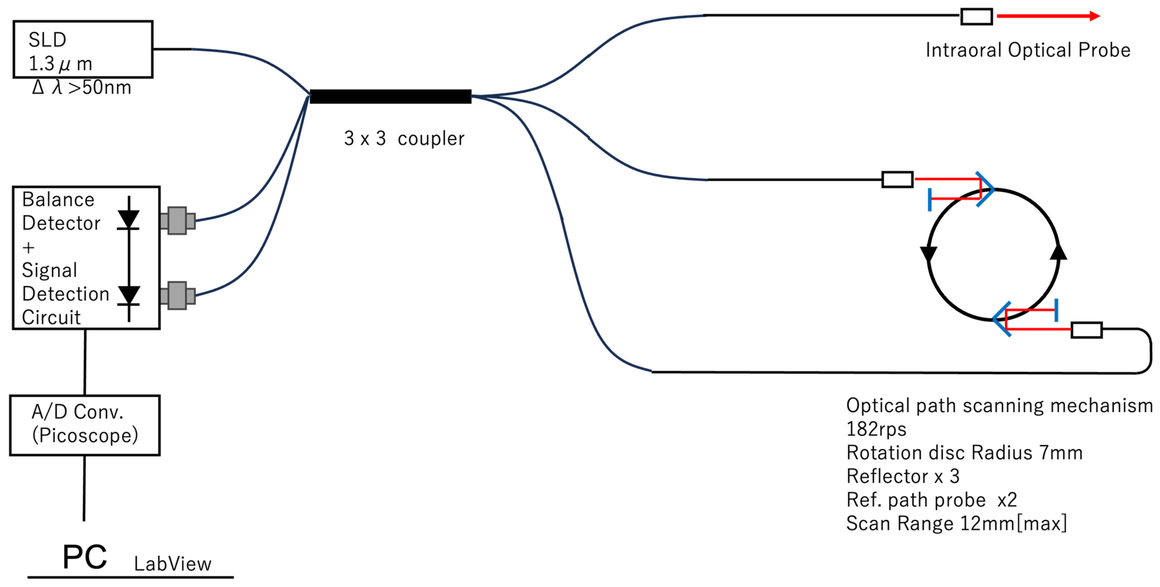
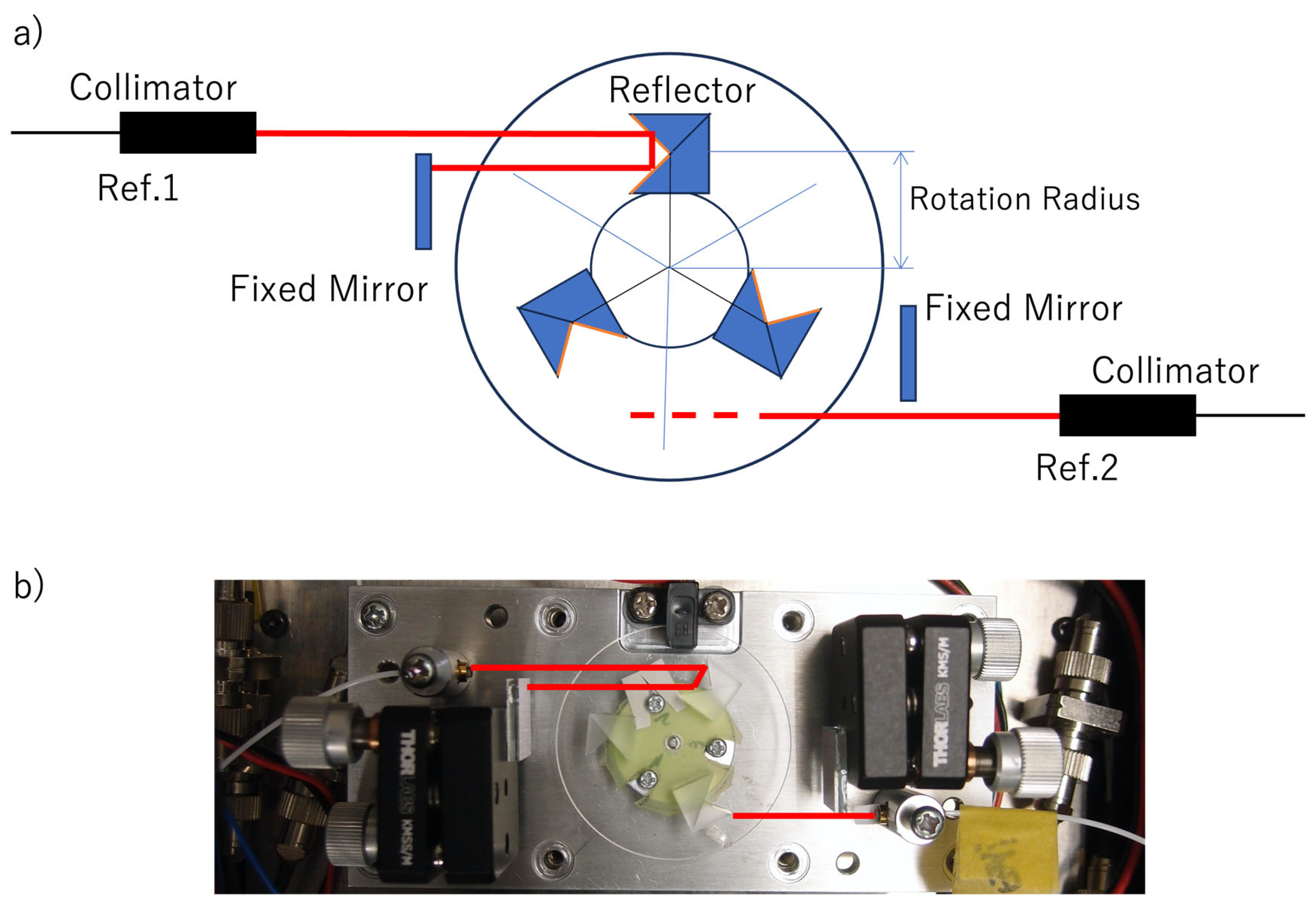
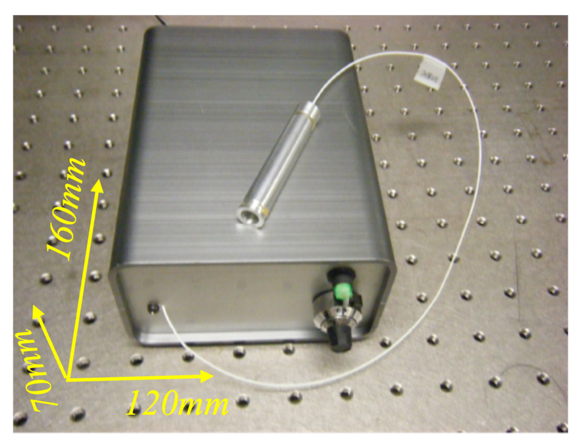
2.1. Portable TD-OCT Scanner
2.2. Probe
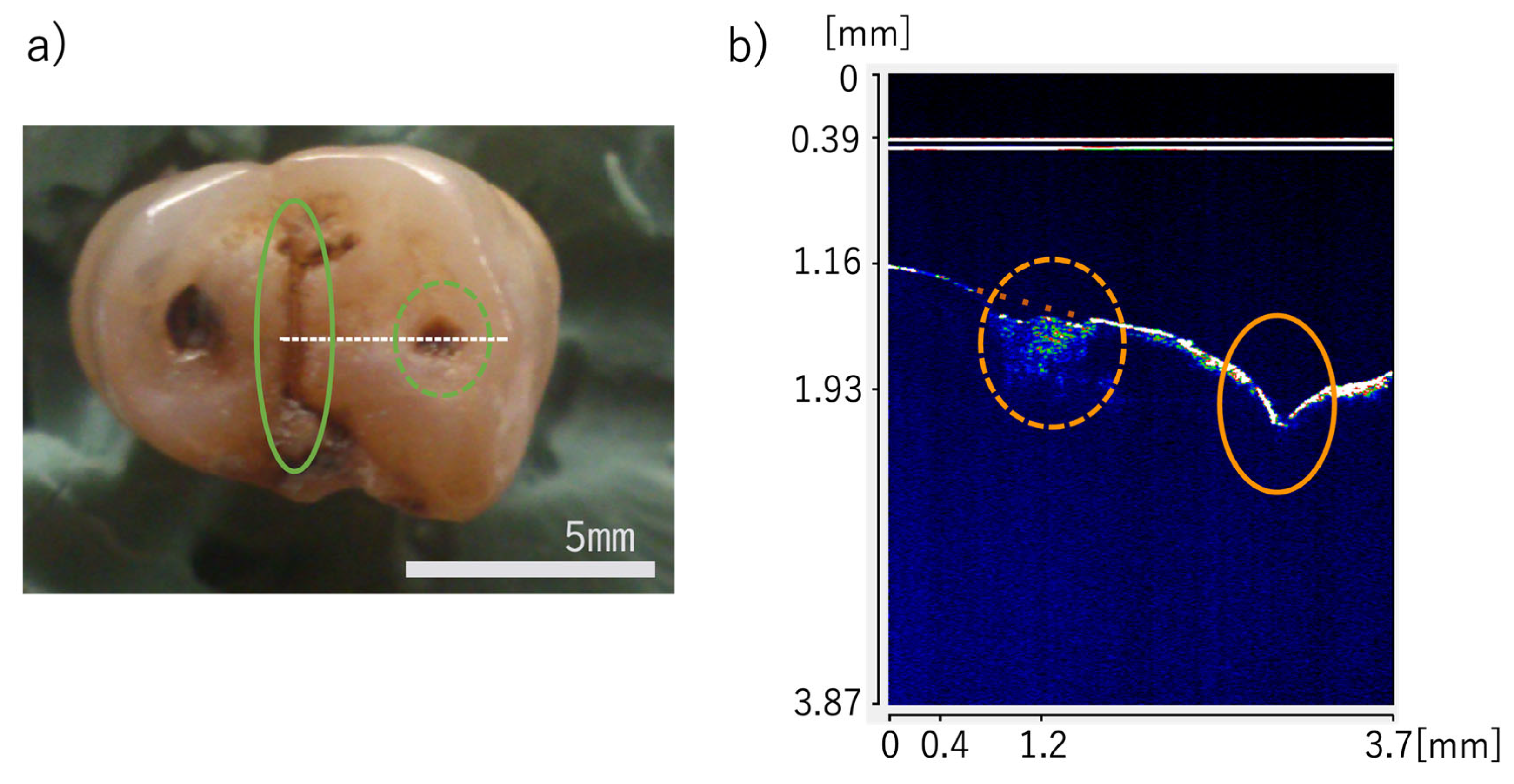
3. OCT Measurement of Teeth and Gum
4. Usage Suggestions
4.1. Speed Enhancement
4.2. Expandability
5. Conclusions
Author Contributions
Funding
Institutional Review Board Statement
Informed Consent Statement
Data Availability Statement
Conflicts of Interest
References
- Japan Dental Association (Ed.) Vision of Dental Care Toward 2040 Dentistry in the Reiwa Era; Japan Dental Association: Tokyo, Japan, 2020; Available online: https://www.jda.or.jp/en/pdf/Vision_of_Dental_Care_Toward_2040-Whole_Version.pdf (accessed on 2 June 2026).
- Lussi, A.; Hellwig, E. Performance of a new laser fluorescence device for the detection of occlusal caries in vitro. J. Dent. 2006, 34, 467–471. [Google Scholar] [CrossRef] [PubMed]
- Rodrigues, J.A.; Hug, I.; Neuhaus, K.W.; Lussi, A. Light-emitting diode and laser fluorescence–based devices in detecting occlusal caries. J. Biomed. Opt. 2011, 16, 107003. [Google Scholar] [CrossRef] [PubMed]
- Wenger, S.; Martin, L.; Gintaute, A.K.; Müller, C.; Nelson, K.; Kernen, F. Optical measurement methods of the 3D-position stability of implant-abutment connections—An in vitro study. BMC Oral Health 2025, 25, 931. [Google Scholar] [CrossRef] [PubMed]
- Pyo, S.W.; Lim, Y.J.; Koo, K.T.; Lee, J. Methods Used to Assess the 3D Accuracy of Dental Implant Positions in Computer-Guided Implant Placement: A Review. J. Clin. Med. 2019, 8, 54. [Google Scholar] [CrossRef] [PubMed]
- Cen, Y.; Huang, X.; Liu, J.; Qin, Y.; Wu, X.; Ye, S.; Du, S.; Liao, W. Application of three-dimensional reconstruction technology in dentistry: A narrative review. BMC Oral Health 2023, 23, 230. [Google Scholar] [CrossRef] [PubMed]
- Gašparovi, B.; Morelato, L.; Lenac, K.; Mauša, G.; Zhurov, A.; Katić, V. Comparing direct measurements and three-dimensional (3D) scans for evaluating facial soft tissue. Sensors 2023, 23, 2412. [Google Scholar] [CrossRef] [PubMed]
- Harumichi, K.; Katsuhiko, T.; Nobuhiro, O.; Ayano, M.; Akito, U.; Shugo, H. 3D dental model measurement system with measurement templates: Toward variable application. Appl. Sci. 2026, 16, 4267. [Google Scholar] [CrossRef]
- Shimada, Y.; Sadr, A.; Burrow, M.F.; Tagami, J.; Ozawa, N.; Sumi, Y. Validation of swept-source optical coherence tomography (SS-OCT) for the diagnosis of occlusal caries. J. Dent. 2010, 38, 655–665. [Google Scholar] [CrossRef] [PubMed]
- Hariri, I.; Sadr, A.; Shimada, Y.; Tagami, J.; Sumi, Y. Effects of structural orientation of enamel and dentine on light attenuation and local refractive index: An optical coherence tomography study. J. Dent. 2012, 40, 387–397. [Google Scholar] [CrossRef] [PubMed]
- Shimada, Y.; Sadr, A.; Nazari, A.; Nakagawa, H.; Otsuki, M.; Tagami, J.; Sumi, Y. 3D evaluation of composite resin restoration at practical training using swept-source optical coherence tomography (SS-OCT). Dent. Mater. J. 2012, 31, 409–417. [Google Scholar] [CrossRef] [PubMed]
- Schneider, H.; Park, K.J.; Häfer, M.; Rüger, C.; Schmalz, G.; Krause, F.; Schmidt, J.; Ziebolz, D.; Haak, R. Dental Applications of Optical Coherence Tomography (OCT) in Cariology. Appl. Sci. 2017, 7, 472. [Google Scholar] [CrossRef]
- Smith, P.W.; Lee, K.; Guo, S.; Zhang, J.; Osann, K.; Chen, Z.; Messadi, D. In vivo diagnosis of oral dysplasia and malignancy using optical coherence tomography: Preliminary studies in 50 patients. Lasers Surg. Med. 2009, 41, 353–357. [Google Scholar] [CrossRef] [PubMed]
- Sato, T.; Tabata, T.; Akabane, K.; Zhao, M.; Khaled, A.H.M.; Hiraishi, N.; Sumi, Y.; Tagami, J.; Shimada, Y. Noninvasive diagnostic methos using optical coherence tomography detected a vulnerable dentin enamel junction created by phosphoric acid etching. Dent. Mater. J. 2023, 42, 552–558. [Google Scholar] [CrossRef] [PubMed]
- Janjua, S.; Jeelani, W.; Khan, M.I.; Qureshi, S.M.; Shaikh, M.S.; Zafa, M.S.; Khurshid, Z. Use of Optical Coherence Tomography in Dentistry. Hindawi Int. J. Dent. 2023, 2023, 4179210. [Google Scholar] [CrossRef] [PubMed]
- Nakajima, Y.; Shimada, Y.; Sadr, A.; Wada, I.; Miyashin, M.; Takagi, Y.; Tagami, J.; Sumi, Y. Detection of occlusal caries in primary teeth using swept source optical coherence tomography. J. Biomed. Opt. 2014, 19, 016020. [Google Scholar] [CrossRef] [PubMed]
- Shimada, Y.; Nakagawa, H.; Sadr, A.; Wada, I.; Nakajima, M.; Nikaido, T.; Otsuki, M.; Tagami, J.; Sumi, Y. Noninvasive cross-sectional imaging of proximal caries using Swept-source optical coherence tomography (SS-OCT) in vivo. J. Biophotonics 2014, 7, 506–513. [Google Scholar] [CrossRef] [PubMed]
- Wada, I.; Shimada, Y.; Ikeda, M.; Sadr, A.; Nakashima, S.; Tagami, J.; Sumi, Y. Clinical assessment of non carious cervical lesion using swept-source optical coherence tomography. J. Biophotonics 2015, 8, 846–854. [Google Scholar] [CrossRef] [PubMed]
- Shimada, Y.; Sadr, A.; Sum, Y.; Tagami, J. Application of Optical Coherence Tomography (OCT) for Diagnosis of Caries, Cracks, and Defects of Restorations. Curr. Oral Health Rep. 2015, 2, 73–80. [Google Scholar] [CrossRef] [PubMed]
- Tsubokawa, M.; Aoki, A.; Kakizaki, S.; Mizutani, K.; Sumi, Y.; Izumi, Y. Application of OCT in Periodontal Therapy. J. Jpn. Soc. Laser Surg. Med. 2018, 39, 37–49. [Google Scholar] [CrossRef]
- Shimada, Y.; Burrow, M.F.; Araki, K.; Zhou, Y.; Hosaka, K.; Sadr, A.; Yoshiyama, M.; Miyazaki, T.; Sumi, Y.; Tagami, J. 3D imaging of proximal caries in posterior teeth using optical coherence tomography. Sci. Rep. 2020, 10, 15754. [Google Scholar] [CrossRef] [PubMed]
- Șurlin, P.; Camen, A.; Stratul, S.I.; Roman, A.; Gheorghe, D.N.; Herăscu, E.; Osiac, E.; Rogoveanu, I. Optical coherence tomography assessment of gingival epithelium inflammatory status in periodontal—Systemic affected patients. Ann. Anat. 2018, 219, 51–56. [Google Scholar] [CrossRef] [PubMed]
- Esperouz, F.; Ciavarella, D.; Lorusso, M.; Santarelli, A.; Muzio, L.L.; Campisi, G.; Russo, L.L. Critical review of OCT in clinical practice for the assessment of oral lesions. Front. Oncol. 2025, 15, 1569197. [Google Scholar] [CrossRef] [PubMed]
- Shiina, T.; Moritani, Y.; Ito, M.; Okamura, Y. Long path scanning mechanism for optical Coherence tomography. Appl. Opt. 2003, 42, 3795–3799. [Google Scholar] [CrossRef] [PubMed]
- Huang, D.; Swason, E.A.; Lin, C.P.; Schuma, J.S.; Stinson, W.G.; Chang, W.; Hee, M.R.; Flotte, T.; Gregory, K.; Puliafito, C.A.; et al. Optical Coherence Tomography. Science 1991, 254, 1178–1181. [Google Scholar] [CrossRef] [PubMed]
- Drexler, W.; Fujimoto, J.G. (Eds.) Optical Coherence Tomography Technology and Applications; Springer: Berlin/Heidelberg, Germany, 2008. [Google Scholar]
- Kalman, J. Fourier-domain optical coherence tomography signal analysis and numerical modeling. Hindawi Int. J. Opt. 2017, 2017, 9586067. [Google Scholar] [CrossRef]
- Grulkowski, I.; Manzanera, S.; Cwiklinski, L.; Sobczuk, F.; Karnowski, K.; Artal, P. Swept source optical coherence tomography and tunable lens technology for comprehensive imaging and biometry of the whole eye. Optica 2018, 5, 52–59. [Google Scholar] [CrossRef]
- Moon, S.; Piao, Z.; Kim, C.S.; Chen, Z. Lens-free endoscopy probe for optical coherence tomography. Opt. Lett. 2013, 38, 2014–2016. [Google Scholar] [CrossRef] [PubMed]
- Huang, X.; Tong, X.L. Study on an all-fiber optical coherence tomography (OCT) probe. Opt. Fiber Technol. 2023, 79, 103345. [Google Scholar] [CrossRef]
- Liu, Y.; Zhang, D.; Li, W.; Zheng, S.; Fan, H.; Lan, T.; Hao, Q.; Huang, Y. Side-viewing axicon-integrated miniature fiber probe for extended depth of focus and ultrahigh lateral resolution endoscopic imaging. Microsyst. Nanoeng. 2025, 11, 235. [Google Scholar] [CrossRef] [PubMed]
- Walther, J.; Golde, J.; Albrecht, M.; Quirk, B.C.; Scolaro, L.; Kirk, R.W.; Gruda, Y.; Schnabel, C.; Tetschke, F.; Joehrens, K.; et al. A handheld fiber-optic probe to enable optical coherence tomography of oral soft tissue. IEEE Trans. Biomed. Eng. 2022, 69, 2276–2282. [Google Scholar] [CrossRef] [PubMed]
- Kushwaha, K.; Ji, M.; Sethi, S.; Jamieson, L.; McLaughlin, R.A.; Li, J. A novel small form-factor handheld optical coherence tomography probe for oral soft tissue imaging. Micromachines 2024, 15, 742. [Google Scholar] [CrossRef] [PubMed]
- Tearney, G.J.; Bouma, B.E.; Fujimoto, J.G. High-speed phase- and group-delay scanning with a grating-based phase control delay line. Opt. Lett. 1997, 22, 1811–1813. [Google Scholar] [CrossRef] [PubMed]
- Su, C.B. Achieving variation of the optical path length by a few millimeters at millisecond rates for imaging of turbid media and optical interferometry: A new technique. Opt. Lett. 1997, 22, 665–667. [Google Scholar] [CrossRef] [PubMed]
- Tanaka, M.; Shiina, T. Micro crack analysis of optical fiber by specialized TD-OCT. Opt. Laser Technol. 2019, 116, 22–25. [Google Scholar] [CrossRef]





| Portable OCT Scanner | Hand-Held Intraoral OCT | Octina | OCTiX | |
|---|---|---|---|---|
| Product | Phenix Dent (Sano, Japan) Prototype | Medical Laser Center Lübeck (Lübeck, Germany) Prototype | Yoshida Dental MFG (Tokyo, Japan) Commercialized | Huvitz (Anyang-si, Republic of Korea) Commercialized |
| OCT type | TD-OCT | SD-OCT (Thorlabs GmbH, Bergkirchen, Germany) | SD-OCT | FD-OCT |
| Device type | Portable | Desktop | Chair Side | Chair Side |
| Probe type | Fiber type | Gun type | Gun type | Bar type |
| Advantage | DC-driven Home Care | High speed (20 k–100 kHz) | High speed (50 kHz) | High speed (No info.) |
| Weakness | Low speed (25–1 kHz) | Hospital Visit | Hospital Visit | Hospital Visit |
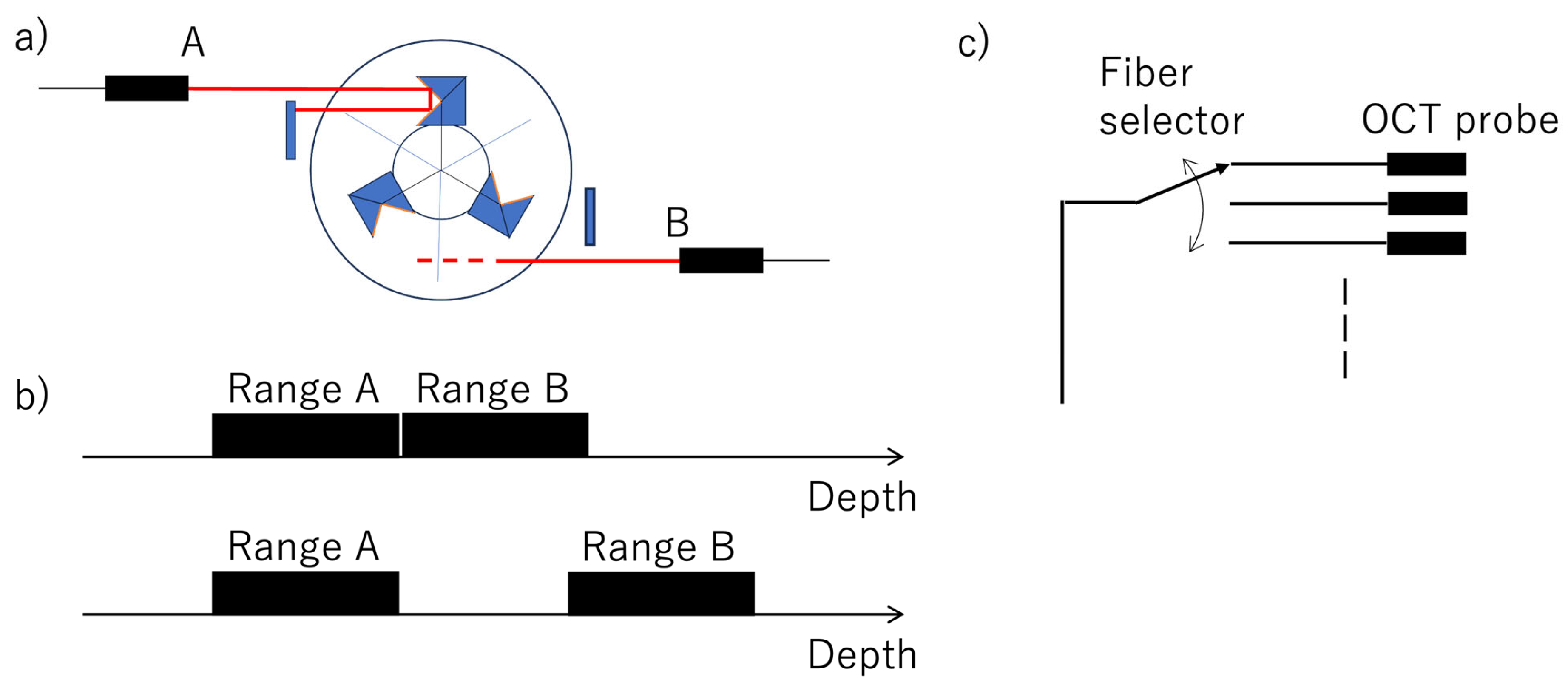
| Portable OCT Scanner | Interferometric Optical Ranging | Optical Interferometry | |
|---|---|---|---|
| Scanning | Rotation of off-axis reflector | Rapid-scanning optical delay (RSOD) | Rotating Cube |
| speed | 25–1000 Hz | 1200 Hz | 22 Hz (45 ms) |
| Scanning range | 14mm (Variable) | 3 mm | 1.5 mm |
| SLD | Anritsu AS3E113HJ10M (Anritsu, Singapore) |
| Coherent length | |
| Scan speed | 1000 rps [max] |
| Scanning range | 12 mm |
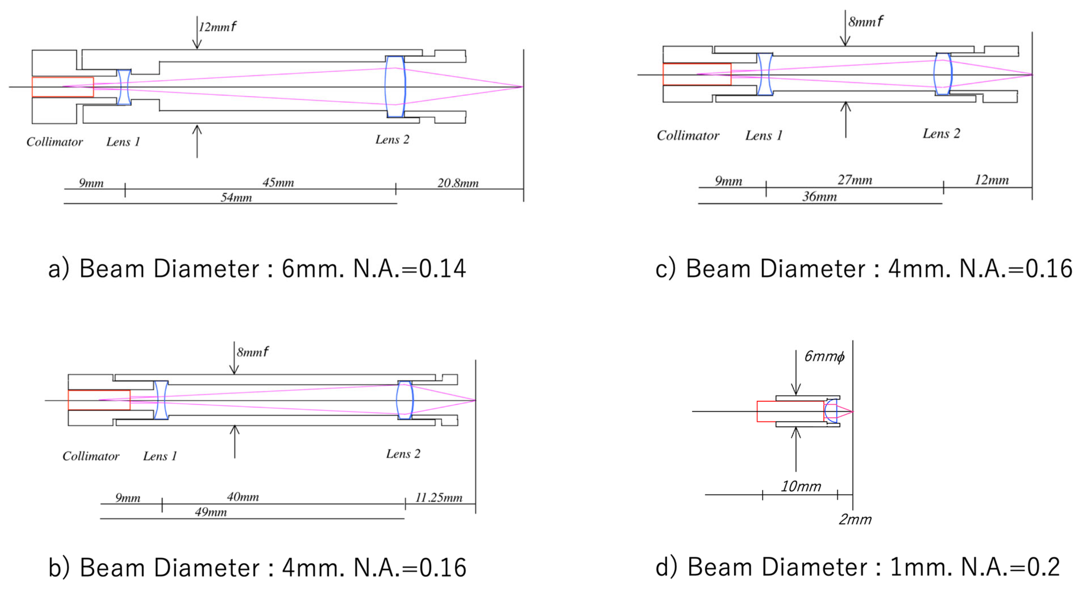
| Probe size | 8 mmφ × 10 mm |
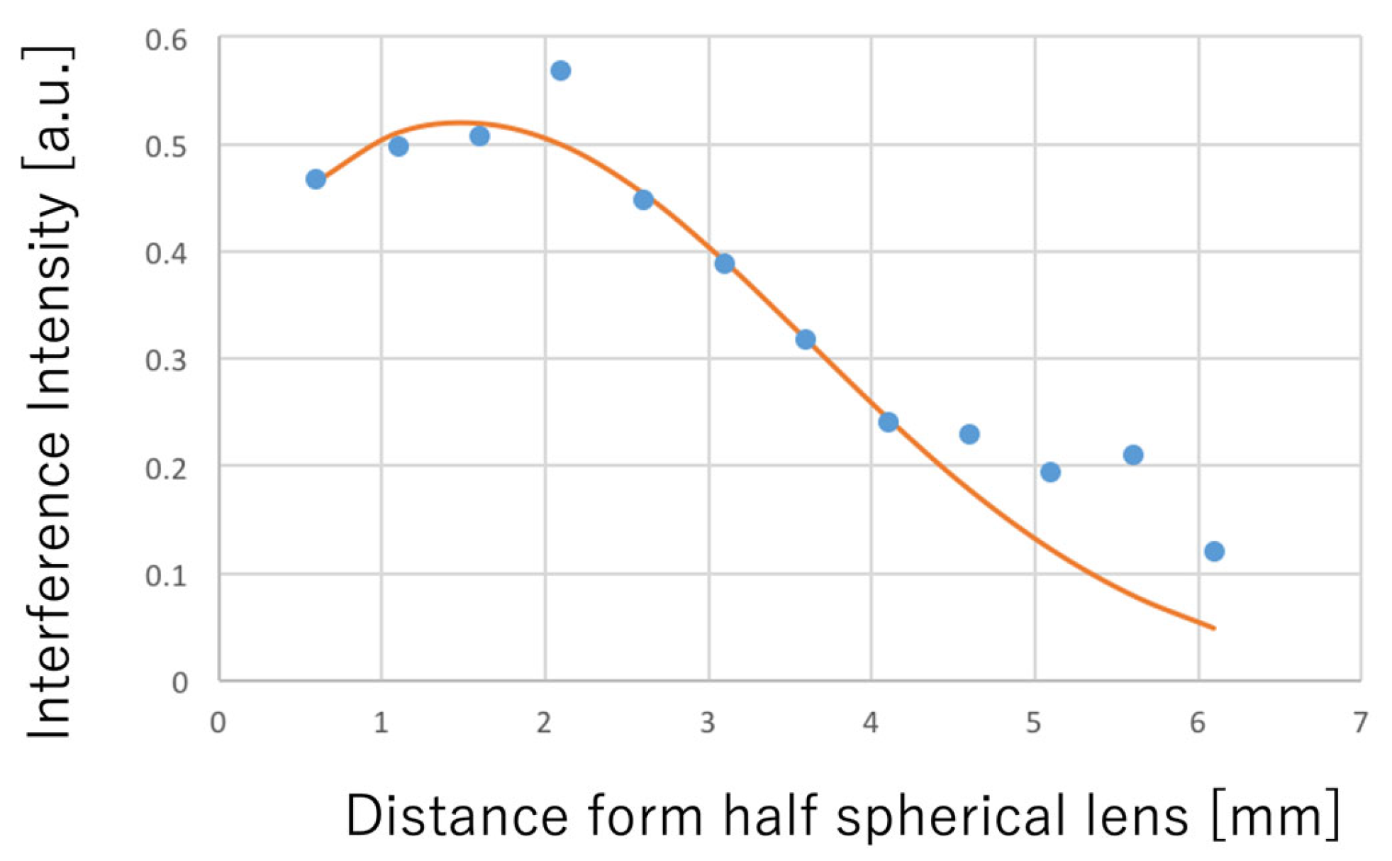
| Probe depth of field | 3 mm |
| OCT size | 200 mm × 150 mm × 100 mm |
| Weight | 1 kg |
| Reflector | Reference Fiber | Radius [mm] | Rotation [rps] | Range [mm] | Scan Speed [scan/s] | |
|---|---|---|---|---|---|---|
| Dental Skin Plant | 1 | 2 | 10 | 25 | 12 | 25 |
| Long range | 1 | 1 | 60 | 4 | >80 | 4 |
| High speed | 3 | 2 | 10 | 125 | 12 | 750 |
| High speed | 3 | 2 | 7 | 183 | >14 | >1000 |
Disclaimer/Publisher’s Note: The statements, opinions and data contained in all publications are solely those of the individual author(s) and contributor(s) and not of MDPI and/or the editor(s). MDPI and/or the editor(s) disclaim responsibility for any injury to people or property resulting from any ideas, methods, instructions or products referred to in the content. |
© 2026 by the authors. Licensee MDPI, Basel, Switzerland. This article is an open access article distributed under the terms and conditions of the Creative Commons Attribution (CC BY) license.
Share and Cite
Shiina, T.; Okaneya, S. Portable TD-OCT Scanner for Dental Use. Bioengineering 2026, 13, 743. https://doi.org/10.3390/bioengineering13070743
Shiina T, Okaneya S. Portable TD-OCT Scanner for Dental Use. Bioengineering. 2026; 13(7):743. https://doi.org/10.3390/bioengineering13070743
Chicago/Turabian StyleShiina, Tatsuo, and Seiroh Okaneya. 2026. "Portable TD-OCT Scanner for Dental Use" Bioengineering 13, no. 7: 743. https://doi.org/10.3390/bioengineering13070743
APA StyleShiina, T., & Okaneya, S. (2026). Portable TD-OCT Scanner for Dental Use. Bioengineering, 13(7), 743. https://doi.org/10.3390/bioengineering13070743