Decoding Visual Pathway Dysfunction with SERF-MEG: A Study in Patients with Optic Neuropathy



Abstract
1. Introduction
2. Materials and Methods
2.1. Research Design and Ethical Authorization
2.2. Participants and Classification
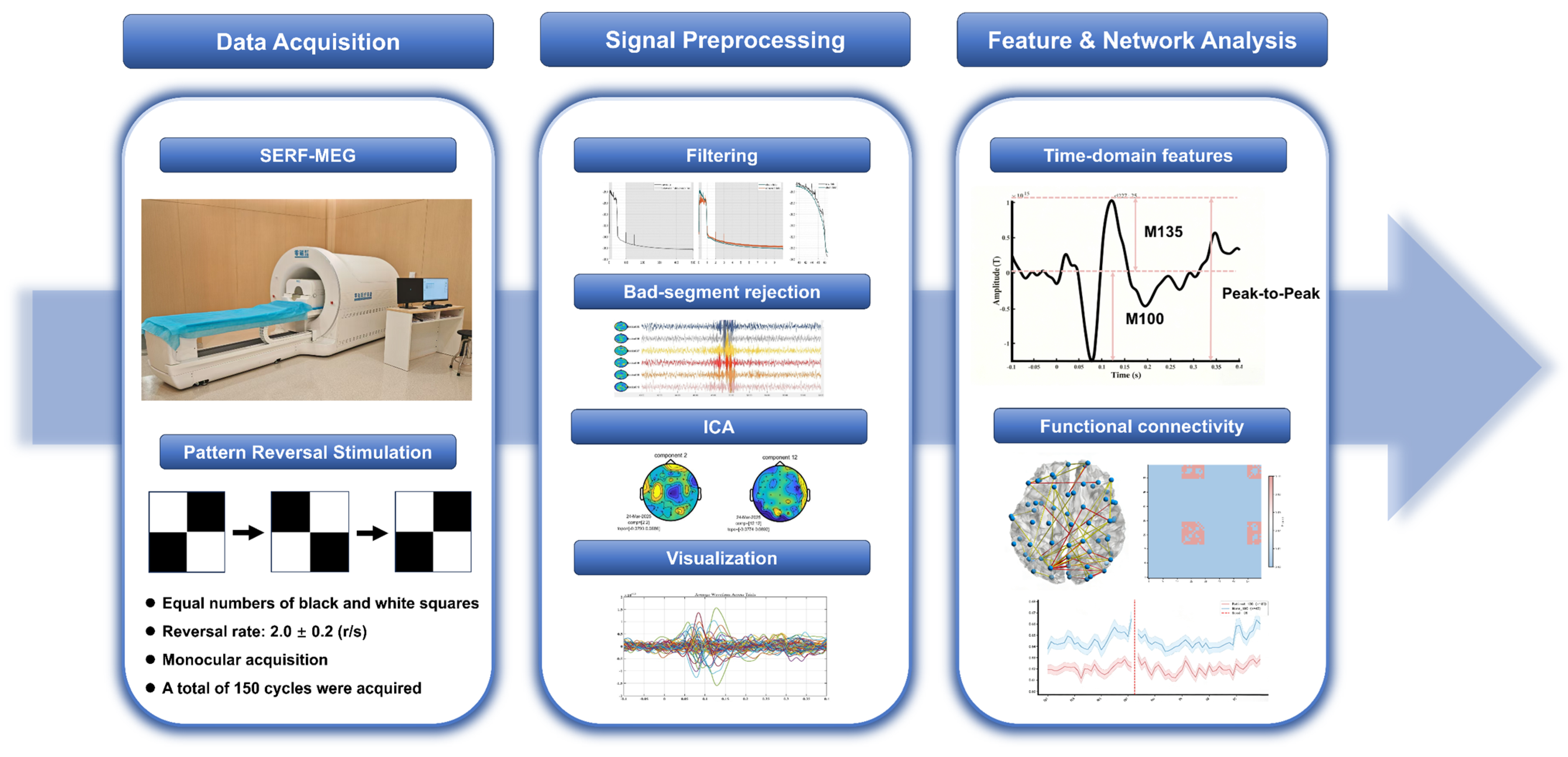
2.3. SERF-MEG Recording
2.4. MEG Signal Preprocessing
2.5. Feature Extraction
2.5.1. Time-Domain and Global Brain Features
2.5.2. Functional Connectivity Analysis
2.6. Statistical Analysis
3. Results
3.1. Clinical Characteristics
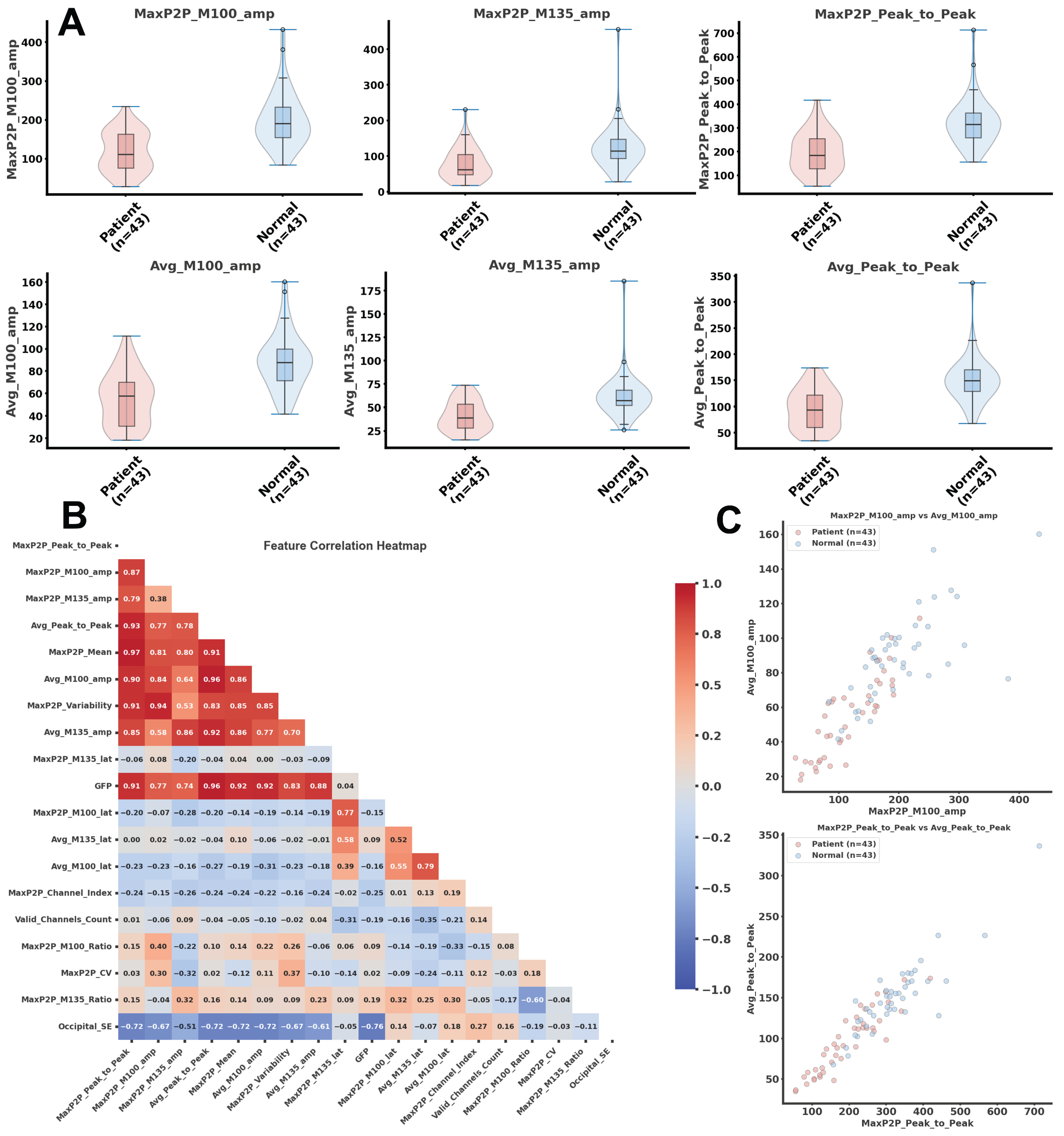
3.2. Global Electrophysiological Differences
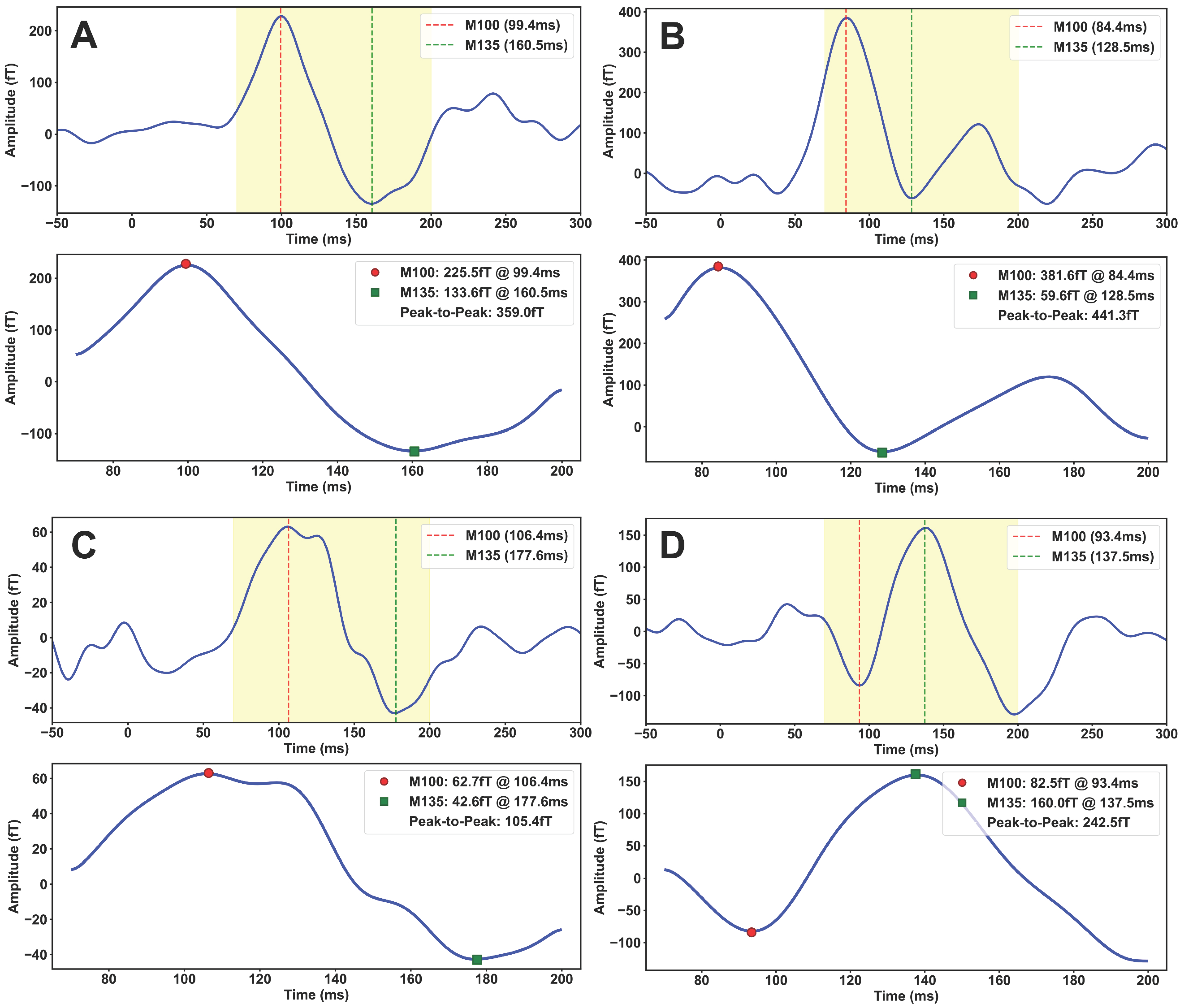
3.3. Time-Domain Feature Analysis
3.4. Feature Correlation Analysis
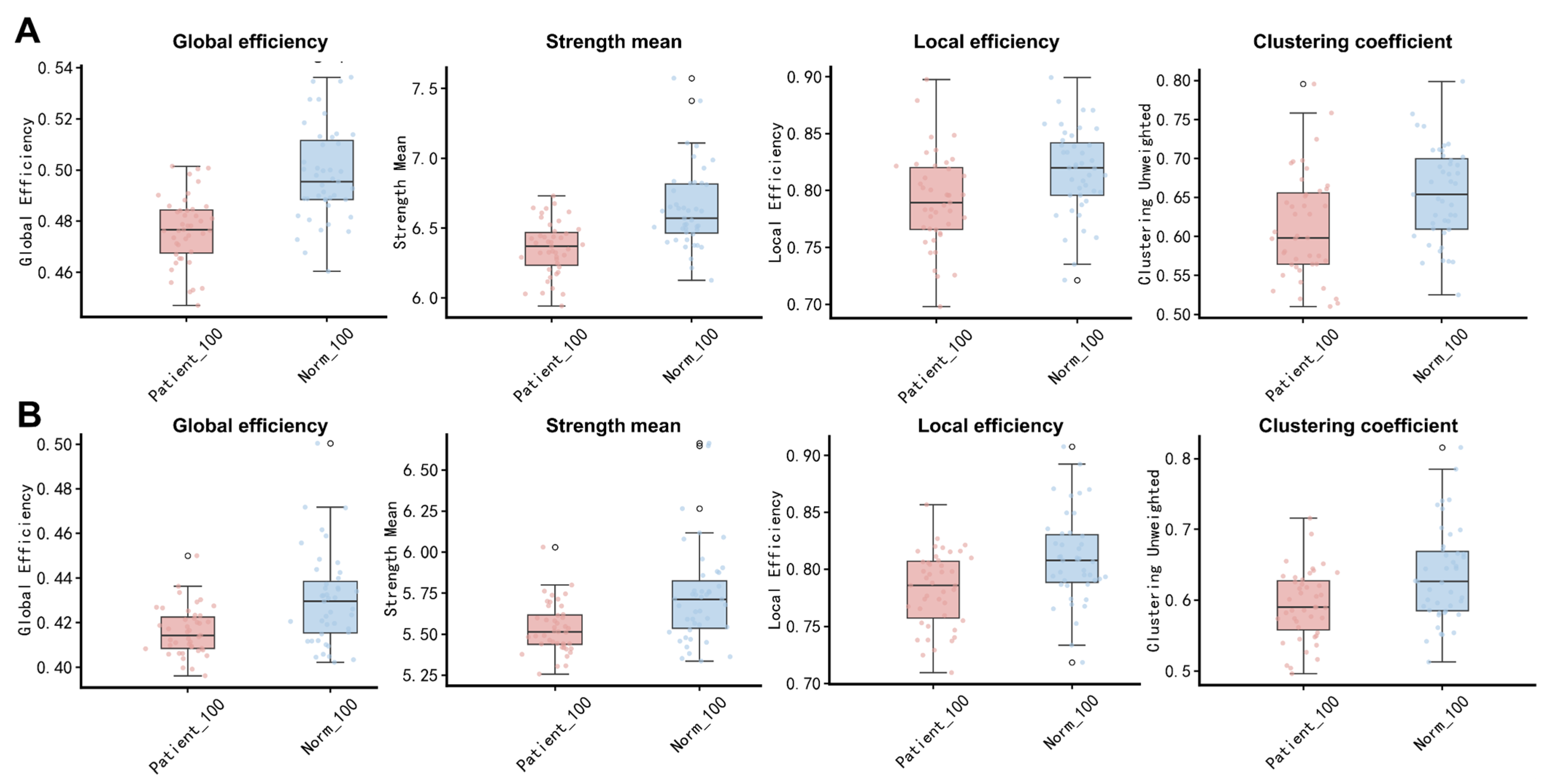
3.5. Overall Alterations in Network Topology
- (1)
- Alpha-Band Network Alterations
- (2)
- Beta-Band Network Alterations
3.6. Visual-Pathway-Specific Connectivity Alterations
- (1)
- Reduced Occipito-Parietal Connectivity
- (2)
- Reduced Occipito-Temporal Connectivity
- (3)
- Reduced Intra-Occipital Coupling
4. Discussion
4.1. Optic Nerve Injury Is Associated with Altered Cortical Visual Responses
4.2. Optic Nerve Injury Is Associated with Frequency-Specific Functional Network Reorganization
4.3. Clinical Relevance, Limitations, and Future Perspectives of SERF-MEG
5. Conclusions
Supplementary Materials
Author Contributions
Funding
Institutional Review Board Statement
Informed Consent Statement
Data Availability Statement
Conflicts of Interest
Abbreviations
| MEG | Magnetoencephalography |
| SERF-MEG | Spin-exchange relaxation-free magnetoencephalography |
| VEP | Visual evoked potential |
| OCT | Optical coherence tomography |
| fMRI | Functional magnetic resonance imaging |
| EEG | Electroencephalography |
| ON | Optic neuritis |
| ION | Ischemic optic neuropathy |
| TON | Traumatic optic neuropathy |
| NMOSD | Neuromyelitis optica spectrum disorder |
| MS | Multiple sclerosis |
| V1 | Primary visual cortex |
| V2 | Secondary visual cortex |
| GFP | Global field power |
| Occipital_SampEn | Occipital sample entropy |
| wPLI | Weighted phase lag index |
| MaxP2P | Maximum peak-to-peak amplitude |
| Avg | Whole-channel average |
| GE | Global efficiency |
| LE | Local efficiency |
| CC | Clustering coefficient |
| Strength_mean | Mean connection strength |
References
- Salvetat, M.L.; Pellegrini, F.; Spadea, L.; Salati, C.; Zeppieri, M. Non-Arteritic Anterior Ischemic Optic Neuropathy (NA-AION): A Comprehensive Overview. Vision 2023, 7, 72. [Google Scholar] [CrossRef] [Scilit]
- Bennett, J.L.; de Seze, J.; Lana-Peixoto, M.; Palace, J.; Waldman, A.; Schippling, S.; Tenembaum, S.; Banwell, B.; Greenberg, B.; Levy, M.; et al. Neuromyelitis optica and multiple sclerosis: Seeing differences through optical coherence tomography. Mult. Scler. J. 2015, 21, 678–688. [Google Scholar] [CrossRef] [Scilit]
- Chen, B.; Zhang, H.; Zhai, Q.; Li, H.; Wang, C.; Wang, Y. Traumatic optic neuropathy: A review of current studies. Neurosurg. Rev. 2022, 45, 1895–1913. [Google Scholar] [CrossRef] [Scilit]
- Song, K.; Wang, Y.; Ren, M.X.; Li, J.; Su, T.; Chen, S.Y.; Shao, Y.; Lv, Y.L. Resting-State Functional Magnetic Resonance Imaging and Functional Connectivity Density Mapping in Patients With Optic Neuritis. Front. Neurosci. 2021, 15, 718973. [Google Scholar] [CrossRef] [Scilit] [PubMed]
- Toosy, A.T.; Mason, D.F.; Miller, D.H. Optic neuritis. Lancet Neurol. 2014, 13, 83–99. [Google Scholar] [CrossRef] [Scilit] [PubMed]
- Zheng, C.; Wang, L.; Xu, X.; Zhou, M.; Liu, K.; Zhang, Y.; Zhao, X.; Lu, L.; Qiu, W.; Zhang, X.; et al. Different visual evoked potentials in neuromyelitis optica spectrum disorder-related optic neuritis and idiopathic demyelinating optic neuritis: A prospective longitudinal analysis. BMC Ophthalmol. 2022, 22, 376. [Google Scholar] [CrossRef] [Scilit] [PubMed]
- Petzold, A.; Fraser, C.L.; Abegg, M.; Alroughani, R.; Alshowaeir, D.; Alvarenga, R.; Andris, C.; Asgari, N.; Barnett, Y.; Battistella, R.; et al. Diagnosis and classification of optic neuritis. Lancet Neurol. 2022, 21, 1120–1134. [Google Scholar] [CrossRef] [Scilit] [PubMed]
- Raz, N.; Chokron, S.; Ben-Hur, T.; Levin, N. Temporal reorganization to overcome monocular demyelination. Neurology 2013, 81, 702–709. [Google Scholar] [CrossRef] [Scilit] [PubMed]
- Cujbă, L.; Banc, A.; Stan, C.; Drugan, T.; Nicula, C. Macular OCT’s Proficiency in Identifying Retrochiasmal Visual Pathway Lesions in Multiple Sclerosis—A Pilot Study. Diagnostics 2024, 14, 1221. [Google Scholar] [CrossRef] [Scilit] [PubMed]
- Baillet, S. Magnetoencephalography for brain electrophysiology and imaging. Nat. Neurosci. 2017, 20, 327–339. [Google Scholar] [CrossRef] [Scilit] [PubMed]
- Boto, E.; Holmes, N.; Leggett, J.; Roberts, G.; Shah, V.; Meyer, S.S.; Muñoz, L.D.; Mullinger, K.J.; Tierney, T.M.; Bestmann, S.; et al. Moving magnetoencephalography towards real-world applications with a wearable system. Nature 2018, 555, 657–661. [Google Scholar] [CrossRef] [Scilit] [PubMed]
- Gross, J. Magnetoencephalography in cognitive neuroscience: A primer. Neuron 2019, 104, 189–204. [Google Scholar] [CrossRef] [Scilit] [PubMed]
- Coelli, S.; Corda, M.; Bianchi, A.M. The time-varying brain: A comprehensive review of dynamic functional connectivity analysis in EEG and MEG. J. Neural Eng. 2025, 22, 051005. [Google Scholar] [CrossRef] [Scilit] [PubMed]
- Samogin, J.; Marino, M.; Porcaro, C.; Wenderoth, N.; Dupont, P.; Swinnen, S.P.; Mantini, D. Frequency-dependent functional connectivity in resting state networks. Hum. Brain Mapp. 2020, 41, 5187–5198. [Google Scholar] [CrossRef] [Scilit] [PubMed]
- Stam, C.J.; van Straaten, E.C. The organization of physiological brain networks. Clin. Neurophysiol. 2012, 123, 1067–1087. [Google Scholar] [CrossRef] [Scilit] [PubMed]
- Backner, Y.; Ben-Shalom, I.; Kuchling, J.; Siebert, N.; Scheel, M.; Ruprecht, K.; Brandt, A.; Paul, F.; Levin, N. Cortical topological network changes following optic neuritis. Neurol. Neuroimmunol. Neuroinflamm. 2020, 7, e687. [Google Scholar] [CrossRef] [Scilit] [PubMed]
- Michalareas, G.; Vezoli, J.; van Pelt, S.; Schoffelen, J.M.; Kennedy, H.; Fries, P. Alpha-Beta and Gamma Rhythms Subserve Feedback and Feedforward Influences among Human Visual Cortical Areas. Neuron 2016, 89, 384–397. [Google Scholar] [CrossRef] [Scilit] [PubMed]
- Palva, J.M.; Palva, S. Functional integration across oscillation frequencies by cross-frequency phase synchronization. Eur. J. Neurosci. 2018, 48, 2399–2406. [Google Scholar] [CrossRef] [Scilit] [PubMed]
- Colclough, G.L.; Brookes, M.J.; Smith, S.M.; Woolrich, M.W. A symmetric multivariate leakage correction for MEG connectomes. Neuroimage 2015, 117, 439–448. [Google Scholar] [CrossRef] [Scilit] [PubMed]
- Vidaurre, D.; Hunt, L.T.; Quinn, A.J.; Hunt, B.A.E.; Brookes, M.J.; Nobre, A.C.; Woolrich, M.W. Spontaneous cortical activity transiently organises into frequency specific phase-coupling networks. Nat. Commun. 2018, 9, 2987. [Google Scholar] [CrossRef] [Scilit] [PubMed]
- De Vico Fallani, F.; Richiardi, J.; Chavez, M.; Achard, S. Graph analysis of functional brain networks: Practical issues in translational neuroscience. Philos. Trans. R. Soc. Lond. B Biol. Sci. 2014, 369, 20130521. [Google Scholar] [CrossRef] [Scilit] [PubMed]
- Rubinov, M.; Sporns, O. Complex network measures of brain connectivity: Uses and interpretations. Neuroimage 2010, 52, 1059–1069. [Google Scholar] [CrossRef] [Scilit] [PubMed]
- Zalesky, A.; Fornito, A.; Bullmore, E.T. Network-based statistic: Identifying differences in brain networks. Neuroimage 2010, 53, 1197–1207. [Google Scholar] [CrossRef] [Scilit] [PubMed]
- Palva, J.M.; Wang, S.H.; Palva, S.; Zhigalov, A.; Monto, S.; Brookes, M.J.; Schoffelen, J.-M.; Jerbi, K. Ghost interactions in MEG/EEG source space: A note of caution on inter-areal coupling measures. Neuroimage 2018, 173, 632–643. [Google Scholar] [CrossRef] [Scilit] [PubMed]
- Borna, A.; Carter, T.R.; Colombo, A.P.; Jau, Y.-Y.; McKay, J.; Weisend, M.; Taulu, S.; Stephen, J.M.; Schwindt, P.D.D. Non-Invasive Functional-Brain-Imaging with an OPM-based Magnetoencephalography System. PLoS ONE 2020, 15, e0227684. [Google Scholar] [CrossRef] [Scilit] [PubMed]
- Boto, E.; Bowtell, R.; Krüger, P.; Fromhold, T.M.; Morris, P.G.; Meyer, S.S.; Barnes, G.R.; Brookes, M.J. On the Potential of a New Generation of Magnetometers for MEG: A Beamformer Simulation Study. PLoS ONE 2016, 11, e0157655. [Google Scholar] [CrossRef] [Scilit] [PubMed]
- Nugent, A.C.; Benitez Andonegui, A.; Holroyd, T.; Robinson, S.E. On-scalp magnetocorticography with optically pumped magnetometers: Simulated performance in resolving simultaneous sources. Neuroimage Rep. 2022, 2, 100093. [Google Scholar] [CrossRef] [Scilit] [PubMed]
- Shah, V.K.; Wakai, R.T. A compact, high performance atomic magnetometer for biomedical applications. Phys. Med. Biol. 2013, 58, 8153–8161. [Google Scholar] [CrossRef] [Scilit] [PubMed]
- Papadopoulou, A.; Pfister, A.; Tsagkas, C.; Gaetano, L.; Sellathurai, S.; D’SOuza, M.; Cerdá-Fuertes, N.; Gugleta, K.; Descoteaux, M.; Chakravarty, M.M.; et al. Visual evoked potentials in multiple sclerosis: P100 latency and visual pathway damage including the lateral geniculate nucleus. Clin. Neurophysiol. 2024, 161, 122–132. [Google Scholar] [CrossRef] [Scilit] [PubMed]
- Lehmann, D.; Skrandies, W. Reference-free identification of components of checkerboard-evoked multichannel potential fields. Electroencephalogr. Clin. Neurophysiol. 1980, 48, 609–621. [Google Scholar] [CrossRef] [Scilit] [PubMed]
- Richman, J.S.; Moorman, J.R. Physiological time-series analysis using approximate entropy and sample entropy. Am. J. Physiol. Heart Circ. Physiol. 2000, 278, H2039–H2049. [Google Scholar] [CrossRef] [Scilit] [PubMed]
- Pfurtscheller, G.; Lopes da Silva, F.H. Event-related EEG/MEG synchronization and desynchronization: Basic principles. Clin. Neurophysiol. 1999, 110, 1842–1857. [Google Scholar] [CrossRef] [Scilit] [PubMed]
- Raz, N.; Dotan, S.; Chokron, S.; Ben-Hur, T.; Levin, N. Demyelination affects temporal aspects of perception: An optic neuritis study. Ann. Neurol. 2012, 71, 531–538. [Google Scholar] [CrossRef] [Scilit] [PubMed]
- Ciapă, M.A.; Donica, V.C.; Costea, C.F.; Bogdănici, C.M. Longitudinal Analysis of P100 Wave Amplitude and Latency in Multiple Sclerosis: A 19-Year Retrospective VEP Study. Diagnostics 2025, 15, 1189. [Google Scholar] [CrossRef] [Scilit] [PubMed]
- Sujanthan, S.; Shmuel, A.; Mendola, J.D. Visually driven functional MRI techniques for characterization of optic neuropathy. Front. Hum. Neurosci. 2022, 16, 943603. [Google Scholar] [CrossRef] [Scilit] [PubMed]
- Caffarra, S.; Joo, S.J.; Bloom, D.; Kruper, J.; Rokem, A.; Yeatman, J.D. Development of the visual white matter pathways mediates development of electrophysiological responses in visual cortex. Hum. Brain Mapp. 2021, 42, 5785–5797. [Google Scholar] [CrossRef] [Scilit] [PubMed]
- Langkilde, A.R.; Frederiksen, J.L.; Rostrup, E.; Larsson, H.B. Functional MRI of the visual cortex and visual testing in patients with previous optic neuritis. Eur. J. Neurol. 2002, 9, 277–286. [Google Scholar] [CrossRef] [Scilit] [PubMed]
- Jensen, O.; Mazaheri, A. Shaping functional architecture by oscillatory alpha activity: Gating by inhibition. Front. Hum. Neurosci. 2010, 4, 186. [Google Scholar] [CrossRef] [Scilit] [PubMed]
- Engel, A.K.; Fries, P. Beta-band oscillations—Signalling the status quo? Curr. Opin. Neurobiol. 2010, 20, 156–165. [Google Scholar] [CrossRef] [Scilit] [PubMed]
- Schoonheim, M.M.; Geurts, J.J.; Landi, D.; Douw, L.; van der Meer, M.L.; Vrenken, H.; Polman, C.H.; Barkhof, F.; Stam, C.J. Functional connectivity changes in multiple sclerosis patients: A graph analytical study of MEG resting state data. Hum. Brain Mapp. 2013, 34, 52–61. [Google Scholar] [CrossRef] [Scilit] [PubMed]
- Tewarie, P.; Schoonheim, M.M.; Stam, C.J.; van der Meer, M.L.; van Dijk, B.W.; Barkhof, F.; Polman, C.H.; Hillebrand, A. Cognitive and clinical dysfunction, altered MEG resting-state networks and thalamic atrophy in multiple sclerosis. PLoS ONE 2013, 8, e69318. [Google Scholar] [CrossRef] [Scilit] [PubMed]
- Allred, J.C.; Lyman, R.N.; Kornack, T.W.; Romalis, M.V. High-sensitivity atomic magnetometer unaffected by spin-exchange relaxation. Phys. Rev. Lett. 2002, 89, 130801. [Google Scholar] [CrossRef] [Scilit] [PubMed]



| Frequency Band | Metric | Control Group | Patient Group | p-Value | Cohen’s d |
|---|---|---|---|---|---|
| Alpha band | Global efficiency | 0.50 ± 0.02 | 0.48 ± 0.01 | <0.01 | –1.33 |
| Strength_mean | 6.64 ± 0.30 | 6.36 ± 0.19 | <0.01 | –1.14 | |
| Local efficiency | 0.82 ± 0.04 | 0.79 ± 0.04 | <0.01 | –0.65 | |
| Clustering coefficient | 0.65 ± 0.06 | 0.61 ± 0.07 | <0.01 | –0.66 | |
| Assortativity | –0.27 ± 0.08 | –0.20 ± 0.09 | <0.01 | +0.81 | |
| Beta band | Global efficiency | 0.43 ± 0.02 | 0.42 ± 0.01 | <0.01 | –0.86 |
| Strength_mean | 5.73 ± 0.30 | 5.54 ± 0.15 | <0.01 | –0.81 | |
| Local efficiency | 0.81 ± 0.04 | 0.78 ± 0.03 | <0.01 | –0.74 | |
| Clustering coefficient | 0.64 ± 0.07 | 0.59 ± 0.05 | <0.01 | –0.59 | |
| Assortativity | –0.24 ± 0.11 | –0.19 ± 0.08 | <0.01 | –0.73 |
| Seed | Affected Network | Representative Target Channels | Cohen’s d | No. of Significant Connections |
|---|---|---|---|---|
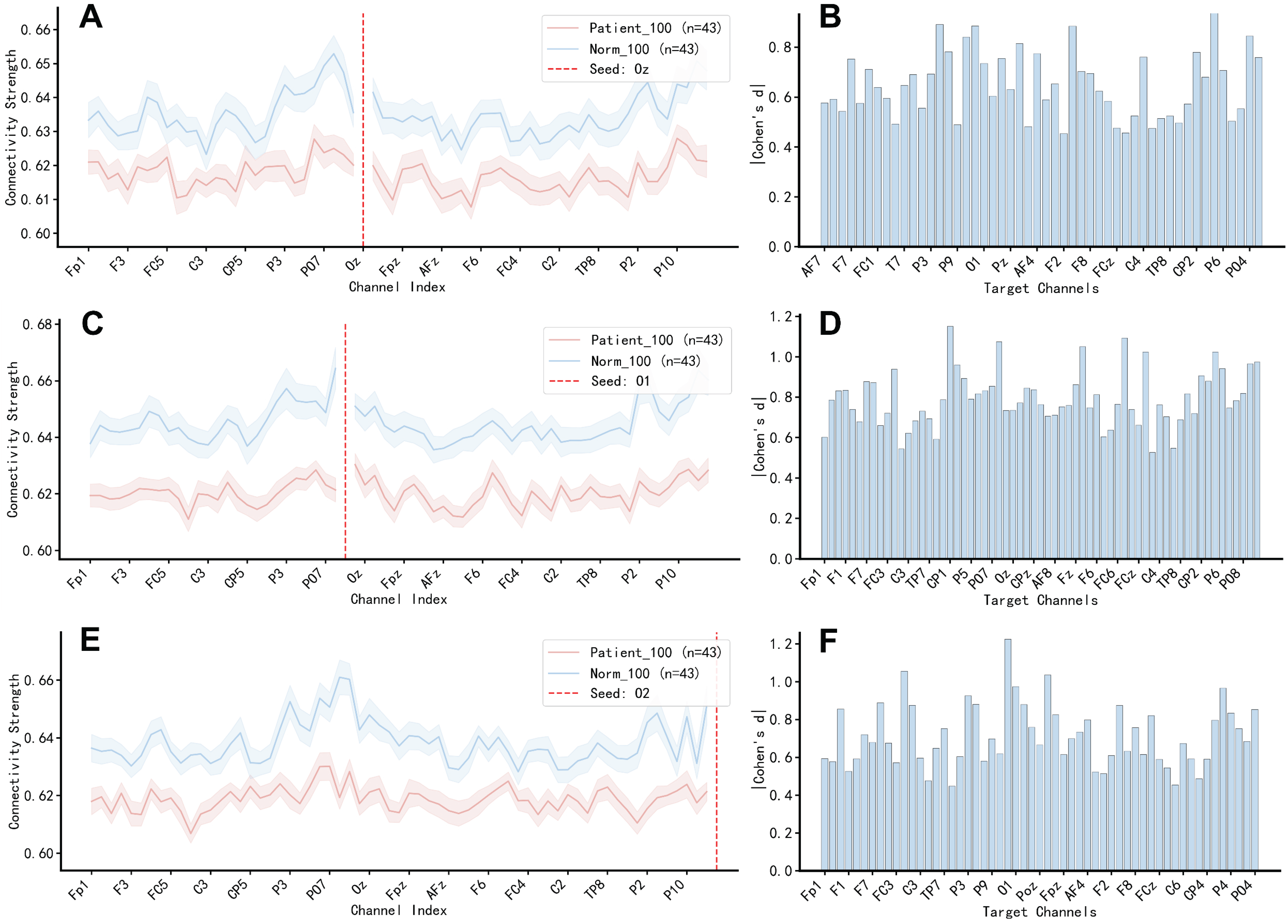
| Oz | Occipito-parietal | P3, P4, PO3, CPz | −0.63 ~ −0.94 | 14 |
| Occipito-temporal | T7, TP7 | −0.65 ~ −0.75 | 4 | |
| Occipito-frontal | F7, F4, FC3 | −0.59 ~ −0.89 | 12 | |
| Inter-occipital | O1, O2 | −0.73 ~ −0.76 | 2 | |
| O1 | Occipito-parietal | CP1, P3, P4, PO3, PO7 | −0.62 ~ −1.15 | 28 |
| Occipito-temporal | T7, TP7, FT7 | −0.69 ~ −0.88 | 6 | |
| Occipito-frontal | Fz, F3, F7, FC1, FC4 | −0.60 ~ −1.09 | 22 | |
| Inter-occipital | O2, Oz | −0.73 ~ −0.97 | 2 | |
| O2 | Occipito-parietal | Pz, P3, P4, PO3 | −0.60 ~ −1.22 | 18 |
| Occipito-temporal | T7, TP7 | −0.65 ~ −0.75 | 4 | |
| Occipito-frontal | FC1, F4, AF3 | −0.57 ~ −1.06 | 16 | |
| Inter-occipital | O1, Oz | −0.76 ~ −0.97 | 2 |
Disclaimer/Publisher’s Note: The statements, opinions and data contained in all publications are solely those of the individual author(s) and contributor(s) and not of MDPI and/or the editor(s). MDPI and/or the editor(s) disclaim responsibility for any injury to people or property resulting from any ideas, methods, instructions or products referred to in the content. |
© 2026 by the authors. Licensee MDPI, Basel, Switzerland. This article is an open access article distributed under the terms and conditions of the Creative Commons Attribution (CC BY) license.
Share and Cite
Wang, H.; Qi, Y.; Lou, Y.; Zhang, X.; Song, X. Decoding Visual Pathway Dysfunction with SERF-MEG: A Study in Patients with Optic Neuropathy. Bioengineering 2026, 13, 694. https://doi.org/10.3390/bioengineering13060694
Wang H, Qi Y, Lou Y, Zhang X, Song X. Decoding Visual Pathway Dysfunction with SERF-MEG: A Study in Patients with Optic Neuropathy. Bioengineering. 2026; 13(6):694. https://doi.org/10.3390/bioengineering13060694
Chicago/Turabian StyleWang, Helei, Yuankun Qi, Yu Lou, Xu Zhang, and Xinda Song. 2026. "Decoding Visual Pathway Dysfunction with SERF-MEG: A Study in Patients with Optic Neuropathy" Bioengineering 13, no. 6: 694. https://doi.org/10.3390/bioengineering13060694
APA StyleWang, H., Qi, Y., Lou, Y., Zhang, X., & Song, X. (2026). Decoding Visual Pathway Dysfunction with SERF-MEG: A Study in Patients with Optic Neuropathy. Bioengineering, 13(6), 694. https://doi.org/10.3390/bioengineering13060694