


Transcriptomic Analysis of Tendon Healing Using an Extracellular Matrix-Coated, Polyurethane Scaffold
 , and
, and
Abstract
1. Introduction
2. Materials and Methods
2.1. Fabrication of the Ctrl-Polyurethane and ECM-Polyurethane Scaffolds
2.2. Establishment of the Large Tendon Defect Model in Rabbits
2.3. RNA Extraction
2.4. RNA-Seq Analysis
2.5. qPCR Analysis
2.6. Histological Characterization for Tendon Healing
2.7. Statistical Analysis
3. Results
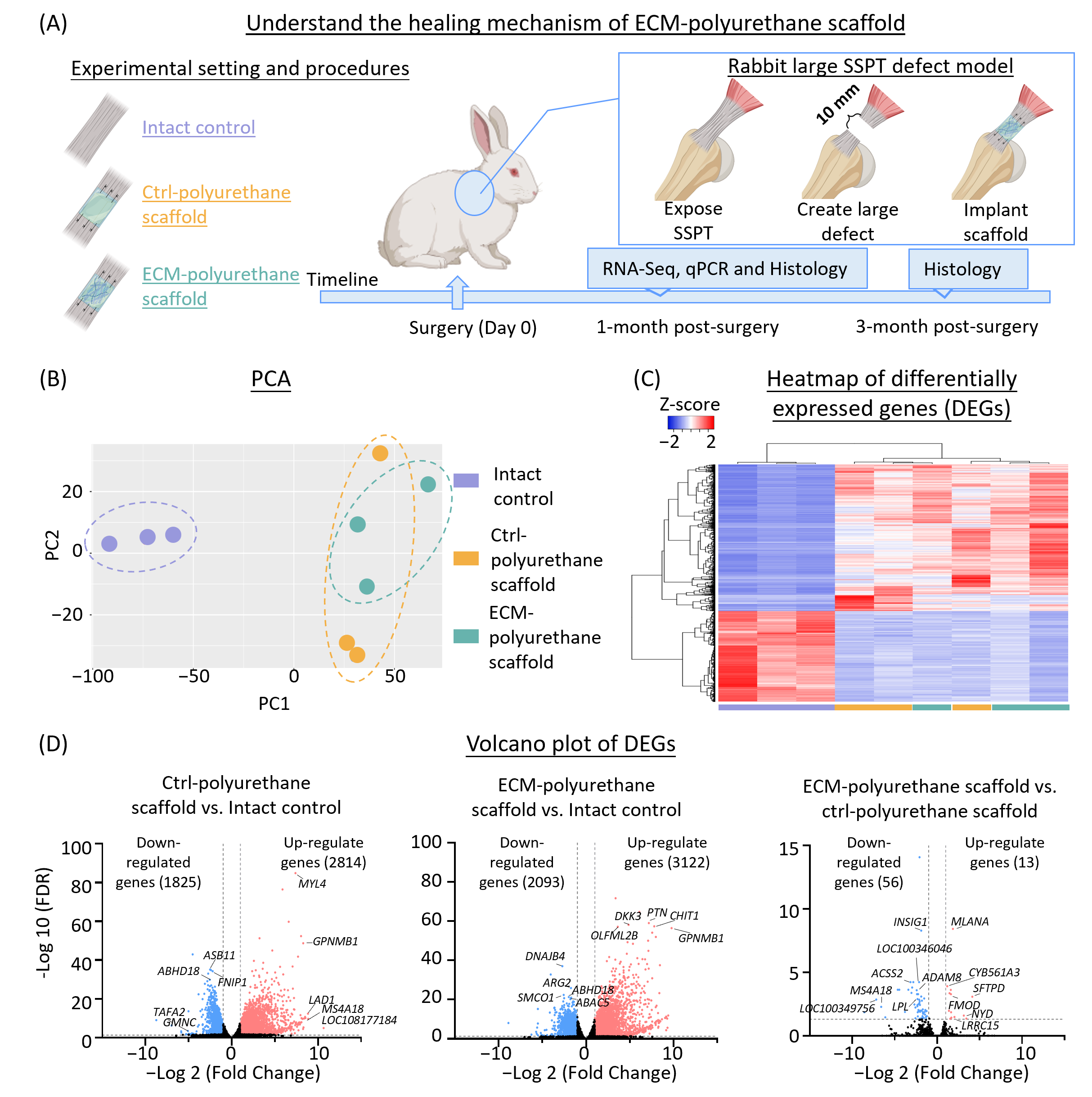
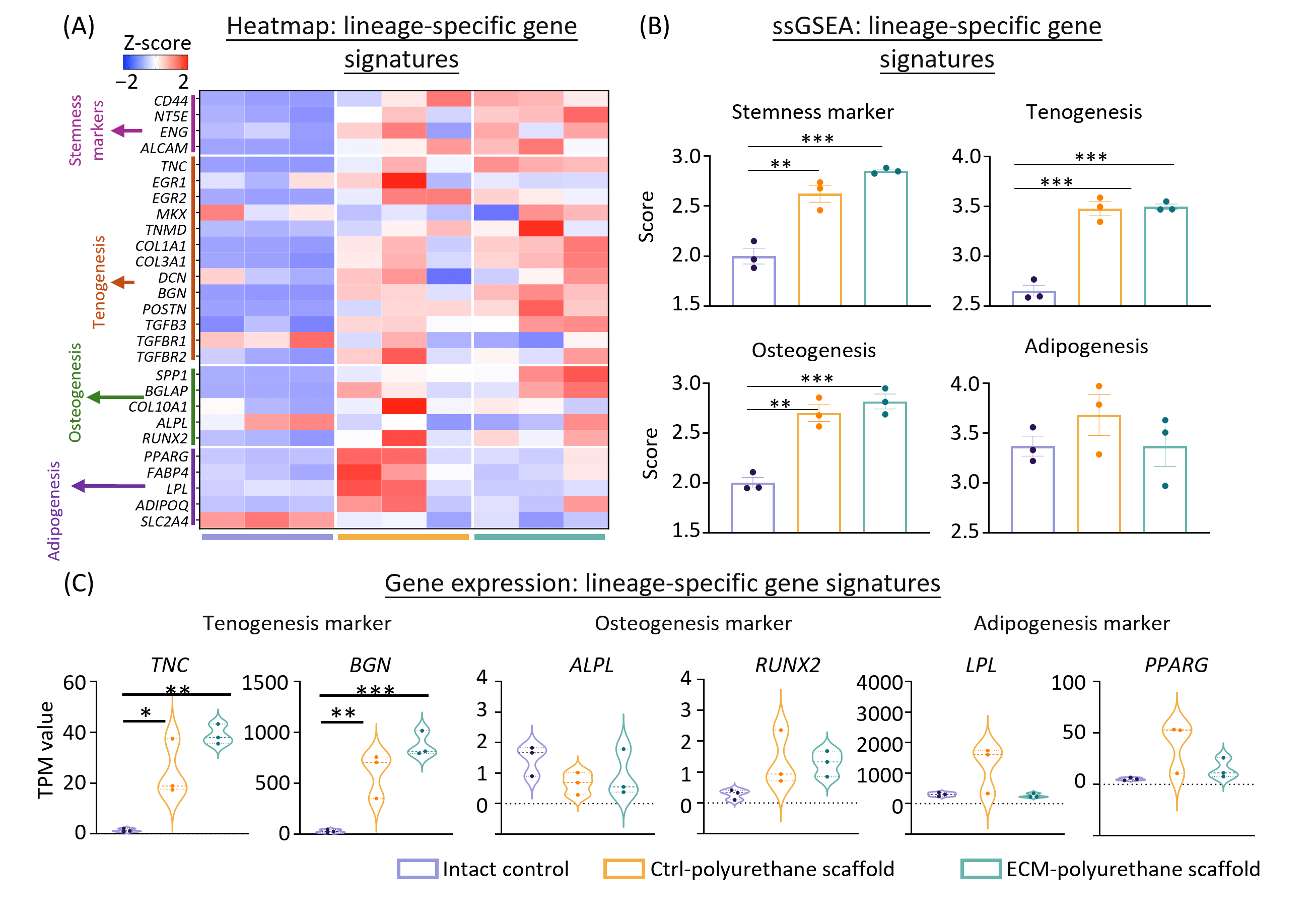
3.1. Overview of Transcriptomic Profiles After the Implantation of Ctrl- and ECM-Polyurethane Scaffolds at One Month Post-Surgery
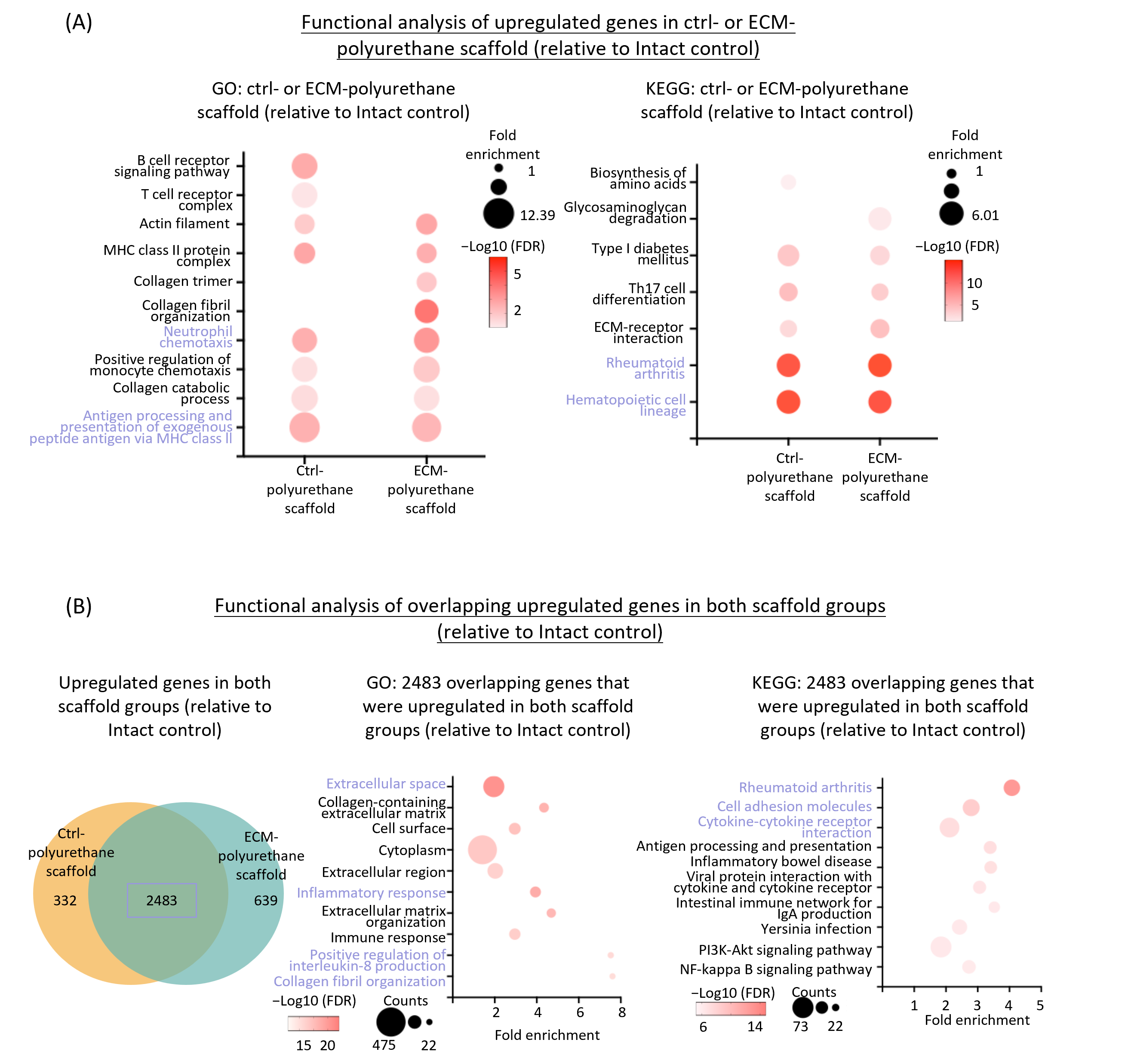
3.2. Similar Immune Activation and Extracellular Matrix Remodeling Signatures in Ctrl- and ECM-Polyurethane Scaffold Groups vs. Intact Control
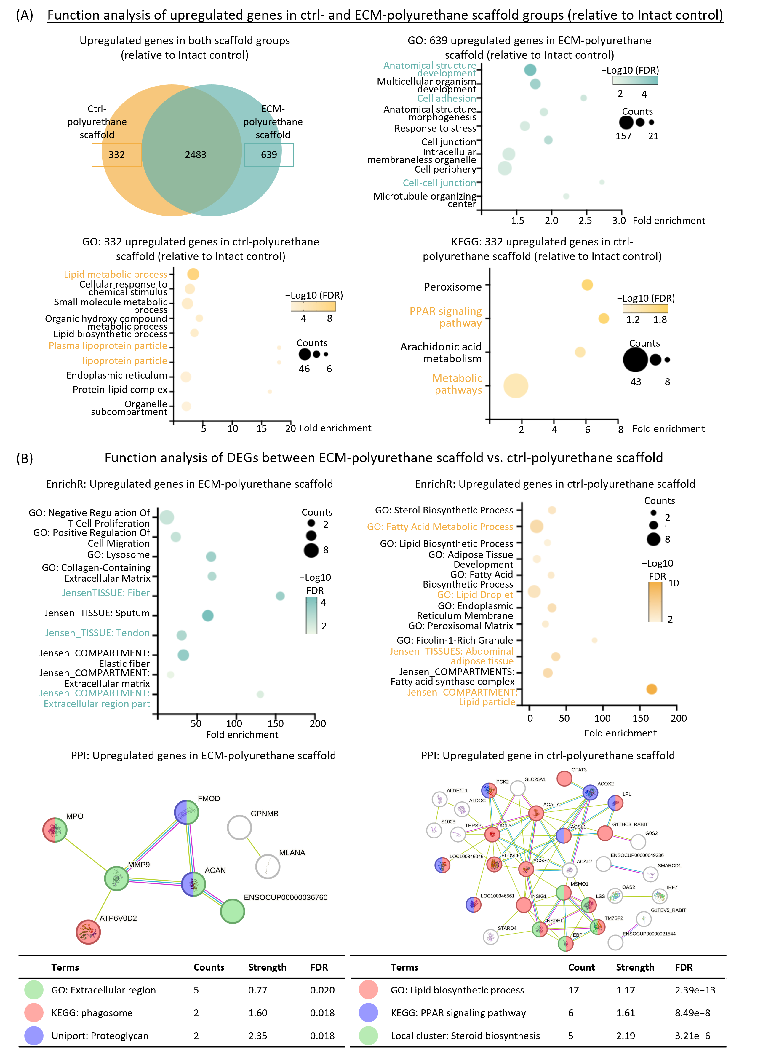
3.3. ECM-Polyurethane Scaffold Group Induces Collagen-Related Programs, Whereas Ctrl-Polyurethane Group Promotes Lipid Biosynthesis
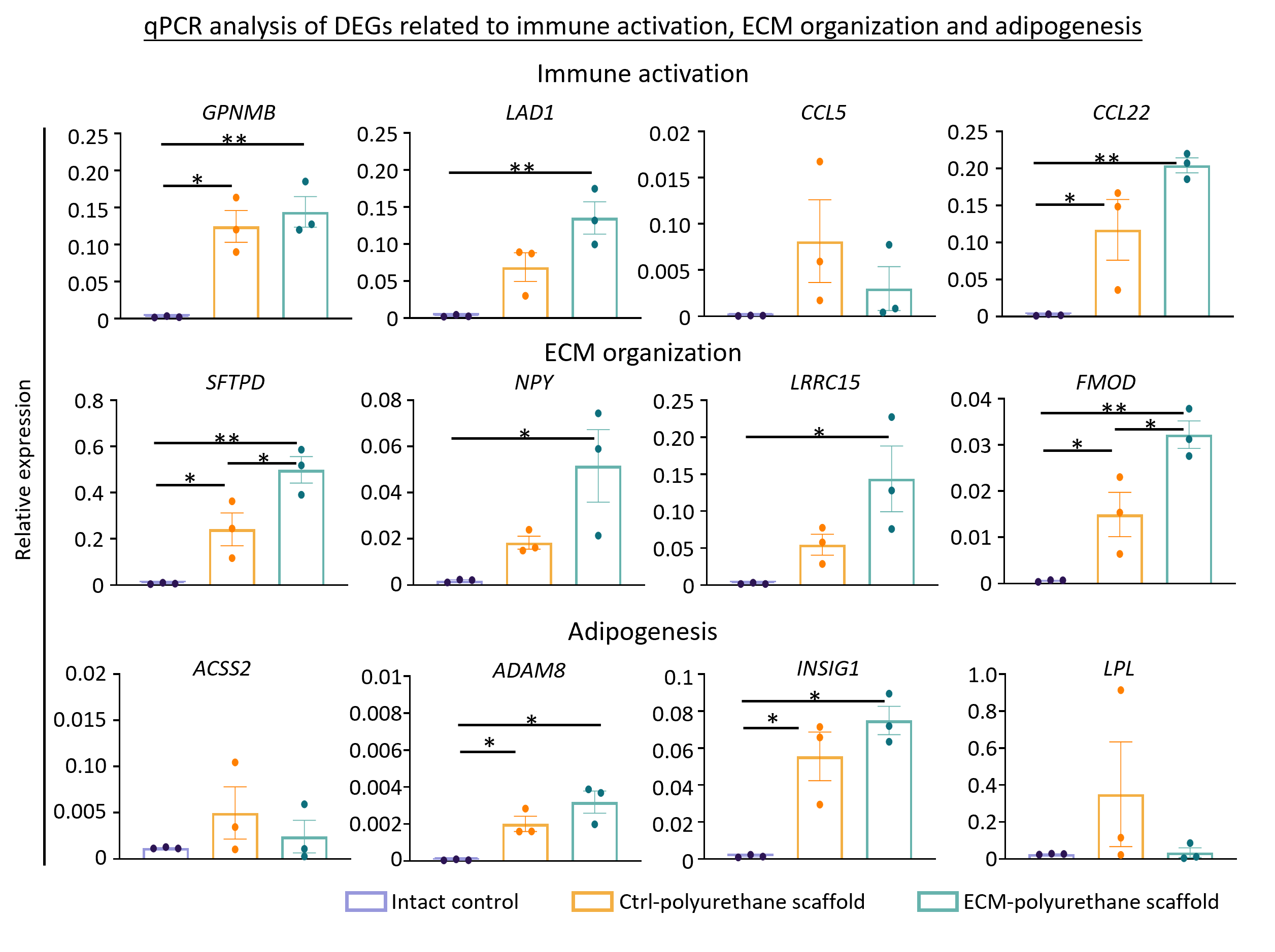
3.4. Validation of Immune Activation, ECM Remodeling and Adipogenesis-Related DEGs in Both Scaffold Groups vs. Intact Control via qPCR
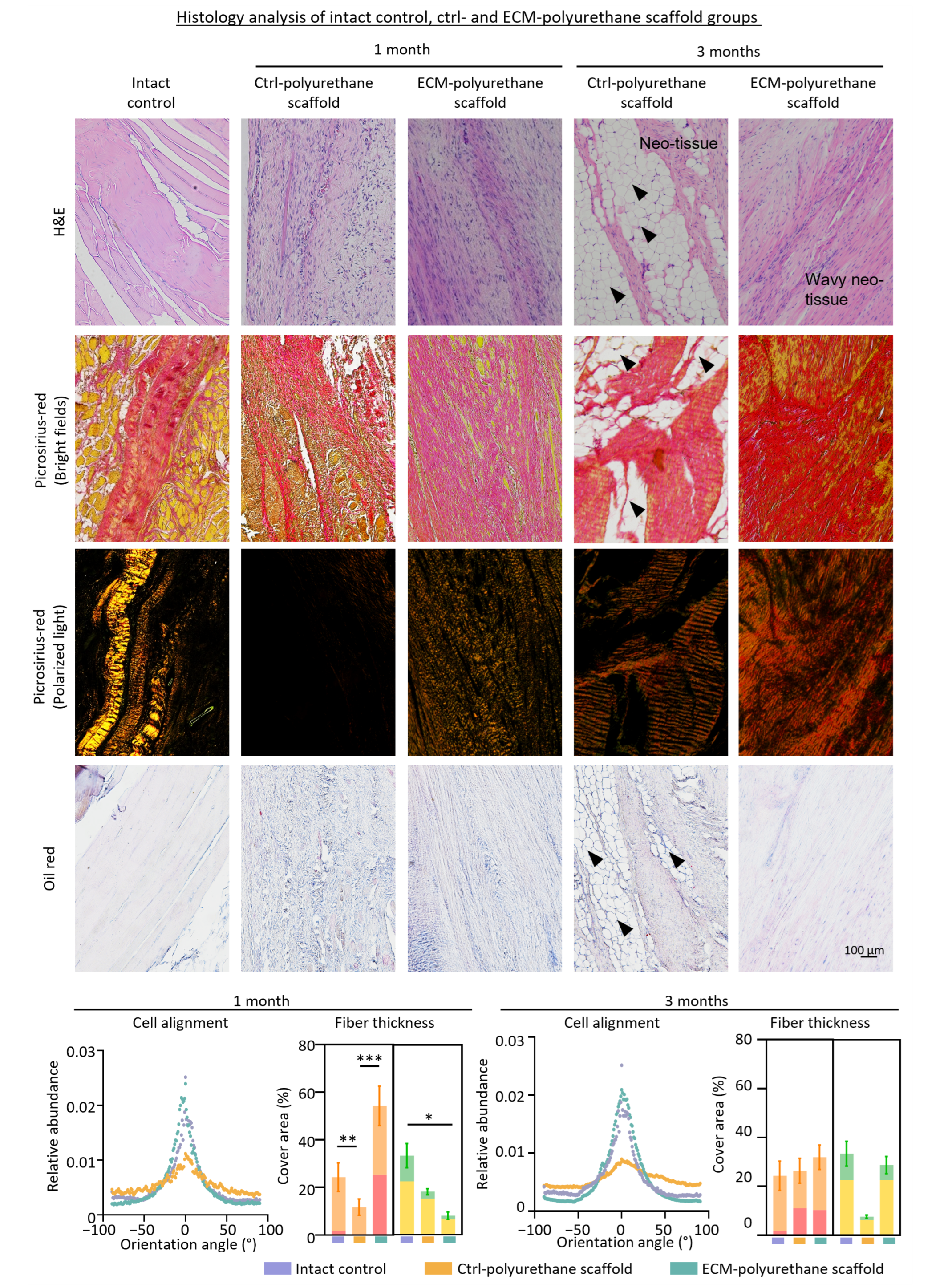
3.5. Implantation of ECM-Polyurethane Scaffold Enhances Rotator Cuff Tendon Healing Whereas Ctrl-Polyurethane Scaffold Leads to Pronounced Fatty Infiltration
4. Discussion
5. Conclusions
Author Contributions
Funding
Institutional Review Board Statement
Informed Consent Statement
Data Availability Statement
Acknowledgments
Conflicts of Interest
References
- Kim, S.J.; Kim, S.H.; Lee, S.K.; Seo, J.W.; Chun, Y.M. Arthroscopic repair of massive contracted rotator cuff tears: Aggressive release with anterior and posterior interval slides do not improve cuff healing and integrity. J. Bone Jt. Surg. Am. 2013, 95, 1482–1488. [Google Scholar] [CrossRef]
- Zhou, H.; Chen, Y.; Yan, W.; Chen, X.; Zi, Y. Advances and challenges in biomaterials for tendon and enthesis repair. Bioact. Mater. 2025, 47, 531–545. [Google Scholar] [CrossRef]
- Galatz, L.M.; Gerstenfeld, L.; Heber-Katz, E.; Rodeo, S.A. Tendon regeneration and scar formation: The concept of scarless healing. J. Orthop. Res. 2015, 33, 823–831. [Google Scholar] [CrossRef]
- Thankam, F.G.; Dilisio, M.F.; Agrawal, D.K. Immunobiological factors aggravating the fatty infiltration on tendons and muscles in rotator cuff lesions. Mol. Cell Biochem. 2016, 417, 17–33. [Google Scholar] [CrossRef] [PubMed]
- Bedi, A.; Bishop, J.; Keener, J.; Lansdown, D.A.; Levy, O.; MacDonald, P.; Maffulli, N.; Oh, J.H.; Sabesan, V.J.; Sanchez-Sotelo, J.; et al. Rotator cuff tears. Nat. Rev. Dis. Prim. 2024, 10, 8. [Google Scholar] [CrossRef]
- Fang, S.; You, M.; Wei, J.; Chen, P. Caveolin-1 is involved in fatty infiltration and bone-tendon healing of rotator cuff tear. Mol. Med. 2023, 29, 33. [Google Scholar] [CrossRef] [PubMed]
- Lake, S.P.; Miller, K.S.; Elliott, D.M.; Soslowsky, L.J. Effect of fiber distribution and realignment on the nonlinear and inhomogeneous mechanical properties of human supraspinatus tendon under longitudinal tensile loading. J. Orthop. Res. 2009, 27, 1596–1602. [Google Scholar] [CrossRef]
- Huang, S.; Rao, Y.; Zhou, M.; Blocki, A.M.; Chen, X.; Wen, C.; Ker, D.F.E.; Tuan, R.S.; Wang, D.M. Engineering an extracellular matrix-functionalized, load-bearing tendon substitute for effective repair of large-to-massive tendon defects. Bioact. Mater. 2024, 36, 221–237. [Google Scholar] [CrossRef] [PubMed]
- Yang, G.; Rothrauff, B.B.; Lin, H.; Gottardi, R.; Alexander, P.G.; Tuan, R.S. Enhancement of tenogenic differentiation of human adipose stem cells by tendon-derived extracellular matrix. Biomaterials 2013, 34, 9295–9306. [Google Scholar] [CrossRef]
- Rao, Y.; Zhu, C.; Suen, H.C.; Huang, S.; Liao, J.; Ker, D.F.E.; Tuan, R.S.; Wang, D. Tenogenic induction of human adipose-derived stem cells by soluble tendon extracellular matrix: Composition and transcriptomic analyses. Stem Cell Res. Ther. 2022, 13, 380. [Google Scholar] [CrossRef]
- Ker, D.F.E.; Wang, D.; Behn, A.W.; Wang, E.T.H.; Zhang, X.; Zhou, B.Y.; Mercado-Pagan, A.E.; Kim, S.; Kleimeyer, J.; Gharaibeh, B.; et al. Functionally Graded, Bone- and Tendon-Like Polyurethane for Rotator Cuff Repair. Adv. Funct. Mater. 2018, 28, 1707107. [Google Scholar] [CrossRef]
- Derwin, K.A.; Badylak, S.F.; Steinmann, S.P.; Iannotti, J.P. Extracellular matrix scaffold devices for rotator cuff repair. J. Shoulder Elb. Surg. 2010, 19, 467–476. [Google Scholar] [CrossRef]
- Lynskey, S.J.; Moody, C.; Ziemann, M.; Gill, S.D.; McGee, S.L.; Page, R.S. From tear to repair: Transcriptomic signatures related to rotator cuff tendon healing outcomes: A prospective clinical and laboratory study. J. Shoulder Elb. Surg. 2025, 35, 602–615. [Google Scholar] [CrossRef]
- Tang, C.; Wang, Z.; Xie, Y.; Fei, Y.; Luo, J.; Wang, C.; Ying, Y.; He, P.; Yan, R.; Chen, Y.; et al. Classification of distinct tendinopathy subtypes for precision therapeutics. Nat. Commun. 2024, 15, 9460. [Google Scholar] [CrossRef] [PubMed]
- Huang, S.; Tam, M.Y.; Ho, W.H.C.; Wong, H.K.; Zhou, M.; Zeng, C.; Xie, D.; Elmer Ker, D.F.; Ling, S.K.; Tuan, R.S.; et al. Establishing a rabbit model with massive supraspinatus tendon defect for investigating scaffold-assisted tendon repair. Biol. Proced. Online 2024, 26, 31. [Google Scholar] [CrossRef]
- Klinger, H.M.; Steckel, H.; Spahn, G.; Buchhorn, G.H.; Baums, M.H. Biomechanical comparison of double-loaded suture anchors using arthroscopic Mason-Allen stitches versus traditional transosseous suture technique and modified Mason-Allen stitches for rotator cuff repair. Clin. Biomech. 2007, 22, 106–111. [Google Scholar] [CrossRef]
- Moriya, T.; Zhao, C.; An, K.N.; Amadio, P.C. The effect of epitendinous suture technique on gliding resistance during cyclic motion after flexor tendon repair: A cadaveric study. J. Hand Surg. Am. 2010, 35, 552–558. [Google Scholar] [CrossRef] [PubMed][Green Version]
- Love, M.I.; Huber, W.; Anders, S. Moderated estimation of fold change and dispersion for RNA-seq data with DESeq2. Genome Biol. 2014, 15, 550. [Google Scholar] [CrossRef] [PubMed]
- Ashburner, M.; Ball, C.A.; Blake, J.A.; Botstein, D.; Butler, H.; Cherry, J.M.; Davis, A.P.; Dolinski, K.; Dwight, S.S.; Eppig, J.T.; et al. Gene ontology: Tool for the unification of biology. The Gene Ontology Consortium. Nat. Genet. 2000, 25, 25–29. [Google Scholar] [CrossRef]
- Gene Ontology, C.; Aleksander, S.A.; Balhoff, J.; Carbon, S.; Cherry, J.M.; Drabkin, H.J.; Ebert, D.; Feuermann, M.; Gaudet, P.; Harris, N.L.; et al. The Gene Ontology knowledgebase in 2023. Genetics 2023, 224, iyad031. [Google Scholar] [CrossRef]
- Thomas, P.D.; Ebert, D.; Muruganujan, A.; Mushayahama, T.; Albou, L.P.; Mi, H. PANTHER: Making genome-scale phylogenetics accessible to all. Protein Sci. 2022, 31, 8–22. [Google Scholar] [CrossRef]
- Xie, Z.; Bailey, A.; Kuleshov, M.V.; Clarke, D.J.B.; Evangelista, J.E.; Jenkins, S.L.; Lachmann, A.; Wojciechowicz, M.L.; Kropiwnicki, E.; Jagodnik, K.M.; et al. Gene Set Knowledge Discovery with Enrichr. Curr. Protoc. 2021, 1, e90. [Google Scholar] [CrossRef]
- Kanehisa, M.; Goto, S. KEGG: Kyoto encyclopedia of genes and genomes. Nucleic Acids Res. 2000, 28, 27–30. [Google Scholar] [CrossRef]
- Huang, D.W.; Sherman, B.T.; Lempicki, R.A. Systematic and integrative analysis of large gene lists using DAVID bioinformatics resources. Nat. Protoc. 2009, 4, 44–57. [Google Scholar] [CrossRef]
- Hanzelmann, S.; Castelo, R.; Guinney, J. GSVA: Gene set variation analysis for microarray and RNA-seq data. BMC Bioinform. 2013, 14, 7. [Google Scholar] [CrossRef] [PubMed]
- Hu, C.; Li, T.; Xu, Y.; Zhang, X.; Li, F.; Bai, J.; Chen, J.; Jiang, W.; Yang, K.; Ou, Q.; et al. CellMarker 2.0: An updated database of manually curated cell markers in human/mouse and web tools based on scRNA-seq data. Nucleic Acids Res. 2023, 51, D870–D876. [Google Scholar] [CrossRef] [PubMed]
- Yin, Z.; Hu, J.J.; Yang, L.; Zheng, Z.F.; An, C.R.; Wu, B.B.; Zhang, C.; Shen, W.L.; Liu, H.H.; Chen, J.L.; et al. Single-cell analysis reveals a nestin(+) tendon stem/progenitor cell population with strong tenogenic potentiality. Sci. Adv. 2016, 2, e1600874. [Google Scholar] [CrossRef]
- Kaji, D.A.; Montero, A.M.; Patel, R.; Huang, A.H. Transcriptional profiling of mESC-derived tendon and fibrocartilage cell fate switch. Nat. Commun. 2021, 12, 4208. [Google Scholar] [CrossRef] [PubMed]
- Szklarczyk, D.; Gable, A.L.; Lyon, D.; Junge, A.; Wyder, S.; Huerta-Cepas, J.; Simonovic, M.; Doncheva, N.T.; Morris, J.H.; Bork, P.; et al. STRING v11: Protein-protein association networks with increased coverage, supporting functional discovery in genome-wide experimental datasets. Nucleic Acids Res. 2019, 47, D607–D613. [Google Scholar] [CrossRef]
- Zhang, W.; Rao, Y.; Wong, S.H.; Wu, Y.; Zhang, Y.; Yang, R.; Tsui, S.K.; Ker, D.F.E.; Mao, C.; Frith, J.E.; et al. Transcriptome-Optimized Hydrogel Design of a Stem Cell Niche for Enhanced Tendon Regeneration. Adv. Mater. 2025, 37, e2313722. [Google Scholar] [CrossRef]
- Zhang, J.; Brown, R.; Hogan, M.V.; Onishi, K.; Wang, J.H. Metformin improves tendon degeneration by blocking translocation of HMGB1 and suppressing tendon inflammation and senescence in aging mice. J. Orthop. Res. 2023, 41, 1162–1176. [Google Scholar] [CrossRef]
- Rich, L.; Whittaker, P. Collagen and picrosirius red staining: A polarized light assessment of fibrillar hue and spatial distribution. J. Morphol. Sci. 2017, 22, 97–104. [Google Scholar]
- Nadkarni, S.K.; Pierce, M.C.; Park, B.H.; de Boer, J.F.; Whittaker, P.; Bouma, B.E.; Bressner, J.E.; Halpern, E.; Houser, S.L.; Tearney, G.J. Measurement of collagen and smooth muscle cell content in atherosclerotic plaques using polarization-sensitive optical coherence tomography. J. Am. Coll. Cardiol. 2007, 49, 1474–1481. [Google Scholar] [CrossRef]
- Faulk, D.M.; Londono, R.; Wolf, M.T.; Ranallo, C.A.; Carruthers, C.A.; Wildemann, J.D.; Dearth, C.L.; Badylak, S.F. ECM hydrogel coating mitigates the chronic inflammatory response to polypropylene mesh. Biomaterials 2014, 35, 8585–8595. [Google Scholar] [CrossRef]
- Sawadkar, P.; Sibbons, P.; Ahmed, T.; Bozec, L.; Mudera, V. Engineering of a functional tendon using collagen as a natural polymer. ACS Biomater. Sci. Eng. 2019, 5, 5218–5228. [Google Scholar] [CrossRef]
- Saade, M.; Araujo de Souza, G.; Scavone, C.; Kinoshita, P.F. The Role of GPNMB in Inflammation. Front. Immunol. 2021, 12, 674739. [Google Scholar] [CrossRef]
- Baer, C.; Kimura, S.; Rana, M.S.; Kleist, A.B.; Flerlage, T.; Feith, D.J.; Chockley, P.; Walter, W.; Meggendorfer, M.; Olson, T.L.; et al. CCL22 mutations drive natural killer cell lymphoproliferative disease by deregulating microenvironmental crosstalk. Nat. Genet. 2022, 54, 637–648. [Google Scholar] [CrossRef]
- Whittaker, P.; Kloner, R.A.; Boughner, D.R.; Pickering, J.G. Quantitative assessment of myocardial collagen with picrosirius red staining and circularly polarized light. Basic. Res. Cardiol. 1994, 89, 397–410. [Google Scholar] [CrossRef] [PubMed]
- Wang, T.; Yu, Z.; Lin, S.; Chen, Z.; Jin, H.; Liang, L.; Zhang, Z.Y. 3D-printed Mg-incorporated PCL-based scaffolds improves rotator cuff tendon-bone healing through regulating macrophage polarization. Front. Bioeng. Biotechnol. 2024, 12, 1407512. [Google Scholar] [CrossRef] [PubMed]
- Ruiz-Alonso, S.; Lafuente-Merchan, M.; Ciriza, J.; Saenz-del-Burgo, L.; Pedraz, J.L. Tendon tissue engineering: Cells, growth factors, scaffolds and production techniques. J. Control. Release 2021, 333, 448–486. [Google Scholar] [CrossRef]
- Voleti, P.B.; Buckley, M.R.; Soslowsky, L.J. Tendon healing: Repair and regeneration. Annu. Rev. Biomed. Eng. 2012, 14, 47–71. [Google Scholar] [CrossRef] [PubMed]
- Sharma, P.; Maffulli, N. Biology of tendon injury: Healing, modeling and remodeling. J. Musculoskelet. Neuronal Interact. 2006, 6, 181–190. [Google Scholar] [PubMed]
- Vasquez-Bolanos, L.S.; Gibbons, M.C.; Ruoss, S.; Wu, I.T.; Esparza, M.C.; Fithian, D.C.; Lane, J.G.; Singh, A.; Nasamran, C.A.; Fisch, K.M.; et al. Transcriptional time course after rotator cuff repair in 6 month old female rabbits. Front. Physiol. 2023, 14, 1164055. [Google Scholar] [CrossRef]
- de Girolamo, L.; Morlin Ambra, L.F.; Perucca Orfei, C.; McQuilling, J.P.; Kimmerling, K.A.; Mowry, K.C.; Johnson, K.A.; Phan, A.T.; Whited, J.L.; Gomoll, A.H. Treatment with Human Amniotic Suspension Allograft Improves Tendon Healing in a Rat Model of Collagenase-Induced Tendinopathy. Cells 2019, 8, 1411. [Google Scholar] [CrossRef]
- Ferrer, G.A.; Miller, R.M.; Yoshida, M.; Wang, J.H.; Musahl, V.; Debski, R.E. Localized Rotator Cuff Tendon Degeneration for Cadaveric Shoulders with and Without Tears Isolated to the Supraspinatus Tendon. Clin. Anat. 2020, 33, 1007–1013. [Google Scholar] [CrossRef]
- Klatte-Schulz, F.; Minkwitz, S.; Schmock, A.; Bormann, N.; Kurtoglu, A.; Tsitsilonis, S.; Manegold, S.; Wildemann, B. Different Achilles Tendon Pathologies Show Distinct Histological and Molecular Characteristics. Int. J. Mol. Sci. 2018, 19, 404. [Google Scholar] [CrossRef]
- Thankam, F.G.; Roesch, Z.K.; Dilisio, M.F.; Radwan, M.M.; Kovilam, A.; Gross, R.M.; Agrawal, D.K. Association of Inflammatory Responses and ECM Disorganization with HMGB1 Upregulation and NLRP3 Inflammasome Activation in the Injured Rotator Cuff Tendon. Sci. Rep. 2018, 8, 8918. [Google Scholar] [CrossRef] [PubMed]

| Forward Primers (5′ to 3′) | Reverse Primers (5′ to 3′) | |
|---|---|---|
| GAPDH | GCAAAGTGGATGTTGTCGCC | TGATGACCAGCTTCCCGTTC |
| GPNMB | TACAAGCTGGGACGGTGTTC | ACACGGTCACCTCCATAAGC |
| LAD1 | CCTTCTCTAACTTTTCCGCTCG | TCCCTGCCTGGAAATCGAC |
| CCL5 | CTATACCAGCGGCAAGTGCT | TCTTCTCTGGGTTGGCACAC |
| CCL22 | GGCCTTTAGTGCTCCTCCAG | TTCTCCCGTTCGTCCACTTG |
| SFTPD | CTCTAGGTCGTTGTGCAACTCA | AAACAACCCCCTCAGTGTCTG |
| NPY | CCACAATGCTCGGGAACAAG | CAGGCATACAAGCAGGGACA |
| LRRC15 | CGACTCTACGACAACCCCTG | TTGTTGAGCAGGAGCCAGTT |
| FMOD | AGGCTCCTGGTTAGGAATTTGG | AAGTGAGCTCTGCTGAGAGTG |
| ACSS2 | TGTCCAGGGTAAGCTGAAAGAG | TGCCACCACAAGTCAATCCC |
| ADAM8 | CATCTCACGAACTCTGGTGGTT | AGAAGCCACAGTCTGGGAAGT |
| INSIG1 | AGGGGGCAGTTTCCTGAGTA | AGCACCCTCAACCTATCCCT |
| LPL | GTGTAACATCGGGGAAGCCA | TTCGTGGGAGCACTTCACAA |
Disclaimer/Publisher’s Note: The statements, opinions and data contained in all publications are solely those of the individual author(s) and contributor(s) and not of MDPI and/or the editor(s). MDPI and/or the editor(s) disclaim responsibility for any injury to people or property resulting from any ideas, methods, instructions or products referred to in the content. |
© 2026 by the authors. Licensee MDPI, Basel, Switzerland. This article is an open access article distributed under the terms and conditions of the Creative Commons Attribution (CC BY) license.
Share and Cite
Rao, Y.; Lauwers, M.; Huang, S.; Zhang, Y.; Ker, D.F.E.; Tuan, R.S.; Wang, D.M. Transcriptomic Analysis of Tendon Healing Using an Extracellular Matrix-Coated, Polyurethane Scaffold. Bioengineering 2026, 13, 652. https://doi.org/10.3390/bioengineering13060652
Rao Y, Lauwers M, Huang S, Zhang Y, Ker DFE, Tuan RS, Wang DM. Transcriptomic Analysis of Tendon Healing Using an Extracellular Matrix-Coated, Polyurethane Scaffold. Bioengineering. 2026; 13(6):652. https://doi.org/10.3390/bioengineering13060652
Chicago/Turabian StyleRao, Ying, Marianne Lauwers, Shuting Huang, Yuyue Zhang, Dai Fei Elmer Ker, Rocky S. Tuan, and Dan Michelle Wang. 2026. "Transcriptomic Analysis of Tendon Healing Using an Extracellular Matrix-Coated, Polyurethane Scaffold" Bioengineering 13, no. 6: 652. https://doi.org/10.3390/bioengineering13060652
APA StyleRao, Y., Lauwers, M., Huang, S., Zhang, Y., Ker, D. F. E., Tuan, R. S., & Wang, D. M. (2026). Transcriptomic Analysis of Tendon Healing Using an Extracellular Matrix-Coated, Polyurethane Scaffold. Bioengineering, 13(6), 652. https://doi.org/10.3390/bioengineering13060652

