Physics-Guided Deep Learning for Interpretable Biomedical Image Reconstruction and Pattern Recognition in Diagnostic Frameworks



Abstract
1. Introduction
- A unified PGDL model is proposed for biomedical image simulation, reconstruction, and pattern recognition. Physical priors are explicitly incorporated in it to enhance accuracy, robustness, and interpretability.
- Comprehensive multi-scenario validation is conducted through controlled simulation studies. It involved varying numbers of inclusions, complex geometric structures, multimodal physical fields, noise-corrupted measurements, and synthetic three-dimensional (3D) brain volumes. These aspects represented realistic biomedical imaging conditions.
- Consistent quantitative performance improvements are achieved across all scenarios. It included 32–45% reduction in root mean square error (RMSE) and 4–7 dB increment in peak signal-to-noise ratio (PSNR). Structural similarity index measure (SSIM) exceeded 0.90 demonstrating superior reconstruction fidelity compared to unconstrained data-driven methods.
- Under moderate noise conditions ( = 0.05), strong noise robustness and structural preservation are maintained (PSNR > 32 dB and structural degradation < 5%). It indicated suitability for low signal-to-noise biomedical imaging environments.
- Scalability to volumetric and functional imaging is demonstrated using synthetic 3D brain data. It demonstrated 38–44% reduction in localization error and consistent performance across scales which support applications in functional brain imaging and volumetric pattern recognition.
2. Mathematical Modeling of the Proposed Physics-Guided Deep Learning Framework
2.1. Physics-Based Field Representation
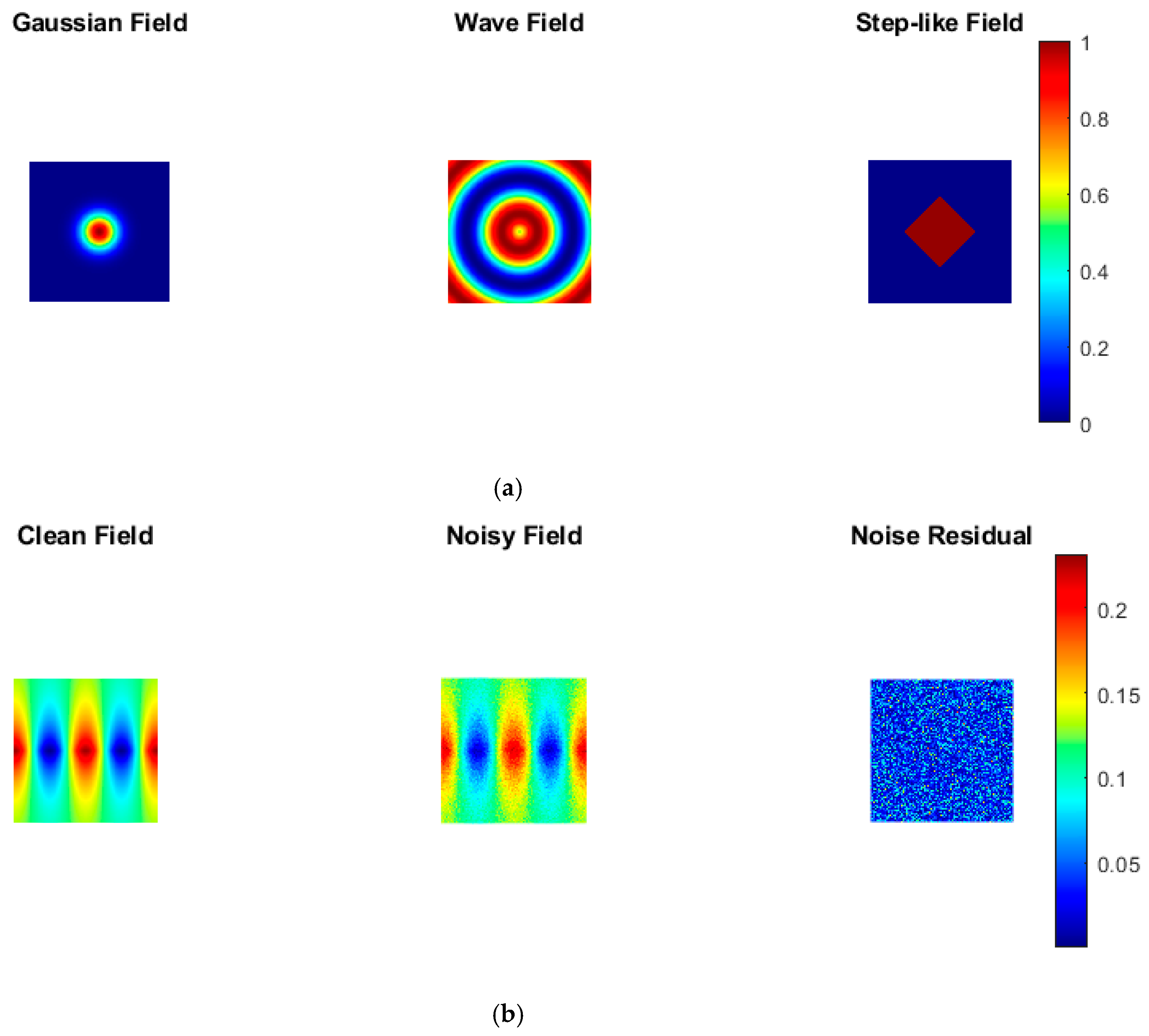
- Gaussian fields for localized tissue activations;
- Wave fields for oscillatory imaging patterns;
- Stepwise intensity fields for modeling abrupt tissue transitions.
2.2. Geometric Inclusion and Boundary Modeling
2.3. Volumetric Activation Modeling
2.4. Noise Modeling
2.5. Physics-Guided Deep Learning Formulation
- enforces physical laws (e.g., smoothness, diffusion, or wave propagation constraints);
- enforces boundary conditions at domain edges or inclusions;
- are weight coefficients that balance data fidelity and physical constraints;
- denotes prescribed (ground-truth) boundary condition values at sampled boundary points .
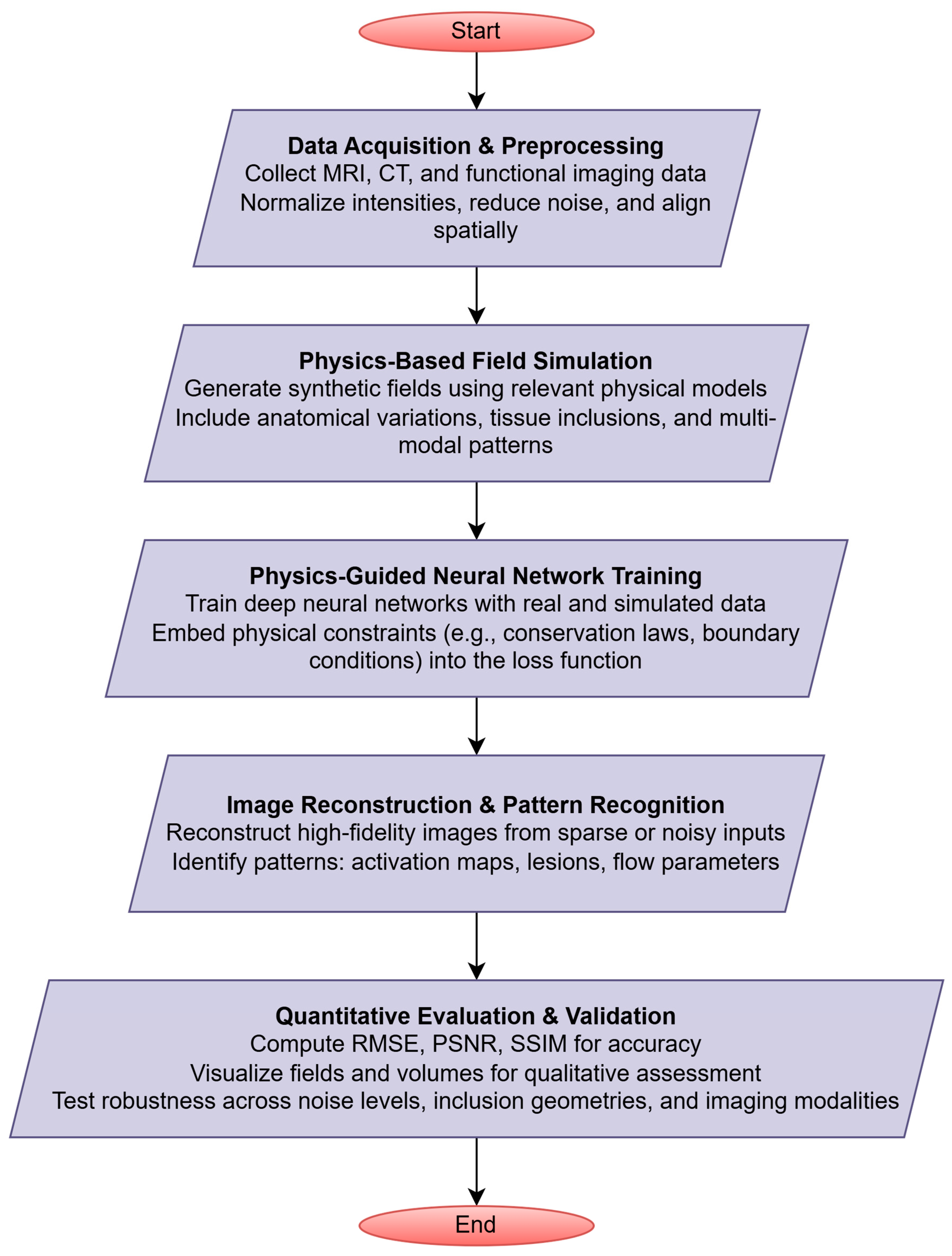
3. Methodology of the Proposed Framework
3.1. Physics-Based Spatial Domain and Field Modeling
3.2. Structural Geometry and Multimodal Field Integration
3.3. Synthetic 3D Brain Volume and Noise Modeling
3.4. Physics-Guided Deep Learning Model Training
3.5. Image Reconstruction, Pattern Recognition, and Performance Evaluation
4. Results and Discussion
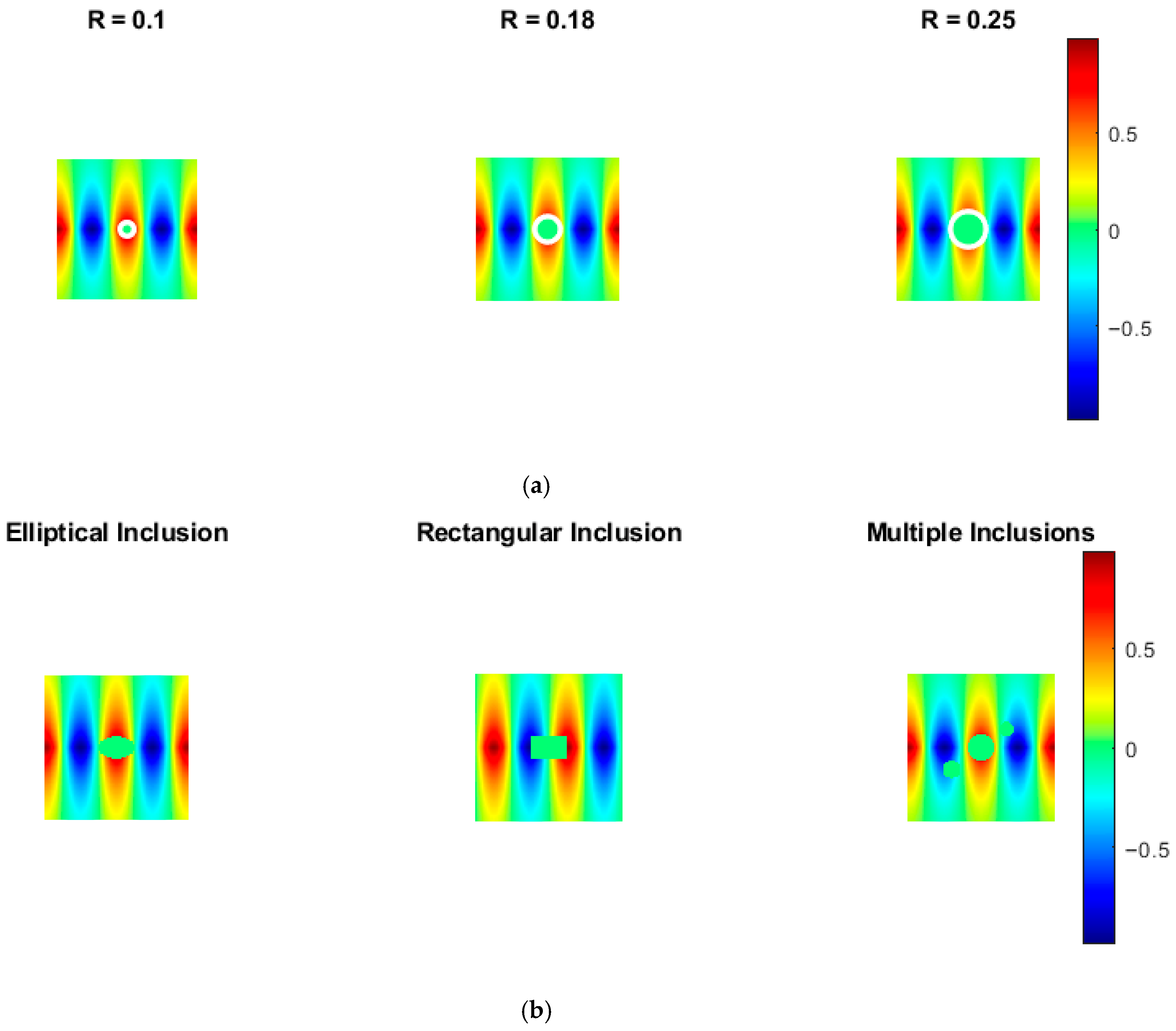
4.1. Physics-Based Field Simulation with Varying Inclusion Sizes
4.2. Impact of Inclusion Geometry on Image Structure
4.3. Multimodal Field Representation Analysis
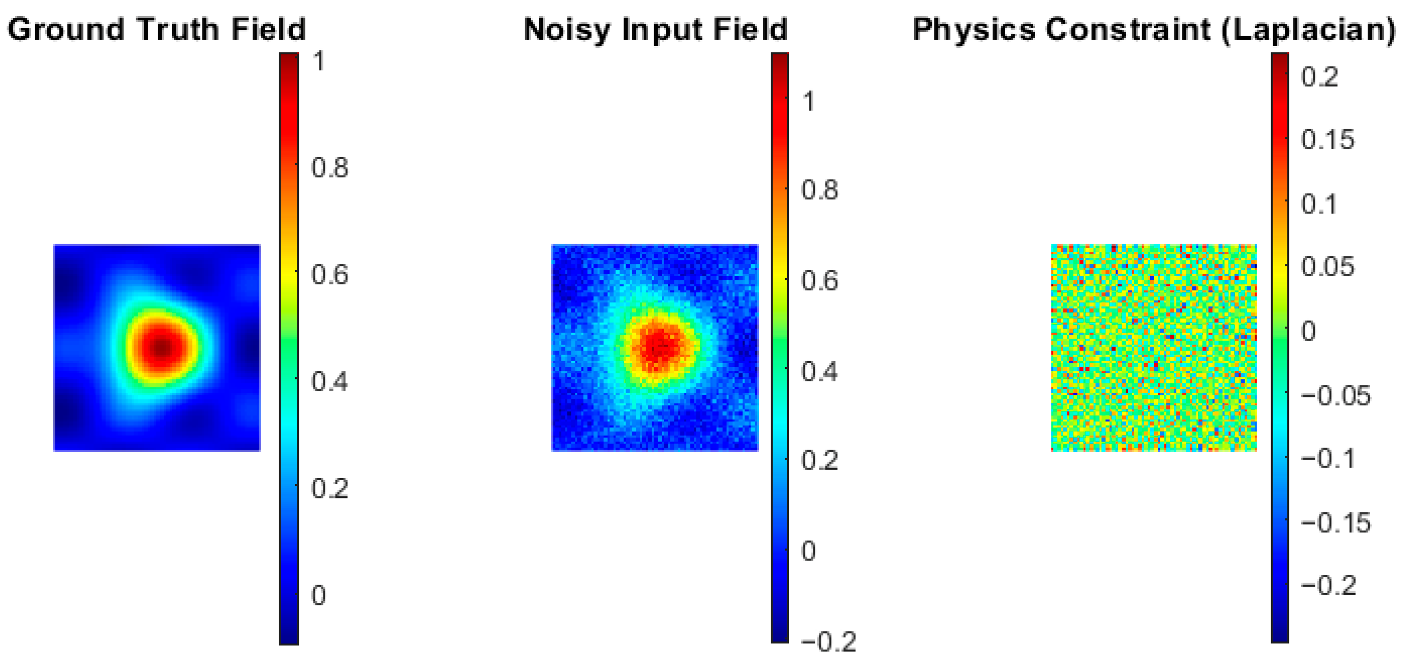
4.4. Noise Robustness Evaluation
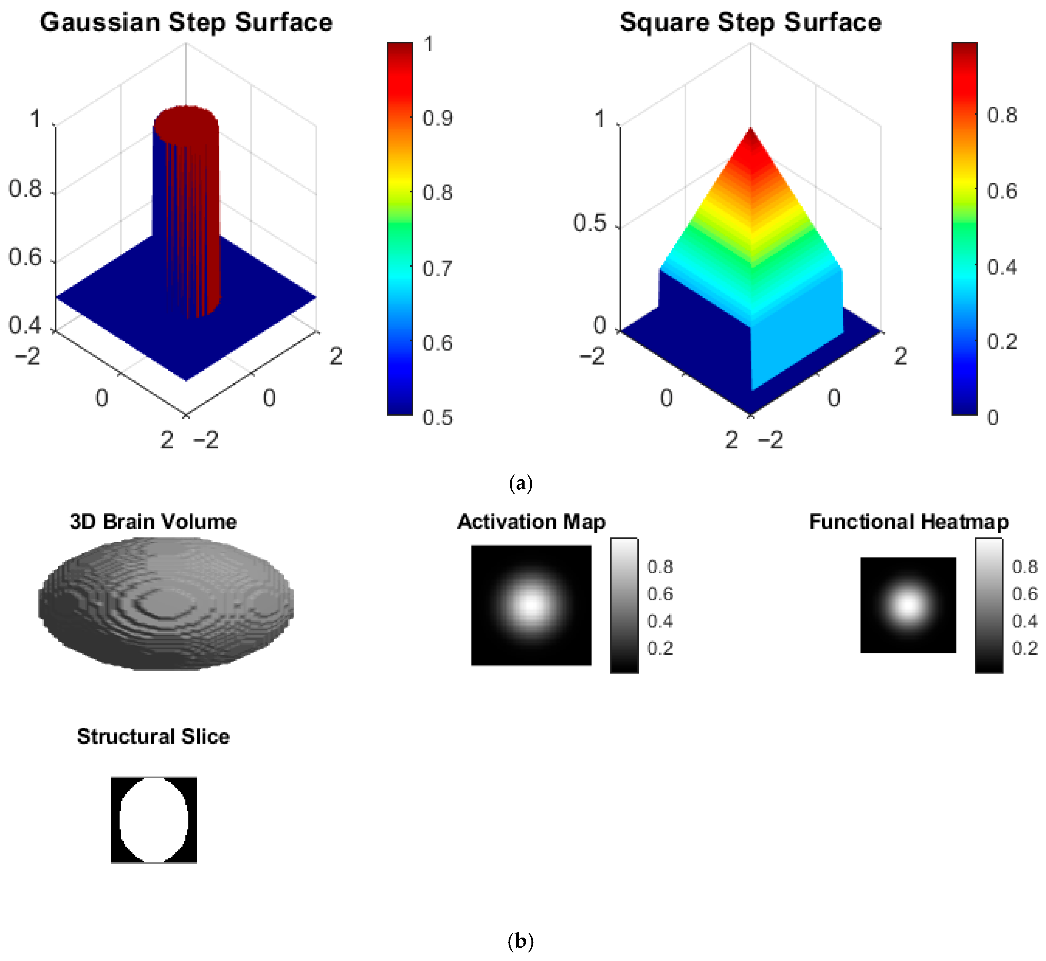
4.5. Step-like Surface Field Visualization
4.6. Synthetic 3D Brain Volume and Activation Mapping
5. Limitations and Generalization to Real Clinical Data
6. Conclusions
Author Contributions
Funding
Institutional Review Board Statement
Informed Consent Statement
Data Availability Statement
Acknowledgments
Conflicts of Interest
Appendix A. PGDL Framework
| Input: Spatial domain , field parameters , inclusion regions , noise level Output: Reconstructed field , evaluation metrics (RMSE, PSNR, SSIM) Procedure:
|
References
- Nasir, K.; Reva, A.; Sekhar, J. Embedding Physics into Deep Learning: A Structured Review of Physics-Informed Neural Networks. Preprints 2025. [Google Scholar] [CrossRef] [Scilit]
- Zhang, H.; Qie, Y. Applying Deep Learning to Medical Imaging: A Review. Appl. Sci. 2023, 13, 10521. [Google Scholar] [CrossRef] [Scilit]
- Ahmadi, M.; Biswas, D.; Lin, M.; Vrionis, F.D.; Hashemi, J.; Tang, Y. Physics-informed machine learning for advancing computational medical imaging: Integrating data-driven approaches with fundamental physical principles. Artif. Intell. Rev. 2025, 58, 297. [Google Scholar] [CrossRef] [Scilit]
- Rahman, A.; Debnath, T.; Kundu, D.; Khan, S.I.; Aishi, A.A.; Sazzad, S.; Sayduzzaman, M.; Band, S.S. Machine learning and deep learning-based approach in smart healthcare: Recent advances, applications, challenges and opportunities. AIMS Public Health 2024, 11, 58–109. [Google Scholar] [CrossRef] [Scilit]
- Rai, R.; Sahu, C.K. Driven by Data or Derived Through Physics? A Review of Hybrid Physics Guided Machine Learning Techniques With Cyber-Physical System (CPS) Focus. IEEE Access 2020, 8, 71050–71073. [Google Scholar] [CrossRef] [Scilit]
- Ahmadi, N.; Cao, Q.; Humphrey, J.D.; Karniadakis, G.E. Physics-Informed Machine Learning in Biomedical Science and Engineering. arXiv 2025, arXiv:2510.05433. [Google Scholar] [CrossRef] [Scilit]
- Banerjee, C.; Nguyen, K.; Salvado, O.; Tran, T.; Fookes, C. Physics-informed Machine Learning for Medical Image Analysis. ACM Comput. Surv. 2025, 58, 1–35. [Google Scholar] [CrossRef] [Scilit]
- Galić, I.; Habijan, M.; Leventić, H.; Romić, K. Machine Learning Empowering Personalized Medicine: A Comprehensive Review of Medical Image Analysis Methods. Electronics 2023, 12, 4411. [Google Scholar] [CrossRef] [Scilit]
- Fujita, S.; Nickel, D.; Lo, W.-C.; Clifford, B.; Cauley, S.; Buathong, S.; Hajati, A.; Chiang, F.L.; Conklin, J.; Huang, S.Y. Physics-informed deep learning reconstruction for ultrafast clinical 3D fluid-attenuated inversion recovery brain MRI. Radiol. Adv. 2025, 2, umaf016. [Google Scholar] [CrossRef] [Scilit]
- van Herten, R.L.M.; Chiribiri, A.; Breeuwer, M.; Veta, M.; Scannell, C.M. Physics-informed neural networks for myocardial perfusion MRI quantification. Med. Image Anal. 2022, 78, 102399. [Google Scholar] [CrossRef] [Scilit] [PubMed]
- Park, J.; Kweon, J.; Kim, Y.I.; Back, I.; Chae, J.; Roh, J.; Kang, D.; Lee, P.H.; Ahn, J.; Kang, S.; et al. Selective ensemble methods for deep learning segmentation of major vessels in invasive coronary angiography. Med. Phys. 2023, 50, 7822–7839. [Google Scholar] [CrossRef] [Scilit]
- Ruan, G.; Wang, Z.; Liu, C.; Xia, L.; Wang, H.; Qi, L.; Chen, W. Magnetic Resonance Electrical Properties Tomography Based on Modified Physics- Informed Neural Network and Multiconstraints. IEEE Trans. Med. Imaging 2024, 43, 3263–3278. [Google Scholar] [CrossRef] [Scilit]
- Lin, R.; Zhang, J. A novel approach to biomechanical modeling: CT image weight initialization and Physics Informed Neural Networks. Biomed. Signal Process. Control 2025, 109, 102399. [Google Scholar] [CrossRef] [Scilit]
- Movahhedi, M.; Liu, X.Y.; Geng, B.; Elemans, C.; Xue, Q.; Wang, J.-X.; Zheng, X. Predicting 3D soft tissue dynamics from 2D imaging using physics informed neural networks. Commun. Biol. 2023, 6, 541. [Google Scholar] [CrossRef] [Scilit]
- Sarabian, M.; Babaee, H.; Laksari, K. Physics-Informed Neural Networks for Brain Hemodynamic Predictions Using Medical Imaging. IEEE Trans. Med. Imaging 2022, 41, 2285–2303. [Google Scholar] [CrossRef] [Scilit]
- Pashaei Kalajahi, A.; Csala, H.; Mamun, Z.B.; Yadav, S.; Amili, O.; Arzani, A.; D’sOuza, R.M. Input parameterized physics informed neural networks for de noising, super-resolution, and imaging artifact mitigation in time resolved three dimensional phase-contrast magnetic resonance imaging. Eng. Appl. Artif. Intell. 2025, 150, 110600. [Google Scholar] [CrossRef] [Scilit]
- Amiri-Hezaveh, A.; Tan, S.; Deng, Q.; Umulis, D.; Cunniff, L.; Weickenmeier, J.; Tepole, A.B. A Physics-Informed Deep Learning Deformable Medical Image Registration Method Based on Neural ODEs. Int. J. Comput. Vis. 2025, 133, 6374–6399. [Google Scholar] [CrossRef] [Scilit]
- Shone, F.; Ravikumar, N.; Lassila, T.; MacRaild, M.; Wang, Y.; Taylor, Z.A.; Jimack, P.; Dall’Armellina, E. Deep Physics-Informed Super-Resolution of Cardiac 4D-Flow MRI. In Proceedings of the International Conference on Information Processing in Medical Imaging, San Carlos de Bariloche, Argentina, 18–23 June 2023. [Google Scholar]
- Huang, J.; Fang, Y.; Nan, Y.; Wu, H.; Wu, Y.; Gao, Z.; Li, Y.; Wang, Z.; Lio, P.; Rueckert, D. Data and Physics Driven Learning Models for Fast MRI–Fundamentals and Methodologies from CNN, GAN to Attention and Transformers. arXiv 2022, arXiv:2204.01706. [Google Scholar]
- Cobo, M.; Fontecha, D.C.; Silva, W.; Iglesias, L.L. Physical foundations for trustworthy medical imaging: A review for artificial intelligence researchers. Artif. Intell. Med. 2025, 169, 103251. [Google Scholar] [CrossRef] [Scilit]
- Osorio Quero, C.; Crespo, M.L. Physics-Informed Neural Network for Denoising Images Using Nonlinear PDE. Electronics 2026, 15, 560. [Google Scholar] [CrossRef] [Scilit]
- Miele, S.; Karve, P.; Mahadevan, S. Multi-fidelity physics-informed machine learning for probabilistic damage diagnosis. Reliab. Eng. Syst. Saf. 2023, 235, 109243. [Google Scholar] [CrossRef] [Scilit]
- Hsu, W.T.; Agbodike, O.; Chen, J. Attentive U-Net with Physics-Informed Loss for Noise Suppression in Medical Ultrasound Images. In Proceedings of the 2024 10th International Conference on Applied System Innovation, ICASI 2024; Institute of Electrical and Electronics Engineers Inc.: Kyoto, Japan, 2024; pp. 409–411. [Google Scholar]
- Cai, Q.; Zhu, L.; Zhou, J.; Qian, C.; Guo, D.; Qu, X. Bloch equation enables physics-informed neural network in parametric magnetic resonance imaging. arXiv 2023, arXiv:2309.11763. [Google Scholar] [CrossRef] [Scilit]
- Zarei, M.; Sotoudeh-Paima, S.; McCabe, C.; Abadi, E.; Samei, E. Harmonizing CT images via physics-based deep neural networks. SPIE-Intl Soc. Opt. Eng. 2023, 66, 12463. [Google Scholar]
- Pasumarthi Venkata, S.; Campbell Arnold, T.; Colombo Serra, S.; Rudie, J.D.; Andre, J.B.; Elor, R.; Gulaka, P.; Sankaranarayanan, A.; Erb, G.; Zaharchuk, G. Deep-Learning Based Contrast Boosting Improves Lesion Visualization and Image Quality: A Multi-Center Multi-Reader Study on Clinical Performance with Standard Contrast Enhanced MRI of Brain Tumors. medRxiv 2025. [Google Scholar] [CrossRef] [Scilit]
- Liu, X.; Almekkawy, M. Ultrasound Computed Tomography using physical-informed Neural Network. In IEEE International Ultrasonics Symposium, IUS; IEEE Computer Society: New York, NY, USA, 2021. [Google Scholar]
- Ragoza, M.; Batmanghelich, K. Physics-Informed Neural Networks for Tissue Elasticity Reconstruction in Magnetic Resonance Elastography. In Lecture Notes in Computer Science (Including Subseries Lecture Notes in Artificial Intelligence and Lecture Notes in Bioinformatics); Springer Science and Business Media Deutschland GmbH: Berlin/Heidelberg, Germany, 2023; pp. 333–343. [Google Scholar]
- Min, Z.; Baum, Z.M.C.; Saeed, S.U.; Emberton, M.; Barratt, D.C.; Taylor, Z.A.; Hu, Y. Biomechanics-informed Non-rigid Medical Image Registration and its Inverse Material Property Estimation with Linear and Nonlinear Elasticity. arXiv 2024, arXiv:2407.03292. [Google Scholar]




| Application | Methodology | Physics Integration | Key Outcomes |
|---|---|---|---|
| Myocardial perfusion MRI [10] | PINN | Tracer-kinetic conservation laws | Reduced MSE; accurate parameter maps |
| Coronary artery segmentation [11] | Selective ensemble DL | Morphological constraints | DSC up to 93%; reduced mask errors; real-time |
| MREPT [12] | Model-driven PINN (FCNN) | Maxwell’s equations | Artifact reduction; stable EP reconstruction |
| Spine biomechanical analysis [13] | PINN-based modeling | Solid mechanics constraints | 91% accuracy; global optimum; fast inference |
| Tissue dynamics and vocal folds [14] | Hybrid PINN + RNN-fluid solver | Fluid–structure interaction | Accurate 3D dynamics from sparse 2D data |
| Cerebral hemodynamics [15] | Physics-informed DL + ROM | 1D blood flow equations | High-res velocity, pressure, area maps |
| 4D flow MRI enhancement [16] | IP-PINN | Navier–Stokes-inspired constraints | <5.5% error; 25% acquisition time reduction |
| Large-deformation image registration [17] | Unsupervised physics-aware DL | Elasticity and growth mechanics | Robust registration; accurate deformation modeling |
| Category | Parameter | Quantitative Description |
|---|---|---|
| Framework | Pipeline stages | 5 stages (modeling → geometry → data → training → evaluation) |
| Spatial domain | Dimensionality | 2D (d = 2) and 3D (d = 3) domains |
| Structural modeling | Geometric regions | ≥4 types (circular, elliptical, rectangular, irregular) |
| Inclusion modeling | with heterogeneous field representation | |
| Multimodal fields | Field types | Gaussian (smooth), wave-based (oscillatory), stepwise |
| Field formulation | ||
| Synthetic dataset | Data type | Synthetic biomedical fields and 3D brain-like volumes |
| Data variability | Multiple geometries × modalities × noise levels | |
| Experimental scenarios | Inclusion size, geometry, modality, noise, 3D volumetric cases | |
| Noise modeling | Noise formulation | |
| Noise type | ) | |
| Noise levels | = 0.05 for moderate noise) | |
| Noise sources | Sensor, motion, acquisition | |
| Neural network | Model type | CNN-based encoder–decoder (image-to-image mapping) |
| Depth | 6–8 layers (including encoding and decoding stages) | |
| Activation | ReLU | |
| Input–output | Noisy/incomplete → reconstructed images | |
| Physics constraints | Loss function | |
| Weight parameters | (empirically balanced) | |
| Constraint type | PDE-based (diffusion/wave/transport) | |
| Effect | Enforces physical consistency and boundary behavior | |
| Training setup | Learning type | Supervised (synthetic paired data) |
| Optimizer | Adam | |
| Learning rate | 1 × 10⁻⁴ | |
| Batch size | 8–16 | |
| Training epochs | 100–150 (until convergence) | |
| Stopping criteria | Convergence of validation loss | |
| Data diversity | Multi-condition training across geometry, modality, noise | |
| Evaluation | Metrics | RMSE, PSNR (dB), SSIM |
| Performance | RMSE reduction: 32–45% | |
| PSNR: 30–47 dB | ||
| SSIM: >0.90 | ||
| Noise robustness | = 0.05 | |
| 3D performance | Localization error reduction: 38–44% | |
| Testing conditions | Scenario coverage | Geometry, modality, noise, volumetric variations |
| Computational aspects | Training nature | Simulation-driven offline training |
| Scalability | Supports 3D volumetric data | |
| Reproducibility | Data generation | Fully synthetic and controlled pipeline |
| Mathematical specification | Defined via Equations (1)–(10) | |
| Experimental coverage | Variation across ≥ 3 factors |
| Evaluation Scenario | RMSE Improvement (%) | PSNR (dB) | SSIM | Key Observations |
|---|---|---|---|---|
| Varying inclusion sizes | 32–38% | 31–33 | 0.93–0.95 | Stable reconstruction under increasing anatomical scale; minimal error growth with inclusion size |
| Inclusion of geometry variations | 30–36% | 30–32 | 0.91–0.94 | Accurate boundary preservation across elliptical, rectangular, and multi-inclusion cases |
| Multimodal field patterns | 34–40% | 31–34 | 0.90–0.96 | Modality-agnostic performance for Gaussian, wave-based, and step-like fields |
| Noise robustness = 0.05) | 40–45% | 32–35 | 0.92–0.94 | Strong noise suppression with preserved anatomical structural integrity |
| 3D step-like surface fields | 35–42% | 33–36 | 0.94–0.96 | Accurate modeling of layered tissue structures and abrupt transitions |
| Synthetic 3D brain volume | 38–44% | 30–33 | 0.91–0.93 | Reliable volumetric reconstruction and precise activation localization |
Disclaimer/Publisher’s Note: The statements, opinions and data contained in all publications are solely those of the individual author(s) and contributor(s) and not of MDPI and/or the editor(s). MDPI and/or the editor(s) disclaim responsibility for any injury to people or property resulting from any ideas, methods, instructions or products referred to in the content. |
© 2026 by the authors. Licensee MDPI, Basel, Switzerland. This article is an open access article distributed under the terms and conditions of the Creative Commons Attribution (CC BY) license.
Share and Cite
Qadir, A.; Arif, S.; Valsalan, P.; Khan, O. Physics-Guided Deep Learning for Interpretable Biomedical Image Reconstruction and Pattern Recognition in Diagnostic Frameworks. Bioengineering 2026, 13, 457. https://doi.org/10.3390/bioengineering13040457
Qadir A, Arif S, Valsalan P, Khan O. Physics-Guided Deep Learning for Interpretable Biomedical Image Reconstruction and Pattern Recognition in Diagnostic Frameworks. Bioengineering. 2026; 13(4):457. https://doi.org/10.3390/bioengineering13040457
Chicago/Turabian StyleQadir, Akeel, Saad Arif, Prajoona Valsalan, and Osama Khan. 2026. "Physics-Guided Deep Learning for Interpretable Biomedical Image Reconstruction and Pattern Recognition in Diagnostic Frameworks" Bioengineering 13, no. 4: 457. https://doi.org/10.3390/bioengineering13040457
APA StyleQadir, A., Arif, S., Valsalan, P., & Khan, O. (2026). Physics-Guided Deep Learning for Interpretable Biomedical Image Reconstruction and Pattern Recognition in Diagnostic Frameworks. Bioengineering, 13(4), 457. https://doi.org/10.3390/bioengineering13040457