Review

Optimal Sites for Upper Extremity Amputation: Comparison Between Surgeons and Prosthetists

by
Brandon Apagüeño
1,
Sara E. Munkwitz
1,
Nicholas V. Mata
1,2,
Christopher Alessia
1,
Vasudev Vivekanand Nayak
3,4,
Paulo G. Coelho
3,4,5,* and
Natalia Fullerton
5
1
University of Miami Miller School of Medicine, Miami, FL 33136, USA
2
Hanger Clinic, Pembroke Pines, FL 33024, USA
3
Department of Biochemistry and Molecular Biology, University of Miami Miller School of Medicine, Miami, FL 33136, USA
4
Dr. John T. Macdonald Foundation Biomedical Nanotechnology Institute (BioNIUM), University of Miami, Miami, FL 33146, USA
5
DeWitt Daughtry Family Department of Surgery, Division of Plastic Surgery, University of Miami Miller School of Medicine, Miami, FL 33136, USA
*
Author to whom correspondence should be addressed.
Bioengineering 2025, 12(7), 765; https://doi.org/10.3390/bioengineering12070765
Submission received: 13 June 2025 / Revised: 8 July 2025 / Accepted: 14 July 2025 / Published: 15 July 2025
(This article belongs to the Section Biomedical Engineering and Biomaterials)

Abstract

Upper extremity amputations significantly impact an individual’s physical capabilities, psychosocial well-being, and overall quality of life. The level at which an amputation is performed influences residual limb function, prosthetic compatibility, and long-term patient satisfaction. While surgical guidelines traditionally emphasize maximal limb preservation, prosthetists often advocate for amputation sites that optimize prosthetic fit and function, highlighting the need for a collaborative approach. This review examines the discrepancies between surgical and prosthetic recommendations for optimal amputation levels, from digit amputations to shoulder disarticulations, and explores their implications for prosthetic design, functionality, and patient outcomes. Various prosthetic options, including passive functional, body-powered, myoelectric, and hybrid devices, offer distinct advantages and limitations based on the level of amputation. Prosthetists emphasize the importance of residual limb length, not only for mechanical efficiency but also for achieving symmetry with the contralateral limb, minimizing discomfort, and enhancing control. Additionally, emerging technologies such as targeted muscle reinnervation (TMR) and advanced myoelectric prostheses are reshaping rehabilitation strategies, further underscoring the need for precise amputation planning. By integrating insights from both surgical and prosthetic perspectives, this review highlights the necessity of a multidisciplinary approach involving surgeons, prosthetists, rehabilitation specialists, and patients in the decision-making process. A greater emphasis on preoperative planning and interprofessional collaboration can improve prosthetic outcomes, reduce device rejection rates, and ultimately enhance the functional independence and well-being of individuals with upper extremity amputations.

1. Introduction

In 2005, an estimated 500,000 people in the U.S. were living with limb loss, a number expected to double by 2050 [1]. Upper extremity amputations are classified as major (above the wrist) or minor (below the wrist) based on the level of limb loss. Major amputations include interscapulothoracic, transhumeral, elbow disarticulation, transradial, and wrist disarticulation [2]. Minor amputations involve phalangeal, metacarpal, and carpal levels [3].
Upper extremity loss significantly impacts physical and psychosocial well-being, daily function, and quality of life [4,5,6]. Unlike lower limb amputations, upper limb loss presents with greater functional challenges, leading to higher disability ratings and lower satisfaction with prosthetic function [4,5,7,8,9]. Although prostheses improve quality of life, rejection rates remain high (30–80%) due to discomfort, limited durability, and inadequate functionality [10,11,12,13,14]. One known contributor to the utility and functional capabilities of an upper extremity prosthesis is the level at which the amputation is performed [15]. Precise and methodical determination of residual amputation stump length has been found to not only improve device proficiency but also to prevent subsequent surgical revisions that may be necessary to achieve an optimal length-to-function ratio [16].
Amputation level plays a crucial role in prosthetic utility and functionality. While general limb length guidelines exist, they often overlook manufacturer-specific variations and evolving prosthetic technology [16]. Surgeons are advised to collaborate with prosthetists to optimize amputation site selection, but this is not always standard practice. This review seeks to bridge the gap between surgical teachings and prosthetists’ recommendations by comparing optimal amputation levels and their compatibility with specific prosthetic devices.

2. Methods

For this review, a literature search using PubMed and Google Scholar was conducted. Studies published from database inception to March 2023 were assessed. Key words included ‘amputation’, ‘disarticulation’, ‘prosthesis’, ‘digit’, ‘hand’, ‘transradial’, ‘transhumeral’, and ‘shoulder’. Eligible sources included peer-reviewed studies, clinical guidelines, and systematic reviews, excluding animal studies and non-English publications. Two reviewers independently screened titles and abstracts, followed by a full-text review. Data extraction focused on surgical and prosthetist recommendations, functional outcomes, and prosthetic compatibility. All photographs provided herein were obtained from the archives of the senior author (N.F.). Informed consent was obtained from all subjects within the study.

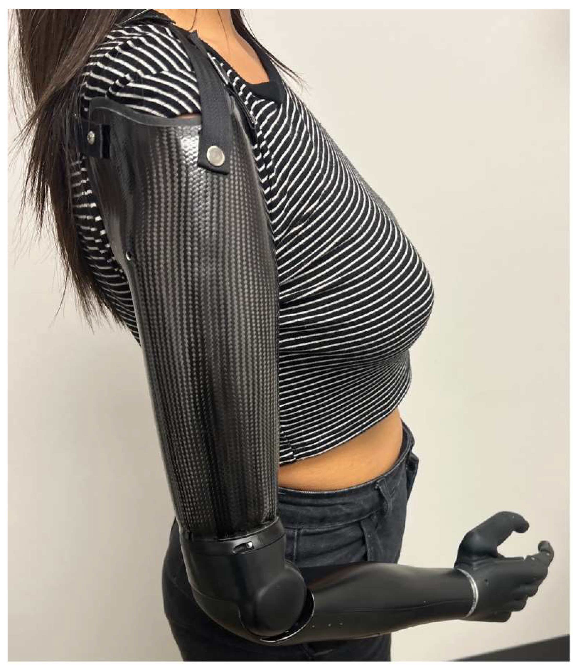
3. Prosthetic Device Types

Multiple prosthetic devices exist, each with its own advantages and disadvantages (Table 1). Passive functional prosthetics (PFPs) enhance aesthetics and assist in simple tasks but provide minimal grasping function. However, they allow digit opposition for fixed grasp and partial digit extension for limited functionality [17]. They are lightweight and cosmetically resemble a natural hand (Figure 1).
Body-powered (BP) prostheses use cables and harnesses to control movement, offering durability, easy maintenance, and greater sensory feedback [18]. They provide a wider range of motion and superior strength for heavy lifting compared to myoelectric (MYO) devices, leading to improved functionality and patient satisfaction [19,20,21]. BP prostheses cost between USD 4000 and USD 10,000, making them more affordable than MYO devices, which can exceed USD 75,000 [22].
Myoelectric (MYO) prostheses use electromyographic (EMG) signals from muscle contractions in the residual limb to control movement of the terminal device [23]. They offer improved fine motor control and improved cosmesis, as they more closely resemble the natural anatomy [19,24,25]. These devices excel in precise movements and are better suited for light tasks that require movement accuracy rather than heavy lifting [20,26,27]. Although the evidence is mixed, MYO devices may also have a positive effect on phantom limb pain [28,29].
Hybrid prostheses combine the fine motor control of MYO devices with the durability of BP models, making them ideal for individuals needing adaptability across various activities [30,31]. They are particularly beneficial for above-elbow amputations, offering enhanced functionality in complex movements like lifting against resistance [32]. Novel hybrid approaches have been introduced, including a recently patented design which features a flexible, bionic hand with palm and finger assemblies with independent bending and transmission components [33]. By enabling coordinated finger movement via fewer actuators, the lightweight prosthesis improved dexterity and digit control [33]. Alternatively, three-dimensional (3D) printing has been employed to create a prosthetic finger with a monolithic structure and flexure hinge [34]. By treating the hinge as a 3D object, the design allowed for a soft and lightweight prosthesis that better mimicked the natural finger and improved user comfort [34].

4. Review of Surgeons’ Versus Prosthetists’ Recommended Amputation Lengths

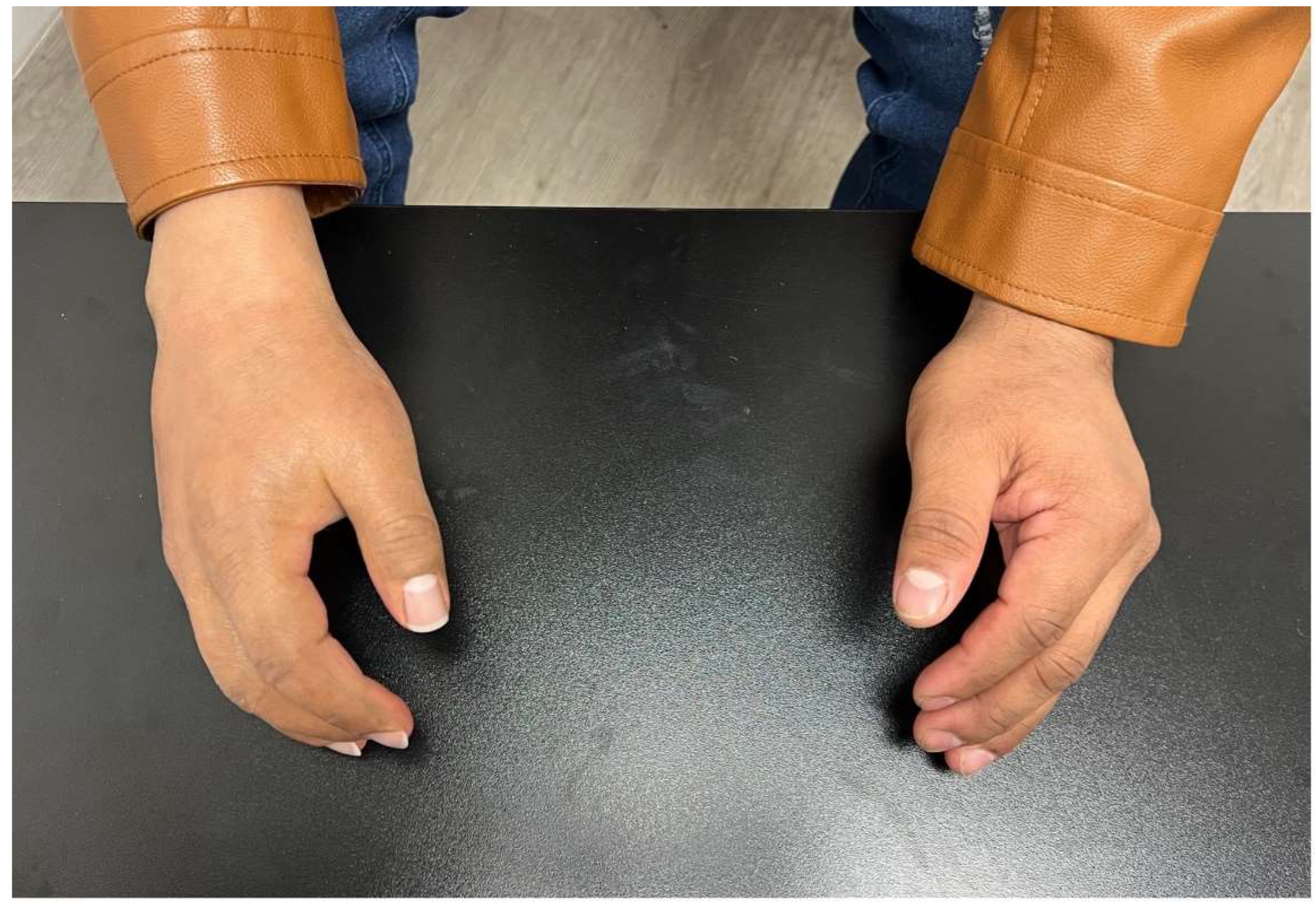
4.1. Digit Amputation

4.1.1. Surgical Recommendations

Finger and thumb amputations are the most common trauma-related amputations in emergency settings [35,36]. Thumb function is directly proportional to stump length, making length preservation critical [37,38]. Losing the thumb at the metacarpophalangeal joint results in a 40% loss of hand function and 36% of upper limb function [39]. Surgical goals focus on removing painful or nonviable tissue while preserving as much functional length as possible [37]. Neuroma formation should be prevented to avoid post-amputation pain [40].
For distal phalanx amputations, preserving the flexor digitorum superficialis (FDS) insertion helps maintain joint flexion [38]. A finger flap is recommended if the wound is ≥2 mm distal to the volar distal interphalangeal (DIP) joint skin crease, while shortening amputation is performed if both digital nerves are damaged [41]. Middle phalanx amputations proximal to the FDS insertion require proximal interphalangeal (PIP) joint disarticulation to prevent loss of motion control. Preserving articular cartilage reduces pain and enhances shock absorption [42]. Extensive central digit amputations may require ray amputation to narrow the residual gap and improve grip function [41].

4.1.2. Prosthetist Recommendations

Similarly, prosthetists prioritize salvaging digit length to maintain pincer grasp. For thumb amputations, passive prosthetics (e.g., Vincent Passive Thumb, Titan Thumb, M-Thumb) enhance aesthetics, while BP prostheses (e.g., Thumb Driver by Naked Prosthetics) improve function in proximal amputations [43].
For amputations distal to the DIP joint, passive prosthetics (e.g., Regal Prosthesis Ltd., Livingskin) offer cosmetic benefits, improving social acceptance despite limited opposition [43]. At levels proximal to the DIP, passive devices (e.g., Point Partial, Point Digit, Grip Lock) remain options, though less cosmetic and without active prehension (Figure 2). At this level, BP prostheses (e.g., PIP Driver, MCP Driver, Partial M-Finger) become available and provide grasp function but are less aesthetically favorable. For either prosthetic type, preserving the joint is crucial to avoid length discrepancies with the contralateral hand [43]. To use a BP prosthesis, the amputation must leave a small segment distal to the joint to allow for engagement of a ring that flexes the prosthesis during movement.

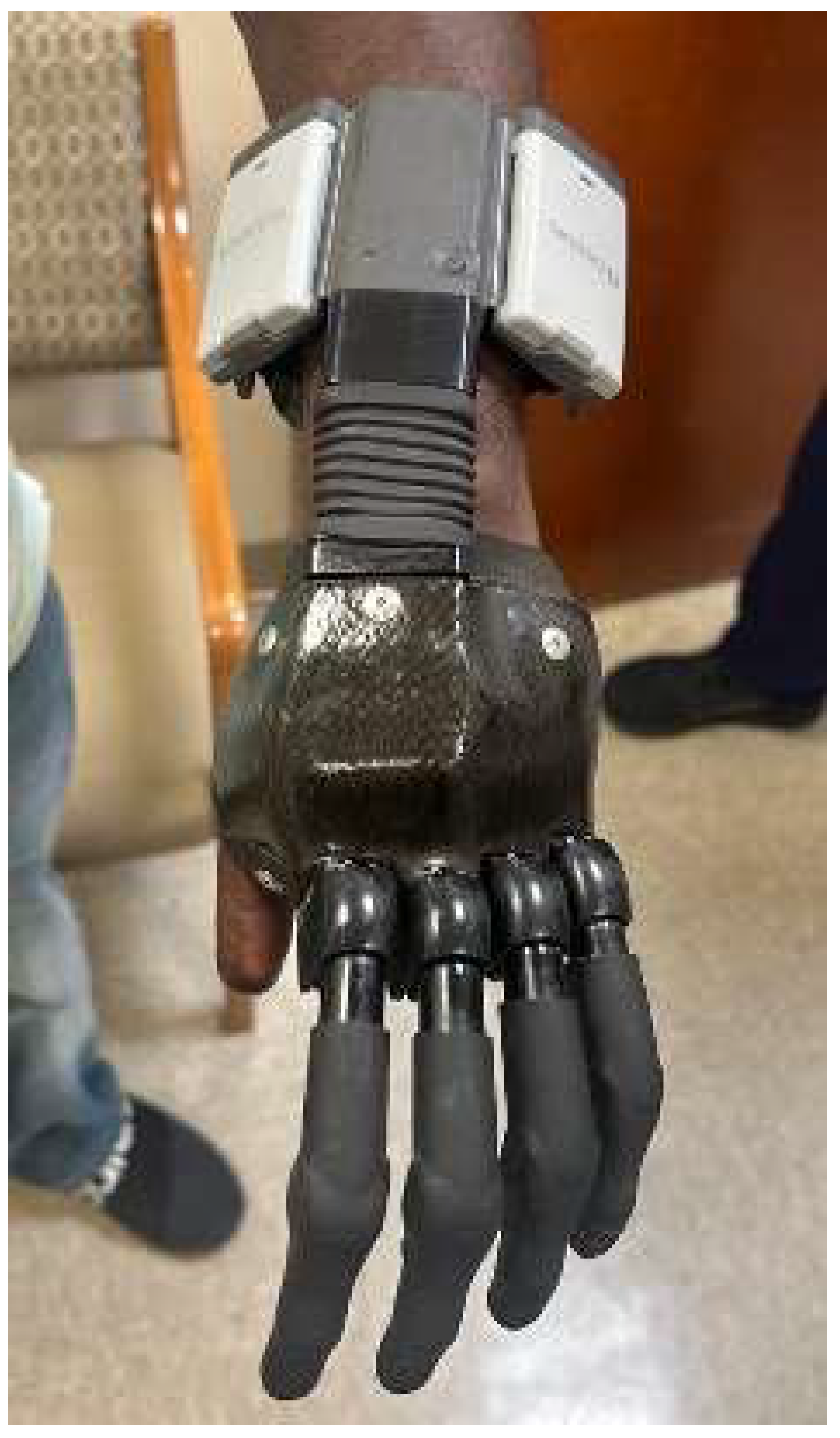
4.2. Partial Hand/Transcarpal Amputation

4.2.1. Surgical Recommendations

Preservation of the length of the metacarpals is recommended, as it allows the residual limb to function as a helper hand [41]. If dorsal wrist extensor insertions are removed, extensor tendons should be reinserted into the residual carpus [41]. In these cases, myocutaneous flaps are preferable to skin flaps for pressure relief, and the associated atrophy will facilitate the placement of passive finger prostheses to assist in grasp [17]. In severe cases involving the thumb or multiple digits, toe-to-hand transfer may be considered [41].

4.2.2. Prosthetist Recommendations

If at least two opposable, sensate digits remain, preserving length is crucial to minimize graft and scar tissue. Thumb preservation is especially important to maintain sensation for grasp with the prosthesis. Techniques to save the thumb include bone lengthening, web deepening, or bone grafting.
Prosthetic options vary by amputation level. Distal amputations with minimal functional loss may benefit from passive functional prosthetics (e.g., Point Digit, Grip Lock, Titan Full, Vincent Passive) with a ratcheting flexion mechanism that locks the prosthetic into various degrees of flexion [40]. BP prosthetics (e.g., Robin-Aids, M-Fingers) are suitable for patients who require more durability and grasping ability for daily activities.
For fine motor control and grasping, MYO prosthetics (e.g., Ossur iDigits, Vincent Partial Hand) are ideal (Figure 3) [43]. Barriers to effective MYO prosthetic control include nonintuitive control mechanisms, signal interference from the small hand muscles, and a limited number of muscle targets sufficiently close to the surface for the prosthetic [44]. The Starfish procedure addresses these issues by repositioning residual interosseous muscles to a more superficial area for better prosthetic control without adding bulk [44,45].
Recently, a machine learning-based approach has been used to improve prosthetic hand control [46]. By using reinforcement learning and computer vision, autonomous object grasping was improved without requiring extensive user training [46]. This highlights the potential for highly responsive prosthetics that reduce reliance on muscle signals.

4.3. Wrist Disarticulation

4.3.1. Surgical Recommendations

Wrist disarticulations are preferred over proximal amputations when sufficient tissue allows for deep tissue and bony coverage [37]. Preserving the ulnar styloid stabilizes the distal radial ulnar joint and enables radial rotation [37]. Additionally, trimming the radial and ulnar styloid prevents pressure points and improves prosthetic fit while preserving the triangular fibrocartilage complex for pronation and supination. In preparation for prosthetic use, special consideration is given to the extensor tendon, as it is essential to maintain muscle tension and stabilization for MYO control of the terminal device [37]. Regarding the nerves, traditional options include burying the nerve in muscle or bone [41].

4.3.2. Prosthetist Recommendations

Wrist disarticulation can be challenging due to limited low-profile wrist and hand components that maintain function and symmetrical limb lengths. Thus, transradial amputations are generally preferred [47]. If wrist disarticulation is necessary, prosthetists recommend a low-profile wrist unit or direct mount of the hand to the prosthesis at the wrist disarticulation level [47]. While this level may restrict supination and pronation, it allows for lower socket trim lines and suspension just proximal to the styloid.

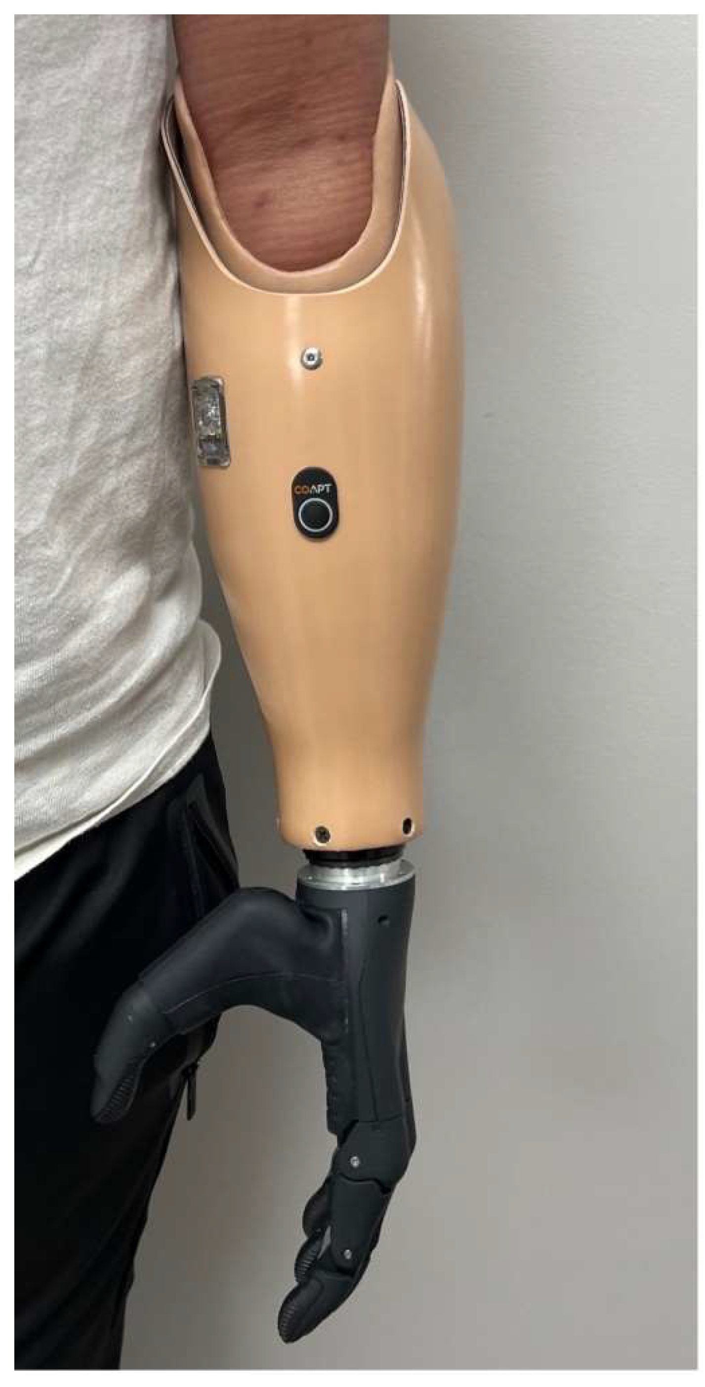
4.4. Transradial Amputation

4.4.1. Surgical Recommendations

In transradial amputations, forearm length is proportional to prosupination [41]. Amputating too close to the elbow limits this motion, so adequate length should be preserved when possible [41]. A minimum of 5 cm from the elbow is needed for prosthetic fitting, while 16–18 cm distal to the olecranon provides a longer lever arm, reducing residual limb stress [41,48]. Intraoperatively, the bicep tendon is transferred from the radius to the ulna to prevent flexion contracture [49]. Dividing the tendon at its radial tuberosity insertion can increase functional length, leaving the brachialis as the primary elbow flexor [41]. Rotary movement of the radius is preserved to prevent synostosis [49]. Myodesis, myoplasty, or myofascial closure stabilizes muscles to minimize stump pain [50]. Myoplasty of flexor and extensor muscles over the radius and ulna aids in muscle stabilization and facilitates later prosthetic fitting [50].

4.4.2. Prosthetist Recommendations

This level gives optimum outcomes for prosthetic fit as it allows for the fitting of all available BP (Figure 4) and MYO prosthetics (Figure 5). The socket must house batteries, controllers, electrodes, and wires for MYO function. Suspension is either suction or anatomically based, depending on patient needs. A stable limb volume is required for MYO prosthesis fitting. To determine the optimal length for amputation, surgeons should use the following formula [47]:
Y = X − 22.5 cm
where Y is the optimal residual limb length (including soft tissue), X is the distance from the lateral epicondyle to the thumb tip (uninvolved limb, elbow at 90°, and 22.5 cm is the average prosthetic terminal device and wrist unit length.
Using this formula, the ulna should be 0.8–1.25 cm shorter than Y, and the radius 1.25–2.5 cm shorter than Y. Surgeons should also consider that shortening the residual limb too much can impair natural supination and pronation, potentially necessitating a prosthetic wrist unit to restore rotational function.
Although often overlooked, prosthetic length estimation must consider socket materials for function, skin protection, and user comfort [51]. Prosthetic socket materials (thermoplastic or high-consistency rubber silicone) add 1–2 cm, while wrist components add 1–2 cm, totaling 2–4 cm [52]. For this reason, it is preferred to have at least 4 cm of the ulna from the olecranon preserved. If the residual limb is shorter than this, a more proximal amputation is advised [48]. Table 2 provides total prosthesis length calculations. Considering these measurements ensures optimal amputation site selection for symmetry and function.
TMR, increasingly used in upper limb amputations, transfers residual mixed or sensory nerves to newly denervated muscles [53,54]. As a result, greater numbers of motor signals are created and amplified, increasing function and range of motion [55]. Additionally, the prevention of disorganized nerve ends reduces neuroma formation and phantom limb pain [56]. For TMR, at least 7–8 cm of distal radius should be resected to ensure adequate soft tissue coverage and space for MYO prosthetic components [55].

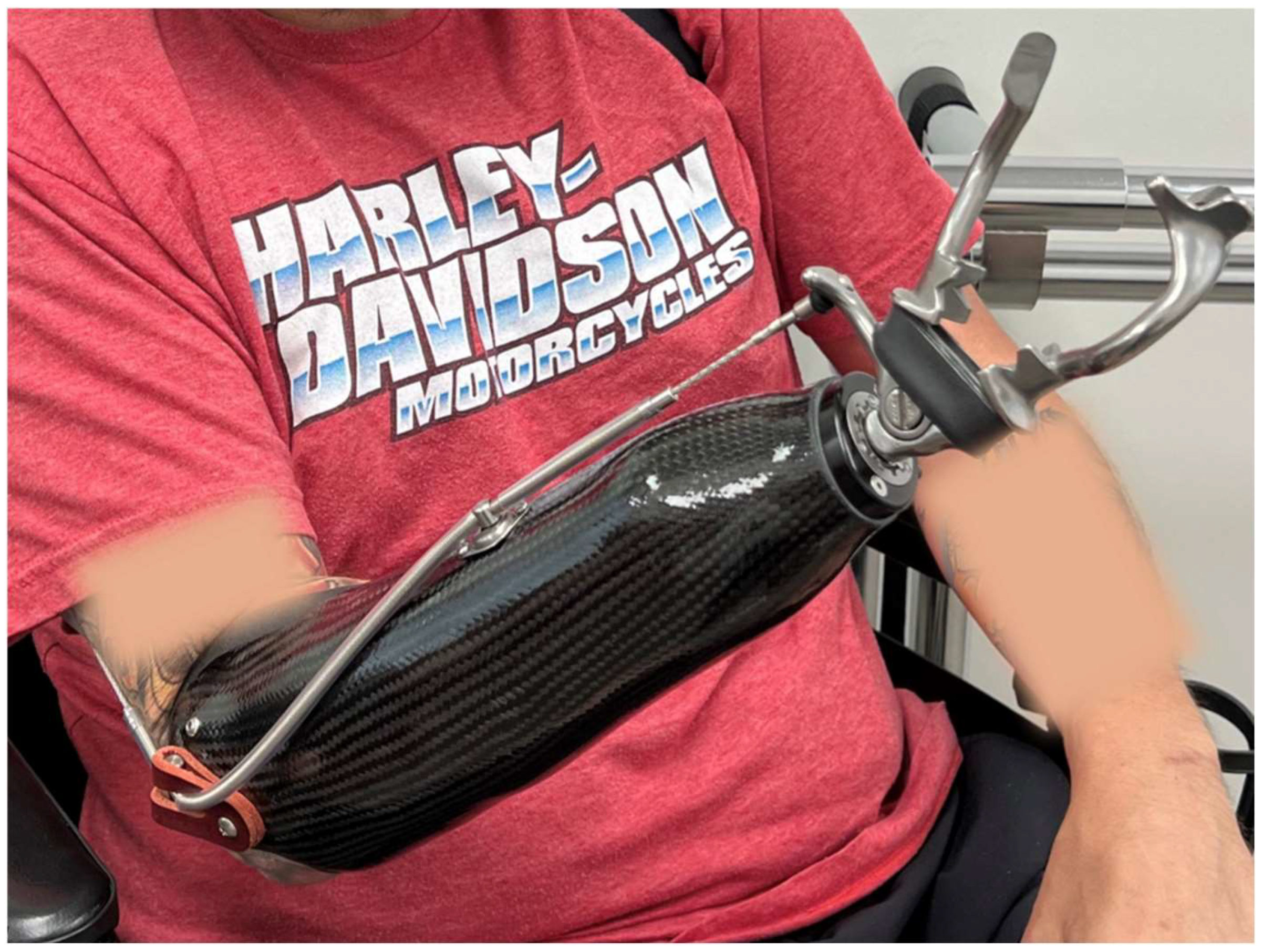
4.5. Elbow Disarticulation

4.5.1. Surgical Recommendations

When the radius and ulna cannot be salvaged, elbow disarticulation preserves shoulder rotation and can be performed if the humeral condyle is intact [31]. While typically less preferable than transradial or transhumeral amputations, it is an option when soft tissue coverage is insufficient to retain the radius [57]. It offers a distinct advantage over transhumeral amputation by maintaining internal and external rotation [49]. Additionally, humeral condyle preservation aids in prosthetic suspension and humeral rotation [41,58]. Successful outcomes depend on muscle stabilization and careful contouring of the condyles to prevent pressure intolerance [49]. However, this level is less favored because the current MYO elbows add 5–6 cm to the upper arm, making prosthetic fitting difficult [49,59]. As a result, elbow disarticulation remains controversial, as amputees are often limited to cable controls and weaker external hinges [60].

4.5.2. Prosthetist Recommendations

Achieving limb length symmetry is challenging at this level due to intact condyles and the longer total limb compared to the contralateral side. Traditional elbow units use a turntable mechanism for humeral rotation, but these amputations require outside locking hinges to lock and unlock the elbow joints. This limits the rotation of the elbow and increases the width [47]. A hybrid prosthesis is preferred if the patient desires more advanced functionality than is possible with the conventional BP unit. This uses outside locking hinges paired with a MYO or multi-articulating hand using a quick-disconnect wrist unit. For those patients who are seeking improved limb length symmetry or wish to use a powered elbow, a humeral midshaft bone shortening osteotomy can be performed. This involves removing about 5 to 6 cm of bone from the midportion of the humerus. Although technically demanding, it preserves the humeral condyles, aiding in prosthetic suspension, and allows for some retained rotational movement [59].

4.6. Transhumeral Amputation

4.6.1. Surgical Recommendations

For transhumeral amputations, residual limb length impacts prosthetic use and fit. Retaining a minimum of 5–7 cm or 25–30% of the humeral length is recommended for optimal fitting [48]. This helps with weight distribution and maintains shoulder/upper arm contour for clothing [25]. Preserving the deltoid, pectoralis major, and latissimus dorsi insertions maximizes range of motion [49]. Sharp humeral edges should be removed.

4.6.2. Prosthetist Recommendations

A mid-length amputation allows for most prosthetic options, which usually include either a BP or MYO elbow (Figure 6). The optimal amputation site is determined by [47]:
Y = X − 14 cm
where Y is the optimal residual limb length (including soft tissue) from the acromion, X is the distance from acromion to distal olecranon (unaffected limb, elbow at 90°), and 14 cm is the average prosthetic elbow unit length.
Prosthetists recommend a hybrid system with a BP elbow and electric lock, which improves efficiency, allowing faster elbow positioning and simultaneous MYO hand control. Harnessing, cabling, and suspension of the prosthetic socket must be maintained, and can enhance prosthetic suspension and rotational control by reshaping the distal humerus. This entails removing a wedge-shaped piece of bone to create an angled portion at the distal humerus. By adjusting the angle of the residual humerus and creating a more anatomically favorable shape, the prosthesis can have better alignment and rotational control, while preserving length. As stated above, a midshaft bone shortening osteotomy of an elbow disarticulation may be performed to aid in rotational control [59].

4.7. Shoulder Disarticulation

4.7.1. Surgical Recommendations

Shoulder disarticulation is performed for tumors or severe injury when the humerus cannot be salvaged [2,61]. To improve cosmesis, the scapula is retained, and in female patients, deltoid preservation helps maintain breast contour [2]. Deltoid preservation is also essential for optimal MYO prosthetic control [2]. The rotator cuff is sutured over the glenoid fossa, while the deltoid is secured to the inferior glenoid and lateral scapular border to fill the subacromial space [2]. After securing all muscles in the glenoid cavity, the flaps are smoothed to refine the shoulder contour [2].
To maximize potential for TMR, the musculocutaneous, median, ulnar, and radial nerves and brachial plexus are dissected [41]. Nerve pairings include the musculocutaneous nerve to the clavicular head of the pectoralis major, the median nerve to the motor nerve of the sternal head of the pectoralis major, the radial nerve to the thoracodorsal or long thoracic nerve, and the ulnar to the sternal head of the pectoralis major or minor [41,62].

4.7.2. Prosthetist Recommendations

One of the most challenging prostheses to fit and wear is a shoulder disarticulation prosthesis. Weight, limited ROM, and battery needs lead many patients to forgo long-term use. An X-Frame socket with a cross-body strap system is recommended for suspension (Figure 7). MYO devices are commonly used for this amputation length, as you lose half of the excursion necessary to control the elbow and terminal devices. Given that BP prosthetics are of minimal use at this level, TMR should highly be considered to optimize MYO components [63].
To guide future practice, recommendations from both surgeons and prosthetists and summaries of the optimal amputation levels for functional prosthetic use are described in Table 3.

5. Conclusions

This paper compares the optimal sites of amputation for upper extremities taught to surgeons with those recommended by prosthetists to enhance function and quality of life through effective prosthetic use (Table 3). Early coordination between surgeons, prosthetists, therapists, and social workers is crucial in the preparation for an upper extremity prosthesis after amputation. Prosthetists take a personalized approach to amputation site selection, considering functional challenges from limb length asymmetry and individual patient preferences. As prosthetic technology advances, their focus has shifted toward achieving symmetrical limb lengths, factoring in measurements for wrist flexion, socket material, and terminal devices. They also prioritize patient-specific needs, recognizing that task importance varies among individuals. A major challenge in upper extremity prosthesis engineering arises when striving for symmetrical limb lengths. While surgeons emphasize maximal limb preservation, prosthetists recommend a tailored approach that accounts for contralateral limb length and prosthetic dimensions. This requires surgeons to consider not only the terminal device but also socket components and other prosthetic elements. Despite recommendations, selecting the optimal amputation site has limitations. Surgeons’ choices are often constrained by the extent of the residual limb after trauma, affecting the generalizability of findings. Prosthesis recommendations may also be biased by prosthetists’ preferred devices and the populations they serve. To reduce this bias, future research should include prosthetists from diverse regions and backgrounds. Collaboration among surgeons, prosthetists, and occupational therapists—starting preoperatively—is crucial for better outcomes. Additionally, input from various prosthetic companies is needed for a more comprehensive understanding. Future studies should address these challenges to advance the field.

Author Contributions

Conceptualization, P.G.C. and N.F.; Methodology, B.A., S.E.M., N.V.M., C.A., V.V.N., P.G.C. and N.F.; Software, B.A., S.E.M., N.V.M., C.A., V.V.N., P.G.C. and N.F.; Validation, P.G.C. and N.F.; Formal Analysis, B.A., S.E.M., N.V.M., C.A., V.V.N., P.G.C. and N.F.; Investigation, B.A., S.E.M., N.V.M., C.A., V.V.N., P.G.C. and N.F.; Resources, P.G.C. and N.F.; Data Curation, B.A., S.E.M., N.V.M., C.A., P.G.C. and N.F.; Writing—Original Draft Preparation, B.A., S.E.M., N.V.M. and C.A.; Writing—Review and Editing, V.V.N., P.G.C. and N.F.; Visualization, P.G.C. and N.F.; Supervision, P.G.C. and N.F.; Project Administration, P.G.C. and N.F. All authors have read and agreed to the published version of the manuscript.

Funding

This research received no external funding.

Institutional Review Board Statement

Not applicable.

Informed Consent Statement

Informed consent was obtained from all subjects involved in the study.

Data Availability Statement

No new data were created or analyzed in this study.

Conflicts of Interest

The authors declare no conflicts of interest.

References

  1. Ziegler-Graham, K.; MacKenzie, E.J.; Ephraim, P.L.; Travison, T.G.; Brookmeyer, R. Estimating the prevalence of limb loss in the United States: 2005 to 2050. Arch. Phys. Med. Rehabil. 2008, 89, 422–429. [Google Scholar] [CrossRef] [PubMed]
  2. Bowker, J.H.; Michael, J.W. Atlas of Limb Prosthetics: Surgical, Prosthetic, and Rehabilitation Principles; Mosby Inc.: Maryland, MO, USA, 1992. [Google Scholar]
  3. Behrend, C.; Reizner, W.; Marchessault, J.A.; Hammert, W.C. Update on advances in upper extremity prosthetics. J. Hand Surg. Am. 2011, 36, 1711–1717. [Google Scholar] [CrossRef] [PubMed]
  4. Tennent, D.J.; Wenke, J.C.; Rivera, J.C.; Krueger, C.A. Characterisation and outcomes of upper extremity amputations. Injury 2014, 45, 965–969. [Google Scholar] [CrossRef] [PubMed]
  5. Ro, J.-S.; Leigh, J.-H.; Jeon, I.; Bang, M.S. Trends in burden of work-related upper limb amputation in South Korea, 2004–2013: A nationwide retrospective cohort study. BMJ Open 2019, 9, e032793. [Google Scholar] [CrossRef] [PubMed]
  6. Østlie, K.; Magnus, P.; Skjeldal, O.H.; Garfelt, B.; Tambs, K. Mental health and satisfaction with life among upper limb amputees: A Norwegian population-based survey comparing adult acquired major upper limb amputees with a control group. Disabil. Rehabil. 2011, 33, 1594–1607. [Google Scholar] [CrossRef] [PubMed]
  7. Flaubert, J.L.; Spicer, C.M.; Jette, A.M. The Promise of Assistive Technology to Enhance Activity and Work Participation; The National Academic Press: Washington, DC, USA, 2017. [Google Scholar]
  8. Jang, C.H.; Yang, H.S.; Yang, H.E.; Lee, S.Y.; Kwon, J.W.; Yun, B.D.; Choi, J.Y.; Kim, S.N.; Jeong, H.W. A survey on activities of daily living and occupations of upper extremity amputees. Ann. Rehabil. Med. 2011, 35, 907–921. [Google Scholar] [CrossRef] [PubMed]
  9. Raichle, K.A.; Hanley, M.A.; Molton, I.; Kadel, N.J.; Campbell, K.; Phelps, E.; Ehde, D.; Smith, D.G. Prosthesis use in persons with lower-and upper-limb amputation. J. Rehabil. Res. Dev. 2008, 45, 961. [Google Scholar] [CrossRef] [PubMed]
  10. Resnik, L.; Borgia, M.; Clark, M. Function and quality of life of unilateral major upper limb amputees: Effect of prosthesis use and type. Arch. Phys. Med. Rehabil. 2020, 101, 1396–1406. [Google Scholar] [CrossRef] [PubMed]
  11. Tintle, L.S.M.; Baechler, L.M.F.; Nanos, C.G.P., III; Forsberg, L.J.A.; Potter, M.B.K. Traumatic and trauma-related amputations: Part II: Upper extremity and future directions. J. Bone Jt. Surg. 2010, 92, 2934–2945. [Google Scholar] [CrossRef] [PubMed]
  12. Wright, T.W.; Hagen, A.D.; Wood, M.B. Prosthetic usage in major upper extremity amputations. J. Hand Surg. 1995, 20, 619–622. [Google Scholar] [CrossRef] [PubMed]
  13. Biddiss, E.A.; Chau, T.T. Upper limb prosthesis use and abandonment: A survey of the last 25 years. Prosthet. Orthot. Int. 2007, 31, 236–257. [Google Scholar] [CrossRef] [PubMed]
  14. Biddiss, E.A.; Chau, T.T. Multivariate prediction of upper limb prosthesis acceptance or rejection. Disabil. Rehabil. Assist. Technol. 2008, 3, 181–192. [Google Scholar] [CrossRef] [PubMed]
  15. Biddiss, E.; Chau, T. Upper-limb prosthetics: Critical factors in device abandonment. Am. J. Phys. Med. Rehabil. 2007, 86, 977–987. [Google Scholar] [CrossRef] [PubMed]
  16. Solarz, M.K.; Thoder, J.J.; Rehman, S. Management of major traumatic upper extremity amputations. Orthop. Clin. 2016, 47, 127–136. [Google Scholar] [CrossRef] [PubMed]
  17. Lake, C. Partial hand amputation: Prosthetic management. Atlas Amputations Limb Defic. Surg. Prosthet. Rehabil. Princ. 2004, 3, 209–217. [Google Scholar]
  18. Bates, T.J.; Fergason, J.R.; Pierrie, S.N. Technological advances in prosthesis design and rehabilitation following upper extremity limb loss. Curr. Rev. Musculoskelet. Med. 2020, 13, 485–493. [Google Scholar] [CrossRef] [PubMed]
  19. Carey, S.L.; Lura, D.J.; Highsmith, M.J. Differences in myoelectric and body-powered upper-limb prostheses: Systematic literature review. J. Rehabil. Res. Dev. 2015, 52, 247–262. [Google Scholar] [CrossRef] [PubMed]
  20. Kejlaa, G. Consumer concerns and the functional value of prostheses to upper limb amputees. Prosthet. Orthot. Int. 1993, 17, 157–163. [Google Scholar] [CrossRef] [PubMed]
  21. Williams, T.W., III. Progress on stabilizing and controlling powered upper-limb prostheses. J. Rehabil. Res. Dev. 2011, 48, ix. [Google Scholar] [CrossRef] [PubMed]
  22. Resnik, L.; Meucci, M.R.; Lieberman-Klinger, S.; Fantini, C.; Kelty, D.L.; Disla, R.; Sasson, N. Advanced upper limb prosthetic devices: Implications for upper limb prosthetic rehabilitation. Arch. Phys. Med. Rehabil. 2012, 93, 710–717. [Google Scholar] [CrossRef] [PubMed]
  23. Uellendahl, J.E. Upper extremity myoelectric prosthetics. Phys. Med. Rehabil. Clin. N. Am. 2000, 11, 639–652. [Google Scholar] [CrossRef]
  24. Silcox, D., 3rd; Rooks, M.D.; Vogel, R.R.; Fleming, L.L. Myoelectric prostheses. A long-term follow-up and a study of the use of alternate prostheses. JBJS 1993, 75, 1781–1789. [Google Scholar] [CrossRef] [PubMed]
  25. Hafshejani, M.K.; Javanshir, M.; Kamali, K.; Ghasemi, M.; Emami, M.; Esmaeeli, S.; Langari, A.; Sattari Naeini, M. The comparison of psychological and social adaptation below elbow amputation men using a mechanical and myoelectric prosthesis by using of TAPES questionnaire. Life Sci. J. 2012, 9, 5583–5587. [Google Scholar]
  26. Simon, A.M.; Lock, B.A.; Stubblefield, K.A. Patient training for functional use of pattern recognition–controlled prostheses. JPO J. Prosthet. Orthot. 2012, 24, 56–64. [Google Scholar] [CrossRef] [PubMed]
  27. Huang, M.E.; Levy, C.E.; Webster, J.B. Acquired limb deficiencies. 3. Prosthetic components, prescriptions, and indications. Arch. Phys. Med. Rehabil. 2001, 82, S17–S24. [Google Scholar] [CrossRef] [PubMed]
  28. Lotze, M.; Grodd, W.; Birbaumer, N.; Erb, M.; Huse, E.; Flor, H. Does use of a myoelectric prosthesis prevent cortical reorganization and phantom limb pain? Nat. Neurosci. 1999, 2, 501–502. [Google Scholar] [CrossRef] [PubMed]
  29. Bouffard, J.; Vincent, C.; Boulianne, É.; Lajoie, S.; Mercier, C. Interactions between the phantom limb sensations, prosthesis use, and rehabilitation as seen by amputees and health professionals. JPO J. Prosthet. Orthot. 2012, 24, 25–33. [Google Scholar] [CrossRef]
  30. Madusanka, D.; Wijayasingha, L.; Gopura, R.; Amarasinghe, Y.; Mann, G. A review on hybrid myoelectric control systems for upper limb prosthesis. In Proceedings of the 2015 Moratuwa Engineering Research Conference (MERCon), Moratuwa, Sri Lanka, 7–8 April 2015; pp. 136–141. [Google Scholar]
  31. Childress, D.S. Upper-limb prosthetics: Control of limb prostheses. In Atlas of Limb Prosthetics: Surgical, Prosthetic, and Rehabilitation Principles; American Academy of Orthopaedic Surgeons: Rosemont, IL, USA, 1992. [Google Scholar]
  32. Billock, J. Upper limb prosthetic management hybrid design approaches. Clin. Prosthet. Orthot. 1985, 9, 23–25. [Google Scholar]
  33. Zhang, H.L.J.; Liu, S.; Zhang, W.; Lu, Y.; Yang, S.; Yin, R.; Shen, D. Flexible Under-Actuated Bionic Hand. CN110216700B. 2018. Available online: https://patents.google.com/patent/CN110216700B/en?oq=CN110216700B%2c+++华东理工大学%2c+仿生假肢手 (accessed on 13 July 2025).
  34. Liu, S.; Zhang, H.; Yin, R.; Chen, A.; Zhang, W. Flexure hinge based fully compliant prosthetic finger. In Proceedings of the SAI Intelligent Systems Conference (IntelliSys) 2016, London, UK, 21–22 September 2016; Springer: Berlin/Heidelberg, Germany, 2018; Volume 2, pp. 839–849. [Google Scholar]
  35. Reavey, P.L.; Stranix, J.T.; Muresan, H.; Soares, M.; Thanik, V. Disappearing digits: Analysis of national trends in amputation and replantation in the United States. Plast. Reconstr. Surg. 2018, 141, 857e–867e. [Google Scholar] [CrossRef] [PubMed]
  36. Win, T.S.; Henderson, J. Management of traumatic amputations of the upper limb. BMJ 2014, 348, g255. [Google Scholar] [CrossRef] [PubMed]
  37. Banerjee, S.N. Rehabilitation Management of Amputees; Williams & Wilkins: Philadelphia, PA, USA, 1982. [Google Scholar]
  38. Ovadia, S.A.; Askari, M. Upper extremity amputations and prosthetics. In Seminars in Plastic Surgery; Thieme Medical Publishers: New York, NY, USA, 2015; pp. 055–061. [Google Scholar]
  39. Dellon, A.L. The moving two-point discrimination test: Clinical evaluation of the quickly adapting fiber/receptor system. J. Hand Surg. 1978, 3, 474–481. [Google Scholar] [CrossRef] [PubMed]
  40. Dellon, A.L. “Think nerve” in upper extremity reconstruction. Clin. Plast. Surg. 1989, 16, 617–627. [Google Scholar] [CrossRef] [PubMed]
  41. Dumanian, G.; Souza, J.; Kuiken, T. Treatment of the upper extremity amputee. In Plastic Surgery: Volume 6: Hand and Upper Extremity; Elsevier: Amsterdam, The Netherlands, 2018; pp. 894–904. [Google Scholar]
  42. Graham, W.P.; Kilgore, E.S.; Whitaker, L.A. Transarticular digital joint amputations: Preservation of the articular cartilage. Hand 1973, 5, 58–62. [Google Scholar] [CrossRef] [PubMed]
  43. Graham, E.M.; Hendrycks, R.; Baschuk, C.M.; Atkins, D.J.; Keizer, L.; Duncan, C.C.; Mendenhall, S.D. Restoring Form and Function to the Partial Hand Amputee: Prosthetic Options from the Fingertip to the Palm. Hand Clin. 2021, 37, 167–187. [Google Scholar] [CrossRef] [PubMed]
  44. Denduluri, S.K.; Rees, A.; Nord, K.M.; Loeffler, B.J.; Gaston, R.G. The Starfish Procedure for Independent Digital Control of a Myoelectric Prosthesis. Tech. Hand Up. Extrem. Surg. 2023, 27, 61–67. [Google Scholar] [CrossRef] [PubMed]
  45. Grier, A.J.; Loeffler, B.J.; Gaston, R.G. Starfish Procedure. Hand Clin. 2021, 37, 447–455. [Google Scholar] [CrossRef] [PubMed]
  46. Odeyemi, J.; Ogbeyemi, A.; Wong, K.; Zhang, W. On automated object grasping for intelligent prosthetic hands using machine learning. Bioengineering 2024, 11, 108. [Google Scholar] [CrossRef] [PubMed]
  47. Pasquina, P.F.; Cooper, R.A. Care of the Combat Amputee; Office of the Surgeon General, United States Army: Falls Church, VA, USA, 2009. [Google Scholar]
  48. Maduri, P.; Akhondi, H. Upper Limb Amputation; StatPearls Publishing: Treasure Island, FL, USA, 2019. [Google Scholar]
  49. Fitzgibbons, P.; Medvedev, G. Functional and clinical outcomes of upper extremity amputation. JAAOS J. Am. Acad. Orthop. Surg. 2015, 23, 751–760. [Google Scholar] [CrossRef] [PubMed]
  50. Lake, C.; Dodson, R. Progressive upper limb prosthetics. Phys. Med. Rehabil. Clin. 2006, 17, 49–72. [Google Scholar] [CrossRef] [PubMed]
  51. Schultz, A.E.; Baade, S.P.; Kuiken, T.A. Expert opinions on success factors for upper-limb prostheses. J. Rehabil. Res. Dev. 2007, 44, 483. [Google Scholar] [CrossRef] [PubMed]
  52. Armstrong, T.W.; Williamson, M.L.; Elliott, T.R.; Jackson, W.T.; Kearns, N.T.; Ryan, T. Psychological distress among persons with upper extremity limb loss. Br. J. Health Psychol. 2019, 24, 746–763. [Google Scholar] [CrossRef] [PubMed]
  53. Kuiken, T.A.; Dumanian, G.A.; Lipschutz, R.D.; Miller, L.A.; Stubblefield, K. The use of targeted muscle reinnervation for improved myoelectric prosthesis control in a bilateral shoulder disarticulation amputee. Prosthet. Orthot. Int. 2004, 28, 245–253. [Google Scholar] [CrossRef] [PubMed]
  54. Mereu, F.; Leone, F.; Gentile, C.; Cordella, F.; Gruppioni, E.; Zollo, L. Control strategies and performance assessment of upper-limb TMR prostheses: A review. Sensors 2021, 21, 1953. [Google Scholar] [CrossRef] [PubMed]
  55. Morgan, E.N.; Potter, B.K.; Souza, J.M.; Tintle, S.M.; Nanos, G.P., III. Targeted muscle reinnervation for transradial amputation: Description of operative technique. Tech. Hand Up. Extrem. Surg. 2016, 20, 166–171. [Google Scholar] [CrossRef] [PubMed]
  56. Souza, J.M.; Cheesborough, J.E.; Ko, J.H.; Cho, M.S.; Kuiken, T.A.; Dumanian, G.A. Targeted muscle reinnervation: A novel approach to postamputation neuroma pain. Clin. Orthop. Relat. Res. 2014, 472, 2984–2990. [Google Scholar] [CrossRef] [PubMed]
  57. Mathew, P.J.; Rakoczy, K.D.; Fullerton, N.; Dodds, S.D.; Hui-Chou, H.G. Elbow joint salvage in pediatric traumatic amputation with targeted muscle reinnervation, external tissue expansion, and free muscle flap–A case report. Orthoplastic Surg. 2022, 9, 9–13. [Google Scholar] [CrossRef]
  58. Luccia, N.D.; Marino, H. Case study: Fitting of electronic elbow on an elbow disarticulated patient by means of a new surgical technique. Prosthet. Orthot. Int. 2000, 24, 247–251. [Google Scholar] [CrossRef] [PubMed]
  59. Cheesborough, J.E.; Smith, L.H.; Kuiken, T.A.; Dumanian, G.A. Targeted muscle reinnervation and advanced prosthetic arms. In Seminars in Plastic Surgery; Thieme Medical Publishers: New York, NY, USA, 2015; pp. 062–072. [Google Scholar]
  60. Bowker, J.H.; Carneiro, R.S.; Clark, C.R.; Gailey, R.S.; Poonekar, P.D.; Mnaymneh, W.; Ouellette, E.A. Atlas of Limb Prosthetics: Surgical, Prosthetic, and Rehabilitation Principles, 2nd ed.; Mosby Year Book: St. Louis, MO, USA, 1992. [Google Scholar]
  61. Levine, E.A.; Warso, M.A.; McCoy, D.M.; TK, D.G. Forequarter amputation for soft tissue tumors. Am. Surg. 1994, 60, 367–370. [Google Scholar] [PubMed]
  62. Gart, M.S.; Souza, J.M.; Dumanian, G.A. Targeted muscle reinnervation in the upper extremity amputee: A technical roadmap. J. Hand Surg. 2015, 40, 1877–1888. [Google Scholar] [CrossRef] [PubMed]
  63. Kulkarni, A.; Luthringer, M.; Fried, A.; Mikosz, M.; Mauro, J.; Vella, G.R.; Lally, T.; Shah, A. Building a Multidisciplinary Clinic Dedicated to Upper-Extremity Limb Loss. J. Hand Surg. 2024, 49, 267–274. [Google Scholar] [CrossRef] [PubMed]
Figure 1. Silicone restoration of the right hand.
Bioengineering 12 00765 g001
Figure 2. Passive functional prostheses of digits 1–3 at varying interphalangeal joints, allowing for passive ratcheting mechanism.
Bioengineering 12 00765 g002
Figure 3. Transcarpal myoelectric prosthesis in patient who had undergone Starfish procedure.
Bioengineering 12 00765 g003
Figure 4. Transradial body-powered prosthetic.
Bioengineering 12 00765 g004
Figure 5. Transradial myoelectric prosthesis.
Bioengineering 12 00765 g005
Figure 6. Transhumeral myoelectric prosthesis.
Bioengineering 12 00765 g006
Figure 7. (A) Anterior and (B) posterior views of X-Frame style socket with cross-body strap system for shoulder disarticulation prosthesis.
Bioengineering 12 00765 g007
Table 1. Differences in the types of prosthetic devices.
TypeAdvantagesDrawbacksExamples
Passive Functional Prosthetics (PFPs)High cosmesis
Waterproof
Lightweight
Minimal functionVincent Passive Thumb, Titan Thumb, M-Thumb, Regal Prosthesis Ltd., Livingskin, Point Partial, Point Digit, Grip Lock, Point Digit, Grip Lock, Titan Full, Vincent Passive
Body-Powered (BP) ProstheticsDurable
Waterproof
Low cosmesis
Heavy
Lower fine motor control
Thumb Driver, PIP Driver, MCP Driver, Partial M-Finger, Robin-Aids, M-Fingers
Myoelectric (MYO) ProstheticsFine motor control
Moderate cosmesis
Lightweight
Fragile
Expensive
Ossur iDigits, Vincent Partial Hand
Table 2. Common terminal device measurements.
Type of Terminal DeviceProsthetic BrandAvailable Sizing
MyohandsCOVVISmall: 6.75″, Medium: 7.75″, Large: 7.75″
BebionicSmall: 6.50″, Medium: 7.50″, Large: 7.875
Taska7.75″ or 8.25″
Taska CX7.25″
Body-Powered HookHosmer 5xAdult: 4.9″
Hosmer 99xAdult Small: 3.9″
Myo ElbowsUtah Power U32.5″ to elbow center, 4″ to ulnar surface
Steeper Espire1.89″
Motion Arm1.5″
Table 3. Summary of surgical recommendations based on amputation site.
Site of AmputationProsthetics RecommendedMinimum Residual LengthMaximum Residual LengthOther Considerations
ThumbPFP or BP prosthesisPreserve as much length as possible
Phalanges Distal to DIPPFPPreserve as much length as possible
Phalanges Proximal to DIPPFP or BP prosthesisConsider more proximal mid-phalanx amputation over disarticulation for digit symmetryEnsure adequate soft tissue coverage to prevent painful stumpsRay amputation can be considered in cases with central digit involvement
MetacarpalsPFP, BP, or MYOMust retain enough length for functional gripConsider thin flap coverageStarfish procedure may be performed if MYO device desired
WristPFP, BP, or MYO prosthesis with low-profile wrist unitNot ideal for prosthetics due to limb length discrepancyPreserve ulnar styloid and extensor tendon
RadiusBP or MYO prosthesisRetain at least 4 cm of the ulna from the olecranonMust allow room for terminal device + socket (~22.5 cm from lateral epicondyle)If TMR is desired, at least 7–8 cm of distal radius should be resected
ElbowBP or hybrid designNot ideal for prosthetics due to limb length discrepancyHumeral midshaft osteotomy can improve symmetry and control while preserving humeral condyles
HumerusHybrid design with BP elbow and myoelectric handRetain at least 5–7 cm (or 25–30%) of humeral length from the acromionMust allow room for terminal device + socket (~14 cm from acromion)Humeral angle or midshaft osteotomy can improve suspension and rotational control
ShoulderMYO prosthesis with X-Frame socketPreserve scapula and deltoid for shoulder contour and prosthetic controlEnsure adequate soft tissue coverage for prosthetic suspensionTMR should be highly considered
Disclaimer/Publisher’s Note: The statements, opinions and data contained in all publications are solely those of the individual author(s) and contributor(s) and not of MDPI and/or the editor(s). MDPI and/or the editor(s) disclaim responsibility for any injury to people or property resulting from any ideas, methods, instructions or products referred to in the content.

